Cnidome and Morphological Features of Pelagia noctiluca (Cnidaria: Scyphozoa) Throughout the Different Life Cycle Stages - Frontiers
←
→
Page content transcription
If your browser does not render page correctly, please read the page content below
ORIGINAL RESEARCH
                                                                                                                                          published: 04 August 2021
                                                                                                                                    doi: 10.3389/fmars.2021.714503

                                            Cnidome and Morphological
                                            Features of Pelagia noctiluca
                                            (Cnidaria: Scyphozoa) Throughout
                                            the Different Life Cycle Stages
                                            Ainara Ballesteros 1* , Carina Östman 2 , Andreu Santín 1 , Macarena Marambio 1 ,
                                            Mridvika Narda 3 and Josep-Maria Gili 1
                                            1
                                              Department of Marine Biology and Oceanography, Institute of Marine Sciences (ICM-CSIC), Barcelona, Spain,
                                            2
                                              Evolutionary Biology Centre (EBC), Department of Organismal Biology, Uppsala University, Uppsala, Sweden, 3 ISDIN,
                                            Innovation and Development, Barcelona, Spain

                                            Pelagia noctiluca is considered the most important jellyfish in the Mediterranean Sea,
                                            due to its abundance and the severity of its stings. Despite its importance in marine
                                            ecosystems and the health problems caused by its massive arrival in coastal areas,
                             Edited by:
                                            little is known about its early life stages and its cnidome has never been described.
                           Rachel Collin,
       Smithsonian Tropical Research        This study of the morphological and anatomical features throughout the life cycle
           Institute (SI), United States    identifies four early stages: two ephyra and two metaephyra stages. Ephyra stage 1,
                       Reviewed by:         newly developed from a planula, has no velar canals, gastric filaments or nematocyst
           Andre Carrara Morandini,
       University of São Paulo, Brazil
                                            batteries. Ephyra stage 2, has velar canals, a cruciform-shaped manubrium and gastric
                      Cheryl L. Ames,       filaments. Metaephyra stage 3 has eight tentacle buds and nematocyst clusters for
           Tohoku University, Japan
                                            the first time. Lastly, in metaephyra stage 4, the eight primary tentacles grow nearly
                  Maria Pia Miglietta,
  Texas A&M University at Galveston,        simultaneously, with no secondary tentacles. Complete nematocyst battery patterns
                        United States       gradually develop throughout the later life stages. Four nematocyst types are identified:
                      Massimo Avian,
            University of Trieste, Italy
                                            a-isorhiza, A-isorhiza, O-isorhiza and eurytele. Of these, a-isorhiza and eurytele are
                  *Correspondence:
                                            the most important throughout the entire life cycle, while A-isorhiza and O-isorhiza
                    Ainara Ballesteros      have a more important role in advanced stages. All nematocysts show a positive
              ballesteros@icm.csic.es
                                            correlation between increasing capsule volumes and increasing body diameter of the
                   Specialty section:       ephyrae, metaephyrae, young medusae and adult medusae. In the early stages, the
         This article was submitted to      volumes of euryteles in the gastric filaments are larger than those in the exumbrella,
                       Marine Biology,
                                            indicating that the capsule volume is critical in the absence of marginal tentacles,
               a section of the journal
          Frontiers in Marine Science       specialized for feeding. This study provides updated information, the most extensive
           Received: 25 May 2021            description to date, including high-resolution photographs and schematic drawings of
           Accepted: 13 July 2021           all the developmental stages in the life cycle of P. noctiluca. Additionally, the first cnidome
         Published: 04 August 2021
                                            characterization is provided for each stage to facilitate accurate identification of this
                             Citation:
            Ballesteros A, Östman C,        species when collected in the water column, and to raise awareness of the potential for
 Santín A, Marambio M, Narda M and          human envenomation.
             Gili J-M (2021) Cnidome
          and Morphological Features        Keywords: cnidome, early stage, life cycle, morphology, nematocyst, Pelagia noctiluca
        of Pelagia noctiluca (Cnidaria:
 Scyphozoa) Throughout the Different        Abbreviations: CDD, central disk diameter; CL, capsule length; CV, capsule volume; CW, capsule width; LM, light
                    Life Cycle Stages.      microscope; LStL, lappet stem length; ML, manubrium length; RA, relative abundance; RLL, rhopalial lappet length; SEM,
           Front. Mar. Sci. 8:714503.       scanning electron microscope; TBD, total body diameter; TMLL, total marginal lappet length; TMLsL, total marginal lappets
    doi: 10.3389/fmars.2021.714503          length.

Frontiers in Marine Science | www.frontiersin.org                                 1                                          August 2021 | Volume 8 | Article 714503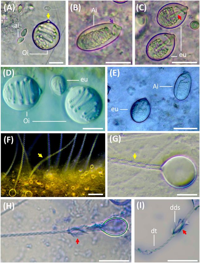
Ballesteros et al.                                                                                          Pelagia noctiluca Morphology and Cnidome

INTRODUCTION                                                                strobilation, gives rise to multiple ephyrae, which will become
                                                                            part of the pelagic life (pelagic life phase). Ephyrae then
The presence of jellyfish blooms has a negative effect on human             grow into sexually reproductive medusae, closing the life cycle
activities (Purcell et al., 2007) and causes socio-economic and             (Fuentes et al., 2011).
public health problems worldwide (Gili and Pagès, 2005; Purcell                 The alternation between sexual and asexual generations does
et al., 2007; Kingsford et al., 2018). Human activities causing             not always occur in scyphozoans (Helm, 2018). Planulae can
eutrophication, global warming, overfishing and increasing                  metamorphose directly to ephyrae, thus lacking the polyp phase.
coastal constructions are considered possible significant reasons           This is the case for Pelagia noctiluca (Rottini Sandrini and Avian,
for jellyfish outbreaks (Purcell et al., 2007; Richardson et al.,           1983; Canepa et al., 2014; Helm et al., 2015; Ramondenc et al.,
2009). Each year, several jellyfish swarms are identified in the            2019), one of the few scyphozoans with life cycle variations
Mediterranean Sea (Brotz and Pauly, 2012) and a possible long-              (Helm, 2018) and the only species within the Pelagiidae family
term increase of this phenomenon has been reported (Brotz et al.,           (Helm et al., 2015). The holoplanktonic life cycle of P. noctiluca is
2012). A first step in the early detection of jellyfish blooms is the       an important difference from the metagenetic life cycle of other
identification of the early life stages (Straehler-Pohl and Jarms,          common Mediterranean jellyfish such as Cotylorhiza tuberculata,
2010; Holst, 2012). Under this premise, an identification system,           Rhizostoma pulmo, and Aurelia aurita (Kikinger, 1992; Fuentes
based on useful keys that describe distinctive morphological                et al., 2011; Matveev et al., 2012). Therefore, studying their
features between early stages of scyphozoan species, has been               holoplanktonic life cycle provides a unique opportunity to
established in the last decade (Straehler-Pohl and Jarms, 2010;             understand the evolutionary simplification and its developmental
Holst, 2012).                                                               implications (Helm et al., 2015).
    Jellyfish stings not only cause discomfort for beach users                  The mauve stinger, P. noctiluca, is recognized as the most
(Cegolon et al., 2013) but also problems for fishers, who may               important jellyfish in the Mediterranean Sea (Brotz and Pauly,
be stung while taking in fishing nets containing numerous                   2012; Canepa et al., 2014) due to its abundance along the basin
jellyfish (Al-Rubiay et al., 2009). Tourism and fisheries are the           and the severity of its sting (Mariottini et al., 2008; Marambio
industries most affected by jellyfish blooms (Purcell et al., 2007).        et al., 2021b). Generally, the sting produces local symptoms such
Though a diversity of jellyfish species exists, like all cnidarians,        as severe pain, burning sensation, erythema, edema and vesicles
they share the same fundamental stinging mechanism involving                (Cegolon et al., 2013; Montgomery et al., 2016; Hall, 2018). As
injection of prey and predator with venom-filled stinging                   systemic symptoms are uncommon, P. noctiluca is considered a
cells called cnidocytes (Mariscal, 1974). Cnidocytes contain                non-life-threatening species (Cegolon et al., 2013). Owing to the
subcellular capsules called cnidocysts, which can be classified             high number of stings seen by lifeguard services (De Donno et al.,
into three main categories: nematocyst, spirocyst and ptychocyst            2014), the efforts of the scientific community are focused on sting
(Mariscal, 1974; Watson and Wood, 1988). The categories of                  prevention and management of the mauve stinger (Morabito
spirocyst and ptychocyst each comprise a specific cnidocyst                 et al., 2014, 2020; Hall, 2018; Ballesteros et al., 2021).
type, but over thirty different cnidocyst types are classified as               The life cycle of P. noctiluca was described in the monograph
nematocysts (Mariscal, 1974), some of which, due to capsule size            ‘Medusae of the British Isles’ (Russell, 1970). The simple,
and shape, tubule length and pattern, including spines, can be              schematic drawings, especially of the early developmental stages,
differentiated into subtypes (Östman, 2000). Some nematocyst                are difficult to compare with living or preserved individuals
types are considered taxonomically diagnostic, whereas others are           in plankton samples (Holst, 2012). Despite the notoriety of
common across jellyfish species (Fautin, 2009). The cnidome, the            P. noctiluca in the Mediterranean Sea (Brotz and Pauly, 2012;
term for the total complement of cnidocytes within a cnidarian              Canepa et al., 2014) and elsewhere (Mariottini et al., 2008), this
specimen (Heins et al., 2015), including their cnidocyst size,              species has not been included in the current reference studies for
abundance and distribution during each developmental life stage,            identification of early planktonic scyphozoan stages (Straehler-
is essential in taxonomic descriptions (Weill, 1934; Calder,                Pohl and Jarms, 2010; Straehler-Pohl et al., 2011; Holst, 2012).
1983; Östman, 2000; Fautin, 2009). The criteria for nematocyst                  Pelagia noctiluca is a non-selective predator (Milisenda
classification have changed with the advent of the scanning                 et al., 2018). Its nematocysts capture a wide variety of prey,
electron microscope (SEM) and improved light microscopic                    copepods being one of the main items (Sabatés et al., 2010;
(LM) resolution (Shostak and Kolluri, 1995). Advances in                    Tilves et al., 2016). While the nematocysts types of adult
microscopic observation techniques have revealed errors in the              P. noctiluca were previously identified (Krasinska, 1914; Weill,
traditional nomenclature (Östman, 2000), hence the need to                  1934; Quadrifoglio et al., 1986; Larson, 1987; Avian et al., 1991;
re-examine and reclassify some cnidocytes.                                  Marchini et al., 2004; Sánchez-Rodríguez and Lucio-Martínez,
    Scyphozoan life cycles vary among species from metagenetic              2011), the complete and detailed cnidome for the entire life cycle
to holoplanktonic (Ceh et al., 2015). A metagenetic life                    has never been described.
cycle implies an alternation between different life forms and                   To provide morphological information on the early life stages
reproductive models (Ceh et al., 2015). Fertilized eggs from adult          and cnidome of P. noctiluca, the goals of our study were to:
medusae metamorphose to planulae, which attach to benthic                   (I) re-examine and classify the nematocysts, applying current
substrates, giving rise to an asexually reproducing polyp (benthic          nematocyst classifications, (II) describe the developmental stages
life phase) (Fuentes et al., 2011). In optimal environmental                during the life cycle, including the cnidome as an important
conditions (Ceh et al., 2015), perpendicular polyp fission, called          taxonomic characteristic and (III) analyze the nematocyst capsule

Frontiers in Marine Science | www.frontiersin.org                       2                                      August 2021 | Volume 8 | Article 714503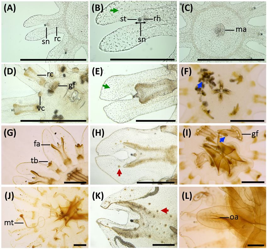
Ballesteros et al.                                                                                                         Pelagia noctiluca Morphology and Cnidome

volumes in relation to body diameter during the different                                 Individuals were fed daily, with a varied diet according to the
development stages.                                                                   different life cycle stages. Ephyrae and metaephyrae were fed with
                                                                                      Brachiounus sp. cultures, newly hatched Artemia sp., chopped
                                                                                      nauplii and Aurelia sp. mucus. Young medusae were fed with
MATERIALS AND METHODS                                                                 Artemia sp. nauplii and small pieces of the jellyfish Aurelia sp.
                                                                                      For the most developed juveniles and adult medusae, eggs of
Culture and Growth of Pelagia noctiluca                                               Merluccius merluccius, Parapenaeus longirostris and pieces of the
Twelve randomly selected, sexually mature jellyfish were                              jellyfish Cassiopea sp. were added to the diet.
collected in April 2019 on the Catalan coast (Port de la Selva,
Spain) during a P. noctiluca bloom, using hand nets and plastic                       Life Cycle Description
jars. The individuals were then transferred to plastic bags full of                   The morphological description was mainly focused on the early
seawater, avoiding air bubbles to ensure good conditions during                       development stages (ephyrae and metaephyrae). On a weekly
transport. The jellyfish were relocated in a kreisel tank (400L                       basis, 15 living individuals were collected from the culture
capacity, 37–38h salinity, 21◦ C temperature and light cycle of                       aquarium for analysis. Individuals were relaxed with a menthol
12 h light/12 h dark) in the Aquarium Experimental Zone (ZAE)                         solution (4% w/v) transferred with the manubrium positioned
at the Institute of Marine Sciences in Barcelona, Spain.                              upward (ventral view) to a glass slide and photographed using
    The next morning, mucus strips with fertilized eggs were                          a binocular loupe (Leica M205 C). The smallest ephyrae were
transferred using a wide plastic pipette to a plastic jug with                        analyzed under a light microscope (ZEISS Axioskop 2 plus) to
natural filtered seawater (37–38h salinity) and abundant                              obtain more accurate images. Afterward, the individuals were
aeration. The plastic jug was situated in a culture room (18◦ C                       carefully returned to the culture.
temperature and light cycle of 12 h light/12 h dark). Within                              To describe the early P. noctiluca stages, terminology
4 days, eggs metamorphosed into planula larvae and later,                             (Figure 1A) and measuring points (Figure 1B) were defined
ephyrae were observed.                                                                for scyphozoan ephyrae as in Straehler-Pohl and Jarms (2010)
    Early life cycle stages (ephyra and metaephyra) were cultured                     and Straehler-Pohl et al. (2011). The total body diameter (TBD),
in plastic jugs with aeration; cultures were placed in the culture                    central disk diameter (CDD), total marginal lappet length
room with the conditions as described above and a complete                            (TMLL), lappet stem length (LStL), rhopalial lappet length (RLL)
cleaning every 3 days, involving removal of individuals one by                        and manubrium length (ML) were measured. In addition, we
one with a plastic pipette and complete replacement of seawater.                      calculated the total marginal lappets length (TMLsL), where s
    For the juvenile and adult medusa stages, open seawater                           stands for the sum of the total marginal lappet length (TMLL) and
circulation in kreisel tanks was selected. The kreisel tank                           the total opposite marginal lappet length (TOMLL) (Figure 1B).
capacities were 50L for young juveniles and 400L for the most                         Data measurements were obtained by photo analysis with the
developed juveniles and adult medusae (37–38h salinity, 21◦ C                         Fiji version of ImageJ software. A total of 30 ephyrae and 30
temperature and light cycle of 12 h light/12 h dark).                                 metaephyrae were measured.

  FIGURE 1 | Anatomy and measuring points defined for ephyra and metaephyra stages. (A) Anatomy of ephyra. (B) Measuring points. CDD, central disk diameter;
  LStL, lappet stem length; ML, manubrium length; RLL, rhopalial lappet length; TBD, total body diameter; TMLL, total marginal lappet length; TMLsL, total marginal
  lappets length; TOMLL, total opposite marginal lappet length. Illustrated by Lau López.

Frontiers in Marine Science | www.frontiersin.org                                 3                                           August 2021 | Volume 8 | Article 714503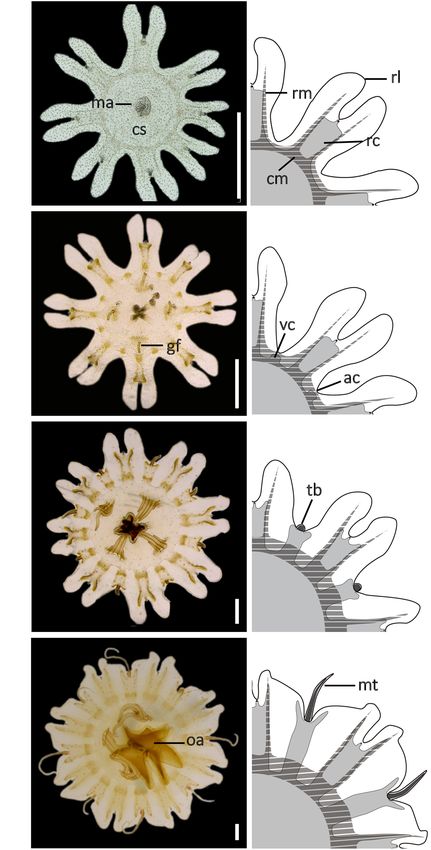
Ballesteros et al.                                                                                              Pelagia noctiluca Morphology and Cnidome

   To compare relative body dimensions during the early
developmental stages, the following ratios (%) were calculated
from measurement data: RLL/TBD × 100, LStL/TBD × 100,
CDD/TBD × 100, RLL/TMLL × 100, LStL/TML × 100,
TMLsL/TBD × 100 and ML/CDD × 100.
   Morphological changes in juvenile and adult medusae, with
a TBD larger than 14.00 mm, were observed directly from
the culture. For detailed analysis, the jellyfish were transferred
to a plastic tray with a small amount of seawater. TBD was
measured with a digital caliper. The individuals were carefully
returned to the culture.

Cnidome Analysis
To describe the cnidome of the early life cycle stages and
the juvenile and adult P. noctiluca medusae, we analyzed
nematocyst distribution, abundance and capsule measurements.
The individuals were not fed overnight. Cnidome observations
were routinely carried out during the early morning hours.
Living ephyrae and metaephyrae were transferred to a
glass slide and covered with a coverslip. Preparations were
examined and photographed with a camera attached to a light
microscope (ZEISS Axioskop 2 plus). In jellyfish with TBD
larger than 14.00 mm, tissues of the body parts exumbrella,
manubrium (oral arms) and marginal tentacles were analyzed.
The TBD was calculated as shown in the morphological
description (Figure 1B).
   Nematocysts were identified according to Weill’s (1934)                FIGURE 2 | Nematocysts during the life cycle of Pelagia noctiluca. Images
classification system with modifications by Mariscal (1974),              from adult medusa, stage 6. (A–E) Undischarged nematocysts. (F–I)
Calder (1974), Östman and Hydman (1997), and Östman                       Discharged nematocysts. (A) O-isorhiza (Oi) with protruding apical capsular
                                                                          tip (yellow arrow) and loosely coiled tubule. Note small a-isorhizas (ai). (B,E)
(2000). The nematocyst nomenclature used was that proposed                A-isorhiza (Ai) with a closely packed tubule, only becoming more loosely
by Watson and Wood (1988). Undischarged capsules were                     coiled apically (violet arrow). (C) Eurytele (eu) with visible inverted shaft (red
measured by photo analysis using the Fiji version of ImageJ               arrow). (D). O-isorhizas (Oi) and euryteles (eu). (F,G) Discharged O-isorhizas
software. Capsule measurements were taken for capsule length              with homotrichous spines in three helical spine-rows (yellow arrows) along the
(CL), capsule width (CW) and capsule volume (CV). As                      tubule. (H,I) Discharged euryteles with distal dilatations of shafts (dds) armed
                                                                          with heterotrichous spines (red arrows) and helical spine pattern on distal
described by Östman and Hydman (1997), the protruding apical              tubule (dt). Note proximal, spineless, narrow shaft in (I). ai, a-isorhiza; Ai,
capsular tip was included in the capsule length measurements              A-isorhiza; dt, distal tubule; dds, distal dilatation of shaft; Oi, O-isorhiza; eu,
(Figure 2A, yellow arrow). Only capsules in a horizontal                  eurytele. Scale bars: 10 µm, scale bar F: 40 µm. (A–D,F–I) photographed by
position showing the protruding apical capsular tip were                  Rubén Duro.
measured. The nematocyst size was calculated from CV as in
Purcell and Mills (1988):
                                                                         Statistical Analyses and Graphs
                                    4
                                CV = π ab2                               For a more detailed assessment of the possible differences
                                    3                                    observed in the cnidome composition between body parts along
where a is the radius of capsule length and b is the radius              the life cycle of P. noctiluca, CV values were grouped by life
of capsule width.                                                        cycle stage (as defined in Section “Morphology and Cnidome
    In total, 3440 nematocysts (1389 a-isorhizas, 318 A-isorhizas,       Descriptions”) and tested for statistical differences between
246 O-isorhizas and 1487 euryteles) from 24 individuals in               groups. All data was first tested for normality and homogeneity
different life cycle stages were examined.                               using the shapiro.test and barlett.test functions availed as part of
    To study the development of nematocyst batteries during the          the basic stats package from the R software platform (R Core
life cycle, the relative abundance (RA) of different nematocyst          Team, 2017). An ANOVA test was performed using the aov
types in the batteries was analyzed in the exumbrella, manubrium         function of the stats package to test for possible differences in
(oral arms) and marginal tentacles. RA was calculated as:                CV in each nematocyst type between: (I) body parts and life
                                                                         cycle stages, (II) body parts within the same life cycle stage
                                        NNT
                               RA =         100                          and (III) life cycle stages (without considering body parts). To
                                        NTN                              determine between which groups those differences occurred,
where NNT is the total number of nematocysts of a single type            post-hoc analyses were performed using the pairwise.t.test
and NTN is the total number of nematocysts in the battery.               function of the stats package.

Frontiers in Marine Science | www.frontiersin.org                    4                                              August 2021 | Volume 8 | Article 714503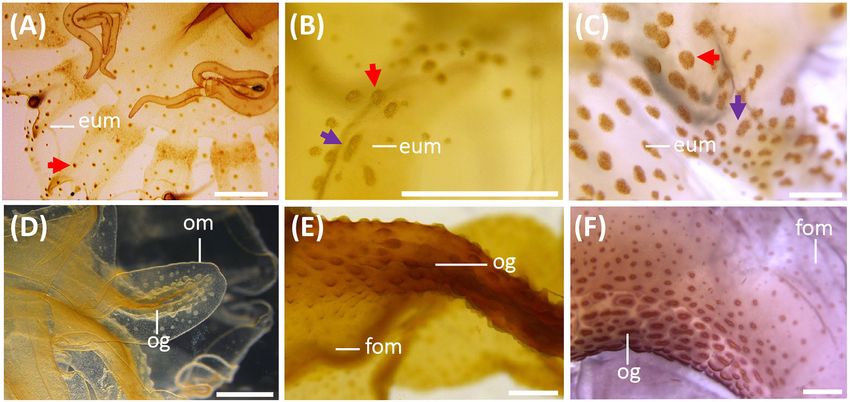
Ballesteros et al. Pelagia noctiluca Morphology and Cnidome
Boxplots were produced to graphically represent differences in
CV between nematocyst types and body parts during the different
life cycle stages of P. noctiluca.
To represent the relative abundance of different nematocyst
types in batteries, data were presented in bar graphs for each
analyzed body part during the life cycle stages.
Finally, to show the differences in CV throughout the
P. noctiluca life cycle for each nematocyst type, data
were represented using scatterplots for each nematocyst
type according to their development stage. Regression
lines were added.
RESULTS
Nematocyst Identification
The cnidome of P. noctiluca included four different nematocyst
types (Figures 2A–C). Three types of homotrichous isorhiza
(haplonemes) (Figures 2A,B,F,G) were identified based on the
isodiametric tubule, armed with spines of similar size and shape,
and lack of well-defined shaft tubule. The fourth nematocyst
type was classified as a heterotrichous microbasic eurytele
(heteroneme), based on the shaft (shorter than 1.5 times the
capsule length) with a clear enlargement of the basal tubule
(Figure 2C, red arrow), and a distal dilation armed with
prominent spines of different sizes and shapes, in contrast to the
smaller spines on the tubule (Figures 2H,I, red arrows).
Other differentiating nematocyst features, such as the
capsule volume and shape, pattern of inverted, coiled
tubule, and spine pattern on everted tubule and shaft
(Figures 2A,B,G), separate a-isorhiza, A-isorhiza and O-isorhiza
from each other and from the eurytele. The small a-isorhiza
has an oval capsule with regular, horizontally, closely packed
tubule coils, except for a few separate coils near the operculum
(Figure 2A). The A-isorhiza capsule is large and oval with the
tubule closely packed in irregular coils, except apically where
it is coiled loosely (Figure 2B, violet arrow). The O-isorhiza
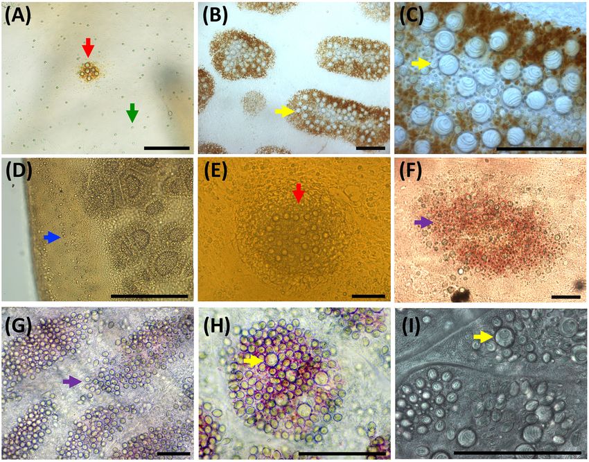
FIGURE 3 | Development of the early stages during the life cycle of Pelagia
capsule is sub-spherical with the broad tubule in regular coils at
noctiluca. (A,B) Ephyra, stage 1. (C,D) Ephyra, stage 2. Note the difference in
a right angle to the capsule axis (Figures 2A,D). The discharged the presence of gastric filaments and velar canals. (E,F) Metaephyra, stage 3.
O-isorhiza shows three helical rows of homotrichous spines on (G,H) Metaephyra, stage 4. (A,C,E,G) Ephyra and metaephyra, ventral view.
the isodiametric tubule (Figures 2F,G, yellow arrows). (B,D,F,H) Ephyra and metaephyra, body quadrant illustrations. White,
The lemon-shaped eurytele capsule has an inverted rod-shaped subumbrella; light gray, gastric system without marginal tentacles; dark gray,
gastric canals inside marginal tentacles; striped, muscular system. ac,
shaft (Figure 2C, red arrow), surrounded by regular tubule coils.
adradial cleft; cm, circular muscle; cs, central stomach; gf, gastric filament;
The discharged eurytele has a differentiated spineless shaft in the ma, manubrium; mt, marginal tentacle; rc, rhopalial canal; rl, rhopalial lappet;
proximal part, narrower than the distal shaft dilatation armed rm, radial muscle; oa, oral arm; tb, tentacle buds; vc, velar canal. Scale bars:
with prominent spines (Figures 2H,I, red arrows). The narrow 1 mm. Illustrated by Lau López.
tubule is armed with small spines.
Morphology and Cnidome Descriptions clefts (Figure 1A). Each marginal lappet is forked distally into
Planula Larva Stage a pair of rhopalial lappets (Figure 3B). A single rhopalium is
Stage 1: The larvae are cone-shaped with a cruciform present per lappet stem, between the pair of rhopalial lappets
manubrium. Only a-isorhizas, scattered in great quantities inside the sense niche (Figures 4A,B). A statocyst, composed
throughout the ectoderm, are identified. of reflective crystals, appears at the rhopalium tip (Figure 4B).
The ephyrae consist of four morphologically identical quadrants
Ephyra Stages (Figures 3B,D). Two different ephyra stages are identified mainly
Ephyrae (Figures 3A–D) present octoradiate bell symmetry, based on the development of the gastric system including absence
formed by eight marginal lappets separated by U-shaped adradial or presence of velar canals and gastric filaments (Figures 3A–C).
Frontiers in Marine Science | www.frontiersin.org 5 August 2021 | Volume 8 | Article 714503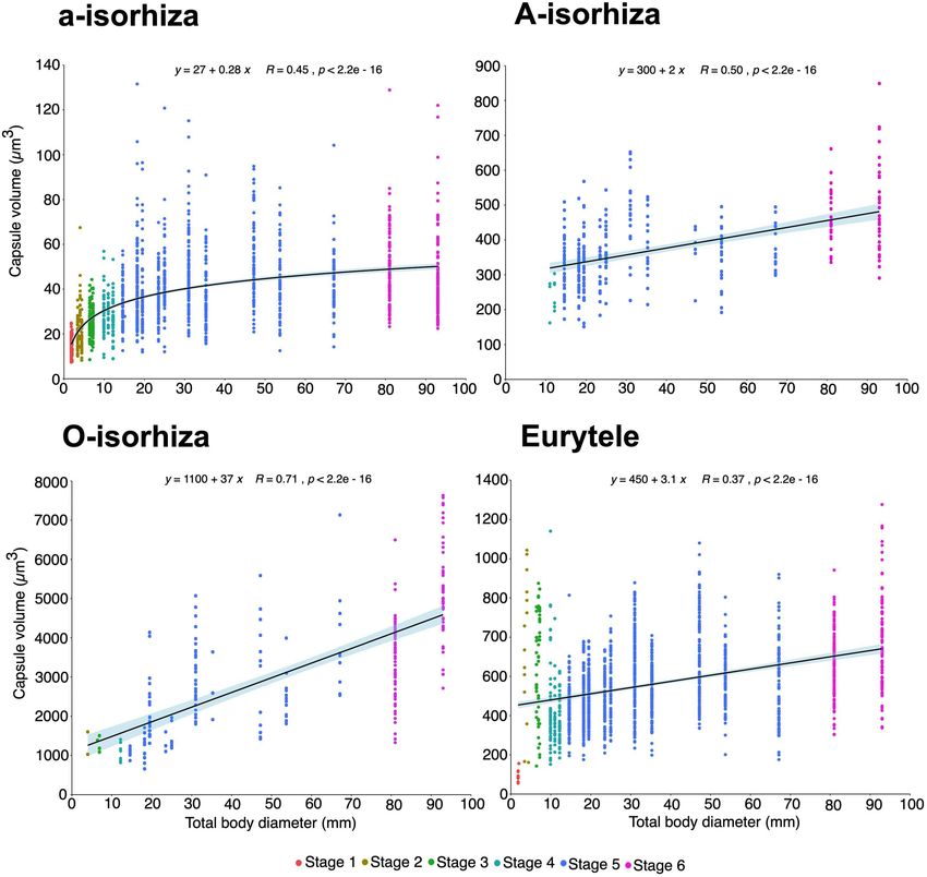
Ballesteros et al.                                                                                                         Pelagia noctiluca Morphology and Cnidome

   Stage 1 (Figures 3A,B, 4A–C): Newly developed ephyrae are                                irregularly distributed euryteles. In the manubrium and nearby
transparent, with round to oval spoon-shaped rhopalial lappets                              areas, a-isorhizas are also present (Table 2). Nematocyst clusters
with rounded tips (Figure 4B). The TBD ranges between 1.80 and                              (batteries/warts) are not identified.
3.00 mm with a mean TBD of ∼ 2.50 mm (Table 1). Additional                                     Stage 2 (Figures 3C,D, 4D–F): Ephyrae are transparent and
measurements are given in Table 1.                                                          brown to ocher-colored, present in areas near the rhopalial
                                                                                            canals, velar canals and at the base of the gastric filament
Gastric system
                                                                                            quadrants. Rhopalial lappets are oval, spoon-shaped to bread
Eight unforked, spade-shaped rhopalial canals (gastric pouches)
                                                                                            knife-shaped with rounded tips (Figure 4E). The ephyrae grow
connect with the central stomach. The terminal rhopalial canal
                                                                                            during the first week reaching between 3.10 and 5.00 mm with
ends at the rhopalium base (Figures 4A,B). Radial muscles, along
                                                                                            a mean TBD of ∼ 4.00 mm (Table 1). Additional measurements
the rhopalial canals, unite with the weakly developed circular
                                                                                            are given in Table 1.
muscle around the central stomach (Figure 3B). In the center
of the gastric cavity there is a cruciform-shaped manubrium                                 Gastric system
(Figures 3A, 4C) with no gastric filaments (Figures 3A, 4C).                                Eight unforked, spade-shaped velar canals (gastric pouches) are
                                                                                            identified (Figures 3D, 4D). Velar canals are connected with
Cnidome
                                                                                            each other and with the rhopalial canals and central stomach.
Single nematocysts are randomly distributed throughout the
                                                                                            Rhopalial canals are unforked and spade-shaped (Figures 4D,E).
ephyra (Figure 4B, green arrow). Abundant small a-isorhizas
                                                                                            In each of the four quadrants, 1 to 3 gastric filaments are present
dominate in the exumbrella ectoderm, followed by a few
                                                                                            above the central stomach (Figures 3C, 4D) with a cruciform-
                                                                                            shaped manubrium (Figure 4F).
                                                                                            Cnidome
                                                                                            Abundant small, single a-isorhizas are randomly scattered
                                                                                            dominating the exumbrella, followed by some O-isorhizas and
                                                                                            euryteles (Figure 4E, green arrows). The manubrium also
                                                                                            contains abundant a-isorhizas. Euryteles are the only nematocyst
                                                                                            type in the gastric filaments (Table 2).

                                                                                            Metaephyra Stages
                                                                                            Metaephyrae (Figures 3E–H) show some differences from
                                                                                            the ephyrae anatomy (Figures 3A–D). Umbrella development
                                                                                            causes some significant changes in the body proportions: CDD
                                                                                            increases and TMLsL decreases with respect to TBD. The
                                                                                            proportion of ML to CDD was closely similar to the ephyra
                                                                                            stages, with the manubrium growing proportionally to the
                                                                                            growth of the individuals in the early stages. As with ephyrae,
                                                                                            the two differentiated metaephyrae are split into 4 identical
                                                                                            morphological quadrants (Figures 3F,H).
                                                                                               Stage 3 (Figures 3E,F, 4G–I): Metaephyrae are faint brown to
                                                                                            ocher-colored. The terminal end of the velar canal and the base
  FIGURE 4 | Morphological changes, development of gastric system and
                                                                                            of the rhopalial canals have an intense brownish color, forming
  cnidae in early stages of the life cycle of Pelagia noctiluca. (A–C) Ephyra,              a distinct ring above the circular muscle. The manubrium is
  stage 1. (D–F) Ephyra, stage 2. (G–I) Metaephyra, stage 3. (J–L) Metaephyra,              completely brown. The oval, rhopalial lappets are spoon-shaped
  stage 4. (A,D,G,J) Rhopalial and velar canals. (B,E,H,K) Rhopalial canals and             to bread knife-shaped with rounded tips (Figure 4H). The
  nematocysts in exumbrella. (C,F,I,L) Central disk. (A) Spade-shaped rhopalial
                                                                                            TBD ranges between 5.10 and 8.00 mm with a mean TBD of
  canals (rc) and sense niche (sn). (B) Sense niche composed of the rhopalium
  (rh) and the statocyst (st) at the tip. (C) Central stomach with manubrium (ma).          ∼ 7.10 mm (Table 1). Additional measurements are given in
  (D) Rhopalial canal (rc) and velar canal (vc). Note initial gastric filaments (gf).       Table 1.
  (E) Spade-shaped rhopalial canal. (F) Brachiounus sp. (blue arrow) inside the
  gastric system. (G) Tentacle buds (tb) above velar canals. (G,H) Fork arms (fa)           Gastric system
  of rhopalium slightly overtop rhopalium. (I) Artemia sp. (blue arrow) caught by           Rhopalial canals are forked. Fork arms present with rounded
  euryteles in gastric filaments (gf). (J) Marginal tentacles (mt) growing                  tips, growing centrifugally into rhopalial lappets from level with
  simultaneously. (K) Sharp-shaped rhopalial canals. (L) Initial oral arms (oa).
  (B,E) Abundant single nematocysts (green arrows). Note absence of
                                                                                            to slightly overstepping the rhopalium tip (Figures 3F, 4G,H).
  nematocyst batteries. (H) First homogenous, round-shaped nematocyst                       Slightly forked velar canals with rounded tips grow in a
  batteries (red arrow). (K) Nematocyst batteries increase in numbers (red                  centrifugal direction until almost reaching the umbrella rim
  arrow), note position close to rhopalial canal. fa, fork arm; gf, gastric filament;       (U-shaped adradial cleft) (Figures 3F, 4G). Eight tentacle buds
  ma, manubrium; mt, marginal tentacle; rc, rhopalial canal; rh, rhopalium; oa,
                                                                                            or incipient tentacles lie above the velar canals (Figures 3F, 4G).
  oral arm; sc, statocyst; sn, sense niche; tb, tentacle bud; vc, velar canal.
  Scale bars: 1 mm, scale bars (B,E): 0.5 mm.
                                                                                            Each body quadrant is formed by 3 to 5 gastric filaments. The
                                                                                            manubrium starts to grow longitudinally (Figure 4I).

Frontiers in Marine Science | www.frontiersin.org                                       6                                     August 2021 | Volume 8 | Article 714503
Ballesteros et al.                                                                                                              Pelagia noctiluca Morphology and Cnidome

TABLE 1 | Morphology, body proportions and cnidome of the early stages of Pelagia noctiluca life cycle.

                                   Ephyra                                                                             Metaephyra

Stage 1                                  Stage 2                                Stage 3                                         Stage 4

TBD range: 1.80–3.00 mm, TBD mean: TBD range: 3.10–5.00 mm, TBD                 TBD range: 5.10–8.00 mm, TBD                    TBD range: 8.10–14.00 mm, TBD
∼ 2.50 mm                          mean: ∼ 4.00 mm                              mean: ∼ 7.10 mm                                 mean: ∼ 11.70 mm
Anatomy:                                 Anatomy:                               Anatomy:                                        Anatomy:
8 marginal lappets                       8 marginal lappets                     8 marginal lappets                              8 marginal lappets
16 round to oval spoon-like rhopalial    16 oval spoon- to bread knife-shaped   16 oval spoon- to bread knife-shaped            16 oval spoon- to bread knife-shaped
lappets, round tips                      rhopalial lappets, round tips          rhopalial lappets, round tips                   broad rhopalial lappets, round tips
No gastric filaments                     1–3 gastric filaments per quadrant     3–5 gastric filaments per quadrant              5–8 gastric filaments per quadrant
Long ML, approx. 40% of CDD              Long ML, approx. 39% of CDD            Tentacle buds or incipient marginal             8 marginal tentacles
(SD = 0.03)                              (SD = 0.05)                            tentacles                                       Long ML, approx. 39% of CDD (SD = 0.04)
                                                                                Long ML, approx. 37% of CDD (SD = 0.03)
Gastric system:                          Gastric system:                        Gastric system:                                 Gastric system:
Rhopalial canals unforked,               Rhopalial canals unforked,             Rhopalial canals forked, round-shaped, fork     Rhopalial canals forked: round to
spade-shaped                             spade-shaped                           tips level to slightly overtop rhopalium tip    sharp-shaped, fork tips overtop rhopalium
No velar canals                          Velar canals unforked, spade-shaped    Velar canals slightly forked, round fork tips   Velar canals forked, round fork tips
Mean body proportions:                   Mean body proportions:                 Mean body proportions:                          Mean body proportions:
RLL ≈ 14% of TBD (SD = 0.01)             RLL ≈ 13% of TBD (SD = 0.01)           RLL ≈ 11% of TBD (SD = 0.01)                    RLL ≈ 9% of TBD (SD = 0.02)
LStL ≈ 13% of TBD (SD = 0.01)            LStL ≈ 13% of TBD (SD = 0.02)          LStL ≈ 8% of TBD (SD = 0.01)                    LStL ≈ 5% of TBD (SD = 0.01)
CDD ≈ 47% of TBD (SD = 0.02)             CDD ≈ 48% of TBD (SD = 0.03)           CDD ≈ 64% of TBD (SD = 0.02)                    CDD ≈ 72% of t TBD (SD = 0.04)
TMLsL ≈ 53% of TBD (SD = 0.02)           TMLsL ≈ 52% of TBD (SD = 0.03)         TMLsL ≈ 36% of TBD (SD = 0.02)                  TMLsL ≈ 28% of TBD (SD = 0.04)
LStL ≈ 47% of TMLL (SD = 0.03)           LStL ≈ 50% of TMLL (SD = 0.05)         LStL ≈ 42% of TMLL (SD = 0.04)                  LStL ≈ 39% of TMLL (SD = 0.05)
RLL ≈ 53% of TMLL (SD = 0.03)            RLL ≈ 50% of TMLL (SD = 0.05)          RLL ≈ 58% of TMLL (SD = 0.08)                   RLL ≈ 61% of TMLL (SD = 0.05)
Cnidome:                                 Cnidome:                               Cnidome:                                        Cnidome:
Exumbrella: randomly scattered small     Exumbrella: randomly scattered small   Exumbrella: randomly scattered small            Exumbrella: randomly scattered small
a-isorhizas dominate, followed by some   a-isorhizas dominate, followed by      a-isorhizas dominate, followed by some          a-isorhizas dominate, followed by some
euryteles                                some euryteles                         O-isorhizas and euryteles                       O-isorhizas,
No nematocyst batteries                  No nematocyst batteries                Small nematocyst batteries                      A-isorhizas and euryteles
                                                                                                                                Nematocyst batteries increase in number

CDD, central disk diameter; LStL, lappet stem length; ML, manubrium length; RLL, rhopalial lappet length; SD, standard deviation; TBD, total body diameter; TMLL, total
marginal lappet length; TMLsL, total marginal lappets length. Scale bars: 1 mm. Illustrated by Lau López.

Cnidome                                                                                a mean TBD of ∼ 11.70 mm (Table 1). Additional measurements
Several homogenous, round-shaped nematocyst batteries appear                           given in Table 1.
as protrusions in the exumbrella ectoderm (Figure 4H,
                                                                                       Gastric system
red arrow). Among the abundant scattered individual
                                                                                       Fork arms of rhopalial canals grow further into the rhopalial
nematocysts, a-isorhizas dominate, followed by O-isorhizas
                                                                                       lappets. Their round to sharp-shaped fork tips overstep
and euryteles (Figure 5A). A strong association is observed
                                                                                       the rhopalium tip (Figures 3H, 4K). Velar canals, forked
between position and size of nematocyst warts and degree
                                                                                       with round-shaped tips, grow in a centrifugal direction into
of pigmentation, from brown to ochre (Figure 4H). Two
                                                                                       marginal lappets, following the complete umbrella shape
parallel rows of nematocyst warts and pigment appears
                                                                                       (Figure 3H) with widening marginal lappets (Figures 3G,H).
at the base of both rhopalial and velar canals, sometimes
                                                                                       Nearly simultaneously, all marginal tentacles grow as extensions
reaching the umbrella margin (Figure 4H). Scattered
                                                                                       to velar canals and parallel to marginal lappets (Figures 3G,H).
nematocyst clusters are also present around the central
                                                                                       Each body quadrant has 5 to 8 gastric filaments. The
stomach. In more developed individuals, a few nematocyst
                                                                                       manubrium is divided distally into 4 incipient oral arms
batteries are identified at the incipient manubrium base,
                                                                                       (Figure 4L), in which an oral groove and thin oral margin are
while a-isorhizas are abundant and randomly distributed.
                                                                                       differentiated (Figure 6D).
Euryteles are still the only nematocyst type present in the gastric
filaments (Table 2).                                                                   Cnidome
    Stage 4 (Figures 3G,H, 4J–L): Coloration is similar to                             Copious, scattered, small a-isorhiza dominate in the exumbrella
the previous stage. Rhopalial lappets are bread knife-shaped                           (Figure 7A, red color). Warts, more obvious than in the
(Figure 4K). The TBD ranged between of 8.10 and 14.00 mm with                          previous stage, increase in nematocyst number, size and

Frontiers in Marine Science | www.frontiersin.org                                  7                                              August 2021 | Volume 8 | Article 714503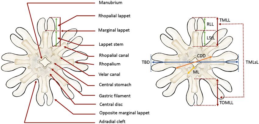
Ballesteros et al. Pelagia noctiluca Morphology and Cnidome
TABLE 2 | Nematocyst capsule, abundance and measurements from different body parts in each stage during Pelagia noctiluca life cycle.
Stage Body Nematocyst Abundance Capsule Capsule Capsule Capsule Volume Capsule
(number of parts type (number) Length Width Length × Width Mean ± SD (µm3 ) Volume
specimens) Mean ± SD Mean ± SD Range (µm) Range (µm3 )
(µm) (µm)
Planula larva 0 Whole a-isorhiza (−) ++++ − − − − −
(3) body
Ephyra 1 Exumbrella a-isorhiza (83) ++++ 3.70 ± 0.39 2.79 ± 0.31 2.24–4.97 × 2.11–3.65 15.42 ± 4.16 7.40–24.87
(3) A-isorhiza (−) absent − − − – −
O-isorhiza (−) absent − − − – −
Eurytele (10) + 6.38 ± 0.74 5.22 ± 0.52 5.12–7.99 × 4.51–6.10 93.38 ± 29.33 55.35–155.80
Manubrium a-isorhiza (11) ++ 3.66 ± 0.33 2.70 ± 0.37 3.16–4.31 × 2.10–3.24 14.39 ± 4.69 7.64–20.45
A-isorhiza (−) absent − − − − −
O-isorhiza (−) absent − − − − −
Eurytele (−) absent − − − − −
Ephyra 2 Exumbrella a-isorhiza (93) ++++ 4.27 ± 0.49 2.93 ± 0.45 3.15–6.02 × 2.00–4.63 20.09 ± 8.82 8.26–67.45
(3) A-isorhiza (−) absent − − − − −
O-isorhiza (3) rare to + 14.52 ± 1.43 13.05 ± 1.40 13.51–15.53 × 12.06– 1315.87 ± 405.92 1028.84–
14.04 1602.40
Eurytele (3) + 9.30 ± 2.01 6.69 ± 0.85 8.09–11.62 × 6.13– 228.42 ± 112.18 161.34–357.93
7.67
Manubrium a-isorhiza (17) +++ 4.35 ± 0.47 3.36 ± 0.39 3.37–5.36 × 2.67–4.23 26.33 ± 7.10 12.58–37.19
A-isorhiza (−) absent − − − − −
O-isorhiza (−) absent − − − − −
Eurytele (−) absent − − − − −
Gastric a-isorhiza (−) absent − − − − −
filaments A-isorhiza (−) absent − − − − −
O-isorhiza (−) absent − − − − −
Eurytele (11) exclusive 14.16 ± 1.25 10.89 ± 1.13 12.18–16.24 × 9.03– 901.77 ± 251.05 520.02–
12.19 1243.65
Metaephyra 3 Exumbrella a-isorhiza (111) ++++ 4.64 ± 0.38 3.18 ± 0.33 3.43–5.51 × 2.55–3.99 25.07 ± 6.40 12.63–44.27
(3) A-isorhiza (−) absent − − − − −
O-isorhiza (4) + 14.31 ± 0.90 13.10 ± 0.71 1.05–15.15 × 12.37– 901.77 ± 251.05 1089.98–
13.79 1507.11
Eurytele (18) ++ 10.66 ± 1.58 8.17 ± 1.38 8.11–13.93 × 5.80– 400.00 ± 196.21 142.89–894.65
11.08
Manubrium a-isorhiza (16) +++ 4.54 ± 0.46 3.37 ± 0.44 3.63–5.33 × 2.12–3.87 28.03 ± 8.44 8.54–40.51
A-isorhiza (−) absent − − − − −
A-isorhiza (−) absent − − − − −
Eurytele (−) absent − − − − −
Gastric a-isorhiza (−) absent − − − − −
filaments A-isorhiza (−) absent − − − − −
O-isorhiza (−) absent − − − − −
Eurytele (35) exclusive 13.92 ± 1.27 10.35 ± 0.83 11.06–16.15 × 8.33– 794.39 ± 180.07 486.82–
11.50 1074.87
Metaephyra 4 Exumbrella a-isorhiza (58) ++++ 4.77 ± 0.46 3.35 ± 0.32 3.76–5.88 × 2.58–4.24 28.45 ± 6.81 16.74–53.26
(3) A-isorhiza (3) rare to + 10.02 ± 1.00 6.62 ± 0.64 8.89–10.76 × 5.90– 234.25 ± 62.74 161.98–274.85
7.10
O-isorhiza (3) absent to + 13.97 ± 1.11 13.26 ± 0.36 13.018– 1289.91 ± 172.06 1168.25–
14.75 × 13.01–13.52 1411.58
Eurytele (50) +++ 10.83 ± 1.23 8.15 ± 1.00 8.61–14.19 × 6.18– 391.54 ± 143.02 172.40–839.77
10.63
Manubrium a-isorhiza (17) ++++ 4.90 ± 0.52 3.38 ± 0.28 3.99–5.64 × 3.04–3.85 29.54 ± 6.16 19.94–42.45
A-isorhiza (6) absent to + 9.90 ± 0.47 6.87 ± 0.56 9.16–10.46 × 6.21–7.45 246.18 ± 43.56 196.47–303.98
O-isorhiza (5) absent to + 13.64 ± 0.80 11.85 ± 0.90 12.65–14.79 × 11.09– 1014.04 ± 212.55 1332.93–
13.12 5070.18
Eurytele (18) +++ 11.46 ± 0.77 7.73 ± 0.53 10.46–12.82 × 6.54– 361.38 ± 64.65 238.51–478.16
8.86
(Continued)
Frontiers in Marine Science | www.frontiersin.org 8 August 2021 | Volume 8 | Article 714503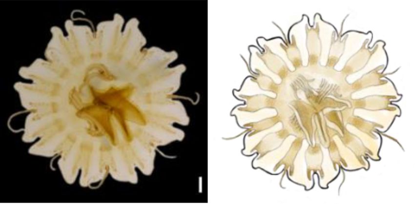
Ballesteros et al. Pelagia noctiluca Morphology and Cnidome
TABLE 2 | Continued
Stage Body Nematocyst Abundance Capsule Capsule Capsule Capsule Volume Capsule
(number of parts type (number) Length Width Length × Width Mean ± SD (µm3 ) Volume
specimens) Mean ± SD Mean ± SD Range (µm) Range (µm3 )
(µm) (µm)
Metaephyra 4 Gastric a-isorhiza (−) absent − − − − −
(3) filaments A-isorhiza (−) absent − − − − −
O-isorhiza (−) absent − − − − −
Eurytele (19) exclusive 13.92 ± 1.27 10.35 ± 0.83 11.06–16.15 × 8.33– 794.39 ± 180.07 486.82–
11.50 1074.87
Tentacles a-isorhiza (33) ++++ 4.62 ± 0.71 3.17 ± 0.58 3.44–6.10 × 2.21–4.25 26.22 ± 12.58 8.90–56.89
A-isorhiza (−) absent − − − − −
O-isorhiza (−) absent − − − − −
Eurytele (34) +++ 10.55 ± 0.95 7.59 ± 0.81 8.81–12.28 × 5.73– 326.38 ± 88.04 151.98–499.06
9.15
Juvenile 5 Exumbrella a-isorhiza (117) ++++ 5.52 ± 0.68 3.45 ± 0.54 4.21–7.57 × 2.16–5.41 37.86 ± 15.38 12.57–105.08
(10) A-isorhiza (3) rare to + 10.61 ± 0.40 6.94 ± 0.21 10.32–10.89 × 6.79– 266.87 ± 5.64 262.89–270.86
7.08
O-isorhiza (71) absent to + 18.55 ± 3.04 16.59 ± 2.56 12.34–25.51 × 11.64– 2859.55 ± 12883.25 874.99–
23.10 7130.14
Eurytele (293) +++ 12.72 ± 1.60 9.09 ± 1.32 9.29–15.10 × 5.98– 579.16 ± 228.41 175.60
10.56 1280.17
Manubrium a-isorhiza (230) ++++ 6.04 ± 0.88 3.69 ± 0.51 4.43–8.47 × 2.69–6.29 44.87 ± 17.19 16.78–131.55
A-isorhiza (33) absent to + 11.66 ± 1.43 7.37 ± 0.86 9.25–13.75 × 5.29– 339.90 ± 94.16 150.48–499.95
8.80 65632–
O-isorhiza (60) absent to ++ 16.51 ± 2.39 14.86 ± 2.32 12.11–21.76 × 10.16– 2036.07 ± 920.02 4621.37
20.14
Eurytele (284) +++ 11.82 ± 1.03 8.35 ± 0.78 9.50–15.10 × 5.98– 440.21 ± 111.59 198.48–806.66
10.56
Tentacles a-isorhiza (383) ++++ 5.69 ± 0.75 3.63 ± 0.54 3.85–8.33 × 2.10–5.82 41.20 ± 17.05 11.95–120.77
A-isorhiza (207) absent to ++ 12.37 ± 0.91 7.36 ± 0.79 10.21–14.49 × 4.94– 357.48 ± 92.41 161.77–652.44
9.29
O-isorhiza (1) absent to + 17.46 15.81 17.46–15.81 2285.11 −
Eurytele (510) +++ 12.92 ± 1.16 9.25 ± 0.94 8.99–16.19 × 5.93– 592.64 ± 159.29 175.71–
11.80 1077.70
Adult 6 Exumbrella a-isorhiza (60) ++++ 6.27 ± 0.65 3.53 ± 0.41 4.72–7.27 × 2.69–4.71 41.51 ± 11.04 23.28–79.40
(2) A-isorhiza* rare to + − − − − −
O-isorhiza (64) absent to ++ 21.99 ± 2.08 19.73 ± 2.17 17.87–26.07 × 15.31– 4619.10 ± 1409.00 2433.59–
24.19 7629.99
Eurytele (46) +++ 13.28 ± 1.17 9.54 ± 0.82 10.50–15.36 × 7.42– 644.14 ± 155.14 302.99–993.32
11.45
Manubrium a-isorhiza (53) ++++ 6.69 ± 0.84 4.01 ± 0.54 5.18–8.02 × 3.05–5.39 58.69 ± 21.37 26.65–122.00
A-isorhiza (11) absent to + 12.95 ± 0.61 7.79 ± 0.50 11.65–14.13 × 7.12– 414.60 ± 65.86 323.40–538.50
8.63
O-isorhiza (31) absent to ++ 19.80 ± 2.02 17.55 ± 2.56 15.32–23.56 × 12.80– 3332.99 ± 1194.03 1327.77–
24.02 6495.87
Eurytele (44) +++ 12.69 ± 1.43 9.53 ± 1.34 10.2–16.71 × 7.27– 632.47 ± 262.44 304.97–
12.99 1476.59
Tentacles a-isorhiza (107) ++++ 5.88 ± 0.60 3.70 ± 0.45 4.39–7.70 × 2.99–5.65 43.42 ± 14.99 22.47–128.86
A-isorhiza (55) absent to ++ 14.27 ± 1.38 8.03 ± 0.62 12.51–18.62 × 6.51–9.54 488.81 ± 113.31 290.98–848.24
O-isorhiza (4) absent to + 22.38 ± 0.55 20.65 ± 0.84 21.59–23.13 × 19.80– 5004.16 ± 404.93 4476.70–
21.96 5653.51
Eurytele (112) +++ 14.07 ± 1.21 10.26 ± 0.86 10.72–17.98 × 7.82– 789.22 ± 185.84 343.08–
12.65 1357.32
Abundance: nematocyst densities marked with increasing number + from one to four (+, ++, +++, ++++); absent, no nematocyst type found in investigated body
parts; rare, nematocyst type not found in each investigated body part; exclusive, the only nematocyst type found in the investigated body part. SD, standard deviation.
*Discharged nematocyst observed.
amount. Note differences in Figures 4H,K (red arrows). Near are present in the incipient oral arms (Figures 5B, 6D)
the rhopalial and velar canals and the central stomach, whereas random, scattered a-isorhizas are present in the
a clear nematocyst pattern appears (Figures 4K, 6A), manubrium base. A-isorhizas and O-isorhizas may be
with clusters of a-isorhizas and euryteles, but often some absent or present, but always in low abundances (Figure 5B).
A-isorhizas and O-isorhizas (Figure 5A). Nematocyst clusters Marginal tentacles contain scattered a-isorhizas and euryteles
Frontiers in Marine Science | www.frontiersin.org 9 August 2021 | Volume 8 | Article 714503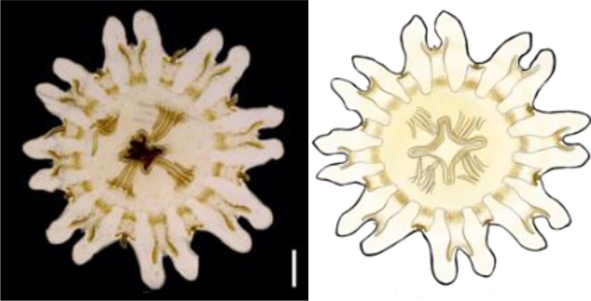
Ballesteros et al.                                                                                                        Pelagia noctiluca Morphology and Cnidome

  FIGURE 5 | Relative abundance of nematocyst types in batteries of different Pelagia noctiluca stages. (A) Exumbrella (50 batteries) (B) Manubrium (40 batteries).
  (C) Tentacles (30 batteries). Note large proportion of a-isorhizas and euryteles, and absence or very low proportion of A-isorhizas and O-isorhizas. Stages:
  metaephyra, stage 3; metaephyra, stage 4; juvenile medusa, stage 5; adult medusa, stage 6. Note: Gastric filaments not included due to the absence of nematocyst
  clusters.

  FIGURE 6 | Nematocyst batteries on exumbrella (eum) (A–C) and on oral arms (D–F) during the life cycle of Pelagia noctiluca. (A,D) Metaephyra, stage 4. (B,E)
  Juvenile medusa, stage 5. (C,F) Adult medusa, stage 6. (A) Homogenous, round-shaped batteries (red arrow). (B,C) Heterogeneous-shaped clusters, from round to
  oval (red arrow), narrow to oval-shaped (violet arrow). (D–F) High abundance of nematocyst warts throughout length of oral groove (og) compared to frilled oral
  margin (fom). eum, exumbrella margin; fom, frilled oral margin; om, oral margin; og, oral groove. Scale bars: 1 mm.

(Table 2). Euryteles are the only nematocyst type in the gastric                      grow, coloration changes, and young medusae turn intense
filaments (Table 2).                                                                  brown. The nematocyst battery pigmentation appears less
                                                                                      prominent due to less contrast between the brown medusa
Juvenile Medusa Stage                                                                 color and the brown nematocyst warts (Figures 6B,E). As
Stage 5 (Figure 8): The juvenile medusa stage displays                                the juvenile reaches the last medusa stage (Figures 6C,F),
a large TBD range (14.10–70.00 mm). As the medusae                                    the intense brown coloration attenuates and the pigmented

Frontiers in Marine Science | www.frontiersin.org                                10                                          August 2021 | Volume 8 | Article 714503
Ballesteros et al. Pelagia noctiluca Morphology and Cnidome
FIGURE 7 | Nematocyst clusters in different body parts. (A–C) Exumbrella. (D–F) Oral arms. (G–I) Marginal tentacles. (A) Abundant a-isorhizas (green arrow)
randomly distributed despite initial nematocyst battery (red arrow) in metaephyra, stage 4. (B,C) Brown pigmented nematocyst batteries from adult medusa, stage 6.
Note abundant O-isorhizas (yellow arrows). (D) Nematocyst line in rim of frilled oral margin (blue arrow) from juvenile medusa, stage 5. (E,F) Round (red arrow) to oval
(violet arrow) nematocyst clusters in the oral groove from juvenile medusa, stage 5 (E) and adult medusa, stage 6 (F). (G–I) Nematocyst batteries in marginal
tentacles from adult medusa, stage 6. (G) Narrow to oval nematocyst batteries (violet arrow) with absence of large O-isorhizas. (H,I) Nematocyst batteries with
presence of O-isorhizas (yellow arrows). Scale bars: 100 µm. (G,H) photographed by Rubén Duro.
nematocyst warts were more notable on the exumbrella and oval-shaped in comparison with the frilled oral arm margin,
manubrium. Marginal tentacles are also brown. Rhopalial lappets (Figure 6E) only with oval-shaped. A clear line of scattered
have rounded tips. a-isorhizas, A-isorhizas and euryteles are present at the rim of the
oral groove and on the frilled margin (Figure 7D, blue arrow).
Gastric system Nematocyst batteries are present in high densities, covering
Velar canals grow further into the marginal lappet, and they a large proportion of tentacle areas; a-isorhizas and euryteles
are completely at same level with the rhopalial canals. Rhopalial dominate. In comparison with other body parts A-isorhizas
lappets continued to grow without formation of additional are highly abundant while O-isorhizas are present (always in
tentacular lappets. Each lappet contains a fork arm of the low abundances) or absent (Figure 5C). The shape of the
rhopalial canal and another of the velar canal. With growing size, nematocyst clusters varies throughout the tentacle. The shape
the gastric pouches enlarge and widen in a centrifugal direction. of the nematocyst warts varies throughout the tentacle. At the
Sixteen clearly visible unbranched, straight radial septa divide base, close to the umbrella, nematocyst batteries are round to
the gastrovascular cavity (Figure 8, ventral view). The marginal oval-shaped while the narrow to oval-shaped cover the remaining
tentacles grow simultaneously and the margin of the oral arms marginal tentacle, including the tip.
are increasingly frilled. The gastric filaments increase in number
and varies in length.
Adult Medusa Stage
Cnidome Stage 6 (Figure 8): Cultured adult medusae have a TBD range
Nematocysts generally appear completely in clusters, but of 71.00–100.00 mm with the typical mauve to pink coloration.
occasionally, some single and loosely scattered nematocysts are The gonads, along with the gastric filaments, occur in the four
observed. In the exumbrella and oral arms, the composition quadrants above the central stomach, and in alternation with the
of the nematocyst batteries is closely similar formed mainly four oral arms (Figure 8, ventral view).
of a-isorhizas and euryteles A-isorhizas and O-isorhizas may
be present or absent (Table 2 and Figures 5A,B). Warts are Gastric system
round to oval-shaped (Figure 6B, red arrow) and narrow to Rhopalial and velar canals grow in a centrifugal direction.
oval-shaped (Figure 6B, violet arrow). Throughout the oral No changes are noted compared to the most developed
groove, abundant nematocyst warts with oval and narrow to juvenile medusae.
Frontiers in Marine Science | www.frontiersin.org 11 August 2021 | Volume 8 | Article 714503Ballesteros et al. Pelagia noctiluca Morphology and Cnidome
FIGURE 8 | Pelagia noctiluca life cycle. Illustrated by Lau López.
Cnidome stages than in early stages contrary to a-isorhizas (Figures 10A–
The battery pattern, including presence of nematocyst types, C). Although O-isorhizas first appear in ephyra stage 2, it is
is closely similar to the most developed juvenile medusae not until the juvenile stage 5 when their CV begins to increase
(Figures 6C,F, 7B,C,G–I). The clusters mainly consist of (Partner test p < 0.05). In contrast, the CV of the A-isorhizas
a-isorhizas and euryteles followed by some O-isorhizas and increases from their first observation in metaephyra stage 4
A-isorhizas. The relative abundance (RA) of the different (Pairwise p < 0.001) (Table 2). The CV of the A-isorhizas
nematocyst types is presented in Figure 5, and their present in the tentacles also increases with the growth of the
measurements in Table 2. jellyfish (Pairwise p < 0.001) (Figure 10B). Full statistical data
are presented in Supplementary Material.
Nematocyst Capsule Volume During the DISCUSSION
Life Cycle
A positive correlation (Pairwise test p < 0.001) is observed Nematocyst Identification
between increasing TBD and increasing capsule volume (CV) for Improvements in microscopic observation techniques have
all nematocyst types during the different developmental stages of changed the criteria for nematocyst classification. It is therefore
P. noctiluca (Figure 9). essential to re-examine, identify and classify them. In our
During the early stages, the mean CV of euryteles in the gastric study, four nematocyst types were identified during the
filaments is larger than in the other parts of the body analyzed life cycle of P. noctiluca: (I) homotrichous a-isorhiza, (II)
(Table 2 and Figure 10D). In metaephyra stage 4, the CV of homotrichous A-isorhiza, (III) homotrichous O-isorhiza and
euryteles from gastric filaments is significantly larger (Pairwise (IV) heterotrichous microbasic eurytele.
test p < 0.001) than in euryteles from exumbrella, manubrium In the present study, the previously termed atrichous isorhizas
and marginal tentacles. (Krasinska, 1914; Weill, 1934; Avian et al., 1991) of P. noctiluca
The presence of A-isorhizas and O-isorhizas within the are reclassified as homotrichous a-isorhizas (Figure 2A) in
batteries varies widely, from present in low abundances to accordance with Östman (1991) and Östman and Hydman
completely absent; with greater presence in advanced developed (1997). Although no discharged a-isorhiza were examined
Frontiers in Marine Science | www.frontiersin.org 12 August 2021 | Volume 8 | Article 714503Ballesteros et al. Pelagia noctiluca Morphology and Cnidome FIGURE 9 | Increase in capsule volume (CV) during the growth of Pelagia noctiluca. Regression lines indicate a positive correlation between increasing total body diameter (TBD) and increasing nematocyst capsule volumes. (A) a-isorhizas (1389 nematocysts). (B) A-isorhizas (318 nematocysts). (C) O-isorhizas (246 nematocysts). (D) Euryteles (1487 nematocysts). Note largest capsule volume during early stages corresponds to euryteles in gastric filaments. Stages: ephyra, stage 1; ephyra, stage 2; metaephyra, stage 3; metaephyra, stage 4; juvenile medusa, stage 5; adult medusa, stage 6. in detail here, the discharged a-isorhizas showed prominent Confirming previous identifications, heterotrichous homotrichous spines on the proximal tubule in Cyanea capillata microbasic euryteles are found in P. noctiluca (Krasinska, (Östman and Hydman, 1997, e.g., Figures 1 and 21G and 21I). 1914; Weill, 1934; Larson, 1987; Avian et al., 1991). Recently, The previously termed atrichous isorhizas (Krasinska, 1914; some authors have suggested that undischarged euryteles are Weill, 1934) and heterotrichous isorhizas (Avian et al., 1991) often indistinguishable from undischarged birhopaloid type are reclassified as homotrichous A-isorhizas (Figure 2B) in line II nematocysts (shaft with two dilatations) (Morandini and with the A-isorhizas, which showed weak, slender spines of the Marques, 2010; Heins et al., 2015), first identified by Östman same size along most of the discharged tubules, in A. aurita and Hydman (1997). For this reason, some studies have (Östman, 1991) and C. capillata (Östman and Hydman, 1997, encompassed eurytele and birhopaloid type II nematocysts into e.g., Figures 22 and 23). Herein, the A-isorhiza tubules were not rhopaloid nematocysts (shaft with unequal diameter) (Morandini examined in detail. The previously termed holotrichous isorhizas and Marques, 2010; Heins et al., 2015; Ames et al., 2020). (Krasinska, 1914; Weill, 1934; Avian et al., 1991) are reclassified Discharged shafts of euryteles have a single distal dilatation as homotrichous O-isorhizas (Figure 2A), in accordance with (Östman and Hydman, 1997, e.g., Figures 4 and 25), whereas Östman and Hydman (1997) (E.g., Figures 3 and 24), based on discharged shafts of birhopaloid type II have two dilatations our observations of the regular pattern of spines in three helical close together (Östman and Hydman, 1997, e.g., Figures 5 rows on the proximal and mid-tubule (Figure 2G). and 26). These shaft differences were clearly shown in LM Frontiers in Marine Science | www.frontiersin.org 13 August 2021 | Volume 8 | Article 714503
You can also read