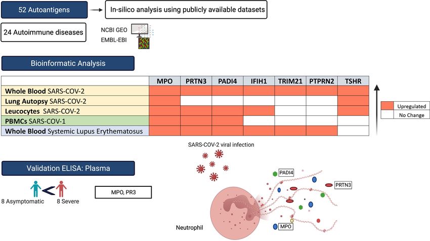
ENHANCED EXPRESSION OF AUTOANTIGENS DURING SARS-COV-2 VIRAL INFECTION
←
→
Page content transcription
If your browser does not render page correctly, please read the page content below
ORIGINAL RESEARCH
                                                                                                                                              published: 30 June 2021
                                                                                                                                     doi: 10.3389/fimmu.2021.686462

                                           Enhanced Expression
                                           of Autoantigens During
                                           SARS-CoV-2 Viral Infection
                                           Narjes Saheb Sharif-Askari 1, Fatemeh Saheb Sharif-Askari 1, Samrein B. M. Ahmed 1,2,
                                           Suad Hannawi 3, Rifat Hamoudi 1,2,4, Qutayba Hamid 1,2,5 and Rabih Halwani 1,2,6*
                                           1 Sharjah Institute of Medical Research, University of Sharjah, Sharjah, United Arab Emirates, 2 Department of Clinical

                                           Sciences, College of Medicine, University of Sharjah, Sharjah, United Arab Emirates, 3 Department of Rheumatology, Ministry
                                           of Health and Prevention, Dubai, United Arab Emirates, 4 Division of Surgery and Interventional Science, University College
                                           London, London, United Kingdom, 5 Meakins-Christie Laboratories, Research Institute of the McGill University Healthy
                                           Center, McGill University, Montreal, QC, Canada, 6 Prince Abdullah Ben Khaled Celiac Disease Research Chair, Department
                          Edited by:       of Pediatrics, Faculty of Medicine, King Saud University, Riyadh, Saudi Arabia
                          Zhiwei Wu,
             Nanjing University, China
                                           Immune homeostasis is disturbed during severe viral infections, which can lead to loss of
                        Reviewed by:
                           Laura Cook,     tolerance to self-peptides and result in short- or long-term autoimmunity. Using publicly
   Peter Doherty Institute for Infection   available transcriptomic datasets, we conducted an in-silico analyses to evaluate the
              and Immunity, Australia
                  Marcelo A. Soares,
                                           expression levels of 52 autoantigens, known to be associated with 24 autoimmune
National Cancer Institute (INCA), Brazil   diseases, during SAR-CoV-2 infection. Seven autoantigens (MPO, PRTN3, PADI4, IFIH1,
                  *Correspondence:         TRIM21, PTPRN2, and TSHR) were upregulated in whole blood samples. MPO and TSHR
                       Rabih Halwani
                                           were overexpressed in both lung autopsies and whole blood tissue and were associated
               rhalwani@sharjah.ac.ae
                                           with more severe COVID-19. Neutrophil activation derived autoantigens (MPO, PRTN3,
                   Specialty section:      and PADI4) were prominently increased in blood of both SARS-CoV-1 and SARS-CoV-2
         This article was submitted to     viral infections, while TSHR and PTPRN2 autoantigens were specifically increased in
                      Viral Immunology,
               a section of the journal
                                           SARS-CoV-2. Using single-cell dataset from peripheral blood mononuclear cells
              Frontiers in Immunology      (PBMCs), we observed an upregulation of MPO, PRTN3, and PADI4 autoantigens
           Received: 26 March 2021         within the low-density neutrophil subset. To validate our in-silico analysis, we measured
            Accepted: 27 May 2021
                                           plasma protein levels of two autoantigens, MPO and PRTN3, in severe and asymptomatic
           Published: 30 June 2021
                                           COVID-19. The protein levels of these two autoantigens were significantly upregulated in
                           Citation:
              Saheb Sharif-Askari N,       more severe COVID-19 infections. In conclusion, the immunopathology and severity of
 Saheb Sharif-Askari F, Ahmed SBM,         COVID-19 could result in transient autoimmune activation. Longitudinal follow-up studies
   Hannawi S, Hamoudi R, Hamid Q
    and Halwani R (2021) Enhanced          of confirmed cases of COVID-19 could determine the enduring effects of viral infection
  Expression of Autoantigens During        including development of autoimmune disease.
         SARS-CoV-2 Viral Infection.
         Front. Immunol. 12:686462.        Keywords: autoantigen, bioinformatics, SARS-CoV-2, COVID-19, autoimmune disease, neutrophil, respiratory viral
   doi: 10.3389/fimmu.2021.686462          infection, lung autopsies

Frontiers in Immunology | www.frontiersin.org                                     1                                          June 2021 | Volume 12 | Article 686462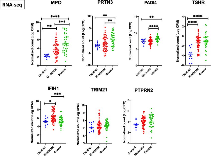
Saheb Sharif-Askari et al.                                                                                          Autoantigens During SARS-CoV-2 Viral Infection

 GRAPHICAL ABSTRACT

INTRODUCTION                                                                            host genetic susceptibility, including human leucocyte antigen
                                                                                        (HLA) polymorphism.
Severe acute respiratory syndrome coronavirus 2 (SARS-CoV-2),                               Alternatively, viral induced inflammation and dysregulation of
the virus causing coronavirus disease 2019 (COVID-19),                                  innate and adaptive immune system, could both directly and
appeared first in Wuhan, China, in December 2019 and has                                 indirectly lead to loss of tolerance to self-peptides and result in
since rapidly spread globally (1, 2). COVID-19 severity level                           short- or long-term autoimmunity (17, 18). Emerging reports are
ranged from asymptomatic infection to life-threatening disease                          associating COVID-19 infection with worsening, relapse or de novo
(3–6). Severe COVID-19 disease has been associated with innate                          induction of several autoimmune diseases including systemic lupus
immune dysregulation, early immunosuppression, lymphopenia,                             erythematosus (19–21), Guillain-Barré syndrome (22, 23),
and cytokine storm (7–10).                                                              vasculitis (24, 25), and multiple sclerosis (26). Further, high
   Multiple factors are involved in the development of                                  prevalence of antinuclear antibodies (ANA) and lupus
autoimmunity, including genetics, age, and environment (11).                            anticoagulant were reported in severe hospitalized SARS-CoV-2
Between the environmental triggers, viral infections, particularly                      infection, which could hint at induction of autoimmunity (27).
those resulting in low interferon production, as it is the case with                        Although the concept of autoimmunity had been explored in
SARS-CoV-2 infection, have long been associated with induction                          previous viral infection (28), its relevance to COVID-19 respiratory
of autoimmunity (11). Similar to many severe viral infections,                          infection deserves more attention; especially that immune
SARS-CoV-2 could trigger the autoimmune reaction through                                derangement during SARS-CoV-2 infection could potentially
multiple mechanism including molecular mimicry, epitope                                 trigger relapse and induction of many new cases. Therefore, the
spreading, bystander activation, and persistence of latent virus                        aim of current study was to utilize publicly available transcriptomic
(12–15). Aforementioned mechanisms could be understood                                  COVID-19 data to evaluate autoimmune activation during
through examination of homology between various antigens of                             COVID-19 infection through measuring the gene expression
SARS-CoV-2 and self-antigens (16). Development of cross-                                levels of 52 autoantigens, known to be associated with 24 different
reactive epitopes are then dependent on viral strain as well as                         autoimmune diseases.

Abbreviations: ANA, Antinuclear antibodies; ANCA, Anti-neutrophil cytoplasmic
autoantibody; ARDS, Acute respiratory distress syndrome; COVID-19, Coronavirus
disease of 2019; DEGs, Differentially expressed genes; EMBL-EBI, European               MATERIAL AND METHODS
Bioinformatics Institute; FFPE, Formalin fixed paraffin embedded; HLA, Human
leukocyte antigen; IAV, Influenza A virus; IQR, Interquartile range; LIMMA, Linear       For the purpose of this study, we used a list of 52 autoantigens
Models for MicroArray data; Log CPM, Log2 counts per million; MPO,                      established by Burbelo et al. (29) for diagnosis of 24 different
Myeloperoxidase; NETs, Neutrophil-derived extracellular traps; NCIB GEO, National       autoimmune disease including Hashimoto’s thyroiditis, ANCA-
Center for Biotechnology Information Gene Expression Omnibus; PBMC, Peripheral
blood mononuclear cells; RA, Rheumatoid arthritis; RMA, Robust Multi-Array
                                                                                        associated vasculitis, rheumatoid arthritis (RA), and Systemic
Average; RSV, Respiratory syncytial virus; SARS-CoV-2, Severe acute respiratory         lupus erythematosus (SLE) (Table 1). The expression of these
syndrome coronavirus 2; SLE, Systemic lupus erythematosus.                              autoantigens in the lungs and whole blood of COVID-19 patients

Frontiers in Immunology | www.frontiersin.org                                       2                                     June 2021 | Volume 12 | Article 686462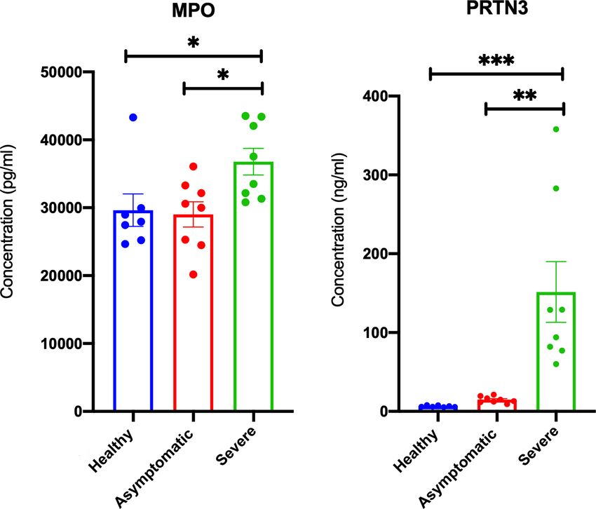
Saheb Sharif-Askari et al. Autoantigens During SARS-CoV-2 Viral Infection TABLE 1 | List of autoantigens and their associated autoimmune disease. Autoantigen Full name Autoimmune disease Location AQP4 aquaporin 4 Neuromyelitis optica 18q11.2 GAD2 glutamate decarboxylase 2 Stiff-person syndrome, T1D 10p12.1 INS insulin Type I diabetes (T1D) 11p15.5 PTPRN protein tyrosine phosphatase receptor type N Type I diabetes 2q35 PTPRN2 protein tyrosine phosphatase receptor type N2 Type I diabetes 7q36.3 SLC30A8 solute carrier family 30 member 8 Type I diabetes 8q24.11 TSHR thyroid stimulating hormone receptor Graves’ disease (GD) 14q24-q31 TPO thyroid peroxidase Hashimoto’s thyroiditis, GD 2p25.3 TG thyroglobulin Hashimoto’s thyroiditis, GD 8q24.22 CHRNA1 cholinergic receptor nicotinic alpha 1 subunit Myasthenia gravis 2q31.1 MUSK muscle associated receptor tyrosine kinase Myasthenia gravis 9q31.3 LRP4 LDL receptor related protein 4 Myasthenia gravis 11p11.2 COL4A3 collagen type IV alpha 3 chain Goodpasture disease 2q36.3 PLA2R1 phospholipase A2 receptor 1 Membranous nephropathy 2q23-q24 THSD7A thrombospondin type 1 domain containing 7A Membranous nephropathy 7p21.3 CYP21A2 cytochrome P450 family 21 subfamily A member 2 Addison’s disease 6p21.33 ATP4A ATPase H+/K+ transporting subunit alpha Autoimmune gastritis 19q13.12 ATP4B ATPase H+/K+ transporting subunit beta Autoimmune gastritis 13q34 CBLIF (GIF) cobalamin binding intrinsic factor Autoimmune gastritis 11q12.1 SEPSECS Sep (O-phosphoserine) tRNA : Sec (selenocysteine) tRNA synthase Autoimmune hepatitis 4p15.2 CYP2D6 cytochrome P450 family 2 subfamily D member 6 Autoimmune hepatitis 22q13.2 FTCD formimidoyltransferase cyclodeaminase Autoimmune hepatitis 21q22.3 DSG1 desmoglein 1 Pemphigus 18q12.1 DSG3 desmoglein 3 Pemphigus 18q12.1 TGM3 transglutaminase 3 Dermatitis herpetiformis 20p13 CSF2 colony stimulating factor 2 Pulmonary alveolar proteinosis 5q31.1 PRTN3 (PR3) proteinase 3 ANCA-associated vasculitis 19p13.3 MPO myeloperoxidase ANCA-associated vasculitis 17q22 IFNG interferon gamma Disseminated non-tuberculosis 12q15 PADI4 peptidyl arginine deiminase 4 Rheumatoid arthritis 1p36.13 TRIM21 tripartite motif containing 21 Sjögren’s syndrome, SLE 11p15.4 RO60 (TROVE2) Ro60, Y RNA binding protein Sjögren’s syndrome, SLE 1q31.2 SSB small RNA binding exonuclease protection factor La Sjögren’s syndrome, SLE 2q31.1 SNRPA small nuclear ribonucleoprotein polypeptide A Systemic lupus erythematosus (SLE) 19q13.2 SNRNP70 small nuclear ribonucleoprotein U1 subunit 70 Systemic lupus erythematosus 19q13.33 SNRPD3 small nuclear ribonucleoprotein D3 polypeptide Systemic lupus erythematosus 22q11.23 HARS1 histidyl-tRNA synthetase 1 Myositis 5q31.3 TARS1 threonyl-tRNA synthetase 1 Myositis 5p13.3 EXOSC9 exosome component 9 Myositis 4q27 EXOSC10 exosome component 10 Myositis 1p36.22 CHD4 chromodomain helicase DNA binding protein 4 Myositis 12p13.31 CHD3 chromodomain helicase DNA binding protein 3 Myositis 17p13.1 IFIH1 interferon induced with helicase C domain 1 Myositis 2q24.2 MORC3 MORC family CW-type zinc finger 3 Myositis 21q22.12 SRP54 signal recognition particle 54 Myositis 14q13.2 TRIM33 tripartite motif containing 33 Myositis 1p13.2 HMGCR 3-hydroxy-3-methylglutaryl-CoA reductase Myositis 5q13.3 FBL fibrillarin Scleroderma 19q13.2 TOP1 DNA topoisomerase I Scleroderma 20q12 POLR3A RNA polymerase III subunit A Scleroderma 10q22.3 DLAT dihydrolipoamide S-acetyltransferase Primary biliary cirrhosis 11q23.1 TGM2 transglutaminase 2 Celiac disease 20q11.23 was then determined in-silico using publicly available datasets. COVID-19 infection was compared to that following infection with We validated autoantigen with mRNA expression equal or more three respiratory viruses: SARS-CoV-1, IAV, and RSV. than 1.5 LogFC change in leucocyte isolated from COIVD-19. RNA-sequencing platforms were used for COVID-19 studies, These datasets were publicly available at National Center for while microarray platforms were used for older datasets of SARS- Biotechnology Information Gene Expression Omnibus (NCIB CoV-1, IAV, and RSV (Table 2). For the COVID-19 lung GEO, http://www.ncbi.nlm.nih.gov/geo) and the European autopsies dataset (PRJNA646224) (32), the authors have Bioinformatics Institute (EMBL-EBI, https://www.ebi.ac.uk). extracted RNA from Formalin fixed paraffin embedded (FFPE) Moreover, single cell transcriptomic datasets of sorted neutrophils tissues from 9 COVID-19 fatal cases, and 10 SARS-CoV-2- were used. In addition, the expression of autoantigens following uninfected individuals who undertook biopsy as part of routine Frontiers in Immunology | www.frontiersin.org 3 June 2021 | Volume 12 | Article 686462
Saheb Sharif-Askari et al.                                                                                             Autoantigens During SARS-CoV-2 Viral Infection

TABLE 2 | Gene expression datasets used in this study.

Groups                  GEO accession           Platform          Sample                  Condition 1                               Condition 2

Microarray Data
                      GSE1739 (30)           GPL201        PBMCs                       Controls (n = 4)    SARS-CoV-1 (n = 10)
                      GSE17156 (31)          GPL571        Whole blood                 Controls (n = 17)   Influenza H3N2
                                                                                                           (n = 17)
                      GSE17156 (31)          GPL571        Whole blood                 Controls (n = 20)   Respiratory syncytial virus (n = 20)
RNA-seq Data
                      PRJNA646224 (32)       GPL21697      Lung autopsies              Controls (n = 10)   Lung autopsies (n = 9)
                      EGAS00001004503        GPL24676      Whole blood                 Controls (n = 10)   COVID-19 (n = 39, 19 mild and 20 severe)
                      (33)
                      GSE157103 (34)         GPL24676      Leukocytes from whole       Controls (n = 10)   moderate COVID-19 (n = 51), severe COVID-19 (n = 49, 12
                                                           blood                                           non-critical and 37 critical)
                      Panousis et al. (35)   GPL11154      Whole blood                 Controls (n = 58)   Active systemic lupus erythematosus (n = 79)
Single-cell RNA-
seq Data
                      GSE150728 (36)         GPL24676      Peripheral blood            Healthy (n = 6)     Severe COVID-19 (n = 7)
                                                           mononuclear cells

SARS-CoV, Severe acute respiratory syndrome coronavirus.

clinical care for lung cancer. For this lung autopsy datasets, we                      data using the Bioconductor package limma-voom (38), and
used processed sequencing data provided by Wu Meng et al. (32).                        presented the results as logCPM. Log-transformed normalized
The authors used DESeq2 to identify differentially expressed                           intensities were also used in Linear Models for MicroArray data
genes between the cases and controls. Benjamini-Horchberg                              (LIMMA) analyses to identify differentially expressed genes between
correction was used for multiple testing (37).                                         diseased and control groups. We used the default Benjamini-
    For COVID-19 whole blood transcriptomic dataset, we used                           Horchberg correction for multiple testing. Raw data from different
processed sequencing data deposited under project number                               studies were never mixed or combined. For each study, the LogFC
EGAS00001004503 (33). In this study, Aschenbrenner et al.                              was obtained separately by analyzing data of diseased and controls.
extracted the RNA from whole blood of 39 COVID-19 patients                             Statistical analyses were performed using R software (v 3.0.2) and
and 10 healthy controls and analyzed it using NovaSeq 6000 (33).                       Prism (v8; GraphPad Software). For all analyses, p-values
Saheb Sharif-Askari et al.                                                                                                Autoantigens During SARS-CoV-2 Viral Infection

of 3, and an enrichment factor >1.5 were collected and grouped                          RESULTS
into clusters based on their membership similarities. The top few
enriched clusters (one term per cluster) were presented. Gene                           MPO and TSHR autoantigens Are
Ontology biology process database was accessed through Enrichr                          Associated With More Severe COVID-19 in
open source, available as a gene set enrichment analysis web server                     Lung Autopsy and Whole Blood
(43, 44). GO biological processes were ranked according to                              Using publicly available transcriptomic datasets, we have
combined score. This score was computed in Enrichr by taking                            determined the expression levels of 52 autoantigens, known to
the log of the p-value from the Fisher exact test and multiplying                       be associated with 24 different autoimmune diseases. The list of
that by the z score of the deviation from the expected rank (43, 44).                   these genes and their associated autoimmune disease is presented
                                                                                        in Table 1. The datasets used in this study are presented in
ELISA                                                                                   Table 2. Expression levels of autoantigens were determined in
The plasma level of MPO and PRTN3 (PR3) for seven non-                                  lung autopsies and whole blood (Figure 1). For lung, RNA-
infected controls, eight severe, and eight asymptomatic COVID-19                        sequencing data was obtained from 9 deceased COVID-19
patients was determined using commercially available human                              patients and 10 negative controls (PRJNA646224) (Figure 1A).
ELISA kit (MPO, Cat # ab119605 and PR3, Cat # ab226902,                                 For whole blood, RNA-sequencing data was extracted from 10
Abcam, Cambridge, MA, USA). The plasma used in our study was                            controls, 19 mild COVID-19, 20 severe COVID-19 (Figures 1B, C).
obtained from COVID-19 patients recruited from Rashid                                   MPO and TSHR were the only two autoantigens overexpressed in
Hospital. Plasma was isolated from blood via standard Ficoll-                           lung autopsies and were shared between the lung and blood of
Paque density gradient centrifugation (Sigma, Histopaque-10771).                        severe COVID-19 patients. In whole blood study, seven
Assays were preformed strictly following the manufacturer’s                             autoantigens were upregulated in blood of severe COVID-19
instructions. Each sample was assayed in duplicate, and values                          patients (MPO, PRTN3, PADI4, IFIH1, TRIM21, PTPRN2, and
were expressed as the mean of 2 measures per sample. One-way                            TSHR) (Figure 1B), while four autoantigens (MPO, PRTN3, IFIH1,
analysis of variance (ANOVA) and post hoc Tukey multiple                                and PADI4) were increased in blood of mild patients (Figure 1C).
comparison analyses were applied.                                                       The upregulation of MPO and PRTN3 was significantly higher in

                     A

                     B                                    C                                         D

 FIGURE 1 | Gene expression of autoantigens in lung autopsies and whole blood of COVID-19 patients. (A) Enhanced expression of MPO and TSHR autoantigens in
 lung autopsies (n = 9 COVID-19 vs n = 10 controls, Dataset: PRJNA646224). (B) For whole blood COVID-19 dataset (EGAS00001004503), seven autoantigens
 were upregulated in severe COVID-19 vs controls comparison (C), four autoantigens were upregulated in mild COVID-19 vs controls. The upregulation of MPO and
 PRTN3 was higher in severe versus mild COVID-19. The sample size presented in B–C EGAS00001004503 dataset was as following; n = 10 controls, n = 19 mild COVID-
 19, and n = 20 severe COVID-19. Results are presented as log fold change of gene expression with adjusted p-values of less than 0.05. The log fold changes were
 compared between severe and mild COVID-19 groups. (D) Upregulation of autoantigens in whole blood of both severe COVID-19 and active systemic lupus erythematosus
 patients. The following datasets were used; EGAS00001004503 (n = 20 severe COVID-19 vs n = 10 controls), and Panousis et al. (35) (n = 79 active SLE vs n = 58 controls).
 Unpaired student t-test was used to compare between each two independent groups. ∗p < 0.05, ∗∗p < 0.01, ∗∗∗∗p < 0.0001.

Frontiers in Immunology | www.frontiersin.org                                       5                                            June 2021 | Volume 12 | Article 686462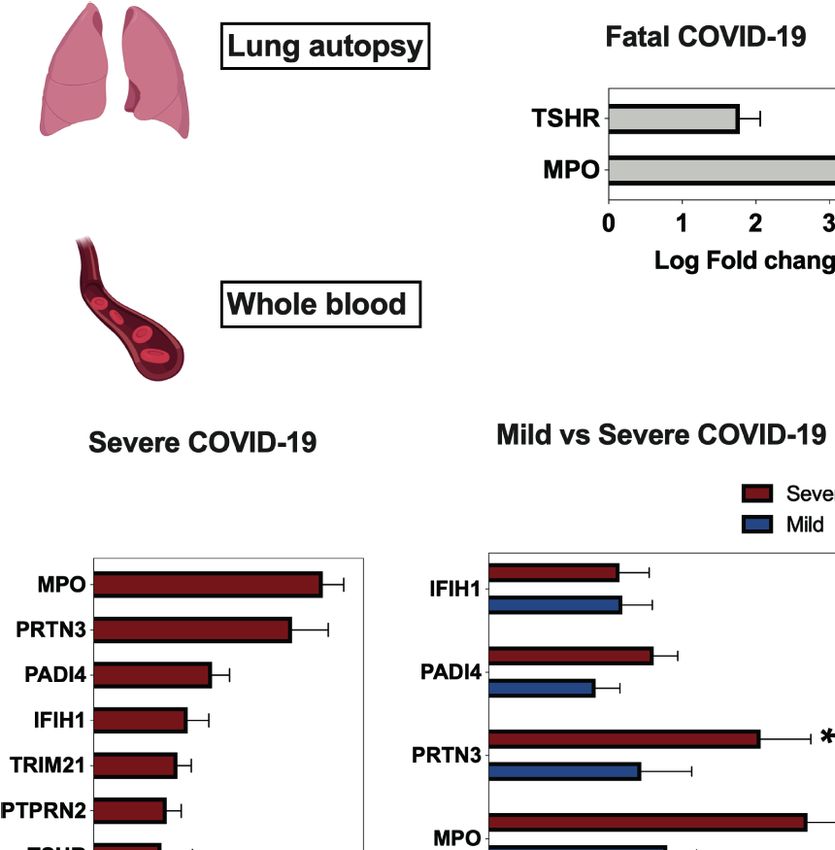
Saheb Sharif-Askari et al.                                                                                        Autoantigens During SARS-CoV-2 Viral Infection

severe versus mild COVID-19 (MPO, 3.4 ± 0.3 vs 1.9±0.3 log fold                   showing that beside autoimmune disease, these genes were
change [LogFC]; p-value = 0.004, and PRTN3 (2.9 ± 0.5 vs 1.6 ± 0.5                associated with other inflammatory and fibrotic conditions
LogFC; p-value = 0.01 as presented in Figure 1C). We also looked at               affecting different organs including lung (Bronchiectasis; MPO,
the levels of the selected seven autoantigens during active systemic              PRTN3, and TRIM21) and kidney (Glomerulonephritis; MPO,
lupus erythematosus (SLE) autoimmune disease using whole blood                    PRTN3, TRIM21, and PADI4). In addition, the GO biological
transcriptomic data deposited by Panousis et al. (35). Of the seven               process database revealed the association of these seven genes with
autoantigens, six markers were also upregulated during active SLE,                interferon alpha signaling, neutrophil activation, and regulation of
however, MPO (3.4 ± 0.3 vs 0.9 ± 0.2 LogFC; p-value
Saheb Sharif-Askari et al. Autoantigens During SARS-CoV-2 Viral Infection
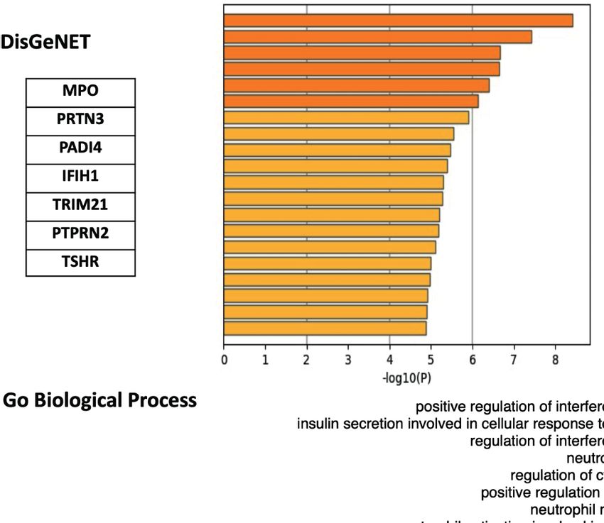
A
B
FIGURE 3 | Gene ontology enrichments identified in the DisGeNET and Gene Ontology biology process databases. (A) Summary of enrichment of DisGeNet
category of Metascape. Terms with a p-value 1.5 (the enrichment factor is the ratio between the observed
counts and the counts expected by chance) are collected and grouped into clusters based on their membership similarities. The top few enriched clusters (one term
per cluster) were presented. (B) Top 10 categories of the GO biological processes. GO analysis associated with the top seven differentially expressed autoantigens
was performed using Enrichr. Bar lines represent cumulative score for the 10 top-ranked categories of GO biological processes. Combined score is computed by
taking the log of the p-value from the Fisher exact test and multiplying that by the z score of the deviation from the expected rank.
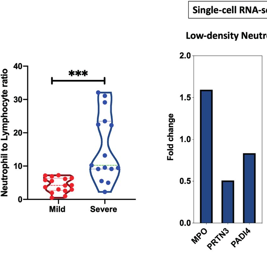
A B C
FIGURE 4 | Neutrophil to lymphocyte ratio and gene expression isolated from severe COVID-19 patients. (A) Neutrophil to lymphocyte ratio from whole blood of 16
mild and 15 severe COIVD-19 patients were compared (PRJNA646224). This ratio was significantly higher in severe COVID-19 (B) Single-cell RNA sequencing was
performed on PBMCs from six severe COVID-19 patients and seven healthy controls (GSE150728). Differential expression of MPO, PRTN3, and PADI4 within the low-
density neutrophils cluster. (C) Increased counts of low-density neutrophil and canonical neutrophil during severe COVID-19 infection. The count value represents the absolute
cell number within the PBMCs. Unpaired student t-test was used to compare between the independent groups. ∗p < 0.05, ∗∗p < 0.01, ∗∗∗p < 0.001.
Frontiers in Immunology | www.frontiersin.org 7 June 2021 | Volume 12 | Article 686462
Saheb Sharif-Askari et al.                                                                                      Autoantigens During SARS-CoV-2 Viral Infection

(GSE150728) of COVID-19 severe patients were used (36). Between                   differential expression and LogFC were obtained by comparing the
the different immune cells three autoantigens, MPO, PRTN3, and                    normalized gene expression of the infected group versus healthy
PADI4, were significantly enriched within the low-density neutrophil               donors (Figure 6A). None of autoantigens were upregulated more
subset (Figure 4B). Moreover, the counts of low-density neutrophil                than one LogFC in IAV and RSV, while five autoantigens in SARS-
and canonical neutrophil were increased during severe COVID-19                    CoV-1 and seven autoantigens in SARS-CoV-2 were upregulated
infection (Figure 4C). To validate our in-silico analysis, we next                more than one LogFC (Figure 6A). MPO, PRTN3 (PR3), and
measured plasma protein levels of two autoantigens of MPO and                     PADI4 were the top shared autoantigens appearing in both
PRTN3 in severe and asymptomatic COVID-19. The protein levels                     coronavirus respiratory infections, with an increase in expression
were estimated using human ELISA assays. The results revealed an                  of more than 1.5 LogFC. We then intersected the differentially
increase of MPO (mean 36,787 ± 1,961 vs mean 29,007 ± 1,860 pg/ml;                expressed genes in all four respiratory infections to obtain the
p-value = 0.038) and PRTN3 (mean 151.5 ± 38 vs mean 14.77 ±                       shared signatures (Figure 6B). Interestingly, TSHR and PTPRN2
1.4 ng/ml; p-value = 0.001) in severe compared to asymptomatic                    autoantigens were specifically increased in SARS-CoV-2
COVID-19 infection (Figure 5). Level of these proteins were not                   (Figure 6B). TSHR was also overexpressed in COVID-19 lung
different in asymptomatic and non-infected controls (Figure 5).                   autopsies. Two genes (IFIH1 and TRIM21) were upregulated in
                                                                                  three viral infections; however, their expression was higher in
Prominent Autoantigens Upregulation in                                            SARS-CoV-2 compared to IAV, and RSV (Figure 6B).
Coronavirus Infections Relative to Other
Viral Infections
We next compared the profile of autoantigen upregulation                           DISCUSSION
observed during SARS-CoV-2 to that detected during other
respiratory viral infection. To do that, we used transcriptomic                   Herein we report the common autoantigens during COVID-19
microarrays and RNA-sequencing data from blood of SARS-                           viral infection. Fifty-two autoantigens, which are linked
CoV-1, influenza A virus (IAV), and respiratory syncytial virus                    with 24 different autoimmune diseases, were tested (29). Seven
(RSV) infected patients at the peak of disease. For each condition,               autoantigens were found to be elevated in COVID-19. The level

 FIGURE 5 | Elevated plasma MPO and PRTN3 levels in severe COIVD-19 patients. An increase in the plasma levels of MPO and PRTN3 in severe compared to
 asymptomatic COVID-19 patients. The protein levels were estimated using human ELISA assays (n = non-infected controls, n = 8 asymptomatic COVID-19, and
 severe n = 8 COVID-19). ∗p < 0.05, ∗∗p < 0.01, ∗∗∗p < 0.001.

Frontiers in Immunology | www.frontiersin.org                                 8                                       June 2021 | Volume 12 | Article 686462
Saheb Sharif-Askari et al. Autoantigens During SARS-CoV-2 Viral Infection
A B
FIGURE 6 | Expression of autoantigens during COVID-19 and other viral infections. The following datasets were used to compare expression of autoantigens
between SARS-CoV and other viral infections; GSE17156 (n = 17 IAV vs n = 17 controls), GSE17156 (n = 20 RSV vs n = 20 controls), GSE1739 (n = 10 SARS-
CoV-1 vs n = 4 controls), and EGAS00001004503 (n = 39 COVID-19 vs n = 10 controls). (A) Log fold change in the expression of autoantigens following infection
with SARS-CoV-1, SARS-CoV-2, IAV, and RSV relative to healthy controls. (B) Intersection of upregulated autoantigen signatures in four different respiratory viral
infections, IAV, RSV, SARS-CoV-1, and SARS-CoV-2. Three top upregulated autoantigens (PADI4, MPO, and PRTN3) are shared between SARS-COV-1 and SARS-
COV-2. For all analyses, p < 0.05 was considered significant. IAV, influenza A virus; RSV, Respiratory syncytial virus.
of three autoantigens, MPO, PRTN3, and PADI4, were higher in The top seven autoantigens upregulated in the blood of severe
the blood of severe compared to mild COVID-19. COVID-19 patients, were associated with a wide range of vascular
Autoimmune disease could be triggered by genetic and and inflammatory autoimmune disorders (Figure 3A). Vasculitis, a
environmental factors; viral infections had been known as a shared condition between fatal COVID-19 and vascular
major environmental cause of transient autoimmunity that could autoimmune diseases such as Anti-neutrophil cytoplasmic
potentially lead to relapse or induction of de novo autoimmune autoantibody (ANCA) vasculitis, is featured by elevation of MPO
disorders (11). These autoimmune disorders emerge weeks post and PRTN3 levels (25, 48). Wilk et al. single cell data identified a
viral infection; hence sensitive serological tests are needed to distinct group of low-density neutrophil; these immune cells were
determine the cause-effect relationship between SARS-CoV-2 only detected in severe COVID-19 complicated with acute
infection and autoimmune disease diagnosis (45–47). respiratory distress syndrome (ARDS) (36). These cells had a
Interestingly, MPO and TSHR were increased in both lung significantly high level of MPO, PRTN3, and PADI4 indicating
autopsies and whole blood of severe COVID-19 patients. that they could be a major source of the observed increase in blood
Comparison of COVID-19 blood transcriptomic with IAV and level of these autoantigens during severe COVID-19. Following
RSV revealed that MPO, PRTN3, and PADI4 were selectively SARS-CoV-2 infection, neutrophil-derived extracellular traps
upregulated in coronavirus infections, SARS-CoV-1 and SARS- (NETs) formation by neutrophils, NETosis, may therefore lead to
CoV-2, while TSHR and PTPRN2 autoantigens were distinctive burst of autoantigens including PADI4, MPO, and PRTN3 in the
to SARS-CoV-2 infection. These two genes (TSHR and context of immunostimulatory molecules.
PTPRN2) did not increase in the mild COVID-19 and hence Confirming previous findings (49, 50), increased MPO level
they were associated with more severe infection. reflected COVID-19 severity. Neutrophil activation leading to net
Following the results obtained through gene ontology formation or NETosis is observed in both viral infections and
enrichment analyses, single cell transcriptomics of PBMCs autoimmune diseases (47, 51, 52). NETosis could then be
revealed the significant increase in MPO, PRTN3, and PADI4 considered as a common pathological modulator of viral
mRNA levels within low-density neutrophils. In addition, analyses infection and autoimmune disease (47, 52, 53). NETosis
of cell counts provided by Wilk et al. study showed significant autoantigens, MPO, PRTN3, and PADI4, were markedly
increase in both low-density and canonical neutrophils during increased in SARS-CoV-1 and SARS-CoV-2 but they did not
severe COVID-19 infection compared to controls (Figure 4C) (36). appear in IAV and RSV. Supporting our findings, a recent
Frontiers in Immunology | www.frontiersin.org 9 June 2021 | Volume 12 | Article 686462Saheb Sharif-Askari et al. Autoantigens During SARS-CoV-2 Viral Infection
investigation by FP Veras et al. showed viable SARS-CoV-2 ability around half of recovered individuals, while joint pain and chest
to directly induce the release of NETs by healthy neutrophils (54). pain lingered in 20–30% of recovered patients (68). Similarly, a
While, in another study, influenza A virus infection did not affect 6-month follow-up study of 1,733 COVID-19 hospitalized
MPO release (55). MPO is a peroxidase enzyme responsible for patients from China reported lasting of fatigue and muscle
intracellular catalytic reactions between hydrogen peroxidase and weakness in more than half of patients, while patients who
chlorides to form hypochlorous acid (56, 57). NETosis leads to were more severely ill during their hospital stay had more
extracellular burst of chromatic, histones, and neutrophil granules persisting long-term symptoms (71).
containing MPO, PRTN3, and PADI4. An exaggerated increase in Emerging case reports have shown that SARS-CoV-2 induces
the level of these antigens during COVID-19 infection could lead long-term immune-inflammatory abnormalities (66, 72). Schenker
to breaking the autoimmune tolerance and the recognition of et al. reported a 65-year-old female patient with de novo reactive
autoantigens by immune sentinel cells. arthritis and cutaneous vasculitis 10 days after recovery of all
NETosis induces inflammation and DNA deployment that COVID-19-related symptoms (72). In a more severe case, a 31-
could trigger autoimmunity. While NET produced by year female was presented with fatal multisystem inflammatory
neutrophils, their clearance is achieved by macrophages syndrome (MIS) 2 weeks post recovery from COVID-19 (65). This
efferocytosis. During ARDS, neutrophil count and lifespan is increase in incidence of autoimmunity was also reported among
significantly increased, and the ability of macrophages to engulf children, where SARS-CoV-2 epidemic was associated with a 30-
NETs and apoptotic cells is significantly decreased, prolonging the fold increase in Kawasaki-like disease (71).
lung injury induced by neutrophil blast. Pharmacologic treatment Long-COVID-19 persisting symptoms involve immune-
could be used to enhance NETs clearance. Macrophage NETs mediated inflammatory disease and neurological abnormalities
efferocytosis could be restored by AMP-activated protein kinase that could suggest possibility of triggering pre-existing or de novo
(AMPK) activator such as Metformin or application of autoimmune reactions weeks or month after COVID-19 recovery
neutralizing antibody against HMGB1 (58). Therefore, such (37, 73). Previous studies have shown that autoantigen gene
medications could be considered to reduce blood levels of these upregulation is often followed by an increase in the respective
autoantigens, and hence lower chance of triggering autoimmunity. autoantibody level (29, 48, 74). Although the increase in
TSHR is expressed by thyroid epithelial cells, and various autoantigen expression observed in this study could only trigger
extra-thyroidal tissue including the adipose, peripheral blood short lasting autoimmunity, follow-up longitudinal studies are
cells, and fibrocytes (59). Derived from monocytes, human needed to establish the long-term enduring effects of SARS-CoV-2
fibrocytes express both thyroglobulin and thyrotropin receptor infection in developing autoimmune diseases.
(60). They are increased during lung injury and have both the
inflammatory characters of macrophages and the tissue
remodeling features of fibroblasts (61). Chronic inflammatory DATA AVAILABILITY STATEMENT
conditions such as autoimmunity, cardiovascular disease, and
asthma promote differentiation of immune cells to circulating The datasets presented in this study can be found in online
fibrocytes and their accumulation at the site of injury (61, 62). repositories. The names of the repository/repositories and accession
TSHR is targeted by autoantibodies during Graves’ disease number(s) can be found in the article/supplementary material.
(63). SARS-CoV-2 infection has been connected with the
initiation and relapse phases of Grave disease (45, 46). Of note,
this disease has emerged in some patients during COVID-19 ETHICS STATEMENT
recovery period. Patients diagnosed were negative for naso-
pharyngeal swab PCR test but were positive for both IgM and The studies involving human participants were reviewed and
IgG SARS-CoV-2 antibodies. This suggests that the observed approved by Dubai Health Authority. The patients/participants
increase of autoantigens during SARS-CoV-2 infection may provided their written informed consent to participate in this study.
trigger autoimmunity. This could lead to initiation or relapse
of autoimmune disorders as a long-term COVID-19 outcome. In
fact, evidence of transient autoimmunity has been reported AUTHOR CONTRIBUTIONS
among long COVID-19 outcomes by several studies (64–66). RaH, QH, NSS-A, FSS-A, conceived and designed the experiments.
Follow-up data from survivors of viral infections have shown NSS-A, FSS-A, RaH, RiH, SH and SA analyzed the data. All authors
appearance of autoimmune disorders within weeks to months contributed to the article and approved the submitted version.
after recovery (67). In addition, TSHR elevation was only
detected in severe COVID-19, which could suggest higher
chance for appearance of autoimmune disorders post severe
COVID-19 infection. FUNDING
The majority of COVID-19 patients are expected to show one
or more residual symptoms months after recovering from the This research has been financially supported by Tissue Injury and
infection (66, 68–70). In a post-acute COVID-19 follow-up study Repair (TIR) group operational grant (Grant code: 150317);
of 179 confirmed cases in Italy, fatigue and dyspnea persisted in COVID-19 research grant (CoV19-0307) and (CoV19-0308),
Frontiers in Immunology | www.frontiersin.org 10 June 2021 | Volume 12 | Article 686462Saheb Sharif-Askari et al. Autoantigens During SARS-CoV-2 Viral Infection
Seed grant (Grant code: 2001090275); and by collaborative Foundation Seed Grant (AJF202019); and by Prince Abdullah Ben
research grant (Grant code: 2001090278) to RH, University of Khalid Celiac Disease Research Chair, under the Vice Deanship of
Sharjah, UAE; and by a Sandooq Al Watan Applied Research & Research Chairs, King Saud University, Riyadh, Kingdom of
Development grant to RH (SWARD-S20-007); and by Al Jalila Saudi Arabia.
REFERENCES 19. Mantovani Cardoso E, Hundal J, Feterman D, Magaldi J. Concomitant New
Diagnosis of Systemic Lupus Erythematosus and COVID-19 With Possible
1. Wu F, Zhao S, Yu B, Chen Y-M, Wang W, Song Z-G, et al. A New Antiphospholipid Syndrome. Just a Coincidence? A Case Report and Review
Coronavirus Associated With Human Respiratory Disease in China. Nature of Intertwining Pathophysiology. Clin Rheumatol (2020) 39(9):2811–5. doi:
(2020) 579(7798):265–9. doi: 10.1038/s41586-020-2008-3 10.1007/s10067-020-05310-1
2. Zhu N, Zhang D, Wang W, Li X, Yang B, Song J, et al. A Novel Coronavirus 20. Slimani Y, Abbassi R, El Fatoiki F-Z, Barrou L, Chiheb S. Systemic Lupus
From Patients With Pneumonia in China, 2019. N Engl J Med (2020) 382 Erythematosus and Varicella-Like Rash Following COVID-19 in a Previously
(8):727–33. doi: 10.1056/NEJMoa2001017 Healthy Patient. J Med Virol (2020) 93(2):1184–7. doi: 10.1002/jmv.26513
3. Chen H, Guo J, Wang C, Luo F, Yu X, Zhang W, et al. Clinical Characteristics 21. Gao Z-W, Wang X, Lin F, Dong K. The Correlation Between SARS-CoV-2
and Intrauterine Vertical Transmission Potential of COVID-19 Infection in Infection and Rheumatic Disease. Autoimmun Rev (2020) 19(7):102557–7.
Nine Pregnant Women: A Retrospective Review of Medical Records. Lancet doi: 10.1016/j.autrev.2020.102557
(2020) 395(10226):809–15. doi: 10.1016/S0140-6736(20)30360-3 22. Toscano G, Palmerini F, Ravaglia S, Ruiz L, Invernizzi P, Cuzzoni MG, et al.
4. Huang C, Wang Y, Li X, Ren L, Zhao J, Hu Y, et al. Clinical Features of Guillain–Barré Syndrome Associated With SARS-Cov-2. N Engl J Med (2020)
Patients Infected With 2019 Novel Coronavirus in Wuhan, China. Lancet 382(26):2574–6. doi: 10.1056/NEJMc2009191
(2020) 395(10223):497–506. doi: 10.1016/S0140-6736(20)30183-5 23. Alberti P, Beretta S, Piatti M, Karantzoulis A, Piatti ML, Santoro P, et al. Guillain-
5. Rothe C, Schunk M, Sothmann P, Bretzel G, Froeschl G, Wallrauch C, et al. Barré Syndrome Related to COVID-19 Infection. Neurol - Neuroimmunol
Transmission of 2019-Ncov Infection From an Asymptomatic Contact in Neuroinflamm (2020) 7(4):e741. doi: 10.1212/NXI.0000000000000741
Germany. N Engl J Med (2020) 382(10):970–1. doi: 10.1056/NEJMc2001468 24. Becker RC. Covid-19-associated Vasculitis and Vasculopathy. J Thromb
6. Holshue ML, DeBolt C, Lindquist S, Lofy KH, Wiesman J, Bruce H, et al. First Thrombolysis (2020) 50(3):499–511. doi: 10.1007/s11239-020-02230-4
Case of 2019 Novel Coronavirus in the United States. N Engl J Med (2020) 382 25. Vacchi C, Meschiari M, Milic J, Marietta M, Tonelli R, Alfano G, et al. Covid-
(10):929–36. doi: 10.1056/NEJMoa2001191 19-associated Vasculitis and Thrombotic Complications: From Pathological
7. Vardhana SA, Wolchok JD. The Many Faces of the anti-COVID Immune Findings to Multidisciplinary Discussion. Rheumatology (2020) 59(12):e147–
Response. J Exp Med (2020) 217(6):e20200678. doi: 10.1084/jem.20200678 50. doi: 10.1093/rheumatology/keaa581
8. Blanco-Melo D, Nilsson-Payant BE, Liu W-C, Uhl S, Hoagland D, Møller R, 26. Zanin L, Saraceno G, Panciani PP, Renisi G, Signorini L, Migliorati K, et al.
et al. Imbalanced Host Response to SARS-CoV-2 Drives Development of Sars-CoV-2 Can Induce Brain and Spine Demyelinating Lesions. Acta
COVID-19. Cell (2020) 181(5):1036–45.e1039. doi: 10.1016/j.cell.2020.04.026 Neurochir (2020) 162(7):1491–4. doi: 10.1007/s00701-020-04374-x
9. Lee JS, Park S, Jeong HW, Ahn JY, Choi SJ, Lee H, et al. Immunophenotyping 27. Gazzaruso C, Carlo Stella N, Mariani G, Nai C, Coppola A, Naldani D, et al.
of COVID-19 and Influenza Highlights the Role of Type I Interferons in High Prevalence of Antinuclear Antibodies and Lupus Anticoagulant in
Development of Severe COVID-19. Sci Immunol (2020) 5(49):eabd1554. doi: Patients Hospitalized for SARS-CoV2 Pneumonia. Clin Rheumatol (2020)
10.1126/sciimmunol.abd1554 39(7):2095–7. doi: 10.1007/s10067-020-05180-7
10. Liao M, Liu Y, Yuan J, Wen Y, Xu G, Zhao J, et al. Single-Cell Landscape of 28. Ercolini AM, Miller SD. The Role of Infections in Autoimmune Disease. Clin
Bronchoalveolar Immune Cells in Patients With COVID-19. Nat Med (2020) Exp Immunol (2009) 155(1):1–15. doi: 10.1111/j.1365-2249.2008.03834.x
26(6):842–4. doi: 10.1038/s41591-020-0901-9 29. Burbelo PD, Iadarola MJ, Alevizos I, Sapio MR. Transcriptomic Segregation of
11. Fujinami RS. Viruses and Autoimmune Disease–Two Sides of the Same Coin? Human Autoantigens Useful for the Diagnosis of Autoimmune Diseases. Mol
Trends Microbiol (2001) 9(8):377–81. doi: 10.1016/S0966-842X(01)02097-2 Diagn Ther (2016) 20(5):415–27. doi: 10.1007/s40291-016-0211-6
12. Fujinami RS, von Herrath MG, Christen U, Whitton JL. Molecular Mimicry, 30. Reghunathan R, Jayapal M, Hsu LY, Chng HH, Tai D, Leung BP, et al.
Bystander Activation, or Viral Persistence: Infections and Autoimmune Expression Profile of Immune Response Genes in Patients With Severe Acute
Disease. Clin Microbiol Rev (2006) 19(1):80–94. doi: 10.1128/CMR.19.1.80- Respiratory Syndrome. BMC Immunol (2005) 6:2. doi: 10.1186/1471-2172-6-2
94.2006 31. Zaas AK, Chen M, Varkey J, Veldman T, Hero AO 3rd, Lucas J, et al. Gene
13. Reddy V, Desai A, Krishna SS, Vasanthapuram R. Molecular Mimicry Expression Signatures Diagnose Influenza and Other Symptomatic
Between Chikungunya Virus and Host Components: A Possible Mechanism Respiratory Viral Infections in Humans. Cell Host Microbe (2009) 6(3):207–
for the Arthritic Manifestations. PLoS Negl Trop Dis (2017) 11(1):e0005238. 17. doi: 10.1016/j.chom.2009.07.006
doi: 10.1371/journal.pntd.0005238 32. Wu M, Chen Y, Xia H, Wang C, Tan CY, Cai X, et al. Transcriptional and
14. Galeotti C, Bayry J. Autoimmune and Inflammatory Diseases Following Proteomic Insights Into the Host Response in Fatal COVID-19 Cases. Proc
COVID-19. Nat Rev Rheumatol (2020) 16(8):413–4. doi: 10.1038/s41584- Natl Acad Sci (2020) 117(45):28336. doi: 10.1073/pnas.2018030117
020-0448-7 33. Aschenbrenner AC, Mouktaroudi M, Krämer B, Oestreich M, Antonakos N,
15. Zhou Y, Han T, Chen J, Hou C, Hua L, He S, et al. Clinical and Autoimmune Nuesch-Germano M, et al. Disease Severity-Specific Neutrophil Signatures in
Characteristics of Severe and Critical Cases of COVID-19. Clin Transl Sci Blood Transcriptomes Stratify COVID-19 Patients. Genome Med (2021) 13
(2020) 13(6):1077–86. doi: 10.1111/cts.12805 (1):7. doi: 10.1186/s13073-020-00823-5
16. Megremis S, Walker TDJ, He X, Ollier WER, Chinoy H, Hampson L, et al. 34. Overmyer KA, Shishkova E, Miller IJ, Balnis J, Bernstein MN, Peters-Clarke
Antibodies Against Immunogenic Epitopes With High Sequence Identity to TM, et al. Large-Scale Multi-Omic Analysis of COVID-19 Severity. Cell Syst
SARS-CoV-2 in Patients With Autoimmune Dermatomyositis. Ann (2020) 12(1):23–40. doi: 10.1016/j.cels.2020.10.003
Rheumatic Dis (2020) 79(10):1383. doi: 10.1136/annrheumdis-2020-217522 35. Panousis NI, Bertsias GK, Ongen H, Gergianaki I, Tektonidou MG, Trachana
17. Canas CA. The Triggering of post-COVID-19 Autoimmunity Phenomena M, et al. Combined Genetic and Transcriptome Analysis of Patients With SLE:
Could be Associated With Both Transient Immunosuppression and an Distinct, Targetable Signatures for Susceptibility and Severity. Ann Rheumatic
Inappropriate Form of Immune Reconstitution in Susceptible Individuals. Dis (2019) 78(8):1079. doi: 10.1136/annrheumdis-2018-214379
Med Hypotheses (2020) 145:110345. doi: 10.1016/j.mehy.2020.110345 36. Wilk AJ, Rustagi A, Zhao NQ, Roque J, Martı́nez-Coló n GJ, McKechnie JL,
18. Bastard P, Rosen LB, Zhang Q, Michailidis E, Hoffmann HH, Zhang Y, et al. et al. A Single-Cell Atlas of the Peripheral Immune Response in Patients With
Autoantibodies Against Type I Ifns in Patients With Life-Threatening Severe COVID-19. Nat Med (2020) 26(7):1070–6. doi: 10.1038/s41591-020-
COVID-19. Science (2020) 370(6515). doi: 10.1126/science.abd4585 0944-y
Frontiers in Immunology | www.frontiersin.org 11 June 2021 | Volume 12 | Article 686462Saheb Sharif-Askari et al. Autoantigens During SARS-CoV-2 Viral Infection
37. Mahase E. Covid-19: What do We Know About “Long Covid”? BMJ (2020) Respiratory Tract After Antigen Stimulation of Gut-Associated Lymphoid
370:m2815. doi: 10.1136/bmj.m2815 Tissue. Am Rev Respir Dis (1989) 140(2):311–6. doi: 10.1164/ajrccm/140.2.311
38. Ritchie ME, Phipson B, Wu DI, Hu Y, Law CW, Shi W, et al. Limma Powers 58. Gré goire M, Uhel F, Lesouhaitier M, Gacouin A, Guirriec M, Mourcin F, et al.
Differential Expression Analyses for RNA-Sequencing and Microarray Impaired Efferocytosis and Neutrophil Extracellular Trap Clearance by
Studies. Nucleic Acids Res (2015) 43(7):e47–7. doi: 10.1093/nar/gkv007 Macrophages in ARDS. Eur Respir J (2018) 52(2):1702590. doi: 10.1183/
39. Dudoit S, Yang YH, Callow MJ, Speed TP. Statistical Methods for Identifying 13993003.02590-2017
Differentially Expressed Genes in Replicated cDNA Microarray Experiments. 59. Williams GR. Extrathyroidal Expression of TSH Receptor. Ann Endocrinol
Stat Sin (2002), 111–39. (Paris) (2011) 72(2):68–73. doi: 10.1016/j.ando.2011.03.006
40. Hughey JJ, Butte AJ. Robust Meta-Analysis of Gene Expression Using the 60. Fernando R, Atkins S, Raychaudhuri N, Lu Y, Li B, Douglas RS, et al. Human
Elastic Net. Nucleic Acids Res (2015) 43(12):e79–9. doi: 10.1093/nar/gkv229 Fibrocytes Coexpress Thyroglobulin and Thyrotropin Receptor. Proc Natl
41. Piñero J, Bravo À, Queralt-Rosinach N, Gutié rrez-Sacristá n A, Deu-Pons J, Acad Sci (2012) 109(19):7427. doi: 10.1073/pnas.1202064109
Centeno E, et al. DisGeNET: A Comprehensive Platform Integrating 61. Reilkoff RA, Bucala R, Herzog EL. Fibrocytes: Emerging Effector Cells in
Information on Human Disease-Associated Genes and Variants. Nucleic Chronic Inflammation. Nat Rev Immunol (2011) 11(6):427–35. doi: 10.1038/
Acids Res (2017) 45(D1):D833–9. doi: 10.1093/nar/gkw943 nri2990
42. Zhou Y, Zhou B, Pache L, Chang M, Khodabakhshi AH, Tanaseichuk O, et al. 62. Smith TJ. TSH-Receptor-Expressing Fibrocytes and Thyroid-Associated
Metascape Provides a Biologist-Oriented Resource for the Analysis of Ophthalmopathy. Nat Rev Endocrinol (2015) 11(3):171–81. doi: 10.1038/
Systems-Level Datasets. Nat Commun (2019) 10(1):1523. doi: 10.1038/ nrendo.2014.226
s41467-019-09234-6 63. Antonelli A, Ferrari SM, Corrado A, Di Domenicantonio A, Fallahi P.
43. Chen EY, Tan CM, Kou Y, Duan Q, Wang Z, Meirelles GV, et al. Enrichr: Autoimmune Thyroid Disorders. Autoimmun Rev (2015) 14(2):174–80. doi:
Interactive and Collaborative HTML5 Gene List Enrichment Analysis Tool. 10.1016/j.autrev.2014.10.016
BMC Bioinf (2013) 14:128. doi: 10.1186/1471-2105-14-128 64. Bastida C, Also MA, Pericas JM, Letang E, Tuset M, Miró JM.
44. Kuleshov MV, Jones MR, Rouillard AD, Fernandez NF, Duan Q, Wang Z, [Rhabdomyolysis and Severe Hepatotoxicity Due to a Drug-Drug
et al. Enrichr: A Comprehensive Gene Set Enrichment Analysis Web Server Interaction Between Ritonavir and Simvastatin. Could We Use the Most
2016 Update. Nucleic Acids Res (2016) 44(W1):W90–7. doi: 10.1093/nar/ Cost-Effective Statin in All Human Immunodeficiency Virus-Infected
gkw377 Patients?]. Enferm Infecc Microbiol Clin (2014) 32(9):579–82. doi: 10.1016/
45. Mateu-Salat M, Urgell E, Chico A. Sars-COV-2 as a Trigger for Autoimmune j.eimc.2014.03.014
Disease: Report of Two Cases of Graves’ Disease After COVID-19. 65. Fox SE, Lameira FS, Rinker EB, Vander Heide RS. Cardiac Endotheliitis and
J Endocrinol Invest (2020) 43(10):1527–8. doi: 10.1007/s40618-020-01366-7 Multisystem Inflammatory Syndrome After Covid-19. Ann Intern Med (2020)
46. Jimé nez-Blanco S, Pla-Peris B, Marazuela M. Covid-19: A Cause of Recurrent 173(12):1025–7. doi: 10.7326/L20-0882
Graves’ Hyperthyroidism? J Endocrinol Invest (2021) 44(2):387–8. doi: 66. Sollini M, Ciccarelli M, Cecconi M, Aghemo A, Morelli P, Gelardi F, et al.
10.1007/s40618-020-01440-0 Vasculitis Changes in COVID-19 Survivors With Persistent Symptoms: An
47. Gupta S, Kaplan MJ. The Role of Neutrophils and NETosis in Autoimmune [18F]FDG-PET/CT Study. Eur J Nucl Med Mol Imaging (2021) 48(5):1460–6.
and Renal Diseases. Nat Rev Nephrol (2016) 12(7):402–13. doi: 10.1038/ doi: 10.1007/s00259-020-05084-3
nrneph.2016.71 67. Toplak N, Avčin T. Influenza and Autoimmunity. Ann New Y Acad Sci (2009)
48. Jones BE, Herrera CA, Agosto-Burgos C, Starmer J, Bass WA, Poulton CJ, 1173(1):619–26. doi: 10.1111/j.1749-6632.2009.04759.x
et al. ANCA Autoantigen Gene Expression Highlights Neutrophil 68. Carfì A, Bernabei R, Landi F for the Gemelli Against C-P-ACSG. Persistent
Heterogeneity Where Expression in Normal-Density Neutrophils Correlates Symptoms in Patients After Acute Covid-19. JAMA (2020) 324(6):603–5. doi:
With ANCA-induced Activation. Kidney Int (2020) 98(3):744–57. doi: 10.1001/jama.2020.12603
10.1016/j.kint.2020.04.037 69. Zhao Y-M, Shang Y-m, Song W-b, Li Q-q, Xie H, Xu Q-f, et al. Follow-Up
49. Guan WJ, Ni ZY, Hu Y, Liang WH, Ou CQ, He JX, et al. Clinical Study of the Pulmonary Function and Related Physiological Characteristics of
Characteristics of Coronavirus Disease 2019 in China. N Engl J Med (2020) COVID-19 Survivors Three Months After Recovery. EClinicalMedicine
382(18):1708–20. doi: 10.1056/NEJMoa2002032 (2020) 25. doi: 10.1016/j.eclinm.2020.100463
50. Chen T, Wu D, Chen H, Yan W, Yang D, Chen G, et al. Clinical Characteristics of 70. van den Borst B, Peters JB, Brink M, Schoon Y, Bleeker-Rovers CP, Schers H,
113 Deceased Patients With Coronavirus Disease 2019: Retrospective Study. BMJ et al. Comprehensive Health Assessment Three Months After Recovery From
(Clin Res ed.) (2020) 368:m10911. doi: 10.1136/bmj.m1091 Acute COVID-19. Clin Infect Dis (2020). doi: 10.1093/cid/ciaa1750
51. Galani I, Andreakos E. Neutrophils in Viral Infections: Current Concepts and 71. Verdoni L, Mazza A, Gervasoni A, Martelli L, Ruggeri M, Ciuffreda M, et al.
Caveats. J Leukoc Biol (2015) 98(4):557–64. doi: 10.1189/jlb.4VMR1114-555R An Outbreak of Severe Kawasaki-Like Disease at the Italian Epicentre of the
52. Turunen S, Huhtakangas J, Nousiainen T, Valkealahti M, Melkko J, Risteli J, SARS-CoV-2 Epidemic: An Observational Cohort Study. Lancet (2020) 395
et al. Rheumatoid Arthritis Antigens Homocitrulline and Citrulline are (10239):1771–8. doi: 10.1016/S0140-6736(20)31103-X
Generated by Local Myeloperoxidase and Peptidyl Arginine Deiminases 2, 3 72. Schenker HM, Hagen M, Simon D, Schett G, Manger B. Reactive Arthritis and
and 4 in Rheumatoid Nodule and Synovial Tissue. Arthritis Res Ther (2016) 18 Cutaneous Vasculitis After SARS-CoV-2 Infection. Rheumatology (2020) 60
(1):239. doi: 10.1186/s13075-016-1140-9 (1):479–80. doi: 10.1093/rheumatology/keaa689
53. Khandpur R, Carmona-Rivera C, Vivekanandan-Giri A, Gizinski A, Yalavarthi S, 73. Long Covid. Available at: https://www.longcovid.org.
Knight JS, et al. Nets are a Source of Citrullinated Autoantigens and Stimulate 74. Keller A, Ludwig N, Comtesse N, Henn W, Steudel WI, Lenhof HP, et al.
Inflammatory Responses in Rheumatoid Arthritis. Sci Transl Med (2013) 5 Combining Gene Expression Signatures and Autoantibody Profiles in Human
(178):178ra40. doi: 10.1126/scitranslmed.3005580 Meningioma. Gene Ther (2009) 16(2):184–9. doi: 10.1038/gt.2008.130
54. Veras FP, Pontelli MC, Silva CM, Toller-Kawahisa JE, de Lima M, Nascimento
DC, et al. Sars-CoV-2-triggered Neutrophil Extracellular Traps Mediate Conflict of Interest: The authors declare that the research was conducted in the
COVID-19 Pathology. J Exp Med (2020) 217(12). doi: 10.1084/jem.20201129 absence of any commercial or financial relationships that could be construed as a
55. Pang G, Clancy R, Cong M, Ortega M, Zhigang R, Reeves G. Influenza Virus potential conflict of interest.
Inhibits Lysozyme Secretion by Sputum Neutrophils in Subjects With Chronic
Bronchial Sepsis. Am J Respir Crit Care Med (2000) 161(3 Pt 1):718–22. doi: Copyright © 2021 Saheb Sharif-Askari, Saheb Sharif-Askari, Ahmed, Hannawi,
10.1164/ajrccm.161.3.9812047 Hamoudi, Hamid and Halwani. This is an open-access article distributed under
56. Sibille Y, Reynolds HY. Macrophages and Polymorphonuclear Neutrophils in the terms of the Creative Commons Attribution License (CC BY). The use, distribution
Lung Defense and Injury. Am Rev Respir Dis (1990) 141(2):471–501. doi: or reproduction in other forums is permitted, provided the original author(s) and the
10.1164/ajrccm/141.2.471 copyright owner(s) are credited and that the original publication in this journal is
57. Wallace FJ, Clancy RL, Cripps AW. An Animal Model Demonstration of cited, in accordance with accepted academic practice. No use, distribution or
Enhanced Clearance of Nontypable Haemophilus Influenzae From the reproduction is permitted which does not comply with these terms.
Frontiers in Immunology | www.frontiersin.org 12 June 2021 | Volume 12 | Article 686462You can also read