Exogenous Fatty Acid Hydroperoxide Perception as Elicitor Is Related to Modulation of Plant Plasma Membrane Structure - Authorea

Page created by Jennifer Clarke
 
CONTINUE READING
Exogenous Fatty Acid Hydroperoxide Perception as Elicitor Is Related to Modulation of Plant Plasma Membrane Structure - Authorea
Posted on Authorea 2 Mar 2021 | The copyright holder is the author/funder. All rights reserved. No reuse without permission. | https://doi.org/10.22541/au.161468800.08766598/v1 | This a preprint and has not been peer reviewed. Data may be preliminary.

                                                                                                                                                                                                                                                              Exogenous Fatty Acid Hydroperoxide Perception as Elicitor Is
                                                                                                                                                                                                                                                              Related to Modulation of Plant Plasma Membrane Structure
                                                                                                                                                                                                                                                              Estelle Deboever1 , Géraldine van Aubel2 , Valeria Rondelli3 , Alexandros Koutsioumpas4 ,
                                                                                                                                                                                                                                                              Marion Mathelie-Guinlet5 , Yves Dufrêne5 , Marc Ongena1 , Laurence Lins1 , Pierre Van
                                                                                                                                                                                                                                                              Cutsem6 , Marie-Laure Fauconnier1 , and Magali Deleu1
                                                                                                                                                                                                                                                              1
                                                                                                                                                                                                                                                                University of Liege
                                                                                                                                                                                                                                                              2
                                                                                                                                                                                                                                                                Fytofend S.A.
                                                                                                                                                                                                                                                              3
                                                                                                                                                                                                                                                                Università degli Studi di Milano
                                                                                                                                                                                                                                                              4
                                                                                                                                                                                                                                                                Heinz Maier-Leibnitz Zentrum
                                                                                                                                                                                                                                                              5
                                                                                                                                                                                                                                                                Université catholique de Louvain
                                                                                                                                                                                                                                                              6
                                                                                                                                                                                                                                                                Université de Namur Departement de Biologie

                                                                                                                                                                                                                                                              March 2, 2021

                                                                                                                                                                                                                                                              Abstract
                                                                                                                                                                                                                                                              Fatty acid hydroperoxides (HPOs) are amphiphilic molecules naturally produced by plants in stressed conditions and involved
                                                                                                                                                                                                                                                              in plant immunity as signalling molecules. Although some studies report their potential use as exogenous biocontrol agents for
                                                                                                                                                                                                                                                              plant protection, evaluation of their efficiency in planta is lacking and no information is available about their mechanism of
                                                                                                                                                                                                                                                              action. In this work, the potential of two HPO forms, 13-HPOD and 13-HPOT, as plant defence elicitors and the underlying
                                                                                                                                                                                                                                                              mechanism of action are investigated. Both HPOs trigger Arabidopsis innate immunity. They increase plant resistance to the
                                                                                                                                                                                                                                                              pathogenic fungi Botrytis cinerea and activate early immunity-related defence responses, like ROS production. As our previous
                                                                                                                                                                                                                                                              study has suggested that HPOs are able to interact with the plant plasma membrane (PPM) lipid fraction, we have further
                                                                                                                                                                                                                                                              investigated the effects of HPOs on biomimetic PPM structure using complementary biophysics tools. Results show that HPO
                                                                                                                                                                                                                                                              insertion into PPM impacts its global structure without solubilizing it. 13-HPOT, with an additional double bond compared to
                                                                                                                                                                                                                                                              13-HPOD, exerts a higher effect by fluidifying and reducing the thickness of the bilayer. Correlation between biological assays
                                                                                                                                                                                                                                                              and biophysical analysis suggests that lipid amphiphilic elicitors that directly act on membrane lipids might trigger early plant
                                                                                                                                                                                                                                                              defence events.

                                                                                                                                                                                                                                                              Exogenous Fatty Acid Hydroperoxide Perception as Elicitor Is Related to Modulation of Plant
                                                                                                                                                                                                                                                              Plasma Membrane Structure
                                                                                                                                                                                                                                                              DEBOEVER Estelle 1,2,8 , VAN AUBEL Géraldine3 ,8 , RONDELLI Valeria4 , KOUTSIOUMPAS Alexandros5 ,
                                                                                                                                                                                                                                                              MATHELIE-GUINLET Marion 6 , DUFRENE Yves F.6 , ONGENA Marc 7 , LINS Laurence1 , VAN CUTSEM
                                                                                                                                                                                                                                                              Pierre 3,8 , FAUCONNIER Marie-Laure 2* and DELEU Magali 1*
                                                                                                                                                                                                                                                              1
                                                                                                                                                                                                                                                               Laboratory of Molecular Biophysics at Interfaces, Gembloux Agro-Bio Tech, University of Liège, 2, Passage
                                                                                                                                                                                                                                                              des Déportés, B-5030 Gembloux, Belgium
                                                                                                                                                                                                                                                              2
                                                                                                                                                                                                                                                               Laboratory of Natural Molecules Chemistry, Gembloux Agro-Bio Tech, University of Liège, 2, Passage des
                                                                                                                                                                                                                                                              Déportés, B-5030 Gembloux, Belgium
                                                                                                                                                                                                                                                              3
                                                                                                                                                                                                                                                               Research Unit in Plant Cellular and Molecular Biology, University of Namur, Rue de Bruxelles, 61, B-5000
                                                                                                                                                                                                                                                              Namur, Belgium

                                                                                                                                                                                                                                                                                                                             1
Exogenous Fatty Acid Hydroperoxide Perception as Elicitor Is Related to Modulation of Plant Plasma Membrane Structure - Authorea
4
                                                                                                                                                                                                                                                                Department of Medical Biotechnologies and Translational Medicine, Università degli Studi di Milano,
                                                                                                                                                                                                                                                              L.I.T.A., Via F.lli Cervi 93, 20090 Segrate, Italy
Posted on Authorea 2 Mar 2021 | The copyright holder is the author/funder. All rights reserved. No reuse without permission. | https://doi.org/10.22541/au.161468800.08766598/v1 | This a preprint and has not been peer reviewed. Data may be preliminary.

                                                                                                                                                                                                                                                              5
                                                                                                                                                                                                                                                               Jülich Centre for Neutron Science (JCNS) at Heinz Maier-Leibnitz Zentrum (MLZ), Forschungszentrum
                                                                                                                                                                                                                                                              Jülich GmbH, Lichtenbergstrasse 1, 85748 Garching, Germany
                                                                                                                                                                                                                                                              6
                                                                                                                                                                                                                                                               Institute of Biomolecular Science and Technology (IBST), Croix du sud 4-5/L7.07.06, B-1348 Louvain-la-
                                                                                                                                                                                                                                                              Neuve, Belgium
                                                                                                                                                                                                                                                              7
                                                                                                                                                                                                                                                               Microbial Processes and Interactions (MiPI), Gembloux Agro-Bio Tech, Université de Liège, 2, Passage des
                                                                                                                                                                                                                                                              Déportés, B-5030 Gembloux, Belgium
                                                                                                                                                                                                                                                              8
                                                                                                                                                                                                                                                                  FytoFend S.A., rue Georges Legrand, 6, B-5032 Isnes, Belgium
                                                                                                                                                                                                                                                              * Correspondence:magali.deleu@uliege.be(M.D.) +marie-laure.fauconnier@uliege.be(M-L. F.)
                                                                                                                                                                                                                                                              Author email address: Deboever Estelle: e.deboever@fytofend.com ; Van Aubel Géraldine:
                                                                                                                                                                                                                                                              geraldine.vanaubel@unamur.be      ;  Rondelli    Valeria:   valeria.rondelli@unimi.it ;    Koutsioumpas
                                                                                                                                                                                                                                                              Alexandros:a.koutsioumpas@fz-juelich.de    ;    Mathelie-Guinlet     Marion:marion.mathelie@uclouvain.be
                                                                                                                                                                                                                                                              ; Dufrêne Yves:yves.dufrene@uclouvain.be ; Ongena Marc:marc.ongena@uliege.be ; Lins Laurence:
                                                                                                                                                                                                                                                              l.lins@uliege.be ; Van Cutsem Pierre: pierre.vancutsem@unamur.be ; Fauconnier Marie-Laure:marie-
                                                                                                                                                                                                                                                              laure.fauconnier@uliege.be; Deleu Magali:magali.deleu@uliege.be.
                                                                                                                                                                                                                                                              Date of submission:
                                                                                                                                                                                                                                                              Number of Tables: 0 - Number of Figures: 5
                                                                                                                                                                                                                                                              Word count: 5329
                                                                                                                                                                                                                                                              Supplementary data: Figures: 2 - Tables: 2
                                                                                                                                                                                                                                                              Short Title : Plant hydroperoxide eliciting activity is linked to PPM modulation
                                                                                                                                                                                                                                                              Highlight : Modulation of the lipid dynamics of plant plasma membrane by exogenous fatty acid hydro-
                                                                                                                                                                                                                                                              peroxide oxylipins triggers early defence events like ROS production leading to plant resistance against
                                                                                                                                                                                                                                                              pathogens
                                                                                                                                                                                                                                                              Abstract: Fatty acid hydroperoxides (HPOs) are amphiphilic molecules naturally produced by plants in
                                                                                                                                                                                                                                                              stressed conditions and involved in plant immunity as signalling molecules. Although some studies report
                                                                                                                                                                                                                                                              their potential use as exogenous biocontrol agents for plant protection, evaluation of their efficiency in
                                                                                                                                                                                                                                                              planta is lacking and no information is available about their mechanism of action. In this work, the potential
                                                                                                                                                                                                                                                              of two HPO forms, 13-HPOD and 13-HPOT, as plant defence elicitors and the underlying mechanism of
                                                                                                                                                                                                                                                              action are investigated. Both HPOs trigger Arabidopsis innate immunity. They increase plant resistance
                                                                                                                                                                                                                                                              to the pathogenic fungi Botrytis cinerea and activate early immunity-related defence responses, like ROS
                                                                                                                                                                                                                                                              production. As our previous study has suggested that HPOs are able to interact with the plant plasma
                                                                                                                                                                                                                                                              membrane (PPM) lipid fraction, we have further investigated the effects of HPOs on biomimetic PPM
                                                                                                                                                                                                                                                              structure using complementary biophysics tools. Results show that HPO insertion into PPM impacts its
                                                                                                                                                                                                                                                              global structure without solubilizing it. 13-HPOT, with an additional double bond compared to 13-HPOD,
                                                                                                                                                                                                                                                              exerts a higher effect by fluidifying and reducing the thickness of the bilayer. Correlation between biological
                                                                                                                                                                                                                                                              assays and biophysical analysis suggests that lipid amphiphilic elicitors that directly act on membrane lipids
                                                                                                                                                                                                                                                              might trigger early plant defence events.
                                                                                                                                                                                                                                                              Keywords: Elicitor, Fatty Acid Hydroperoxide, Molecular Mechanism, Oxidative Burst, Oxylipin, Plant
                                                                                                                                                                                                                                                              Defence, Plant Plasma Membrane.
                                                                                                                                                                                                                                                              Abbreviations : (13-HPOD) 13-hydroperoxy- 9,11-octadecadienoic acid; (13-HPOT) 13-hydroperoxy-
                                                                                                                                                                                                                                                              9,11,15-octadecatrienoic acid; (AFM) atomic force microscopy; (CMC) critical micelle concentrati-
                                                                                                                                                                                                                                                              on; (COS) chitooligosaccharides, (d62 DPPC) 1,2-dipalmitoyl-d62-sn-glycero-3-phosphocholine; (DAMP)

                                                                                                                                                                                                                                                                                                                    2
Exogenous Fatty Acid Hydroperoxide Perception as Elicitor Is Related to Modulation of Plant Plasma Membrane Structure - Authorea
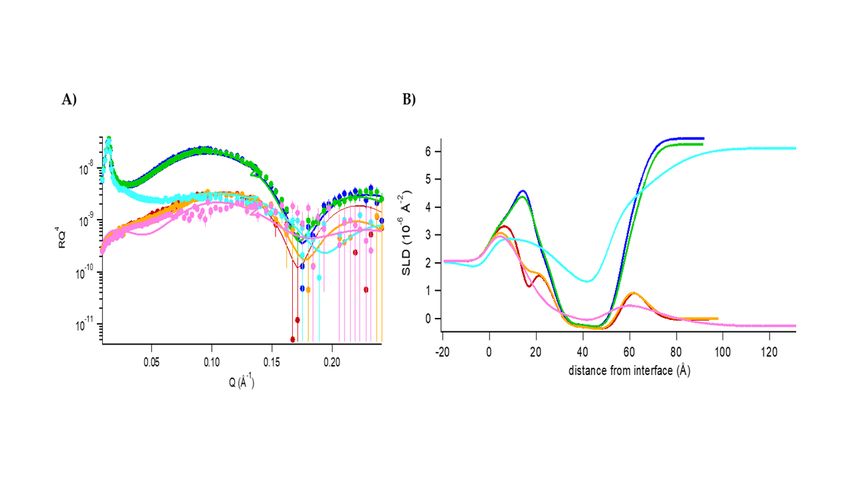
damage-associated molecular pattern; (DMSO) dimethylsulfoxide; (Flg22) flagellin; (GIPC) glycosyl-inositol-
                                                                                                                                                                                                                                                              phosphoryl-ceramide; (GluCer) glucosylceramide; (GP) generalized polarization; (HPO) fatty acid hydroper-
                                                                                                                                                                                                                                                              oxide; (ISR) induced systemic resistance; (LOX-1) Lipoxidase from Glycine max (soybean) type I-B; (MLV)
Posted on Authorea 2 Mar 2021 | The copyright holder is the author/funder. All rights reserved. No reuse without permission. | https://doi.org/10.22541/au.161468800.08766598/v1 | This a preprint and has not been peer reviewed. Data may be preliminary.

                                                                                                                                                                                                                                                              multilamellar vesicles; (MS) Murashige and Skoog medium; (NR) neutron reflectometry; (OGA) oligoga-
                                                                                                                                                                                                                                                              lacturonides; (PAMP) pathogen-associated molecular pattern; (PLPC) 1-palmitoyl-2-linoleoyl-sn-glycero-3-
                                                                                                                                                                                                                                                              phosphocholine; (POX) peroxidase; (PPM) plant plasma membrane; (PRR) pattern recognition receptor;
                                                                                                                                                                                                                                                              (RBOH) respiratory burst oxidase homolog; (RLU) relative light unit; (RT) room temperature; (SAR) sys-
                                                                                                                                                                                                                                                              temic acquired resistance; (SLB) supported lipid bilayer; (SLD) scattering length density; (SUV) small
                                                                                                                                                                                                                                                              unilamellar vesicle; (THSD) Tukey honest significant differences; (TRIS) tri(hydroxymethyl)aminomethane.

                                                                                                                                                                                                                                                              Introduction
                                                                                                                                                                                                                                                              Since several chemical pesticides have been shown to be detrimental on human health and ecosystems,
                                                                                                                                                                                                                                                              considerable research has been done to find more environment-friendly plant protection solutions (Carvalho,
                                                                                                                                                                                                                                                              2017; Gay, 2012; Hernández et al., 2013; Wang et al., 2013). Elicitors, defined as molecules able to stimulate
                                                                                                                                                                                                                                                              defence responses in a host plant, are one of the emerging alternatives (Henry et al., 2012; Paré et al., 2005;
                                                                                                                                                                                                                                                              Thakur and Sohal, 2013). They can have a biological origin, be derived from plants (termed as Damage-
                                                                                                                                                                                                                                                              Associated Molecular Patterns (DAMPs)) or microorganisms (referred as Microbe-Associated Molecular
                                                                                                                                                                                                                                                              Patterns (MAMPs) or Pathogen-Associated Molecular Patterns (PAMPs)) (Malik et al., 2020; Yu et al.,
                                                                                                                                                                                                                                                              2017) or even be chemically synthesized (Luzuriaga-Loaiza et al., 2018). They can be of different chemical
                                                                                                                                                                                                                                                              natures like carbohydrate polymers, lipids, peptides and proteins (Boller and Felix, 2009; Jogaiah et al., 2019;
                                                                                                                                                                                                                                                              Pršić and Ongena, 2020; Thakur and Sohal, 2013). Their perception by plant cells results in protection based
                                                                                                                                                                                                                                                              on the activation of signalling cascades and defence mechanisms leading to the induction of plant immunity,
                                                                                                                                                                                                                                                              like the systemic acquired resistance (SAR) and the induced systemic resistance (ISR) (Malik et al., 2020;
                                                                                                                                                                                                                                                              Pršić and Ongena, 2020).
                                                                                                                                                                                                                                                              In addition to be a selective barrier between the cell and the extracellular medium, the plasma membrane is a
                                                                                                                                                                                                                                                              sensor for modification of cellular environment and plays thus a key role in the recognition process of bioactive
                                                                                                                                                                                                                                                              molecules. While many danger signals or invasion patterns are recognized by specific pattern recognition
                                                                                                                                                                                                                                                              receptors within PPM (Pršić and Ongena, 2020; Schellenberger et al., 2019), some amphiphilic elicitors, like
                                                                                                                                                                                                                                                              surfactin from Bacillus and rhamnolipids from Pseudomonas , have been strongly suggested to be perceived
                                                                                                                                                                                                                                                              by the lipid fraction of PPM (Gerbeau-Pissot et al., 2014; Henry et al., 2011; Luzuriaga-Loaiza et al., 2018;
                                                                                                                                                                                                                                                              Monnier et al., 2018). Recognition of elicitors by the plant cells first triggers early defence responses among
                                                                                                                                                                                                                                                              which the release of reactive oxygen species (ROS) (superoxide anion (O2 * ¯), hydrogen peroxide (H2 O2 )
                                                                                                                                                                                                                                                              and hydroxyl radical (* OH)), also known as the oxidative burst (Camejo et al., 2019; Mittler, 2017; Wang
                                                                                                                                                                                                                                                              et al., 2019; Zaid and Wani, 2019).
                                                                                                                                                                                                                                                              HPOs are amphiphilic molecules naturally produced by plants in response to (a)biotic stresses by the ox-
                                                                                                                                                                                                                                                              idative catabolism of polyunsaturated fatty acids. They belong to the large family of plant oxylipins (Blee,
                                                                                                                                                                                                                                                              2002; Genva et al., 2019; Wasternack and Feussner, 2018). For years, HPOs, such as 9 or 13-hydroperoxy-
                                                                                                                                                                                                                                                              9,11,15-octadecatrienoic acid (9 or 13-HPOT) and 9 or 13-hydroperoxy- 9,11-octadecadienoic acid (9 or 13-
                                                                                                                                                                                                                                                              HPOD), have been extensively studied for their signalling properties (Deboever et al., 2020a). More recently,
                                                                                                                                                                                                                                                              HPOs have emerged as a promising plant defence solution and their exogenous application to protect plants
                                                                                                                                                                                                                                                              against phytopathogens has been considered (Deboever et al., 2020b; Graner et al., 2003; Prost et al., 2005).
                                                                                                                                                                                                                                                              They display direct biocide activity against various plant bacteria and fungi (Prost et al., 2005). In our
                                                                                                                                                                                                                                                              previous study (Deleu et al., 2019), we have also shown that HPOs are able to interact with PPM lipids and
                                                                                                                                                                                                                                                              to perturb their lateral organization. We have hypothesized that by this interaction, HPOs could activate
                                                                                                                                                                                                                                                              cellular signaling involved in plant defense mechanism.
                                                                                                                                                                                                                                                              In the present study, we first explored the potential of exogenously applied HPOs to protect Arabidopsis
                                                                                                                                                                                                                                                              thaliana plants againstBotrytis cinerea by a systemic signalling mechanism. Their eliciting activity was
                                                                                                                                                                                                                                                              evaluated by measuring the ROS production byArabidopsis thaliana cells in their presence. In a second part,

                                                                                                                                                                                                                                                                                                                     3
the molecular mechanism of HPO perception by the PPM was further investigated on plant biomimetic lipid
Posted on Authorea 2 Mar 2021 — The copyright holder is the author/funder. All rights reserved. No reuse without permission. — https://doi.org/10.22541/au.161468800.08766598/v1 — This a preprint and has not been peer reviewed. Data may be preliminary.

                                                                                                                                                                                                                                                              systems by using a panel of complementary biophysical tools. More particularly, we analysed the effects of
                                                                                                                                                                                                                                                              HPOs on the transversal organization and on the structure of the PPM bilayer.

                                                                                                                                                                                                                                                              Experimental procedures
                                                                                                                                                                                                                                                              Materials
                                                                                                                                                                                                                                                              As described in our previous works (Deboever et al., 2020b; Deleu et al., 2019; Fauconnier and Marlier,
                                                                                                                                                                                                                                                              1996), HPOs were enzymatically synthetized from the reaction of LOX-1 on linoleic (13-HPOD) or linolenic
                                                                                                                                                                                                                                                              acid (13-HPOT). The purity (higher than 98%) was checked by high-performance liquid chromatography.
                                                                                                                                                                                                                                                              For deuterated 13-HPOD, we used only deuterated reactants and solvents. Nuclear magnetic resonance and
                                                                                                                                                                                                                                                              mass spectrometry were used for a full chemical characterization of the samples (data not shown).
                                                                                                                                                                                                                                                              1-palmitoyl-2-linoleoyl-sn-glycero-3-phosphocholine (PLPC), β-sitosterol, C16 glucosyl(ß) ceramide
                                                                                                                                                                                                                                                              (d18:1/16:0) (GluCer), lipoxidase from Glycine max (soybean) type I-B (LOX-1), the linoleic and α-
                                                                                                                                                                                                                                                              linolenic acids, 6-Dodecanoyl-N,N-dimethyl-2-naphthylamine (Laurdan), horseradish peroxidases, luminol
                                                                                                                                                                                                                                                              were purchased from Sigma-Aldrich (Belgium). 1,2-dipalmitoyl-d62-sn-glycero-3-phosphocholine (d62 DPPC)
                                                                                                                                                                                                                                                              was purchased from Avanti Polar Lipids (Italy). Deuterium oxide (D2 O) of 99.8% purity was purchased
                                                                                                                                                                                                                                                              from ARMAR (Europa) GmbH. Chloroform and methanol were both purchased from Scharlau Lab
                                                                                                                                                                                                                                                              Co. Dimethylsulfoxide (DMSO) and tri(hydroxymethyl)aminomethane (TRIS) were provided by Sigma
                                                                                                                                                                                                                                                              Chemical. The ultrapure water was produced by Millipore systems available in our laboratory, the resistivity
                                                                                                                                                                                                                                                              was 18.2 M΄Ω cm. The active substance COS-OGA was provided by FytoFend S.A. (Belgium) under the
                                                                                                                                                                                                                                                              composition FytoSave® (12.5 g/L COS-OGA). Botrytis cinerea was grown on oat-based medium (25 g/L
                                                                                                                                                                                                                                                              oat flour, 12 g/L agar) at room temperature.

                                                                                                                                                                                                                                                              Induction of ISR in Arabidopsis thalianaseedlings
                                                                                                                                                                                                                                                              The capacity of HPOs to trigger ISR was tested on A. thalianainfected by B. cinerea according to the
                                                                                                                                                                                                                                                              procedure described in (Ongena et al., 2000). Seeds were sterilized with ethanol (70% v/v) and bleach
                                                                                                                                                                                                                                                              (15% v/v) before multiple rinsing with sterile water, sowed in a square Petri Dish filled with agar medium
                                                                                                                                                                                                                                                              (6-8 g/L) and transferred to a growth room at 22°C under a 16 h light/8 h dark photoperiod. After one
                                                                                                                                                                                                                                                              week, seedlings were transferred to a sterile Araponics system filled with hydroponic solution (5 mL/10 L
                                                                                                                                                                                                                                                              of Hydroponic Nutrient Solution 3-part Mix). After approximately 5 weeks in the growth room, the plants
                                                                                                                                                                                                                                                              were transferred to 10 mL vials containing 10 mL hydroponic and kept in the dark wrapped in aluminium
                                                                                                                                                                                                                                                              foil then transferred to adapt for one day before elicitation. The next day, half of the plants were treated
                                                                                                                                                                                                                                                              in vials with 10 mL hydroponic solution supplemented with 20 mM HPOs in 1% DMSO. The other half
                                                                                                                                                                                                                                                              (control) was treated with 10 mL hydroponic solution with 1% DMSO. After 24 h, four leaves of each plant
                                                                                                                                                                                                                                                              were infected with B. cinerea . A 3-μL droplet containing 2 500 spores was deposited on the adaxial face of
                                                                                                                                                                                                                                                              each leaf. Four days after inoculation, the disease was scored as the percentage of B. cinerea lesions having
                                                                                                                                                                                                                                                              extended beyond the inoculum drop zone to produce spreading lesions (Ongena et al., 2007, 2000). Three
                                                                                                                                                                                                                                                              independent experiments were carried out, with 8 plants per treatment.

                                                                                                                                                                                                                                                              Production of H2 O2 by Arabidopsis thaliana
                                                                                                                                                                                                                                                              Photoautotrophic cell suspensions from A. thaliana strain Landsberg erecta ecotype were cultured on a
                                                                                                                                                                                                                                                              rotary shaker at 100 rpm, in Murashige and Skoog (MS) medium (4.4 g/L) with 0.5 mg/L naphthalene
                                                                                                                                                                                                                                                              acetic acid, 0.05 mg/L kinetin, pH 5.7 and maintained at 24°C with approximately 2% CO2 under a 16 h/8
                                                                                                                                                                                                                                                              h light/dark photoperiod. H2 O2 production was assessed using luminol-dependent chemiluminescence on
                                                                                                                                                                                                                                                              seven-day-old cells directly after the addition of the elicitors in the growth medium, according to the method
                                                                                                                                                                                                                                                              described by Baker and Mock (Baker and Mock, 2004). Luminescence (relative light units, (RLU)) was

                                                                                                                                                                                                                                                                                                                    4
measured every three min for 90 min. Eight technical replicates were carried out for each test compound and
Posted on Authorea 2 Mar 2021 — The copyright holder is the author/funder. All rights reserved. No reuse without permission. — https://doi.org/10.22541/au.161468800.08766598/v1 — This a preprint and has not been peer reviewed. Data may be preliminary.

                                                                                                                                                                                                                                                              three independent measurements were performed. Results were expressed as means ± standard deviations
                                                                                                                                                                                                                                                              of the area under the H2 O2 production curves. ROS production values were analysed using Tukey Honest
                                                                                                                                                                                                                                                              Significant Differences (THSD) test for multiple comparisons (p< 0.1).

                                                                                                                                                                                                                                                              Calcein leakage
                                                                                                                                                                                                                                                              PLPC/sito/GluCer (60:20:20) small unilamellar vesicles (SUVs) were prepared as described previously (De-
                                                                                                                                                                                                                                                              boever et al., 2020b; Deleu et al., 2013, 2019). PLPC, sitosterol and GluCer in proportion 60:20:20 were
                                                                                                                                                                                                                                                              dissolved in a chloroform/methanol mixture (2/1, v/v). The solvent was evaporated under a gentle stream
                                                                                                                                                                                                                                                              of nitrogen to obtain a dried lipid film which was maintained under vacuum overnight. 10 mM calcein in 10
                                                                                                                                                                                                                                                              mM TRIS-HCl buffer pH 7.4 was added to hydrate the dried lipid film. The lipid dispersion was maintained
                                                                                                                                                                                                                                                              at 37°C for at least 1 h and vortexed every 10 min. 5 cycles of freeze-thawing were applied to spontaneously
                                                                                                                                                                                                                                                              form multilamellar vesicles. To obtain SUVs, this suspension was sonicated to clarity (5 cycles x 2 min) using
                                                                                                                                                                                                                                                              a titanium probe with 400W amplitude keeping the suspension in an ice bath. Finally, generated titanium
                                                                                                                                                                                                                                                              particles were removed from SUV solution by centrifuging during 10 min at 6200 rpm. The unencapsulated
                                                                                                                                                                                                                                                              calcein was removed from the SUV dispersion by the Sephadex G65 mini-column separation technique (Fu
                                                                                                                                                                                                                                                              and Singh, 1999). The actual phospholipid content of each preparation was determined by phosphorus assay
                                                                                                                                                                                                                                                              (Bartlett, 1958) and the concentration of liposomes was adjusted for each type of experiment to 5 μM in 10
                                                                                                                                                                                                                                                              mM TRIS-HCl buffer at pH 7.4.
                                                                                                                                                                                                                                                              Fluorescence was measured as previously described in (Bartlett, 1958) with a Perkin Elmer (model LS50B)
                                                                                                                                                                                                                                                              fluorescence spectrometer equipped with polarizers. Total amount of calcein release was determined by
                                                                                                                                                                                                                                                              adding Triton-X100 (0.2%) to a liposome suspension that dissolved the lipid membrane without interfering
                                                                                                                                                                                                                                                              with the fluorescence signals. The emission and excitation wavelengths were set at 517 nm and 467 nm,
                                                                                                                                                                                                                                                              respectively. A fluorescence signal of 750 μL of SUV was first recorded as a baseline, followed by the addition
                                                                                                                                                                                                                                                              of 13-HPOD/T (at t=30 sec) in 7 different concentrations while continuing the recording for 900 s. The
                                                                                                                                                                                                                                                              amount of calcein released after time t was calculated according to (Shimanouchi et al., 2009):

                                                                                                                                                                                                                                                                                                                         (It − Io )
                                                                                                                                                                                                                                                                                                       RF (%) = 100
                                                                                                                                                                                                                                                                                                                        (Imax − Io )

                                                                                                                                                                                                                                                              where RF is the fraction of calcein released, Io , It and Imax are the fluorescence intensities measured at
                                                                                                                                                                                                                                                              the beginning of the experiment, at time t and after the addition of 0.2% Triton X-100, respectively. All
                                                                                                                                                                                                                                                              experiments were carried out at least three times, each time with freshly prepared SUVs.

                                                                                                                                                                                                                                                              Laurdan generalized polarization
                                                                                                                                                                                                                                                              For Laurdan generalized polarization experiments, multilamellar vesicles (MLVs) were prepared based on
                                                                                                                                                                                                                                                              (Deboever et al., 2020b; Parasassi and Gratton, 1995). PLPC, sitosterol and GluCer in proportion 60:20:20
                                                                                                                                                                                                                                                              were dissolved in a chloroform/methanol mixture (2/1, v/v). HPOs were added to the lipid mixture to reach
                                                                                                                                                                                                                                                              a lipid:HPO molar ratio of 5:1. The solvent was evaporated under a gentle stream of nitrogen to obtain a
                                                                                                                                                                                                                                                              dried lipid film which was maintained under vacuum overnight. The resulting film was hydrated with 10 mM
                                                                                                                                                                                                                                                              Tris-HCl buffer at pH 7.4 prepared from Milli-Q water and 1 μL of Laurdan solution prepared in DMSO was
                                                                                                                                                                                                                                                              added to reach a final concentration of 5 nM. The lipid dispersion was maintained at a temperature well
                                                                                                                                                                                                                                                              above the transition phase temperature of the lipid for at least 1 h and vortexed every 10 min.
                                                                                                                                                                                                                                                              Fluorescence of Laurdan in MLVs was monitored at various temperatures (between 20 and 50°C by steps
                                                                                                                                                                                                                                                              of 5°C) with a Perkin Elmer LS50B fluorescence spectrometer. Samples were placed in 10 mm pathlength
                                                                                                                                                                                                                                                              quartz cuvettes under continuous stirring and the cuvette holder was thermostated with a circulating bath.
                                                                                                                                                                                                                                                              Samples were equilibrated at each temperature for 10-15 min prior to the measurements.

                                                                                                                                                                                                                                                                                                                    5
The excitation wavelength was set to 360 nm (slit = 2.5 nm), and at least 10 measurements of emission
Posted on Authorea 2 Mar 2021 — The copyright holder is the author/funder. All rights reserved. No reuse without permission. — https://doi.org/10.22541/au.161468800.08766598/v1 — This a preprint and has not been peer reviewed. Data may be preliminary.

                                                                                                                                                                                                                                                              intensities at 440 nm and 490 nm were recorded and averaged for each sample and the blank (DMSO) at
                                                                                                                                                                                                                                                              each temperature. An emission spectrum from 400 nm to 600 nm (slit = 4.5 nm) was also recorded for each
                                                                                                                                                                                                                                                              sample-temperature combination. Generalized polarization (GP) of Laurdan was then calculated according
                                                                                                                                                                                                                                                              to (Harris et al., 2002; Parasassi et al., 1992):

                                                                                                                                                                                                                                                                                                                    I440 − I490
                                                                                                                                                                                                                                                                                                            GP =
                                                                                                                                                                                                                                                                                                                    I440 + I490

                                                                                                                                                                                                                                                              where I440 and I490 are the blank-subtracted emission intensities at 440 nm and 490 nm, respectively. All
                                                                                                                                                                                                                                                              experiments were carried out at least three times, each time with freshly prepared MLVs.

                                                                                                                                                                                                                                                              Neutron reflectometry
                                                                                                                                                                                                                                                              Neutron reflectometry (NR) measurements were performed at the MARIA neutron reflectometer (Mattauch
                                                                                                                                                                                                                                                              et al., 2018) operated by Jülich Centre for Neutron Science at Heinz Maier-Leibnitz Zentrum in Garching
                                                                                                                                                                                                                                                              (Germany) while using custom temperature-regulated (through a connected Julabo F12-ED circulator) liquid
                                                                                                                                                                                                                                                              cells (Koutsioubas, 2016). Two different wavelengths were used, 10 Å for the low-q region and 5 Å for the
                                                                                                                                                                                                                                                              high-q region up to 0.25 Å-1 , with a 10% wavelength spread. Using a peristaltic pump combined with
                                                                                                                                                                                                                                                              valves (flow rate ˜0.5 mL/min) solvent exchange was possible without moving the measuring cells from the
                                                                                                                                                                                                                                                              instrument.
                                                                                                                                                                                                                                                              Specular NR measures the thickness and scattering length density (SLD) profile of layered structures along
                                                                                                                                                                                                                                                              the surface normal (z). The SLD distribution along the normal, represented as ρ(z), is specific of the chemical
                                                                                                                                                                                                                                                              composition of materials along the normal and depends on the coherent nuclear scattering lengths (bi ) of
                                                                                                                                                                                                                                                              its constituent atoms and their number density along the normal (ni (z)) so that ρ(z) = [?]i bi ni (z). In
                                                                                                                                                                                                                                                              reflectivity data measurements, the intensity of reflected neutrons is recorded relatively to the incident beam
                                                                                                                                                                                                                                                              as a function of the momentum transfer vector (q = 4πsin θ/λ), where θ is the incidence angle and λ the
                                                                                                                                                                                                                                                              wavelength of incident neutrons. The variation of reflectivity as a function of momentum transfer R(qz ) is
                                                                                                                                                                                                                                                              related to the square modulus of the one-dimensional Fourier transform of the SLD profile along the normal
                                                                                                                                                                                                                                                              to the interface (ρ(qz )) through the relation:

                                                                                                                                                                                                                                                                                                        R(qz ) ∼ (16π /qz )|ρ(qz )|

                                                                                                                                                                                                                                                              Following the characterization by neutron reflectivity of silicon/solution interface, we deposited by vesicle
                                                                                                                                                                                                                                                              fusion the membrane of interest (Koutsioubas et al., 2017). After its full characterization in 150 mM
                                                                                                                                                                                                                                                              NaCl solutions in D2 O and H2 O, 2 μg of 13-HPOD/T (a concentration lower than their critical micelle
                                                                                                                                                                                                                                                              concentration (CMC = 25.4 ± 1.9 μM and 24.0 ± 1.3 μM for 13-HPOT and 13-HPOD, respectively, according
                                                                                                                                                                                                                                                              to (Deleu et al., 2019)), deuterated or not depending on the membrane studied, were injected in the measuring
                                                                                                                                                                                                                                                              cell (6 mL total volume). Reflectivity was measured after letting the systems equilibrate for 1 h, again in
                                                                                                                                                                                                                                                              the two contrasts condition with 10 mM Tris (pH 7.4).
                                                                                                                                                                                                                                                              To analyse the specular reflection data, the interface is modelled as a series of parallel layers where each layer
                                                                                                                                                                                                                                                              is characterized by an average SLD and a thickness. Based on these parameters, a model reflectivity profile
                                                                                                                                                                                                                                                              is calculated by means of the optical matrix method (Névot and Croce, 1980). The interfacial roughness
                                                                                                                                                                                                                                                              between two consecutive layers is included in the model by the Abeles method, as described by Nevot and
                                                                                                                                                                                                                                                              Croce (Névot and Croce, 1980).
                                                                                                                                                                                                                                                              Finally, the calculated model profile is compared to the measured profile and the quality of the fit is assessed
                                                                                                                                                                                                                                                              by using the χ2 in minimum-squares method. NR is a technique suited to collect structural information about
                                                                                                                                                                                                                                                              the different layers of the studied membrane (Rondelli et al., 2019). Thus, the silicon support and the bulk
                                                                                                                                                                                                                                                              water are seen as bulk infinite layer, the silicon oxide layer, the water layer between the silicon oxide and the

                                                                                                                                                                                                                                                                                                                      6
membrane and the diverse hydrophilic/hydrophobic layers of the lipid membranes are modelled as defined
Posted on Authorea 2 Mar 2021 — The copyright holder is the author/funder. All rights reserved. No reuse without permission. — https://doi.org/10.22541/au.161468800.08766598/v1 — This a preprint and has not been peer reviewed. Data may be preliminary.

                                                                                                                                                                                                                                                              layers with a proper thickness, roughness with respect to the previous layer, compactness, composition and
                                                                                                                                                                                                                                                              consequently contrast. Supported lipid bilayers (SLB) were formed using both the same lipid mixture as
                                                                                                                                                                                                                                                              previously (PLPC/sito/GluCer in molar ratio 60/20/20) and d62 DPPC. Injections were done at 47°C and
                                                                                                                                                                                                                                                              measurements at room temperature (RT). The reflectivity profile of the silicon support and of the samples
                                                                                                                                                                                                                                                              has been measured in different contrasts (H2 O and D2 O) and data analysis was performed with the fit
                                                                                                                                                                                                                                                              program MOTOFIT (Nelson, 2006). SLD used for the specific components are reported in Supplementary
                                                                                                                                                                                                                                                              DataTable S1 .

                                                                                                                                                                                                                                                              Atomic force microscopy
                                                                                                                                                                                                                                                              To probe the nanoscale effects of HPOs on lipid membranes, SLB (ternary mixture of PLPC/sito/GluCer
                                                                                                                                                                                                                                                              (60/20/20)) were reconstructed on freshly cleaved mica substrates by allowing the fusion of a 2 mM lipid
                                                                                                                                                                                                                                                              vesicles solution (V = 100 μL) at 55°C for 45 min. Samples were then left for thermalization at room
                                                                                                                                                                                                                                                              temperature for 30 min without dewetting and immersed in 3 mL Tris buffer (pH 7.5).
                                                                                                                                                                                                                                                              To avoid damaging the samples, atomic force microscopy (AFM) images were obtained in the quantitative
                                                                                                                                                                                                                                                              imaging (QI) mode of a JPK Nanowizard III setup, with a minimal applied force of 200 pN and a speed
                                                                                                                                                                                                                                                              of 50 μm/s. Soft sharpened silicon nitride cantilevers (MSCT, Bruker) were used and calibrated before any
                                                                                                                                                                                                                                                              experiment using the thermal noise method (k ˜ 0.02 N/m). HPOs, prepared in Tris buffer, were injected
                                                                                                                                                                                                                                                              to reach a final concentration of 3 μM below their critical micellar concentration. AFM images were then
                                                                                                                                                                                                                                                              recorded at different time points in different areas to follow the HPOs impact on the lipid bilayer.

                                                                                                                                                                                                                                                              Results
                                                                                                                                                                                                                                                              In planta protective effect of HPOs against phytopathogens
                                                                                                                                                                                                                                                              The capacity of HPOs to induce systemic resistance in A. thaliana against B. cinerea was tested under
                                                                                                                                                                                                                                                              controlled conditions by treating plant roots with HPO solutions and inoculating plant leaves with the
                                                                                                                                                                                                                                                              pathogen. The disease severity with HPO treatment was measured after 4 days and compared to controls
                                                                                                                                                                                                                                                              (treatment with water containing 1% DMSO).
                                                                                                                                                                                                                                                              Treatment with both HPOs provided similar obvious disease reduction as shown by the decreased size of the
                                                                                                                                                                                                                                                              lesions (Figure 1 ). About 40% of the plants had no symptoms. Given the experimental design (treatment
                                                                                                                                                                                                                                                              and infection on two different plant organs), this cannot be a direct biocidal effect but rather a systemic
                                                                                                                                                                                                                                                              signalling in the plant. One can therefore wonder about the signalling mechanism initiated by HPOs and
                                                                                                                                                                                                                                                              more particularly their initial perception by the plant cells and the responses they induce.

                                                                                                                                                                                                                                                              Perception of HPOs and early defenses responses activation
                                                                                                                                                                                                                                                              Very often, ROS production is a biphasic process with a first transient phase within minutes after the infection
                                                                                                                                                                                                                                                              and a second more intense and sustained phase that can last for many hours (Wang et al., 2019). This first
                                                                                                                                                                                                                                                              wave, which is linked to the activation of early defence responses, has been investigated to determine whether
                                                                                                                                                                                                                                                              the two HPOs are perceived by plants and can induce an immune response.
                                                                                                                                                                                                                                                              Cell suspensions cultures are a valuable model system for studying elicitor-induced defence reactions in plants
                                                                                                                                                                                                                                                              and they easily allow studying early signalling events like oxidative bursts (Jogaiah et al., 2019; Khonon et
                                                                                                                                                                                                                                                              al., 2011). Here, photoautotrophic A. thalianacell suspensions were used to detect H2 O2 production after
                                                                                                                                                                                                                                                              treatment with HPOs. Extracellular ROS accumulation was detected using a luminol-based assay (Monnier
                                                                                                                                                                                                                                                              et al., 2018; van Aubel et al., 2016). The elicitor FytoSave® was used as positive control as its active
                                                                                                                                                                                                                                                              substance, COS-OGA made of pectin-derived oligogalacturonides (OGA) and chitooligosaccharides (COS),
                                                                                                                                                                                                                                                              is known to induce a significant production of ROS at a concentration of 25 ppm (Ledoux et al., 2014; van

                                                                                                                                                                                                                                                                                                                     7
Aubel et al., 2018, 2016). A range of six different concentrations (0.5 μM to 100 μM) was tested and ROS
Posted on Authorea 2 Mar 2021 — The copyright holder is the author/funder. All rights reserved. No reuse without permission. — https://doi.org/10.22541/au.161468800.08766598/v1 — This a preprint and has not been peer reviewed. Data may be preliminary.

                                                                                                                                                                                                                                                              production was monitored for 90 min.
                                                                                                                                                                                                                                                              The ROS production after 13-HPOD or 13-HPOT treatment was concentration-dependent (Figure 2A).It
                                                                                                                                                                                                                                                              was higher than the controls, negative or positive, for concentrations 50 μM and 100μM. In those cases, the
                                                                                                                                                                                                                                                              response to 13-HPOT treatment is higher than the 13-HPOD one. Also, the oxidative burst peak occurred
                                                                                                                                                                                                                                                              quicker (30 min instead of 40 min) for 13-HPOT than for 13-HPOD (Figure 2B). Comparatively to COS-
                                                                                                                                                                                                                                                              OGA, the kinetics of ROS production induced by HPOs in plant cells was slower (within 5 min only for
                                                                                                                                                                                                                                                              COS-OGA vs 30-40 min for HPOs) but the oxidative burst lasted longer with a total duration of 60-70
                                                                                                                                                                                                                                                              min before returning to the basal level. Such long lasting response profile was also observed with synthetic
                                                                                                                                                                                                                                                              rhamnolipid bolaforms, for which it was suggested that their perception occurred via the lipid fraction of
                                                                                                                                                                                                                                                              the plasma membrane (Luzuriaga-Loaiza et al., 2018). On the contrary, the elicitor flagellin, known to be
                                                                                                                                                                                                                                                              perceived by membrane pattern recognition receptors (Smith and Heese, 2014), induces a quicker oxidative
                                                                                                                                                                                                                                                              burst initiated within 4-6 min with a peak at ˜10min (Yu et al., 2017), similarly to the OGA in the COS-
                                                                                                                                                                                                                                                              OGA composition (Smith and Heese, 2014; van Aubel et al., 2013). The observation of different kinetic
                                                                                                                                                                                                                                                              profiles for HPOs would suggest that a protein receptor is not directly involved in their recognition. Due to
                                                                                                                                                                                                                                                              the ability of HPOs to interact with PPM lipids (Deleu et al., 2019), we hypothesized that the HPOs would
                                                                                                                                                                                                                                                              rather be recognized via the lipid phase of the membrane.

                                                                                                                                                                                                                                                              Changes of PPM biophysical properties induced by HPOs
                                                                                                                                                                                                                                                              The interaction of HPOs with PPM characteristic lipids has already been found to modify the lateral orga-
                                                                                                                                                                                                                                                              nization of membrane bilayer in terms of lipid domain size and distribution (Deleu et al., 2019). It is also
                                                                                                                                                                                                                                                              known that the plant sphingolipid GluCer is a privileged partner for the interaction and that 13-HPOT has
                                                                                                                                                                                                                                                              a higher interaction affinity than 13-HPOD.
                                                                                                                                                                                                                                                              In the present study, further analysis of the effects of HPOs interaction with lipids on PPM structure was
                                                                                                                                                                                                                                                              carried out. Simplified biomimetic models with two different lipid compositions were studied, the first
                                                                                                                                                                                                                                                              mimicking the PPM, namely PLPC:sito:GluCer (60/20/20), and the second made of d62 DPPC, a classic
                                                                                                                                                                                                                                                              deuterated model.

                                                                                                                                                                                                                                                              Effect of HPOs on membrane permeability
                                                                                                                                                                                                                                                              First, we have investigated the ability of HPOs to permeabilize model membranes by measuring the release
                                                                                                                                                                                                                                                              of calcein. If the membrane is permeabilized by a bioactive molecule, the self-quenched calcein initially
                                                                                                                                                                                                                                                              encapsulated within the LUV is released into the external medium and gives rise to an increase of fluorescence
                                                                                                                                                                                                                                                              emission. Very little permeabilization effect was observed (values less than 10%) for both HPOs on the
                                                                                                                                                                                                                                                              PLPC:sito:GluCer membrane model (Figure S1 ) suggesting that HPOs would not derive their mode of
                                                                                                                                                                                                                                                              action from a mechanism of solubilization of the membrane or pore formation, but rather a more subtle
                                                                                                                                                                                                                                                              modification of the membrane organization that could lead to the activation of a signalling cascade.

                                                                                                                                                                                                                                                              Change in bilayer fluidity induced by HPOs
                                                                                                                                                                                                                                                              The effect of HPOs on the bilayer fluidity was investigated by monitoring the lipid phase-dependent emission
                                                                                                                                                                                                                                                              spectrum shift of Laurdan, a fluorescent probe that readily locates at the hydrophilic/hydrophobic interface
                                                                                                                                                                                                                                                              of bilayers (Harris et al., 2002; Sanchez et al., 2007). Its fluorescence depends on the physical state of the
                                                                                                                                                                                                                                                              environment. When present in a gel phase bilayer, its maximum fluorescence intensity is close to 440 nm
                                                                                                                                                                                                                                                              emission wavelength. When the bilayer is in a fluid state, the Laurdan maximum fluorescence is observed at
                                                                                                                                                                                                                                                              higher wavelengths (around 490 nm). This ”red-shift” phenomenon is due to a higher quantity and mobility
                                                                                                                                                                                                                                                              of the water molecules located around the probe. This is directly related to the lower order within the bilayer
                                                                                                                                                                                                                                                              and is measured by the Generalized Polarization (GP): a decreasing GP value corresponds to a higher fluidity
                                                                                                                                                                                                                                                              of the bilayer (Parasassi et al., 1991). This method has been previously applied for investigating the effect

                                                                                                                                                                                                                                                                                                                    8
of drugs, natural herbicides or other elicitors on lipid membrane organization (Deleu et al., 2013; Furlan et
Posted on Authorea 2 Mar 2021 — The copyright holder is the author/funder. All rights reserved. No reuse without permission. — https://doi.org/10.22541/au.161468800.08766598/v1 — This a preprint and has not been peer reviewed. Data may be preliminary.

                                                                                                                                                                                                                                                              al., 2020; Lebecque et al., 2019; Sautrey et al., 2014).
                                                                                                                                                                                                                                                              The effect of HPOs on PLPC:sito:GluCer MLV membrane fluidity was investigated for a range of tempera-
                                                                                                                                                                                                                                                              tures from 20°C to 50°C (Figure 3 ). In presence of 13-HPOT, the Laurdan GP values decreased significantly
                                                                                                                                                                                                                                                              compared to those observed on pure MLVs with no significant effect of temperature. This indicates a flui-
                                                                                                                                                                                                                                                              difying effect of 13-HPOT on the bilayer. On the contrary, 13-HPOD did not induce significant change in
                                                                                                                                                                                                                                                              lipid order at any temperature as its curve almost superimposes to that of pure PLPC:sito:GluCer vesicles.

                                                                                                                                                                                                                                                              Effect of HPOs on the bilayer transversal structure
                                                                                                                                                                                                                                                              The effect of HPOs on the transversal structure of PPM was analysed by NR, a technique of choice to
                                                                                                                                                                                                                                                              study the transverse structure of layered samples within a few Å resolution (Mattauch et al., 2018) and to
                                                                                                                                                                                                                                                              evidence the structural effects of the interaction of incoming molecules on biological membranes (Rondelli et
                                                                                                                                                                                                                                                              al., 2018, 2016). Neutrons interaction with matter depends on the isotopic species. Therefore, neutron-based
                                                                                                                                                                                                                                                              experiments can profit by the use of deuterated molecules to enhance the visibility of molecules within a
                                                                                                                                                                                                                                                              mixed complex system. As the lipids representative of the PPM are not commercially available in their
                                                                                                                                                                                                                                                              deuterated form, d62 DPPC was used to form SLB and to highlight the presence and location of the H-
                                                                                                                                                                                                                                                              bringing HPOs within the membrane.Figure 4A-D show the reflectivity curves together with their fittings
                                                                                                                                                                                                                                                              in two contrasts and the corresponding fit parameters are summarized in Figure 4E . NR spectra were
                                                                                                                                                                                                                                                              not drastically changed by the addition of HPOs. However, the spectra revealed that HPOs always insert
                                                                                                                                                                                                                                                              into the outer membrane leaflet of the d62 DPPC SLB without flipping into the inner layer attached to the
                                                                                                                                                                                                                                                              silicon block. A slight modification of the SLD profiles was observed while adding 13-HPOT but not with
                                                                                                                                                                                                                                                              13-HPOD. This gave rise to a small but significant decrease of the membrane thickness (approximately 2.5
                                                                                                                                                                                                                                                              Å) and roughness without modification of the solvent penetration, i.e ., no alteration of the bilayer. To
                                                                                                                                                                                                                                                              confront these results, obtained on a d62 DPPC bilayer, to a more realistic PPM model, another experiment
                                                                                                                                                                                                                                                              was performed with the deuterated version of 13-HPOD and the ternary mixture of non-deuterated lipids
                                                                                                                                                                                                                                                              representative of PPM. It confirms that 13-HPOD interacts with PPM SLB and localizes on top of the outer
                                                                                                                                                                                                                                                              leaflet without no major change of the membrane organization as observed from SLD profile and NR spectra
                                                                                                                                                                                                                                                              (Figure S2 and Table S2 ).

                                                                                                                                                                                                                                                              Lateral erosion of plant lipid bilayers by HPOs
                                                                                                                                                                                                                                                              To further analyse the effect of HPOs on the lipid bilayer organization, atomic force microscopy (AFM) was
                                                                                                                                                                                                                                                              used to investigate their impact on the lateral nanoscale morphology of SLB. As shown in Figure 5A , the
                                                                                                                                                                                                                                                              ternary mixture of plant lipids reconstituted in SLB did not reveal any phase separation within the thickness
                                                                                                                                                                                                                                                              resolution limit of AFM (0.1 nm), rather homogeneous smooth patches (bright areas) distributed on the mica
                                                                                                                                                                                                                                                              (dark areas). Though large patches were mainly found to cover the entire scanned area, defaults in the SLB
                                                                                                                                                                                                                                                              were used as a “visualization control” to confirm the presence of the lipid bilayer. Its thickness of ˜4-5 nm,
                                                                                                                                                                                                                                                              determined by measuring section profiles on the AFM height images, is in agreement with previous studies
                                                                                                                                                                                                                                                              (Figure 5B ) (Dufrêne and Lee, 2000; Mingeot-Leclercq et al., 2008). The presence of the lipid bilayer was
                                                                                                                                                                                                                                                              further confirmed by recording AFM force curves on areas of high vs low heights. A typical breakthrough
                                                                                                                                                                                                                                                              of the lipid bilayer in the bright areas was observed while no such force signature was found in dark areas
                                                                                                                                                                                                                                                              without lipid bilayers and associated with mica (Figure 5C ).
                                                                                                                                                                                                                                                              After confirming the presence of the SLB, the sample was incubated with either 13-HPOT or 13-HPOD and
                                                                                                                                                                                                                                                              AFM images were recorded every 10-15 min on a defined area. Incubation 13-HPOT or 13-HPOD resulted in
                                                                                                                                                                                                                                                              a time-dependent alteration of the lipid patches (Figure 5D ). Results showed that very small SLB patches
                                                                                                                                                                                                                                                              (green arrows) disappeared after the addition of 13-HPOT, but that it had not a drastic impact on the large
                                                                                                                                                                                                                                                              ones. On the contrary, 13-HPOD completely removed part of a large angular domain after 75 min (see green
                                                                                                                                                                                                                                                              arrows). Nonetheless, after 75 min treatment with 13-HPOT, most of the lipid domains were thinner by
                                                                                                                                                                                                                                                              approximately 2 nm as compared to the initial ones, suggesting that 13-HPOT flattened lipids or part of the
                                                                                                                                                                                                                                                              upper leaflet in a time- and zone-dependent way, which was not observed for 13-HPOD (Figure 5E-F ).

                                                                                                                                                                                                                                                                                                                    9
In brief, AFM studies revealed three major effects of HPOs on plant mimetic lipid bilayers (i) ”erosion”
Posted on Authorea 2 Mar 2021 — The copyright holder is the author/funder. All rights reserved. No reuse without permission. — https://doi.org/10.22541/au.161468800.08766598/v1 — This a preprint and has not been peer reviewed. Data may be preliminary.

                                                                                                                                                                                                                                                              of angular protrusions of large lipid domains, (ii) total erosion of small domains, and (iii) reduction in the
                                                                                                                                                                                                                                                              thickness of the bilayer between 0.5 and 2 nm. 13-HPOT has also a greater effect on membrane organization
                                                                                                                                                                                                                                                              and bilayer thickness than 13-HPOD.

                                                                                                                                                                                                                                                              General discussion and conclusion
                                                                                                                                                                                                                                                              In the present study, we show for the first time the potential of the exogenous application of acyl-
                                                                                                                                                                                                                                                              hydroperoxides 13-HPOD and 13-HPOT to protect plants against phytopathogens. Both forms of HPOs
                                                                                                                                                                                                                                                              applied on A. thaliana roots strongly reduce the size of the lesions further to the inoculation ofB. cinerea
                                                                                                                                                                                                                                                              on leaves. The protection effect without direct contact with the phytopathogen suggests their capacity to
                                                                                                                                                                                                                                                              stimulate the plant immune system. Other phyto-oxylipins, the jasmonic acid precursor 12-oxo-phytodienoic
                                                                                                                                                                                                                                                              acid and an [?]-ketol of octadecadienoic acid were recently identified as mobile signals responsible of ISR,
                                                                                                                                                                                                                                                              originated in the plant roots and travelling into the plant vasculature (Wang et al., 2020). Our results show
                                                                                                                                                                                                                                                              that exogenous application of 13-HPOD and 13-HPOT on A. thaliana cell suspension induces an importantin
                                                                                                                                                                                                                                                              vitro oxidative burst, known as a hallmark of elicitor recognition by the plant cells (Yu et al., 2017). Beyond
                                                                                                                                                                                                                                                              their role as signals, we thus clearly demonstrate that HPO are recognized by the plant cells triggering a
                                                                                                                                                                                                                                                              signalling cascade leading to ISR and plant protection against pathogens.
                                                                                                                                                                                                                                                              The extracellular ROS production is initiated earlier and lasts longer that the one observed with well-known
                                                                                                                                                                                                                                                              proteic elicitors like flagellin for which recognition phenomenon involves direct interaction with membrane
                                                                                                                                                                                                                                                              proteic receptors (Gomez-Gomez and Boller, 2002). But the kinetic profile is similar to the one observed
                                                                                                                                                                                                                                                              for other amphiphilic lipid elicitors like surfactin and rhamnolipids (Henry et al., 2011; Jourdan et al., 2009;
                                                                                                                                                                                                                                                              Luzuriaga-Loaiza et al., 2018; Ma et al., 2017) for which a mechanism linked to the perception by the lipid
                                                                                                                                                                                                                                                              of the plasma membrane was suggested.
                                                                                                                                                                                                                                                              From our previous work (Deleu et al., 2019), we know that HPOs can interact with the lipid fraction of
                                                                                                                                                                                                                                                              PPM. In the present study, NR analyses show that both HPOs are more preferably inserted in the outer
                                                                                                                                                                                                                                                              leaflet of the bilayer. This interaction modifies the global morphology of the bilayer as shown by AFM
                                                                                                                                                                                                                                                              where bilayer erosion is observed for both HPOs. 13-HPOT has a higher impact on the PPM structure, but
                                                                                                                                                                                                                                                              does not affect the integrity of the membrane according to the calcein release assays. Its insertion further
                                                                                                                                                                                                                                                              reduces the thickness of the bilayer according to the NR and AFM data and fluidifies it more according to
                                                                                                                                                                                                                                                              the Laurdan GP data than 13-HPOD. This difference between 13-HPOT and 13-HPOD could be explained
                                                                                                                                                                                                                                                              by the presence of an additional double bond in 13-HPOT which gives it a greater structure rigidity. We
                                                                                                                                                                                                                                                              postulate that this might force the lipids of the membrane outer leaflet to further reorganize compared to
                                                                                                                                                                                                                                                              a more flexible molecule like 13-HPOD, and consequently could have a stronger impact on the dynamics of
                                                                                                                                                                                                                                                              the membrane. The higher binding affinity of 13-HPOT compared to 13-HPOD for PPM bilayer (Deleu et
                                                                                                                                                                                                                                                              al., 2019) could also enhance this reorganization effect.
                                                                                                                                                                                                                                                              The correlation between the higher ROS production and the higher impact on PPM lipid bilayer structure
                                                                                                                                                                                                                                                              for 13-HPOT compared to 13-HPOD is in favour of our hypothesis that the PPM lipid fraction plays a key
                                                                                                                                                                                                                                                              role in the recognition of HPOs giving rise to plant defence mechanisms. In the study of Sandor et al. (2016),
                                                                                                                                                                                                                                                              it is demonstrated on A. thaliana and tobacco cells, that the induction of ROS by various elicitors including
                                                                                                                                                                                                                                                              cryptogein, flagellin and an oligosaccharide, is concomitant to the increase in the relative proportion of
                                                                                                                                                                                                                                                              membrane ordered domains (Sandor et al., 2016). According to them, the recognition of the elicitor at the
                                                                                                                                                                                                                                                              plasma membrane level triggers the production of ROS which in turn reorganizes the membrane leading to an
                                                                                                                                                                                                                                                              increase of ordered domains. But in the case of cryptogein, they have also suggested an inverse event sequence.
                                                                                                                                                                                                                                                              Thanks to its capacity to interact with membrane sterols (Gerbeau-Pissot et al., 2014) and to mechanically
                                                                                                                                                                                                                                                              trap them, cryptogein gives rise to a higher membrane fluidity which stimulates the ROS production. In
                                                                                                                                                                                                                                                              agreement with this study, our results suggest that elicitors that directly act on membrane lipid dynamics and
                                                                                                                                                                                                                                                              more particularly on the membrane fluidity are able to trigger early defence events like ROS production. But
                                                                                                                                                                                                                                                              the complete molecular mechanistic view between the change of the membrane dynamics and the occurrence

                                                                                                                                                                                                                                                                                                                    10
of the oxidative burst is not yet identified. The formation of specific membrane lipid domains recruiting key
Posted on Authorea 2 Mar 2021 — The copyright holder is the author/funder. All rights reserved. No reuse without permission. — https://doi.org/10.22541/au.161468800.08766598/v1 — This a preprint and has not been peer reviewed. Data may be preliminary.

                                                                                                                                                                                                                                                              signalling proteins (Gronnier et al., 2018) could be implicated. From our previous study (Deleu et al., 2019),
                                                                                                                                                                                                                                                              we also know that plant membrane sphingolipids are privileged partners for HPO interaction. Therefore,
                                                                                                                                                                                                                                                              the presence of glycosyl-inositol-phosphoryl-ceramides (GIPCs), the plant sphingolipids exclusively located
                                                                                                                                                                                                                                                              in the outer leaflet of PPM and involved in the inter-leaflet coupling (Gronnier et al., 2016), could also play
                                                                                                                                                                                                                                                              a role in the signal transduction.
                                                                                                                                                                                                                                                              In addition to their eliciting activity evidenced in the present study, HPOs also retain some antimicrobial
                                                                                                                                                                                                                                                              activity against various phytopathogens (Deboever et al., 2020b). The dual effect of HPOs as well as the
                                                                                                                                                                                                                                                              possibility to produce them at low cost (Fauconnier and Marlier, 1996) make them attractive compounds to
                                                                                                                                                                                                                                                              be used as alternative to conventional pesticides for plant protection.
                                                                                                                                                                                                                                                              Author Contributions: Experiments design, E.D., M.D., V.R., A.K., M.M.M., M.O. and G.V.A..; exper-
                                                                                                                                                                                                                                                              iments and data analysis, E.D., M.D., M.M.M., V.R. and G.V.A.; HPOs synthesis, and purification, E.D.;
                                                                                                                                                                                                                                                              writing—original draft preparation, E.D.; writing—review and editing, all authors.
                                                                                                                                                                                                                                                              Data availability statement : The data supporting the findings of this study are available from the
                                                                                                                                                                                                                                                              corresponding author, Magali Deleu, upon request.
                                                                                                                                                                                                                                                              Funding: E.D. is supported by a ¡¡ Fonds pour la formation a la Recherche dans l’Industrie et dans
                                                                                                                                                                                                                                                              l’Agriculture ¿¿ (FRIA) grant (5100617F) from the FRS-FNRS (Fonds National de la Recherche Scientifique,
                                                                                                                                                                                                                                                              Belgium). M.D., M. O. and L.L. thank the FRS-FNRS for their position as Senior Research Associates and for
                                                                                                                                                                                                                                                              grant CDR (J.0014.08 and J.0086.18 projects). Work at the Universite Catholique de Louvain was supported
                                                                                                                                                                                                                                                              by the National Fund for Scientific Research (FNRS) and the Research Department of the Communaute
                                                                                                                                                                                                                                                              Francaise de Belgique (Concerted Research Action). Y.F.D. is a Research Director at the FNRS. This
                                                                                                                                                                                                                                                              research was funded also by the ‘Medical Biotechnologies and Translational Medicine Department’ of the
                                                                                                                                                                                                                                                              ‘Universita degli Studi di Milano’, grant number ‘PSR2018’ to V.R. This article was published with the
                                                                                                                                                                                                                                                              support of the “Fondation Universitaire de Belgique”.
                                                                                                                                                                                                                                                              Acknowledgments: The authors thank the financial support via the project from University of Liege (ARC-
                                                                                                                                                                                                                                                              FIELD project 13/17-10). Authors also thank beamline MARIA Julich Centre for Neutron Science (JCNS)
                                                                                                                                                                                                                                                              at Heinz Maier-Leibnitz Zentrum (MLZ, Garching, Germany) for allocation of beamtime. Acknowledgements
                                                                                                                                                                                                                                                              are also due to FytoFend’s research team for their logistic support in bioassays. Final thanks to Jelena Prisc
                                                                                                                                                                                                                                                              for technical support and nice discussion on ISR and ROS experiment data measured in planta .
                                                                                                                                                                                                                                                              Conflicts of Interest: The authors declare no conflict of interest. The funders had no role in the design
                                                                                                                                                                                                                                                              of the study; in the collection, analyses, or interpretation of data; in the writing of the manuscript, or in the
                                                                                                                                                                                                                                                              decision to publish the results.

                                                                                                                                                                                                                                                              References
                                                                                                                                                                                                                                                              Baker, C.J., Mock, N.M., 2004. A method to detect oxidative stress by monitoring changes in the extracel-
                                                                                                                                                                                                                                                              lular antioxidant capacity in plant suspension cells. Physiol. Mol. Plant Pathol. 64, 255–261.
                                                                                                                                                                                                                                                              Bartlett, G.R., 1958. Calorimetric Assay Phosphorylated for Free Glyceric Acids. J. Biol. Chem. 234,
                                                                                                                                                                                                                                                              469–471.
                                                                                                                                                                                                                                                              Blee, E., 2002. Impact of phyto-oxylipins in plant defense. Trends Plant Sci. 7, 315–321.
                                                                                                                                                                                                                                                              Boller, T., Felix, G., 2009. A Renaissance of Elicitors: Perception of Microbe-Associated Molecular Patterns
                                                                                                                                                                                                                                                              and Danger Signals by Pattern-Recognition Receptors. Annu. Rev. Plant Biol. 60, 379–406.
                                                                                                                                                                                                                                                              Camejo, D., Guzman-cedeno, A., Vera-macias, L., Jimenez, A., 2019. Oxidative post-translational modi fi
                                                                                                                                                                                                                                                              cations controlling plant-pathogen interaction. Plant Physiol. Biochem. 144, 110–117.
                                                                                                                                                                                                                                                              Carvalho, F.P., 2017. Pesticides, environment, and food safety. Food Energy Secur. 6, 48–60.

                                                                                                                                                                                                                                                                                                                     11
Deboever, E., Deleu, M., Mongrand, S., Lins, L., Fauconnier, M.-L., 2020a. Plant–Pathogen Interactions:
Posted on Authorea 2 Mar 2021 — The copyright holder is the author/funder. All rights reserved. No reuse without permission. — https://doi.org/10.22541/au.161468800.08766598/v1 — This a preprint and has not been peer reviewed. Data may be preliminary.

                                                                                                                                                                                                                                                              Underestimated Roles of Phyto-oxylipins. Trends Plant Sci. 25, 22–34.
                                                                                                                                                                                                                                                              Deboever, E., Lins, L., Ongena, M., De Clerck, C., Deleu, M., Fauconnier, M.-L., 2020b. Linolenic fatty
                                                                                                                                                                                                                                                              acid hydroperoxide acts as biocide on plant pathogenic bacteria : biophysical investigation of the mode of
                                                                                                                                                                                                                                                              action. Bioorg. Chem. 100, 1–25.
                                                                                                                                                                                                                                                              Deleu, M., Deboever, E., Nasir, M.N., Crowet, J.-M., Dauchez, M., Ongena, M., Jijakli, H., Fauconnier,
                                                                                                                                                                                                                                                              M.-L., Lins, L., 2019. Linoleic and linolenic acid hydroperoxides interact differentially with biomimetic plant
                                                                                                                                                                                                                                                              membranes in a lipid specific manner. Colloids Surfaces B Biointerfaces 175, 384–391.
                                                                                                                                                                                                                                                              Deleu, M., Lorent, J., Lins, L., Brasseur, R., Braun, N., El Kirat, K., Nylander, T., Dufrene, Y.F., Mingeot-
                                                                                                                                                                                                                                                              Leclercq, M.P., 2013. Effects of surfactin on membrane models displaying lipid phase separation. Biochim.
                                                                                                                                                                                                                                                              Biophys. Acta - Biomembr. 1828, 801–815.
                                                                                                                                                                                                                                                              Dufrene, Y.F., Lee, G.U., 2000. Advances in the characterization of supported lipid films with the atomic
                                                                                                                                                                                                                                                              force microscope. Biochim. Biophys. Acta - Biomembr. 1509, 14–41.
                                                                                                                                                                                                                                                              Fauconnier, M.L., Marlier, M., 1996. An efficient procedure for the production of fatty acid hydroperoxides
                                                                                                                                                                                                                                                              from hydrolyzed flax seed oil and soybean lipoxygenase. Biotechnol. Tech. 10, 839–844.
                                                                                                                                                                                                                                                              Fu, F.N., Singh, B.R., 1999. Calcein permeability of liposomes mediated by type a botulinum neurotoxin
                                                                                                                                                                                                                                                              and its light and heavy chains. J. Protein Chem. 18, 701–707.
                                                                                                                                                                                                                                                              Furlan, L., Laurin, Y., Botcazon, C., Rodr, N., Buchoux, S., 2020. Contributions and Limitations of
                                                                                                                                                                                                                                                              Biophysical Approaches to Study of the Interactions between Amphiphilic Molecules and the Plant. Plants
                                                                                                                                                                                                                                                              9, 1–24.
                                                                                                                                                                                                                                                              Gay, H., 2012. Before and after silent spring: From chemical pesticides to biological control and integrated
                                                                                                                                                                                                                                                              pest management-Britain, 1945-1980. Ambix 59, 88–108.
                                                                                                                                                                                                                                                              Genva, M., Obounou Akong, F., Andersson, M.X., Deleu, M., Lins, L., Fauconnier, M.L., 2019. New
                                                                                                                                                                                                                                                              insights into the biosynthesis of esterified oxylipins and their involvement in plant defense and developmental
                                                                                                                                                                                                                                                              mechanisms. Phytochem. Rev. 8, 343–359.
                                                                                                                                                                                                                                                              Gerbeau-Pissot, P., Der, C., Thomas, D., Anca, I.A., Grosjean, K., Roche, Y., Perrier-Cornet, J.M., Mon-
                                                                                                                                                                                                                                                              grand, S., Simon-Plas, F., 2014. Modification of plasma membrane organization in tobacco cells elicited by
                                                                                                                                                                                                                                                              cryptogein. Plant Physiol. 164, 273–286.
                                                                                                                                                                                                                                                              Gomez-Gomez, L., Boller, T., 2002. Flagellin perception: A paradigm for innate immunity. Trends Plant
                                                                                                                                                                                                                                                              Sci. 7, 251–256.
                                                                                                                                                                                                                                                              Graner, G., Hamberg, M., Meijer, J., 2003. Screening of oxylipins for control of oilseed rape (Brassica napus)
                                                                                                                                                                                                                                                              fungal pathogens. Phytochemistry 63, 89–95.
                                                                                                                                                                                                                                                              Gronnier, J., Gerbeau-Pissot, P., Germain, V., Mongrand, S., Simon-Plas, F., 2018. Divide and Rule: Plant
                                                                                                                                                                                                                                                              Plasma Membrane Organization. Trends Plant Sci. 23, 899–917.
                                                                                                                                                                                                                                                              Gronnier, J., Germain, V., Gouguet, P., Cacas, J.L., Mongrand, S., 2016. GIPC: Glycosyl inositol phospho
                                                                                                                                                                                                                                                              ceramides, the major sphingolipids on earth. Plant Signal. Behav. 11, 1–7.
                                                                                                                                                                                                                                                              Harris, F.M., Best, K.B., Bell, J.D., 2002. Use of laurdan fluorescence intensity and polarization to distin-
                                                                                                                                                                                                                                                              guish between changes in membrane fluidity and phospholipid order. Biochim. Biophys. Acta - Biomembr.
                                                                                                                                                                                                                                                              1565, 123–128.
                                                                                                                                                                                                                                                              Henry, G., Deleu, M., Jourdan, E., Thonart, P., Ongena, M., 2011. The bacterial lipopeptide surfactin
                                                                                                                                                                                                                                                              targets the lipid fraction of the plant plasma membrane to trigger immune-related defence responses. Cell.
                                                                                                                                                                                                                                                              Microbiol. 13, 1824–1837.

                                                                                                                                                                                                                                                                                                                    12
Henry, G., Thonart, P., Ongena, M., 2012. PAMPs, MAMPs, DAMPs and others: an update on the diversity
Posted on Authorea 2 Mar 2021 — The copyright holder is the author/funder. All rights reserved. No reuse without permission. — https://doi.org/10.22541/au.161468800.08766598/v1 — This a preprint and has not been peer reviewed. Data may be preliminary.

                                                                                                                                                                                                                                                              of plant immunity elicitors. Biotechnol. Agron. Societe . . . 16, 12.
                                                                                                                                                                                                                                                              Hernandez, A.F., Parron, T., Tsatsakis, A.M., Requena, M., Alarcon, R., Lopez-Guarnido, O., 2013. Toxic
                                                                                                                                                                                                                                                              effects of pesticide mixtures at a molecular level: Their relevance to human health. Toxicology 307, 136–145.
                                                                                                                                                                                                                                                              Jogaiah, S., Govind, S.R., Shetty, H.S., 2019. Role of Oomycete Elicitors in Plant Defense Signaling. In:
                                                                                                                                                                                                                                                              Bioactive Molecules in Plant Defense. Springer International Publishing, pp. 59–74.
                                                                                                                                                                                                                                                              Jourdan, E., Henry, G., Duby, F., Dommes, J., Barthelemy, J.P., Thonart, P., Ongena, M., 2009. Insights
                                                                                                                                                                                                                                                              into the defense-related events occurring in plant cells following perception of surfactin-type lipopeptide from
                                                                                                                                                                                                                                                              Bacillus subtilis. Mol. Plant-Microbe Interact. 22, 456–468.
                                                                                                                                                                                                                                                              Khonon, M.A.R., Okuma, E., Hossain, M.O., Munemasa, S., Uraji, M., Nakamura, Y., Mori, I.C., Mu-
                                                                                                                                                                                                                                                              rata, Y., 2011. Involvement of extracellular oxidative burst in salicylic acid-induced stomatal closure in
                                                                                                                                                                                                                                                              Arabidopsis. Plant Cell Environ. 34, 434–443.
                                                                                                                                                                                                                                                              Koutsioubas, A., 2016. Combined Coarse-Grained Molecular Dynamics and Neutron Ref ectivity Charac-
                                                                                                                                                                                                                                                              terization of Supported Lipid Membranes. J. Phys. Chem. 120, 11474–11483.
                                                                                                                                                                                                                                                              Koutsioubas, A., Appavou, M.S., Lairez, D., 2017. Time-Resolved Neutron Reflectivity during Supported
                                                                                                                                                                                                                                                              Membrane Formation by Vesicle Fusion. Langmuir 33, 10598–10605.
                                                                                                                                                                                                                                                              Lebecque, S., Lins, L., Dayan, F.E., Fauconnier, M., 2019. Interactions Between Natural Herbicides and
                                                                                                                                                                                                                                                              Lipid Bilayers Mimicking the Plant Plasma Membrane. Front. Pharmacol. 10, 1–11.
                                                                                                                                                                                                                                                              Ledoux, Q., Van Cutsem, P., Markό, I.E., Veys, P., 2014. Specific localization and measurement of hydrogen
                                                                                                                                                                                                                                                              peroxide in Arabidopsis thaliana cell suspensions and protoplasts elicited by COS-OGA. Plant Signal. Behav.
                                                                                                                                                                                                                                                              9, 1–6.
                                                                                                                                                                                                                                                              Luzuriaga-Loaiza, W.P., Schellenberger, R., De Gaetano, Y., Obounou Akong, F., Villaume, S., Crouzet, J.,
                                                                                                                                                                                                                                                              Haudrechy, A., Baillieul, F., Clément, C., Lins, L., Allais, F., Ongena, M., Bouquillon, S., Deleu, M., Dorey,
                                                                                                                                                                                                                                                              S., 2018. Synthetic Rhamnolipid Bolaforms trigger an innate immune response in Arabidopsis thaliana. Sci.
                                                                                                                                                                                                                                                              Rep. 8, 1–13.
                                                                                                                                                                                                                                                              Ma, Z., Ongena, M., Höfte, M., 2017. The cyclic lipopeptide orfamide induces systemic resistance in rice to
                                                                                                                                                                                                                                                              Cochliobolus miyabeanus but not to Magnaporthe oryzae. Plant Cell Rep. 36, 1731–1746.
                                                                                                                                                                                                                                                              Malik, A.N.A., Kumar, I.S., Nadarajah, K., 2020. Elicitor and Receptor Molecules : Orchestrators of Plant
                                                                                                                                                                                                                                                              Defense and Immunity. Int. J. Mol. Sci. 21, 963–997.
                                                                                                                                                                                                                                                              Mattauch, S., Koutsioubas, A., Ru, U., Korolkov, D., Fracassi, V., Daemen, J., Schmitz, R., Bussmann, K.,
                                                                                                                                                                                                                                                              Suxdorf, F., Wagener, M., Ka, P., Kleines, H., Fleischhauer-fuss, L., 2018. The high-intensity reflectometer
                                                                                                                                                                                                                                                              of the Julich Centre for Neutron Science : MARIA research papers. J. Appl. Cristallogr. 51, 1–9.
                                                                                                                                                                                                                                                              Mingeot-Leclercq, M.P., Deleu, M., Brasseur, R., Dufrene, Y.F., 2008. Atomic force microscopy of supported
                                                                                                                                                                                                                                                              lipid bilayers. Nat. Protoc. 3, 1654–1659.
                                                                                                                                                                                                                                                              Mittler, R., 2017. ROS Are Good. Trends Plant Sci. 22, 11–19.
                                                                                                                                                                                                                                                              Monnier, N., Furlan, A., Botcazon, C., Dahi, A., Mongelard, G., Cordelier, S., Clement, C., Dorey, S.,
                                                                                                                                                                                                                                                              Sarazin, C., Rippa, S., 2018. Rhamnolipids From Pseudomonas aeruginosa Are Elicitors Triggering Brassica
                                                                                                                                                                                                                                                              napus Protection Against Botrytis cinerea Without Physiological Disorders. Front. Plant Sci. 9, 1–14.
                                                                                                                                                                                                                                                              Nelson, A., 2006. Co-refinement of multiple-contrast neutron / X-ray reflectivity data using MOTOFIT. J.
                                                                                                                                                                                                                                                              Appl. Cristallogr. 39, 273–276.
                                                                                                                                                                                                                                                              Nevot, L., Croce, P., 1980. Caracterisation des surfaces par reflexion rasante de rayons X. Application a
                                                                                                                                                                                                                                                              l’etude du polissage de quelques verres silicates. Rev. Phys. Appliquee 15, 761–779.

                                                                                                                                                                                                                                                                                                                    13
Ongena, M., Daayf, F., Jacques, P., Thonart, P., Benhamou, N., Paulitz, T.C., Belanger, R.R., 2000.
Posted on Authorea 2 Mar 2021 — The copyright holder is the author/funder. All rights reserved. No reuse without permission. — https://doi.org/10.22541/au.161468800.08766598/v1 — This a preprint and has not been peer reviewed. Data may be preliminary.

                                                                                                                                                                                                                                                              Systemic induction of phytoalexins in cucumber in response to treatments with fluorescent pseudomonads.
                                                                                                                                                                                                                                                              Plant Pathol. 49, 523–530.
                                                                                                                                                                                                                                                              Ongena, M., Jourdan, E., Adam, A., Paquot, M., Brans, A., Joris, B., Arpigny, J.L., Thonart, P., 2007.
                                                                                                                                                                                                                                                              Surfactin and fengycin lipopeptides of Bacillus subtilis as elicitors of induced systemic resistance in plants.
                                                                                                                                                                                                                                                              Environ. Microbiol. 9, 1084–1090.
                                                                                                                                                                                                                                                              Parasassi, T., De Stasio, G., Ravagnan, G., Rusch, R.M., Gratton, E., 1991. Quantitation of lipid phases in
                                                                                                                                                                                                                                                              phospholipid vesicles by the generalized polarization of Laurdan fluorescence. Biophys. J. 60, 179–189.
                                                                                                                                                                                                                                                              Parasassi, T., Di Stefano, M., Ravagnan, G., Sapora, O., Gratton, E., 1992. Membrane aging during cell
                                                                                                                                                                                                                                                              growth ascertained by laurdan generalized polarization. Exp. Cell Res. 202, 432–439.
                                                                                                                                                                                                                                                              Parasassi, T., Gratton, E., 1995. Membrane lipid domains and dynamics as detected by Laurdan fluorescence.
                                                                                                                                                                                                                                                              J. Fluoresc. 5, 59–69.
                                                                                                                                                                                                                                                              Pare, P.W., Farag, M.A., Krishnamachari, V., Zhang, H., Ryu, C.M., Kloepper, J.W., 2005. Elicitors and
                                                                                                                                                                                                                                                              priming agents initiate plant defense responses. Photosynth. Res. 85, 149–159.
                                                                                                                                                                                                                                                              Prost, I., Dhondt, S., Rothe, G., Vicente, J., Rodrigez, M.J., Kift, N., Carbonne, F., Griffiths, G., Esquerre-
                                                                                                                                                                                                                                                              Tugaye, M.-T., Rosahl, S., Castresana, C., Hamberg, M., Fournier, J., 2005. Evaluation of the Antimicrobial
                                                                                                                                                                                                                                                              Activities of Plant Oxylipins Supports Their Involvement in Defense against Pathogens. Plant Physiol. 139,
                                                                                                                                                                                                                                                              1902–1913.
                                                                                                                                                                                                                                                              Pršić, J., Ongena, M., 2020. Elicitors of Plant Immunity Triggered by Beneficial Bacteria. Front. Plant Sci.
                                                                                                                                                                                                                                                              11, 1–12.
                                                                                                                                                                                                                                                              Rondelli, V., Brocca, P., Motta, S., Messa, M., Colombo, L., Salmona, M., Fragneto, G., Cantu, L., Del
                                                                                                                                                                                                                                                              Favero, E., 2016. Amyloidβ Peptides in interaction with raft-mime model membranes: A neutron reflectivity
                                                                                                                                                                                                                                                              insight. Sci. Rep. 6, 1–11.
                                                                                                                                                                                                                                                              Rondelli, V., Cola, E. Di, Koutsioubas, A., Alongi, J., Ferruti, P., Ranucci, E., Brocca, P., 2019. Mucin
                                                                                                                                                                                                                                                              Thin Layers : A Model for Mucus-Covered Tissues. Int. J. Mol. Sci. 20, 1–15.
                                                                                                                                                                                                                                                              Rondelli, V., Del Favero, E., Brocca, P., Fragneto, G., Trapp, M., Mauri, L., Ciampa, M.G., Romani,
                                                                                                                                                                                                                                                              G., Braun, C.J., Winterstein, L., Schroeder, I., Thiel, G., Moroni, A., Cantu, L., 2018. Directional K
                                                                                                                                                                                                                                                              + channel insertion in a single phospholipid bilayer: Neutron reflectometry and electrophysiology in the
                                                                                                                                                                                                                                                              joint exploration of a model membrane functional platform. Biochim. Biophys. Acta - Gen. Subj. 1862,
                                                                                                                                                                                                                                                              1742–1750.
                                                                                                                                                                                                                                                              Sanchez, S.A., Tricerri, M.A., Gunther, G., Gratton, E., 2007. Laurdan Generalized Polarization : from
                                                                                                                                                                                                                                                              cuvette to microscope. Mod. Res. Educ. Top. Microsc. 1007–1014.
                                                                                                                                                                                                                                                              Sandor, R., Der, C., Grosjean, K., Anca, I., Noirot, E., Leborgne-Castel, N., Lochman, J., Simon-Plas, F.,
                                                                                                                                                                                                                                                              Gerbeau-Pissot, P., 2016. Plasma membrane order and fluidity are diversely triggered by elicitors of plant
                                                                                                                                                                                                                                                              defence. J. Exp. Bot. 67, 5173–5185.
                                                                                                                                                                                                                                                              Sautrey, G., Zimmermann, L., Deleu, M., Delbar, A., Machado, L.S., Jeannot, K., Van Bambeke, F., Buyck,
                                                                                                                                                                                                                                                              J.M., Decout, J.L., Mingeot-Leclercq, M.P., 2014. New amphiphilic neamine derivatives active against
                                                                                                                                                                                                                                                              resistant Pseudomonas aeruginosa and their interactions with lipopolysaccharides. Antimicrob. Agents
                                                                                                                                                                                                                                                              Chemother. 58, 4420–4430.
                                                                                                                                                                                                                                                              Schellenberger, R., Touchard, M., Clément, C., Baillieul, F., Cordelier, S., Crouzet, J., Dorey, S., 2019.
                                                                                                                                                                                                                                                              Apoplastic invasion patterns triggering plant immunity: plasma membrane sensing at the frontline. Mol.
                                                                                                                                                                                                                                                              Plant Pathol. 20, 1602–1616.
                                                                                                                                                                                                                                                              Shimanouchi, T., Ishii, H., Yoshimoto, N., Umakoshi, H., Kuboi, R., 2009. Calcein permeation across phos-
                                                                                                                                                                                                                                                              phatidylcholine bilayer membrane: Effects of membrane fluidity, liposome size, and immobilization. Colloids

                                                                                                                                                                                                                                                                                                                    14
You can also read