Exploring the Effects of 630 nm Wavelength of Light-Emitting Diode Irradiation on the Proliferation and Migration Ability of Human Biceps Tendon ...
←
→
Page content transcription
If your browser does not render page correctly, please read the page content below
Original Article                         Clinics in Orthopedic Surgery 2023;15:166-174 • https://doi.org/10.4055/cios22132

       Exploring the Effects of 630 nm Wavelength
         of Light-Emitting Diode Irradiation on the
       Proliferation and Migration Ability of Human
               Biceps Tendon Fibroblast Cells
         Ji Hyeon Ryu, PhD, Jisu Park, MS, Ji Won Kim, MS, Yong-Il Shin, MD*, Sang Don Lee, MD†,
                               Youngkwang Oh, MD‡, Suk-Woong Kang, MD‡
       Research Institute for Convergence of Biomedical Science and Technology, Pusan National University Yangsan Hospital, Yangsan,
                       *Department of Rehabilitation Medicine, Pusan National University School of Medicine, Yangsan,
                               †
                                Department of Urology, Pusan National University School of Medicine, Yangsan,
‡
 Department of Orthopedic Surgery, Research Institute for Convergence of Biomedical Science and Technology, Pusan National University Yangsan
                                                           Hospital, Yangsan, Korea

   Background: Light-emitting diode (LED)-based photobiomodulation is used as an inducer of cell regeneration. Although numerous
   in vitro and in vivo orthopedic studies have been conducted, the ideal LED wavelength range for tendon healing has not yet been
   determined. This study, thus, focused on the effects of LED of a 630 nm wavelength on the cell viability, proliferation, and migra-
   tion of human biceps tendon fibroblast cells.
   Methods: Human tendon fibroblast cell culture was performed using the biceps tendon of patients who had undergone biceps
   tenodesis. Human biceps tendon fibroblasts from two patients (male, aged 42 and 69 years) were isolated and cultured. The cell
   type was confirmed by a morphological analysis and using tendon and fibroblast specific markers. They were then split into three
   groups, with each receiving a different irradiation treatment: no LED treatment (control), 630 nm LED, and 630 nm + 880 nm LED for
   20 minutes each. After the LED treatment, cell viability, proliferation, and migration assays were performed, and the results were
   compared between the groups.
   Results: Twenty-four hours after LED treatment, cell viability and proliferation were significantly increased in the 630 nm LED and
   630 nm + 880 nm LED treatment groups compared to that in the control group (p < 0.05). Under the same conditions, compared
   with the control group, the 630 nm LED alone treatment group showed a 3.06 ± 0.21 times higher cell migration rate (p < 0.05), and
   the 630 nm + 880 nm LED combination treatment group showed a 2.88 ± 0.20 times higher cell migration rate (p < 0.05) in three-
   dimensional migration assay.
   Conclusions: In human tendon fibroblast cells, 20 minutes of LED treatment at 630 nm and 630 nm + 880 nm exhibited significant
   effects on cell proliferation and migration. Our findings suggest the potential of LED therapy as an adjuvant treatment for tendon
   healing, and hence, further research is warranted to standardize the various parameters to further develop and establish this as a
   reliable treatment regimen.
   Keywords: Biceps tendon, Fibroblast cells, Proliferation, Migration, Light-emitting diode irradiation

Received April 11, 2022; Revised May 24, 2022; Accepted May 24, 2022
Correspondence to: Suk-Woong Kang, MD
Department of Orthopedic Surgery, Pusan National University Yangsan Hospital, 20 Geumo-ro, Mulgeum-eup, Yangsan 50612, Korea
Tel: +82-55-360-2125, Fax: +82-55-360-2155, E-mail: redmaniak@naver.com
                                                          Copyright © 2023 by The Korean Orthopaedic Association
            This is an Open Access article distributed under the terms of the Creative Commons Attribution Non-Commercial License (http://creativecommons.org/licenses/by-nc/4.0)
                           which permits unrestricted non-commercial use, distribution, and reproduction in any medium, provided the original work is properly cited.
                                                    Clinics in Orthopedic Surgery • pISSN 2005-291X eISSN 2005-4408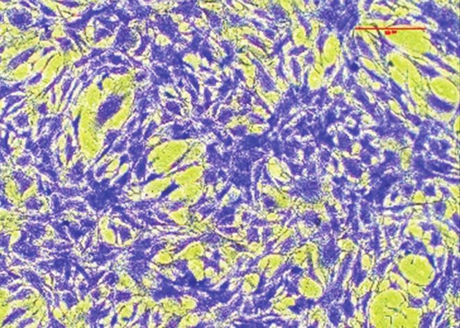
167
                           Ryu et al. Photobiomodulation with Wavelength of 630 nm Light-Emitting Diode
                                    Clinics in Orthopedic Surgery • Vol. 15, No. 1, 2023 • www.ecios.org

Tendon-related diseases and injuries constitute a signifi-              gration of human biceps tendon fibroblasts (HBFs).
cant proportion of musculoskeletal disorders. A growing
geriatric population and an escalation in various sporting
activities have been the main causes for the yearly increase
                                                                                                     METHODS
in prevalence of such diseases, which also puts a strain                Culturing of HBFs
on the social cost of the nation.1) Hence, there has been               The human biceps tendon tissues were collected from
a greater focus on studies related to tendon degenerative               two patients (men, aged 42 years and 69 years) undergo-
lesions and injuries, which have provided extensive infor-              ing biceps tenodesis, with consent of the patients, and
mation about the mechanical and biological properties of                approval of the Institutional Review Board of Pusan Na-
tendons.2) Moreover, this has also led to the development               tional University Yangsan Hospital was obtained (No.
of newer surgical and conservative therapies that can pre-              PNUYH-04-2021-014). Tissues were washed three times
vent major disabilities caused by tendon diseases.                      with phosphate-buffered saline (PBS; Welgene, Gyeong-
       In the treatment of degenerative tendinopathy or                 san, Korea), minced with a sterile scalpel, and then placed
after tendon repair surgery, it is important to enhance                 in a 6-well tissue culture plate (SPL Life Sciences Co. Ltd.,
healing and regeneration. For this, various methods, such               Pocheon, Korea) in Dulbecco’s Modified Eagle Medium
as extracorporeal shockwave therapy,3) prolotherapy,4)                  (Gibco, Grand Island, NY, USA) supplemented with 10%
platelet-rich plasma injections,5) stem cell therapy,6) poly-           fetal bovine serum (FBS; Gibco). They were treated with
deoxyribonucleotide,7) and atelocollagen treatments,8) have             100 IU/mL penicillin and 100 µg/mL streptomycin and
been attempted, each with its own rationale. In addition,               cultured in a humidified 5% CO2 incubator at 37°C for 2
the biologics of bioaugmentation methods, such as mes-                  weeks until they reached 90% confluence. The cells were
enchymal stem cells and growth factors, are being intro-                then trypsinized (0.02% trypsin, 0.02% ethylenediamine-
duced for tissue healing during tendon surgery.6) However,              tetraacetic acid [EDTA] in PBS) for 5 minutes and then
the treatment of tendon diseases and injuries remains pre-              centrifuged for 5 minutes at 200 × g, and the culture ex-
cisely unestablished; hence, it is quite challenging to treat.          panded by a second passage. The cells were then harvested
       Photobiomodulation (PBM) treatments involve low-                 with trypsin/EDTA and cryopreserved. These cryopre-
level light therapy (LLLT) or light-emitting diode (LED)                served third-passage cells were later thawed and used for
therapy using red and near infrared light wavelengths.9)                all experiments in the current study.
In PBM, the photon of the LED light source is absorbed
by a chromophore or photoacceptor in the cell tissue and                Irradiation of Cells by LED
promotes the metabolic activity of the cell. Light absorbed             The following experimental setup was designed to observe
by photoreceptors, such as intracellular cytochrome c                   the effect of PBM on HBFs. The light irradiation module
oxidase, increases reactive oxygen species (ROS) and ad-                was attached to the lid of a 6-well culture plate, and each
enosine triphosphate (ATP) synthesis and releases nitric                well was irradiated by the LED in the top-bottom direc-
oxide (NO). ROS is involved in gene transcription related               tion. Light of a wavelength of 630 nm and 880 nm was
to growth factor production, cell proliferation, and cell               used to irradiate the HBFs. This setup irradiated the cells
motility. It promotes photodegradation of NO, enzyme ac-                with an intensity between 10–100 mW/cm2 per cell per
tivation, mitochondrial metabolism, and ATP production.                 well. Additionally, the light irradiation module was de-
As a result, the overall metabolic activity of the cell is high,        signed to have a frequency of 10–100 Hz and irradiated
and hence, light plays a major role in cell regeneration.10,11)         cells by combining the wavelength, intensity, and frequen-
Based on this evidence, PBM has been employed in vari-                  cy during the experiment (Fig. 1A).
ous therapeutic areas, such as wound healing and nerve                         Cells were seeded onto a 6-well plate (SPL Life Sci-
and bone treatments.9,12,13) Several in vivo studies related            ences Co. Ltd) at density of 1 × 10⁵ cells/mL along with
to tendons have been conducted, and positive effects on                 culture medium. After 18 hours, the culture medium was
tendon healing in cases of injured tendons or tendinitis                replaced with fresh medium, and then the cells were ex-
have been reported.14) However, it is not yet widely used in            posed to LED at 630 nm (10 mW/cm2, 100 Hz) or 630 nm
the field of orthopedics, in particular, clinical applications          (10 mW/cm2, 100 Hz) + 880 nm (40 mW/cm2, 100 Hz) for
involving tendon regeneration. Thus, with the intention                 20 minutes.15) Control cells were treated in the same man-
of developing PBM as a routine treatment procedure, the                 ner, excluding the LED irradiation.
purpose of this study was to verify the effect of LED at a
630 nm wavelength on the survival, proliferation, and mi-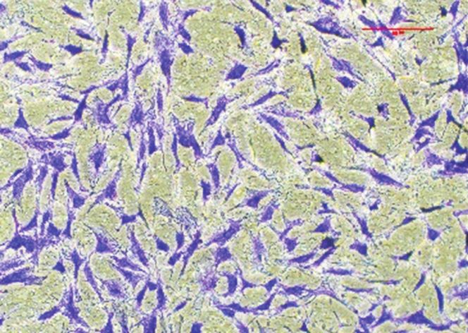
168
Ryu et al. Photobiomodulation with Wavelength of 630 nm Light-Emitting Diode
Clinics in Orthopedic Surgery • Vol. 15, No. 1, 2023 • www.ecios.org
A Schematic (front) Front Back
Applied to the 6-well plate LED conditions applied to cells
B C NC Vimentin -SMA D
Tenascin C
Tenomodulin
-Actin
B
C
BF
LF
-2
M
H
H
AS
BS
D
BE
H
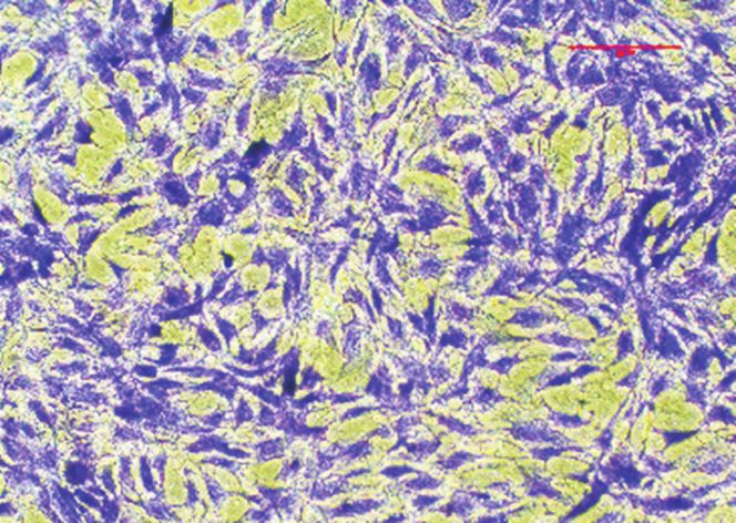
Fig. 1. Identification of fibroblast or tendon by a specific protein marker in human biceps tendon fibroblasts (HBFs) primary cells. (A) Photographs
showing the lighting condition during exposure of HBFs to light-emitting diode (LED) light in an incubator. An LED irradiation was placed on the 6-well
cell culture plate and maintained inside the incubator for 20 minutes. (B) Phase-contrast appearance of primary cell growth from connective tissue
fragments. Elongated spindle-shaped morphology of fibroblast cells. Scale bar represents 100 μm. (C) Fibroblast positively stained for the marker
vimentin (red) or alpha smooth muscle actin (α-SMA; green). Images were taken of cells at passage 2. Scale bar represents 30 μm. (D) Western blotting
expression of tendon marker, tenascin C, and tenomodulin in HBFs. Diseased human lung fibroblast (DHLF), human bronchial epithelial cells (BEAS-2B),
and human bronchial smooth muscle cells (HBSMCs) were used as negative controls (NCs). PCB: printed curcuit board, LCD: liquid crystal display.
Cell Viability Assay MTT solution was added to each well and incubated for
In order to investigate the effect of LED treatment on the 2 hours at 37°C in dark. The supernatant was removed,
viability of HBFs, cell viability was determined using a and the formazan crystals in each well were dissolved in 1
3-(4,5-dimethylthiazole2-yl)-2,5-diphenyl tetrazolium mL of dimethyl sulfoxide (Sigma-Aldrich, St. Louis, MO,
bromide (MTT) reagent (5 mg/mL in PBS, Duchefa Bio- USA) for 30 minutes at 20°C–25°C by shaking. Optical
chemie, Haarlem, The Netherlands). The MTT assay pro- densities at 570 nm were then measured using a micro-
cedure was performed as previously mentioned.16) In brief, plate reader (Tecan, Infinite M200, Grödig, Austria). Data
HBFs were seeded at 1 × 10⁵ cells/mL in 6-well plates and were calculated as a percentage of viable cells in compari-
cultured for 24 hours before LED irradiation. On the fol- son with the control.
lowing day, the culture medium was replaced with serum-
free medium, and then the cells were exposed to LED at Cell Proliferation Assay
630 nm (10 mW/cm2, 100 Hz) or 630 nm (10 mW/cm2, Cell proliferation was determined by 5-bromo-2’-deoxy-
100 Hz) + 880 nm (40 mW/cm2, 100 Hz) for 20 minutes uridine (BrdU, Sigma-Aldrich) incorporation as described
(Fig. 2A). Control cells were treated in the same manner, previously.17) Briefly, HBFs were grown on round cover
excluding the LED irradiation. After 24 hours, 5 μg/mL glass coated with poly-L-lysine (Sigma-Aldrich) for 24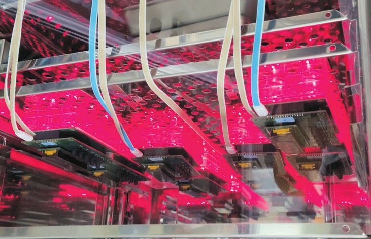
169
                                 Ryu et al. Photobiomodulation with Wavelength of 630 nm Light-Emitting Diode
                                            Clinics in Orthopedic Surgery • Vol. 15, No. 1, 2023 • www.ecios.org

A                          Day 0                                 1                                2                                                 3

                           Cell seeding                    Starvation
                                                                                    LED irradiation for 20 min                                  Analysis

B                Control                            630 nm                       630 nm + 880 nm

                                                                                                                                                             *         *

                                                                                                                Cell viability (%)
                                                                                                                                         100

                                                                                                                                          50

                                                                                                                                           0
                                                                                                                                               Control     630 nm    630 nm
                                                                                                                                                                    + 880 nm

C                Control                            630 nm                       630 nm + 880 nm

                                                                                                                BrdU-positive cell (%)
                                                                                                                                         350
                                                                                                                                                                       *
                                                                                                                                         300
                                                                                                                                         250                 *
                                                                                                                                         200
                                                                                                                                         150
                                                                                                                                         100
                                                                                                                                          50
                                                                                                                                           0
                                                                                                                                               Control     630 nm    630 nm
                                                                                                                                                                    + 880 nm

Fig. 2. Increased human biceps tendon fibroblasts (HBFs) cell viability and proliferation by light-emitting diode (LED) irradiation. (A) Experimental
scheme of LED irradiation for cell viability and proliferation. (B) Effect of LED on HBFs cell viability. Cells were exposed to 630 nm LED or 630 nm + 880 nm LED
irradiation for 20 minutes. Cell viability was measured by 3-(4,5-dimethylthiazole-2-yl)-2,5-diphenyl tetrazolium bromide assay. Data are shown as the
mean ± standard deviation (SD) obtained from three independent experiments (n = 3). (C) 5-Bromo-2'-deoxyuridine (BrdU) immunofluorescence staining
was performed in HBFs after treatment with LED irradiation for 20 minutes. Immunocytochemistry showed localization of BrdU-labeled nuclear (green)
and nuclear staining (blue). Bar graphs represent the quantification of BrdU-positive cells. Data are shown as the mean ± SD of three independent
experiments (n = 3). Scale bar represents 50 μm. *p < 0.05, compared with control.

hours at 1 × 10⁵ cells/mL in 6-well plates. After 24 hours,                        globin G (IgG) (1:500 dilution; Thermo Fisher Scientific,
the culture medium was replaced with serum-free me-                                Rockford, IL, USA) in the dark for 1 hour. BrdU-positive
dium, and then the cells were exposed to LED at 630 nm                             cells were counted in five distinct fields per slide and ex-
(10 mW/cm2, 100 Hz) or 630 nm (10 mW/cm2, 100 Hz) +                                pressed as a percentage of total cells counted. Total DNA
880 nm (40 mW/cm2, 100 Hz) for 20 minutes. Next day,                               was determined using 4’,6-diamidino-2-phenylindole
HBFs were incubated at 37°C for 3 hours with 10 μM of                              (DAPI).
BrdU. Cells were fixed with 4% paraformaldehyde for 30
minutes, washed three times with PBS, and permeabilized                            Cell Migration Assays
by incubation in 0.2% Triton X-100 in PBS for 30 minutes.                          Migration assays were performed as described previously.18)
To measure BrdU incorporation, cells were incubated in 2                           Briefly, a culture-insert (ibid, Martinsried, Germany) was
M hydrochloric acid (HCl) for 30 minutes at 37°C in or-                            attached to the 6-well plate. The cells (7.5 × 10⁵ cells/mL; 80 μL)
der to denature the DNA. Cells were then incubated for 5                           were seeded into each well and incubated for 24 hours in culture
minutes in 0.1 M sodium borate (pH 8.5) to neutralize the                          media with 25 μg/mL mitomycin C (Sigma) for 30 minutes
residual acid and washed three times with PBS. Cells were                          to inhibit cell division and proliferation. The culture-insert
incubated in a blocking solution containing 1% normal goat                         was taken out, the cells were irradiated, and migration
serum for 1 hour at 20°C–25°C and then incubated with                              was analyzed in 0, 12, and 24 hours. The migration of
monoclonal anti-BrdU (1 : 100 dilution; Thermo Fisher                              cells into the cell-free gap created by the removal of the
Scientific, Waltham, MA, USA) in blocking solution at 4°C                          culture-insert was monitored at indicated time points and
for 18 hours. After washing with PBS thrice, the cells were                        photographed using a microscope (Nikon, Tokyo, Japan).
incubated with Alexa Fluor 488 goat anti-mouse immuno-                             Through quantitative assessments of the data, the velocity
170
                         Ryu et al. Photobiomodulation with Wavelength of 630 nm Light-Emitting Diode
                                  Clinics in Orthopedic Surgery • Vol. 15, No. 1, 2023 • www.ecios.org

of migration was determined. All data presented are from              Signaling, Danvers, MA, USA), and β-actin (1 : 3000; Sig-
at least three independent experiments performed in trip-             ma). The membranes were then washed three times with
licate. The trans-well migration assay was performed using            Tris-buffered saline with Tween-20 and incubated with
the CytoSelect Cell Migration Assay Kit containing poly-              secondary antibodies conjugated with horseradish peroxi-
carbonate membrane inserts (8 μm-pore membrane; Cell                  dase (1 : 5000, Cell Signaling) for 1 hour. Immunodetection
Biolabs, San Diego, CA, USA) according to the manufac-                was performed using an enhanced chemiluminescence kit
turer’s instructions. The trans-well migration procedures             (Amersham Pharmacia, Piscataway, NJ, USA) with Fusion
were as described previously.18) A cell suspension contain-           Solo X (Vilber Lourmat, Collegien, France) for detection.
ing 1 × 10⁵ cells/mL in serum-free medium was prepared,               The relative protein level was calculated using β-actin as a
and 2 mL of the cell suspension was added to the upper                loading control.
chamber of each insert. The lower chamber containing
medium with 10% FBS allowed cell migration towards                    Immunocytochemistry
the lower face of the trans-well culture inserts. Cells were          HBFs were cultured on round cover glass coated with
irradiated with 630 nm or 630 nm + 880 nm LED for 20                  poly-L-lysine for 24 hours at 2.5 × 10⁴ cells/well (500 μL)
minutes and then incubated for 12 hours at 37°C with 5%               in 24-well plates. The cells were fixed with 4% paraformal-
CO2. Non-migrating cells on the inner side of the trans-              dehyde in 0.1 M PBS for 30 minutes, washed three times
well culture inserts were gently removed with a cotton-               with 1X PBS, and preincubated in a blocking buffer con-
tipped swab. Migrated cells remaining on the bottom sur-              taining 1% normal goat serum and 0.1% Triton X-100 for
face were stained with 0.1% crystal violet for 15 minutes.            2 hours at 20°C–25°C under gentle rotation. The cells were
Photomicrographs of five individual fields per insert were            incubated with monoclonal anti-actin, α-smooth muscle-
taken using a microscope (Nikon) and analyzed using Im-               FITC antibody (1 : 500, Sigma), and vimentin (1 : 500, Cell
ageJ software (version 1.49; National Institutes of Health,           Signaling) in blocking solution and then incubated at 4°C
Bethesda, MD, USA) to calculate the average number of                 for 18 hours. Samples excluding the addition of primary
cells that had migrated.                                              antibodies were used as negative controls. After washing
                                                                      three times in PBS, the cells were incubated in the dark for
Western Blotting                                                      1.5 hours with Alexa Fluor 594-conjugated anti-rabbit IgG
A Western blot analysis was performed as described pre-               fluorescent secondary antibody diluted 1 : 500 in PBS. The
viously.19) Protein lysates were made from HBFs using                 cells were washed three times and stained with 1 μg/mL
radioimmunoprecipitation assay buffer (Thermo Fisher                  of DAPI for nuclei staining. The stained cells were wet-
Scientific) containing a protease inhibitor cocktail (Xpert           mounted on glass slides and observed using a confocal
protease inhibitor cocktail solution, GenDEPOT) and                   laser scanning microscope (K1-Fluo; Nanoscope Systems,
phosphatase inhibitors (Xpert phosphatase inhibitor cock-             Daejeon, Korea).
tail solution, GenDEPOT). The cell lysates were incubated
for 30 minutes on ice with intermittent vortexing and                 Statistical Analysis
were clarified by centrifugation at 16,609 × g (13,000 rpm;           The data are expressed as mean ± standard deviation.
Hanil, Incheon, Korea) at 4°C for 20 minutes. After cen-              Significant differences between groups were analyzed us-
trifugation, the supernatant was separated and stored at              ing an unpaired Student t-test (OriginPro2020; OriginLab
–70°C until use. Protein concentration in cell lysates was            Corp., Northampton, MA, USA). A p < 0.05 was consid-
quantified using a Pierce bicinchoninic acid protein as-              ered as the criterion for statistical significance.
say kit (Thermo Fisher Scientific). Cell lysates were boiled
in 5X sample buffer and separated by 8%–15% sodium
dodecyl sulfate-polyacrylamide gel electrophoresis, and
                                                                                                    RESULTS
the proteins were transferred onto polyvinylidene difluo-             Identification of HBFs
ride membranes (Millipore, Darmstadt, Germany). The                   Morphological observations of cells isolated from human
membranes were blocked with 5% skimmed milk in TBST                   biceps tendon tissue and subsequently cultured showed
(50 mM Tris HCl pH 7.4, 150 mM NaCl, 0.1% Tween 20)                   spindle- or star-shaped features typical of fibroblasts (Fig.
and incubated with specific primary antibodies at 4°C for             1B). Expression of fibroblast markers (vimentin and alpha
18 hours. The primary antibodies used were as follows:                smooth muscle actin) and tendon-specific markers (tenas-
Tenascin C (1 : 1000; Abcam, Cambridge, MA, USA),                     cin C and tenomodulin proteins) were confirmed by im-
tenomodulin (1 : 1000; Abcam), vimentin (1 : 1000; Cell               munofluorescence assay and Western blotting, respectively
171
                              Ryu et al. Photobiomodulation with Wavelength of 630 nm Light-Emitting Diode
                                         Clinics in Orthopedic Surgery • Vol. 15, No. 1, 2023 • www.ecios.org

(Fig. 1C and D).                                                             rate of 2.87 ± 0.91 folds and higher (p < 0.05).

Effect of LED Treatment on the Viability and                                 Effect of LED Treatment on the Migration of HBFs
Proliferation of HBFs Cells                                                  Cells
The LED treatment group had a significantly increased                        In order to replicate cell migration to wound site, the
survival rate than the control group; however, there was                     migration in 2D was observed in monolayer cells, and
no significant difference between the 630 nm LED alone                       the effect of LED treatment was assessed. The monolayer
treatment group (106.76% ± 2.30%) and the 630 nm and                         cultured HBFs cells were treated with LED for 20 minutes,
880 nm combination treatment group (108.04% ± 0.90%)                         and then the distance the HBFs cells moved during the
(Fig. 2B). As shown in Fig. 2C, The proliferation rate of                    12 hours and 24 hours period was measured (Fig. 3A).
HBF cells was significantly increased in the LED-treated                     The cell migration was unaffected until 12 hours after
group compared to the control group. The 630 nm LED                          LED treatment: however, at 24 hours, it was significantly
treatment resulted in a 2.21 ± 0.76-fold increased cell pro-                 increased in the 630 nm LED alone group (p < 0.05) and
liferation rate (p < 0.05), and the 630 nm + 880 nm LED                      the 630 nm + 880 nm LED treatment group compared
combination treatment group showed a cell proliferation                      with the control group. (p < 0.05) (Fig. 3A). To further

A               Control                         630 nm                      630 nm + 880 nm
                                                                                                                                 150

                                                                                                             Migrated cell (%)
                                                                                                                                                   *         *
                                                                                                                                 100
0 hr

                                                                                                                                  50

                                                                                                                                   0
                                                                                                                                       Control   630 nm    630 nm
                                                                                                                                                          + 880 nm
12 hr
24 hr

B               Control                         630 nm                      630 nm + 880 nm
                                                                                                                                 350
                                                                                                          Migrated cell (%)

                                                                                                                                 300               *         *
                                                                                                                                 250
24 hr

                                                                                                                                 200
                                                                                                                                 150
                                                                                                                                 100
                                                                                                                                  50
                                                                                                                                   0
                                                                                                                                       Control   630 nm    630 nm
                                                                                                                                                          + 880 nm

Fig. 3. Light-emitting diode (LED) irradiation-enhanced human biceps tendon fibroblasts (HBFs) migration. Cells were exposed to 630 nm LED or 630 nm
+ 880 nm LED irradiation for 20 minutes. (A) Representative phase-contrast images at 0, 12, and 24 hours, showing the migration of HBFs into the cell-
free gap, left by the removal of the culture-insert. Bar graphs represent the quantification of migrated cells. Data are shown as the mean ± standard
deviation (SD) of three independent experiments (n = 3). (B) Representative microscopic images of migrating cells by trans-well migration assays. Bar
graphs represent the quantification of migrated cells. Data are shown as the mean ± SD of three independent experiments (n = 3). *p < 0.05, compared
with control.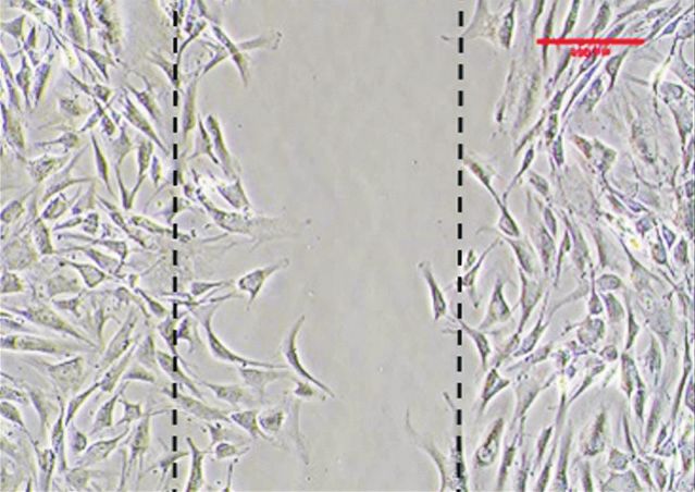
172
                          Ryu et al. Photobiomodulation with Wavelength of 630 nm Light-Emitting Diode
                                   Clinics in Orthopedic Surgery • Vol. 15, No. 1, 2023 • www.ecios.org

confirm these results, a three-dimensional migration assay             light and near-infrared light (600–1,100 nm). There are
was carried out using trans-wells, and we observed similar             effective reports on blue and green light (400–500 nm) in
results. The 630 nm LED alone treatment group showed a                 some studies; however, more research is needed because
3.06 ± 0.21 times higher cell migration rate (p < 0.05), and           the boundary with ultraviolet (< 400 nm), which causes
the 630 nm + 880 nm LED combination treatment group                    DNA damage, is vague. Furthermore, at wavelengths be-
showed a 2.88 ± 0.20 times higher cell migration rate than             low 600 nm, the absorption and scattering of light in tissue
the control group (p < 0.05) (Fig. 3B).                                are higher than red light, and at wavelengths greater than
                                                                       1,100 nm, water absorbs infrared light. Therefore, red and
                                                                       near-infrared light (600–1,100 nm) is used in PBM.24,25)
                      DISCUSSION                                       Lam26) found that when fibroblasts were irradiated with
The results of this study allowed us to confirm the initial            light with a wavelength of 633 nm, procollagen synthesis
effect of LED treatment on fibroblasts using HBF culture.              was increased four times compared with the baseline. In
Cell viability, proliferation, and migration all showed sig-           another study, irradiation at 830 nm accelerated fibroblast-
nificant differences in the 630 nm LED and 630 + 880 nm                myofibroblast transformation and mast cell degranulation.
LED treatment groups compared with the control group.                  Additionally, the chemotaxis and phagocytic activity of
PBM using LLLT or LED therapy was first introduced                     leukocytes and macrophages were enhanced by cell stimu-
by Endre Mester in the early 1960s.20) PBM accelerated                 lation.27) Hence, Russell et al.28) suggested that there could
healing, mediated by increased cell activity generated by              be a synergistic effect when 633 nm and 830 nm were ap-
stimulation of the mitochondrial and cell membrane pho-                plied simultaneously. Hence, in our study, not only 630 nm
toreceptors synthesis of ATP.9) Such modulations on these              treatment but also 630 nm and 880 nm combined irradia-
cells can promote fibroblast proliferation, growth factor              tion was used. However, in our study, no synergistic effect
synthesis, collagen production, and angiogenesis.9,10,20)              was observed in the group treated with 630 nm and 880
Based on this rationale, various studies and animal ex-                nm wavelengths simultaneously. Moreover, since the 880
periments have been conducted in the field of orthopedic               nm alone treatment did not affect cell viability, the 880 nm
surgery. According to Lopes Silva et al.,13) a positive effect         alone group was excluded from the study results. In some
of LLLT/LED was reported on tendon damage. Rosso et                    other studies, only 635 nm was considered as a potential
al.21) showed that PBM has beneficial effects on the recov-            effective option for bone regeneration in comparison of
ery of nerve lesions. It has also been suggested by in vitro           808 nm, 635 nm, and 405 nm. However, since it shows
experiments that PBM may facilitate tissue homeostasis,                various results depending on the light conditions and ir-
thus stimulating the components of the articular tissue                radiation time, a more specific PBM modality should be
and promoting chondroprotective effects.22) Our experi-                made in the future.28,29)
ments used fibroblasts obtained from human tendons on                         This study focused only on the effects of wavelength
the basis of previously established studies, and we observed           because it is one of the major factors affecting cell activity.
> 2-fold cell proliferation and ≥ three-fold cell migration in         However, other conditions, such as light source, energy
the LED-treated group compared with the control group.                 density, light pulse structure, and LED application time
As mentioned earlier, PBM treatment promotes reduction                 can also have a significant effect and need to be investi-
of inflammatory cells, increase of proliferation of fibro-             gated in future studies. Furthermore, because the assays
blasts, stimulation of angiogenesis, formation of granu-               were all performed in vitro, the relevance of these results
lation tissue, and increase of collagen synthesis. One of              in a clinical scenario needs to be assessed. PBM is pres-
the strengths of our study is that it revealed the statistical         ently used in the field of dermatology, where direct contact
significance of PBM treatment on fibroblast proliferation,             of tissues with light is possible. However, in orthopedic
which is one of the effects of PBM treatment.                          cases, further research is needed to determine whether
        Low-level lasers were used for PBM in the 1960s.               light can penetrate the tissues present under the skin, such
Since the 1990s, LED lights have replaced lasers and have              as ligaments, nerves, and bones. The present study, while
been proven to have the same medical benefits.23) LED                  elucidating the effects of PBM on cell activity, has not
lights also provide the advantage of easy adjustability of             examined inflammatory effects among others and hence
light intensity, longevity, and homogeneity of light dose              opens up several avenues for future research to establish
at an optimal intensity.23) It also allows the use of lights           PBM as a routine treatment procedure.
with various wavelengths independently or at the same                         LED irradiation at 630 nm and 630 nm + 880 nm
time. The wavelengths commonly used in PBM are red                     for 20 minutes significantly affected cell proliferation and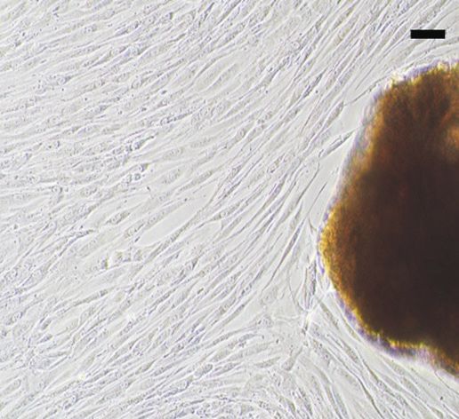
173
                            Ryu et al. Photobiomodulation with Wavelength of 630 nm Light-Emitting Diode
                                      Clinics in Orthopedic Surgery • Vol. 15, No. 1, 2023 • www.ecios.org

migration in human tendon fibroblast cells. LED therapy,                  dation of Korea (MSIT, NRF-2021R1F1A1053630) and
thus, offers promising potential for use as an adjunct treat-             by the Ministry of Health and Welfare of Korea (grant no.
ment for tendon healing, and hence, further research on                   HI17C2397).
various conditions is essential to establish this as a routine
treatment regimen.
                                                                                                             ORCID
                                                                          Ji Hyeon Ryu            https://orcid.org/0000-0003-0342-3108
               CONFLICT OF INTEREST                                       Jisu Park               https://orcid.org/0000-0001-6444-4688
No potential conflict of interest relevant to this article was            Ji Won Kim              https://orcid.org/0000-0002-3839-7213
reported.                                                                 Yong-Il Shin            https://orcid.org/0000-0001-7894-0930
                                                                          Sang Don Lee            https://orcid.org/0000-0001-9459-3887
                                                                          Youngkwang Oh           https://orcid.org/0000-0001-7722-2099
                ACKNOWLEDGEMENTS                                          Suk-Woong Kang          https://orcid.org/0000-0003-0883-2461
This research was funded by the National Research Foun-

                                                            REFERENCES

1.   Hopkins C, Fu SC, Chua E, et al. Critical review on the                   underlying mechanism and clinical applications. J Clin Med.
     socio-economic impact of tendinopathy. Asia Pac J Sports                  2020;9(6):1724.
     Med Arthrosc Rehabil Technol. 2016;4:9-20.
                                                                          10. de Freitas LF, Hamblin MR. Proposed mechanisms of pho-
2.   Yang G, Rothrauff BB, Tuan RS. Tendon and ligament re-                   tobiomodulation or low-level light therapy. IEEE J Sel Top
     generation and repair: clinical relevance and developmental              Quantum Electron. 2016;22(3):7000417.
     paradigm. Birth Defects Res C Embryo Today. 2013;99(3):
                                                                          11. Farivar S, Malekshahabi T, Shiari R. Biological effects of low
     203-22.
                                                                              level laser therapy. J Lasers Med Sci. 2014;5(2):58-62.
3.   Leone L, Vetrano M, Ranieri D, et al. Extracorporeal shock
                                                                          12. Soleimani M, Abbasnia E, Fathi M, Sahraei H, Fathi Y, Kaka
     wave treatment (ESWT) improves in vitro functional activi-
                                                                              G. The effects of low-level laser irradiation on differentia-
     ties of ruptured human tendon-derived tenocytes. PLoS
                                                                              tion and proliferation of human bone marrow mesenchymal
     One. 2012;7(11):e49759.
                                                                              stem cells into neurons and osteoblasts: an in vitro study.
4.   Reeves KD, Sit RW, Rabago DP. Dextrose prolotherapy: a                   Lasers Med Sci. 2012;27(2):423-30.
     narrative review of basic science, clinical research, and best
                                                                          13. Lopes Silva R, Pessoa DR, Mariano RR, Castro A, de Olivei-
     treatment recommendations. Phys Med Rehabil Clin N Am.
                                                                              ra RA, Ferraresi C. Systematic review of photobiomodula-
     2016;27(4):783-823.
                                                                              tion therapy (PBMT) on the experimental calcaneal tendon
5.   Kim HJ, Nam HW, Hur CY, et al. The effect of platelet rich               injury in rats. Photochem Photobiol. 2020;96(5):981-97.
     plasma from bone marrow aspirate with added bone mor-
                                                                          14. Marcos RL, Arnold G, Magnenet V, Rahouadj R, Magdalou J,
     phogenetic protein-2 on the Achilles tendon-bone junction
                                                                              Lopes-Martins RA. Biomechanical and biochemical protec-
     in rabbits. Clin Orthop Surg. 2011;3(4):325-31.
                                                                              tive effect of low-level laser therapy for Achilles tendinitis. J
6.   Zhang C, Wu J, Li X, Wang Z, Lu WW, Wong TM. Current                     Mech Behav Biomed Mater. 2014;29:272-85.
     biological strategies to enhance surgical treatment for rota-
                                                                          15. Sousa D, Goncalves M, Politti F, et al. Photobiomodulation
     tor cuff repair. Front Bioeng Biotechnol. 2021;9:657584.
                                                                              with simultaneous use of red and infrared light emitting di-
7.   Gwak DW, Hwang JM, Kim AR, Park D. Does polydeoxyri-                     odes in the treatment of temporomandibular disorder: study
     bonucleotide has an effect on patients with tendon or liga-              protocol for a randomized, controlled and double-blind
     ment pain?: a PRISMA-compliant meta-analysis. Medicine                   clinical trial. Medicine (Baltimore). 2019;98(6):e14391.
     (Baltimore). 2021;100(19):e25792.
                                                                          16. Riss TL, Moravec RA, Niles AL, et al. Cell viability assays.
8.   Kim JH, Kim DJ, Lee HJ, Kim BK, Kim YS. Atelocollagen                    In: Markossian S, Grossman A, Brimacombe K, et al., eds.
     injection improves tendon integrity in partial-thickness                 Assay guidance manual. Bethesda (MD): Eli Lilly & Com-
     rotator cuff tears: a prospective comparative study. Orthop J            pany and the National Center for Advancing Translational
     Sports Med. 2020;8(2):2325967120904012.                                  Sciences; 2016.
9.   Dompe C, Moncrieff L, Matys J, et al. Photobiomodulation-            17. Mead TJ, Lefebvre V. Proliferation assays (BrdU and EdU)
174
                            Ryu et al. Photobiomodulation with Wavelength of 630 nm Light-Emitting Diode
                                      Clinics in Orthopedic Surgery • Vol. 15, No. 1, 2023 • www.ecios.org

    on skeletal tissue sections. Methods Mol Biol. 2014;1130:                  knee of rats. Lasers Med Sci. 2022;37(3):1677-86.
    233-43.
                                                                          24. Yeh NG, Wu CH, Cheng TC. Light-emitting diodes—their
18. Pijuan J, Barcelo C, Moreno DF, et al. In vitro cell migration,           potential in biomedical applications. Renew Sustain Energy
    invasion, and adhesion assays: from cell imaging to data                  Rev. 2010;14(8):2161-6.
    analysis. Front Cell Dev Biol. 2019;7:107.
                                                                          25. Wang Y, Huang YY, Wang Y, Lyu P, Hamblin MR. Photobio-
19. Kim YM, Ko SH, Shin YI, et al. Light-emitting diode irra-                 modulation (blue and green light) encourages osteoblastic-
    diation induces AKT/mTOR-mediated apoptosis in human                      differentiation of human adipose-derived stem cells: role of
    pancreatic cancer cells and xenograft mouse model. J Cell                 intracellular calcium and light-gated ion channels. Sci Rep.
    Physiol. 2021;236(2):1362-74.                                             2016;6:33719.
20. Whelan HT, Houle JM, Whelan NT, et al. The NASA light-                26. Lam TS. Laser stimulation of collagen synthesis in human
    emitting diode medical program-progress in space flight                   skin fibroblast cultures. Lasers Life Sci. 1986;1:61-77.
    and terrestrial applications. AIP Conf Proc. 2000;504:37-43.
                                                                          27. Osanai T, Shiroto C, Mikami Y, et al. Measurement of GaA-
21. Rosso M, Buchaim DV, Kawano N, Furlanette G, Pomini                       lAs diode laser action on phagocytic activty of human neu-
    KT, Buchaim RL. Photobiomodulation therapy (PBMT) in                      trophils as a possible therapeutic dosimetry determinant.
    peripheral nerve regeneration: a systematic review. Bioengi-              Laser Ther. 1990;2(3):123-33.
    neering (Basel). 2018;5(2):44.
                                                                          28. Russell BA, Kellett N, Reilly LR. A study to determine the
22. Tani A, Chellini F, Giannelli M, Nosi D, Zecchi-Orlandini S,              efficacy of combination LED light therapy (633 nm and 830
    Sassoli C. Red (635 nm), near-infrared (808 nm) and violet-               nm) in facial skin rejuvenation. J Cosmet Laser Ther. 2005;
    blue (405 nm) photobiomodulation potentiality on human                    7(3-4):196-200.
    osteoblasts and mesenchymal stromal cells: a morphological
                                                                          29. Keszler A, Lindemer B, Hogg N, Weihrauch D, Lohr NL.
    and molecular in vitro study. Int J Mol Sci. 2018;19(7):1946.
                                                                              Wavelength-dependence of vasodilation and NO release
23. Tim CR, Martignago C, Assis L, et al. Effects of photobio-                from S-nitrosothiols and dinitrosyl iron complexes by far
    modulation therapy in chondrocyte response by in vitro                    red/near infrared light. Arch Biochem Biophys. 2018;649:47-
    experiments and experimental model of osteoarthritis in the               52.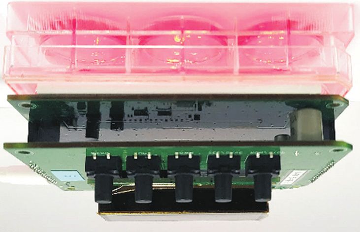
You can also read


