Lidocaine liposome modified with folic acid cells via targeting the PI3K/AKT pathway
←
→
Page content transcription
If your browser does not render page correctly, please read the page content below
EXPERIMENTAL AND THERAPEUTIC MEDICINE 22: 1025, 2021

                      Lidocaine liposome modified with folic acid
                   suppresses the proliferation and motility of glioma
                      cells via targeting the PI3K/AKT pathway
                          DEDONG LI1*, XUEWEI YANG2*, BO LI1, CHENYI YANG3, JIAN SUN1,
                               MINGDONG YU1, HAIYUN WANG3 and YUECHUN LU1

                   1
                    Department of Anesthesiology, Second Hospital of Tianjin Medical University, Tianjin 300211;
                   2
                    Department of Anesthesiology, Tianjin Union Medical Center, Tianjin 300191; 3Department of
                            Anesthesiology, Tianjin Third Central Hospital, Tianjin 300052, P.R. China

                                       Received September 4, 2020; Accepted May 28, 2021

                                                   DOI: 10.3892/etm.2021.10457

Abstract. Glioma is life‑threatening tumor of the central           Introduction
nervous system. Although lidocaine is usually used as local
anesthetic, it also has antitumor effects. However, its clinical    Glioma is one of the most prevalent tumors of the central
application in glioma is hampered by limited distribution to the    nervous system, with an age‑adjusted (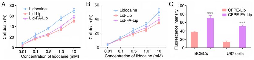
2               LI et al: LIDOCAINE LIPOSOME SUPPRESSES GLIOMA CELLS VIA TARGETING THE PI3K/AKT PATHWAY

the cell surface of multiple types of cancer cells, including        A yellow solid was obtained, which was purified by flash
U87 and MDA‑MB‑231 cells (18,19). Additionally, the expres‑          chromatography to yield the FA‑modified ligand (compound
sion of the FR on normal cells is limited, which make it an          4; 0.20 g, 30%). HRMS: (ESI+) calculated for C62H95N7O14Na
attractive focus for efficient glioma‑targeting. In addition, the    [M+Na]+, 1,161.6937; found, 1,161.6930. Elemental analysis:
FR is also highly expressed by cerebral capillary endothelial        Calculated, C, 64.06; H, 8.24; N, 8.43; found C, 64.18; H, 8.33;
cells (19). Therefore, FA may have superior ability to cross the     N, 8.29.
BBB and efficiently target glioma.
    To verify the hypothesized ability of FA to cross the            Prep a ra t ion a n d ch a ra cteriza t ion of liposo m es.
BBB and investigate the antitumor mechanisms of lidocaine,           Lidocaine‑loaded liposomes were prepared using the thin‑film
an innovative lidocaine‑loaded liposome modified with                hydration method (20,21). The ratio of the components was
FA (Lid‑FA‑Lip) was developed in the present study. This             optimized as follows: i) Conventional liposome (Lip), soybean
modified liposome was designed based on the assumption               phosphatidylcholine (SPC; Shanghai Taiwei Pharmaceutical
that FA will markedly increase the ability of the liposome to        Co., Ltd.)/cholesterol in a molar ratio of 62:33; ii) FA‑modified
penetrate the BBB and thereby improve the antitumor effect.          liposome (FA‑Lip), SPC/cholesterol/compound 4 in a molar
Whether Lid‑FA‑Lip can suppress the motility of glioma cells         ratio of 62:33:3. In brief, all the lipid materials were dissolved
and stimulate apoptosis was also investigated. In addition, the      in a mixed solvent comprising CH2Cl2 and methanol (2:1 v/v),
antitumor effects of Lid‑FA‑Lip on the tumor growth of glioma        and the solution was then warmed on a rotary evaporator
cells in mice and the contribution of PI3K/AKT pathway               at 37˚C to remove the solvent and form a thin film. After
suppression to the underlying mechanism were analyzed.               drying in a vacuum for 10 h, the film was hydrated with PBS
                                                                     (pH 7.4) at 20˚C for 30 min and sonicated intermittently
Materials and methods                                                at 80 W for 80 sec to obtain the liposomes. An appropriate
                                                                     amount of lidocaine (weight ratio of procaine/lipid, 1/30)
Chemistry                                                            or 1,2‑dioleoyl‑sn‑glycero‑3‑phosphoethanolamine‑N-
Synthesis of cholesterol tosylate (compound 2). A solution of        carboxyfluorescein (CFPE; final concentration, 20 µg/ml) was
cholesterol (compound 1; 1.00 g, 2.59 mmol) in pyridine (5 ml)       added to the solution of lipid before removing the solvent to
was prepared, and a solution of p‑toluenesulfonyl chloride           prepare lidocaine‑loaded liposomes (Lid‑Lip and Lid‑FA‑Lip)
(0.74 g, 3.88 mmol) in pyridine (5 ml) was added. After stir‑        or CFPE‑labeled liposomes (CFPE‑Lip and CFPE‑FA‑Lip).
ring the mixture for 10 h at 50˚C, the solvent was removed and           The entrapment efficiency [EE (%)] and drug loading effi‑
the residue was re‑dissolved with ethyl acetate (20 ml). The         ciency [DL (%)] of lidocaine by the liposomes were determined
solution was washed with 1 mol/l HCl and saturated NaCl. The         using high performance liquid chromatography (HPLC). The
solvent was then removed to yield compound 2 (1.19 g, 85%),          detection was performed on an Agilent 1200 HPLC System
which was used for the next step without further purification.       with an Ultimate LP‑C18 column (4.6x250 mm, 5 µm) (both
Melting point, 127‑129˚C. High‑resolution mass spectrom‑             from Agilent Technologies, Inc.) at 25˚C. The mobile phase
etry (HRMS): Electrospray ionization (ESI+) calculated for           comprised water and methanol (70:30 v/v) with a flow rate of
C34H52O3S Na [M+Na]+, 563.3535; found, 563.3532. Elemental           1.0 ml/min. A 10‑µl aliquot of the lidocaine‑containing sample
analysis: Calculated, C, 75.51; H, 9.69; S, 5.93; found, C, 75.43;   was injected and the detection wavelength was 210 nm. The
H, 9.78; S, 6.11.                                                    EE (%) and DL (%) were calculated according to the following
                                                                     equations: EE (%)=weight of encapsulated lidocaine/total
Synthesis of octaethylene glycol monocholesteryl ether               weight of lidocaine, and DL (%)=weight of encapsulated
(compound 3). To a solution of compound 2 (1.00 g, 1.85 mmol)        lidocaine/total weight of liposome. In addition, the size and
in dioxane (15 ml) was added octaethylene glycol (6.85 g,            ζ potential of Lid‑Lip and Lid‑FA‑Lip were detected using a
18.49 mmol). The reaction mixture was refluxed for 20 h. The         Malvern Zeta sizer Nano ZS90 (Malvern Panalytical).
solvent was then removed and the residue was re‑dissolved
with dichloromethane (CH 2Cl 2; 30 ml). The solution was             Release by Lid‑Lip and Lid‑FA‑Lip in vitro. The release
washed with saturated NaCl. After removing the CH2Cl2, the           behavior of lidocaine from the Lid‑Lip and Lid‑FA‑Lip
residue was purified by chromatography to give compound 3            liposomes was assessed using dialysis. Brief ly, the
(0.66 g, 48%) as colorless oil. HRMS: (ESI+) calculated for          lidocaine‑loaded liposomes were put into a dialysis bag
C43H78O9Na [M+Na]+, 761.5544; found, 761.5540. Elemental             (molecular weight cut‑off, 8,000‑14,000 Da) with 50 ml PBS
analysis: Calculated, C, 69.88; H, 10.64; found, C, 69.76; H,        containing 0.1% Tween-80 (v/v) as release medium. The
10.79.                                                               bags were shaken at 37˚C with gentle oscillation (45 rpm).
                                                                     At various time points between 0 and 48 h, an 0.1‑ml sample
Synthesis of FA‑modified ligand (compound 4). To a                   was extracted and replaced with fresh medium. The release
solution of FA (0.50 g, 1.13 mmol) and compound 3 (0.42 g,           behavior from free lidocaine was also analyzed as a control.
0.57 mmol) in dimethyl sulfoxide (DMSO; 10 ml) was                   Subsequently, the amount of lidocaine was detected by the
added 1‑(3‑dimethylaminopropyl)‑3‑ethylcarbodiimide                  aforementioned HPLC method.
hyd r o ch lo r id e ( E D C; 0.11 g, 0. 57 m m ol) a n d
N‑hydroxysuccinimide (NHS; 65 mg, 0.57 mmol). The                    Stability of Lid‑Lip and Lid‑FA‑Lip in vitro. The stability
resulting mixture was stirred at room temperature for 24 h.          of the Lid‑Lip and Lid‑FA‑Lip liposomes was determined
The by‑product was removed by filtration. The product was            by the measurement of turbidity variations in the presence
precipitated from the filtrate by the addition of diethyl ether.     of fetal bovine serum (FBS; Thermo Fisher Scientific, Inc.).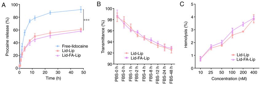
EXPERIMENTAL AND THERAPEUTIC MEDICINE 22: 1025, 2021                                           3

This comprised mixing each liposome formulation with an            BCECs were maintained in DMEM with 10% FBS (Gibco;
equal volume of FBS, and then continuously shaking (50 rpm)        Thermo Fisher Scientific, Inc.) and seeded on 6‑well plates at
at 37˚C. The transmittance was measured between 0 and 48 h         a density of 1x10⁶ cells/well and incubated for 7 days at 37˚C.
using a microplate reader (Bio‑Rad model 550; Bio‑Rad              The transendothelial electric resistance (TEER) of the BBB
Laboratories, Inc.) at a wavelength of 750 nm. The transmit‑       model was measured using a Millicell ERS volt‑ohm meter
tance in PBS was defined as 100%.                                  (EMD Millipore). Only BCEC monolayers with TEER values
                                                                   >200 Ω were used for further experiments. Additionally, the
Hemolysis assays. A female BALB/c mouse (22 g;                     U87 cells were plated on another 6‑well plate (1x10⁶ cells/well).
4 weeks old; weight, 18‑22 g) was supplied by Beijing Vital        The cell culture inserts with BCEC monolayers were trans‑
River Laboratory Animal Technology Co., Ltd. The mouse             ferred to the plates containing the U87 cells and the cells were
was kept in a 20˚C environment with 40‑60% humidity and a          co‑cultured for another 24 h. Subsequently, the CFPE‑labeled
12‑h cycle of day and night. Free food and water were provided     liposomes (CFPE‑Lip and CFPE‑FA‑Lip; final concentration
during the feeding process, and clean and hygienic feeding         of CFPE, 2 µg/ml) were added to the cell culture inserts (donor
conditions were maintained. The mouse was anesthetized by          chamber) of the BBB model and incubated at 37˚C for 4 h.
the intraperitoneal injection of pentobarbital sodium at a dose    The BCECs and U87 cells were then each washed with cold
of 50 mg/kg. Fresh blood (0.1 ml) was collected from the orbit,    PBS three times, trypsinized and finally resuspended in 0.5 ml
and centrifuged (4˚C) at 2,000 x g for 5 min. The supernatant      PBS. The fluorescent intensity of the two types of cells was
was discarded and the precipitated red blood cells were            measured using a flow cytometer (Cytomics FC500; Beckman
washed with PBS until the supernatant was colorless. The cells     Coulter, Inc.) and the flow cytometer's integral analysis soft‑
were then re‑suspended in PBS to a concentration of 2% (w/v).      ware (FlowJo 10; Becton, Dickinson and Company).
Afterwards, Lid‑Lip and Lid‑FA‑Lip were diluted with PBS to
provide liposome samples with lipid concentrations of 10, 25,      Wound healing assays. A wound healing assay was performed
50, 100, 200 and 400 nM. Subsequently, each liposome sample        to evaluate the effects of the liposomal formulations on cell
(0.4 ml) was mixed with red blood cell suspension (0.1 ml) and     migration. U87 cells at a confluence of 100% and treated with
the mixture was incubated at 37˚C for 2 h with gentle shaking      Lid‑Lip or Lid‑FA‑Lip (1 mM lidocaine; 37˚C; 24 h) were
(50 rpm). After centrifuging (4˚C) at 6,000 x g for 10 min, the    wounded by scraping with a 10‑µl pipette tip and washed
absorbance of the supernatant at 540 nm (A540) was measured        twice. The cells were then cultured with serum‑free DMEM to
using a microplate reader. The hemolysis rate of each sample       stimulate wound healing. Photographic images were captured
was calculated as follows: Hemolysis (%)=(A540 sample‑A540         by an Olympus light microscope before culture and at the
negative)/(A540 positive‑A540 negative) x100. The absorbances      24‑h time point to evaluate the migration of the U87 cells.
of PBS and Triton X‑100 mixed with cell solution were defined      The images were then analyzed using ImageJ 8.0 software
as 0 and 100%, respectively.                                       (National Institutes of Health).

Cell culture. Brain capillary endothelial cells (BCECs)            Cell apoptosis assays. Cell apoptosis was evaluated using
and the U87 glioblastoma of unknown origin cell line were          Annexin V‑FITC and propidium iodide (PI) staining (Sangon
obtained from The Cell Bank of Type Culture Collection of          Biotech Co., Ltd.). U87 cells were treated with Lid‑Lip
the Chinese Academy of Sciences. The cells were cultured           or Lid‑FA‑Lip for 48 h (0‑10 mM lidocaine; 37˚C), then
in Dulbecco's modified Eagle's medium (DMEM; Gibco;                centrifuged (200 x g) at 4˚C for 3 min and washed with PBS.
Thermo Fisher Scientific, Inc.) supplemented with 10% FBS          Subsequently, the cells were re‑suspended in 100 µl binding
and 1% penicillin/streptomycin (HyClone; Cytiva).                  buffer with 5 µl Annexin V‑FITC and incubated at room
                                                                   temperature for 10 min. Then, 5 µl PI solution was added and
MTT assay. Cytotoxicity was analyzed using a standard              the cells were incubated for another 5 min at room tempera‑
MTT‑based colorimetric assay. In brief, BCECs and U87 cells        ture. The proportion of apoptotic cells was analyzed using a
were cultured in DMEM supplemented with 10% FBS at 37˚C            flow cytometer (Cytomics FC500; Beckman Coulter, Inc.) and
in a humidified incubator containing 5% CO2. The cells were        the flow cytometer's integral analysis software.
seeded into 96‑well plates at a density of ~5x10³ cells/well and
cultured for 24 h. Fresh medium containing lidocaine, Lid‑Lip      Immunoblot assay. Following treatment of U87 cells with
or Lid‑FA‑Lip was applied to the cells with lidocaine concen‑      Lid‑FA‑Lip (1 mM lidocaine, 37˚C for 24 h), total protein
trations ranging from 0.01‑10 mM. The cells were incubated         was extracted with RIPA buffer (Beyotime Institute of
for another 24 h at 37˚C, and then MTT (5 mg/ml) was added         Biotechnology) and protein determination was performed
to each well with further culturing for another 4 h at 37˚C.       using the BCA method. Protein samples (10 µg loaded per
After this, the cells were lysed using 200 µl DMSO and the         lane) were separated on 10% SDS‑PAGE and transferred onto
absorbance at 490 nm (A490) was measured using a microplate        PVDF membranes (250 mA; 2 h), which were then blocked
reader. The percentages of surviving cells were calculated         with 5% milk in TBS with 0.05% Tween-20 at 25˚C for 2 h. The
using the following equation: Survival (%)=[(A490 treated          PVDF membranes were subsequently treated with primary
cells)/(A490 control cells)] x100.                                 antibodies targeting the following proteins (all from Abcam):
                                                                   Bcl‑2 (ab32124; dilution, 1:500), matrix metalloproteinase 2
Transendothelial migration in a BBB model. Millicell Hanging       (MMP2; ab92536; dilution, 1:500), Ki67 (ab92742; dilution,
Cell Culture Inserts (Corning, Inc.) were used to build an         1:1,000), phosphorylated (p‑)PI3Kp85 (phospho Y607;
in vitro BBB model as previously described (22). In brief,         ab182651; dilution, 1:1,000), PI3Kp85 (ab135253; dilution,
4                 LI et al: LIDOCAINE LIPOSOME SUPPRESSES GLIOMA CELLS VIA TARGETING THE PI3K/AKT PATHWAY

Figure 1. Synthesis of FA‑modified ligand. Compound 1 (cholesterol) was esterified with TsCl in pyridine to yield compound 2, followed by etherification
reaction with octaethylene glycol to obtain compound 3. The esterification of compound 3 with folic acid in the presence of EDC and NHS produced ligand 4.
FA, folic acid; EDC, 1‑(3‑dimethylaminopropyl)‑3‑ethylcarbodiimide hydrochloride; NHS, N‑hydroxysuccinimide; r.t., room temperature.

1:500), p‑AKT (phospho T308; ab38449; dilution, 1:1,000),                      two groups. Statistical comparisons among multiple groups
AKT (ab18785; dilution, 1:1,000) and GAPDH (ab8245;                            were performed via one‑way analysis of variance followed by
dilution, 1:3,000) at room temperature for 2 h. The membranes                  Tukey's post hoc tests. P
EXPERIMENTAL AND THERAPEUTIC MEDICINE 22: 1025, 2021                                                                5

Table I. Characterization of Lid‑Lip and Lid‑FA‑Lip (n=3).

Liposomes                   Size (nm)                       PDI                       EE (%)                     DL (%)                   ζ potential (mV)

Lid‑Lip                    108.97±5.6                  0.189±0.043                  88.51±4.03                 2.75±0.17                      ‑5.31±0.28
Lid‑FA‑Lip                 112.35±9.4                  0.196±0.069                  89.76±6.98                 2.83±0.49                      ‑8.65±0.77

Lid‑Lip, conventional lidocaine‑carrying liposome; Lid‑FA‑Lip, FA‑modified lidocaine‑carrying liposome; PDI, polydispersity index; EE,
entrapment efficiency; DL, drug loading efficiency.

Figure 2. Characterization of the liposomes in vitro. (A) Lidocaine release profiles of free lidocaine, Lid‑Lip and Lid‑FA‑Lip in PBS buffer (pH 7.4) containing
0.1% Tween-80 over 48 h. (B) Variations in the transmittance of Lid‑Lip and Lid‑FA‑Lip in 50% FBS. (C) Hemolysis percentage for Lid‑Lip and Lid‑FA‑Lip.
Results are presented as the mean ± standard deviation (n=3). ***P
6                 LI et al: LIDOCAINE LIPOSOME SUPPRESSES GLIOMA CELLS VIA TARGETING THE PI3K/AKT PATHWAY

Figure 4. Lidocaine liposome modified with FA inhibits the migration of glioma cells and stimulates cell apoptosis. (A) Wound closure assay was performed
to evaluate the migration of U87 cells after treatment with lidocaine‑loaded liposomes. Scale bar, 5 µm. (B) Quantitative analysis of the wound closure assay.
(C) Flow cytometry assays of Annexin V‑FITC and PI staining were performed to evaluate the apoptosis of U87 cells, and a comparison of quantified apoptosis
rates among different groups is shown. (D) Representative immunoblots showing the expression of Ki67, MMP2 and Bcl‑2 in U87 cells following treatment
with Lid‑FA‑Lip. (E‑G) Results for the quantitative analysis of (E) Ki67, (F) MMP2 and (G) Bcl‑2 expression by western blotting. Results are presented as the
mean ± standard deviation (n=3). *P
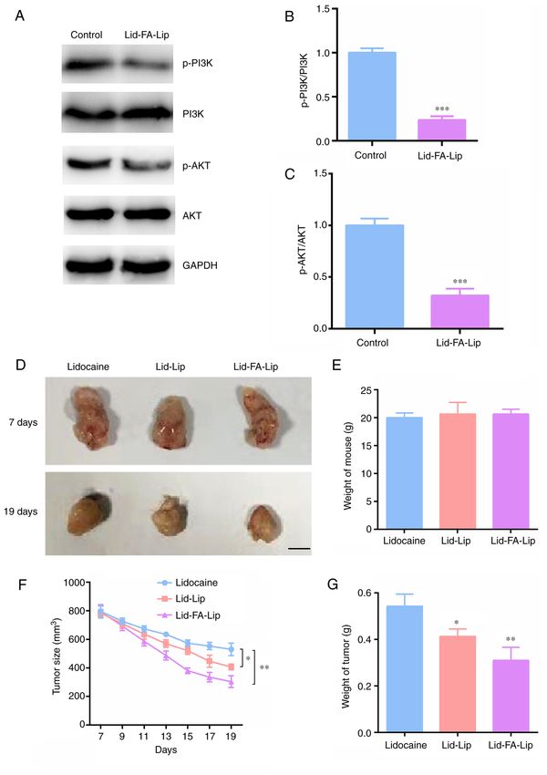
EXPERIMENTAL AND THERAPEUTIC MEDICINE 22: 1025, 2021                                                            7

Figure 5. Lidocaine liposome modified with FA inhibits the tumor growth of glioma cells via the PI3K/AKT pathway. (A) Representative immunoblots showing
p‑PI3K, PI3K, p‑AKT and AKT levels in U87 cells following treatment with Lid‑FA‑Lip. Quantitative analysis of the (B) p‑PI3K/PI3K and (C) p‑AKT/AKT
ratios. (D) Representative photographs of tumors formed by U87 cells, isolated from nude mice following the treatment with different lidocaine formulations
(n=6/group). The upper images are the photographs of the tumors at 7 days, and the lower images show the tumors at 19 days. Scale bar, 5 mm. (E) Weights
of the mice were measured. (F) Tumor growth curves for the control and the liposomal treatment groups are shown. (G) Tumor weights were measured and
compared. Results are presented as the mean ± standard deviation. *P
8              LI et al: LIDOCAINE LIPOSOME SUPPRESSES GLIOMA CELLS VIA TARGETING THE PI3K/AKT PATHWAY

significantly decreased AKT phosphorylation levels compared
with those of the untreated control (Fig. 5A and C). These
results suggest that the antitumor activity of Lid‑FA‑Lip in
glioma is mediated via targeting the PI3K/AKT pathway.
    The antitumor activity of Lid‑FA‑Lip was also validated
in vivo. The findings of the xenograft experiment confirm the
effectiveness of this type of targeted therapy. Smaller tumors
were observed in mice from the Lid‑FA‑Lip treatment group
compared with the Lid‑Lip and control groups. Representative
images of tumors from each group are shown in Fig. 5D.
The results confirm that, although there was no significant
change in the weight of the mice (Fig. 5E), the tumor volume
(Fig. 5F) and tumor weight (Fig. 5G) of the mice treated with
Lid‑FA‑Lip was significantly reduced. This demonstrates the
antitumor effects of Lid‑FA‑Lip.

Discussion
                                                                     Figure 6. Schematic illustration of Lid‑FA‑Lip crossing the BBB and
Glioma is a craniocerebral malignancy affecting glial cells (1),     targeting glioma cells. FA is an effective ligand for crossing the BBB and
                                                                     glioma treatment, and Lid‑FA‑Lip exhibited antitumor effects via targeting
and accounts for ~60% of all intracranial tumors (24).               the PI3K/AKT pathway. Lid‑FA‑Lip, FA‑modified lidocaine‑carrying
Although the BBB results in glioma having an extremely               liposome; BBB, blood‑brain barrier; FA, folic acid.
low rate of metastasis it also hinders the treatment of glioma
using therapeutic drugs. Therefore, the development of novel
targeted drug delivery systems for glioma is necessary.
    Lidocaine is widely used as a local anesthetic. In addition      with a reduction in MMP2 expression compared with that in
to having anesthetic effects, lidocaine has also been reported       the control group. Though flow cytometry and western blotting
to have inhibitory effects on tumors (25‑27). A previous study       assays, it was noted that Lid‑FA‑Lip treatment significantly
demonstrated that lidocaine can inhibit vascular endothelial         stimulated the apoptosis of glioma cells, with a reduction in
growth factor‑A‑induced angiogenesis, and therefore may be           the expression levels of Bcl‑2, an anti‑apoptotic biomarker.
able to suppress cancer progression (28). Additionally, lido‑        Tumor angiogenesis can affect the prognosis of patients with
caine has been reported to inhibit the proliferation and invasion    tumors. Endothelial cells are important in the formation of
of hepatocellular carcinoma cells by inhibiting the PI3K/Akt         new blood vessels that feed the tumor with nutrients. Notably,
pathway (23). Similarly, another study demonstrated that the         the present confirmed that Lid‑FA‑Lip had superior antitumor
inhibitory effect of lidocaine on lung cancer cell proliferation     effects mediated via targeting the PI3K/AKT pathway and
is mediated via PI3K/AKT and EGFR pathways (29). These               thereby suppressed tumor growth. The scheme in Fig. 6
studies, together with the findings of the present study, indicate   illustrates the suggested mechanism. The PI3K/AKT pathway
the potential role of lidocaine in tumor therapy.                    affects multiple cellular processes, including cell proliferation,
    In the present study, an FA‑cholesterol derivative was           migration, invasion and apoptosis (30,31), and is involved in
designed and synthesized, which was characterized by                 the progression of various cancers (32). A number of proteins
HMRS and elemental analysis to confirm its structure. A              promote the progression and metastasis of cancers, including
lidocaine‑loaded liposomal drug delivery system modified             lung cancer and pancreatic cancer, via the PI3K/AKT pathway,
with this ligand was prepared. Analysis of the characteris‑          and drugs targeting the PI3K/AKT pathway have been used in
tics of the liposome in vitro suggested that it had a suitable       the clinic or in clinical trials (33). The PI3K/AKT axis is also
size and PDI, which should contribute to the achievement of          widely known to be involved the regulation of glioma progres‑
passive targeting via enhanced permeability and retention            sion (29). This pathway affects the proliferation, migration and
effects. In addition, the slow release behavior exhibited in the     apoptosis of glioma cells (29), and the results of the present
drug release assay demonstrates that Lid‑FA‑Lip significantly        study indicate that Lid‑FA‑Lip exerts antitumor effects via this
improved the release behavior of lidocaine with a sustained          pathway; Lid‑FA‑Lip reduced PI3K and AKT phosphorylation
release effect. The hemolysis rate was
EXPERIMENTAL AND THERAPEUTIC MEDICINE 22: 1025, 2021 9
and inhibited tumor growth in vitro. Therefore, it may be 5. Finck T, Gempt J, Krieg SM, Meyer B, Zimmer C, Wiestler B,
Kirschke JS and Sollmann N: Assessment of the extent of
concluded that FA is an effective ligand for transporting resection in surgery of high‑grade glioma‑evaluation of black
lidocaine through the BBB and treating glioma, and the blood sequences for intraoperative magnetic resonance imaging
antitumor effects of Lid‑FA‑Lip are mediated via targeting the at 3 tesla. Cancers (Basel) 12: 1580, 2020.
6. Opoku‑Darko M, Lang ST, Artindale J, Cairncross JG, Sevick RJ
PI3K/AKT pathway. and Kelly JJP: Surgical management of incidentally discovered
diffusely infiltrating low‑grade glioma. J Neurosurg 129: 19‑26,
Acknowledgements 2018.
7. Pisapia DJ: The updated World Health Organization glioma
classification: Cellular and molecular origins of adult infiltrating
Not applicable. gliomas. Arch Pathol Lab Med 141: 1633‑1645, 2017.
8. de Blank P, Fouladi M and Huse JT: Molecular markers and
targeted therapy in pediatric low‑grade glioma. J Neurooncol 150:
Funding 5‑15, 2020.
9. Yan C, Wang J, Yang Y, Ma W and Chen X: Molecular
This study was supported by the 2019 Middle‑aged and biomarker‑guided anti‑angiogenic targeted therapy for malignant
glioma. J Cell Mol Med 23: 4876‑4882, 2019.
Young Training Fund Project of the Anesthesiology Branch 10. Li J, Chai Z, Lu J, Xie C, Ran D, Wang S, Zhou J and Lu W:
of Tianjin Medical Association (grant no. TJMZJJ‑2019‑03) αvβ3‑targeted liposomal drug delivery system with attenuated
and the Youth Fund of the Second Hospital of Tianjin Medical immunogenicity enabled by linear pentapeptide for glioma
therapy. J Control Release 322: 542‑554, 2020.
University (grant no. 2018ydey14). 11. He Y, Wu C, Duan J, Miao J, Ren H and Liu J: Anti‑Glioma
effect with targeting therapy using folate modified nano‑micelles
Availability of data and materials delivery curcumin. J Biomed Nanotechnol 16: 1‑13, 2020.
12. Ye L, Zhang Y, Chen YJ and Liu Q: Anti‑tumor effects of
lidocaine on human gastric cancer cells in vitro. Bratisl Lek
The datasets used and/or analyzed during the current study are Listy 120: 212‑217, 2019.
available from the corresponding author on reasonable request. 13. Li YC, Wang Y, Li DD, Zhang Y, Zhao TC and Li CF: Procaine
is a specific DNA methylation inhibitor with anti‑tumor effect
for human gastric cancer. J Cell Biochem 119: 2440‑2449, 2018.
Authors' contributions 14. Zuckerman LM, Frames WL, Mirshahidi HR, Williams NL,
Shields TG, Otoukesh S and Mirshahidi S: Antiproliferative
effect of bupivacaine on patient‑derived sarcoma cells. Mol Clin
DL, XY, BL and CY carried out the molecular biology experi‑ Oncol 13: 7, 2020.
ments and drafted the manuscript. DL, XY, JS, MY and HW 15. Unami A, Shinohara Y, Ichikawa T and Baba Y: Biochemical and
participated in the design of the study and performed the microarray analyses of bupivacaine‑induced apoptosis. J Toxicol
Sci 28: 77‑94, 2003.
statistical analysis. DL, XY and YL conceived of the study, 16. Zhao Y, Zhang L, Peng Y, Yue QM, Hai L, Guo L, Wang QT
participated in its design and coordination and helped to draft and Wu Y: GLUT1‑mediated venlafaxine‑thiamine disulfide
the manuscript. DL and YL confirm the authenticity of all the system‑glucose conjugates with ‘lock‑in’ function for central
nervous system delivery. Chem Biol Drug Des 91: 707‑716,
raw data. All authors read and approved the final manuscript. 2018.
17. Tsou YH, Zhang XQ, Zhu H, Syed S and Xu X: Drug Delivery
Ethics approval and consent to participate to the brain across the blood-brain barrier using nanomaterials.
Small 13: e1701921, 2017. Erratum in: Small 14: e1801588, 2018.
18. Yang Y, Zhao Z, Xie C and Zhao Y: Dual‑targeting liposome
All procedures performed in the current study were approved modified by glutamic hexapeptide and folic acid for bone
by the Ethics Committee of the Second Hospital of Tianjin metastatic breast cancer. Chem Phys Lipids 228: 104882, 2020.
19. Akal ZU, Alpsoy L and Baykal A: Superparamagnetic iron
Medical University. oxide conjugated with folic acid and carboxylated quercetin for
chemotherapy applications. Ceram Int 42: 9065‑9072, 2016.
Patient consent for publication 20. Zhao Z, Chen C, Xie C and Zhao Y: Design, synthesis and
evaluation of liposomes modified with dendritic aspartic acid for
bone‑specific targeting. Chem Phys Lipids 226: 104832, 2020.
Not applicable. 21. Zhao Z, Zhao Y, Xie C, Chen C, Lin D, Wang S, Lin D, Cui X,
Guo Z and Zhou J: Dual‑active targeting liposomes drug delivery
system for bone metastatic breast cancer: Synthesis and biolog‑
Competing interests ical evaluation. Chem Phys Lipids 223: 104785, 2019.
22. Su Z, Xing L, Chen Y, Xu Y, Yang F, Zhang C, Ping Q and
The authors declare that they have no competing interests. Xiao Y: Lactoferrin‑Modified Poly(ethylene glycol)‑Grafted
BSA nanoparticles as a dual‑targeting carrier for treating brain
gliomas. Mol Pharm 11: 1823‑1834, 2014.
References 23. Zhang Y, Jia J, Jin W, Cao J, Fu T, Ma D and Zhang Y: Lidocaine
inhibits the proliferation and invasion of hepatocellular carci‑
1. Dimou J and Kelly J: The biological and clinical basis for noma by downregulating USP14 induced PI3K/Akt pathway.
early referral of low grade glioma patients to a surgical Pathol Res Pract 216: 152963, 2020.
neuro‑oncologist. J Clin Neurosci 78: 20‑29, 2020. 24. Habimana‑Griffin L, Ye D, Carpenter J, Prior J, Sudlow G,
2. Liu M and Wang L: Prognostic significance of preoperative Marsala L, Mixdorf M, Rubin J, Chen H and Achilefu S:
serum albumin, albumin‑to‑globulin ratio, and prognostic Intracranial glioma xenograft model rapidly reestablishes
nutritional index for patients with glioma: A meta‑analysis. blood‑brain barrier integrity for longitudinal imaging of tumor
Medicine (Baltimore) 99: e20927, 2020. progression using fluorescence molecular tomography and
3. Huang J, Yu J, Tu L, Huang N, Li H and Luo Y: Isocitrate contrast agents. J Biomed Opt 25: 1‑13, 2020.
dehydrogenase mutations in glioma: From basic discovery to 25. Du J, Zhang L, Ma H, Wang Y and Wang P: Lidocaine
therapeutics development. Front Oncol 9: 506, 2019. suppresses cell proliferation and aerobic glycolysis by regulating
4. Zhang X, Zhao L, Zhai G, Ji J and Liu A: Erratum: Multifunctional circHOMER1/miR‑138‑5p/HEY1 axis in colorectal cancer.
polyethylene glycol (PEG)‑Poly (Lactic‑Co‑Glycolic Acid) Cancer Manag Res 12: 5009‑5022, 2020.
(PLGA)‑based nanopar ticles loading doxor ubicin and 26. Siekmann W, Tina E, Von Sydow AK and Gupta A: Effect of
tetrahydrocurcumin for combined chemoradiotherapy of glioma. lidocaine and ropivacaine on primary (SW480) and metastatic
Med Sci Monit 26: e926333, 2020. (SW620) colon cancer cell lines. Oncol Lett 18: 395‑401, 2019.10 LI et al: LIDOCAINE LIPOSOME SUPPRESSES GLIOMA CELLS VIA TARGETING THE PI3K/AKT PATHWAY
27. Zhou D, Wang L, Cui Q, Iftikhar R, Xia Y and Xu P: Repositioning 32. Xiao Y, Deng T and Wang D: Davanone terpenoid inhibits cispl‑
lidocaine as an anticancer drug: The role beyond anesthesia. atin‑resistant acute myeloid leukemia cancer cell growth by inducing
Front Cell Dev Biol 8: 565, 2020. caspase‑dependent apoptosis, loss of mitochondrial membrane
28. Suzuki S, Mori A, Fukui A, Ema Y and Nishiwaki K: Lidocaine potential, inhibition of cell migration and invasion and targeting
inhibits vascular endothelial growth factor‑A‑induced angiogen‑ PI3K/AKT/MAPK signalling pathway. J BUON 25: 1607‑1613, 2020.
esis. J Anesth 34: 857‑864, 2020. 33. Liu Z, Mo H, Sun L, Wang L, Chen T, Yao B, Liu R, Niu Y, Tu K,
29. Sun H and Sun Y: Lidocaine inhibits proliferation and metastasis Xu Q and Yang N: Long noncoding RNA PICSAR/miR‑588/EIF6
of lung cancer cell via regulation of miR‑539/EGFR axis. Artif axis regulates tumorigenesis of hepatocellular carcinoma by
Cells Nanomed Biotechnol 47: 2866‑2874, 2019. activating PI3K/AKT/mTOR signaling pathway. Cancer Sci 111:
30. Cierniak S, Koktysz R, Jesiotr M, Gasowska‑Bodnar A and 4118‑4128, 2020.
Bodnar L: Expression of the PI3K/AKT/mTOR pathway as a
prognostic factor in patients with advanced high grade serous This work is licensed under a Creative Commons
ovarian carcinoma treated with neoadjuvant chemotherapy. Eur Attribution-NonCommercial-NoDerivatives 4.0
J Gynaecol Oncol 40: 744‑751, 2019. International (CC BY-NC-ND 4.0) License.
31. Zuo X, Li L and Sun L: Plantamajoside inhibits hypoxia‑induced
migration and invasion of human cervical cancer cells through
the NF‑κ B and PI3K/akt pathways. J Recept Signal Transduct
Res 41: 339‑348, 2021.You can also read