THE HAME SCAFFOLD POSITIVELY REGULATES MPKB PHOSPHORYLATION TO PROMOTE DEVELOPMENT AND SECONDARY METABOLISM IN ASPERGILLUS NIDULANS
←
→
Page content transcription
If your browser does not render page correctly, please read the page content below
www.nature.com/scientificreports

                        OPEN       The HamE scaffold positively
                                   regulates MpkB phosphorylation
                                   to promote development
Received: 18 September 2018
Accepted: 24 October 2018          and secondary metabolism in
                                   Aspergillus nidulans
Published: xx xx xxxx

                                   Dean Frawley1, Betim Karahoda             1
                                                                                 , Özlem Sarikaya Bayram1 & Özgür Bayram1,2

                                   Mitogen-activated protein kinase (MAPK) pathways are conserved signalling cascades in eukaryotes
                                   which regulate a myriad of processes in fungi from sexual reproduction to stress responses. These
                                   pathways rely on recruitment of three kinases on a scaffold protein to facilitate efficient kinase
                                   phosphorylation and subsequent downstream signalling to the nucleus. The model filamentous
                                   fungus Aspergillus nidulans utilises a MAPK pathway termed the pheromone module to regulate both
                                   development and secondary metabolism. This complex consists of the MAP3K (SteC), MAP2K (MkkB),
                                   MAPK (MpkB) and adaptor protein SteD. To date, there has been no scaffold protein identified for this
                                   MAPK pathway. In this study, we characterised a protein termed HamE, which we propose as a scaffold
                                   that regulates kinase phosphorylation and signalling in the pheromone module. Mass spectrometry
                                   analysis and BIFC experiments revealed that HamE physically interacts with both MkkB and MpkB and
                                   transiently interacts with SteC. Deletion of hamE or any of the pheromone module kinases results in
                                   reduced sporulation and complete abolishment of cleistothecia production. Mutants also exhibited
                                   reductions in expression of secondary metabolite gene clusters, including the velvet complex and
                                   sterigmatocystin genes. HamE acts as a positive regulator of MpkB phosphorylation, allowing for HamE
                                   to subsequently regulate development and secondary metabolism.

                                   For eukaryotic organisms to rapidly respond to the myriad of environmental stimuli they encounter, an array
                                   of protein signalling cascades are utilised1,2. Examples of conserved signalling pathways in eukaryotes are
                                   mitogen-activated protein kinase (MAPK) cascades3. These signalling cascades consist of three protein kinases
                                   (MAPKKK, MAPKK and MAPK) that phosphorylate one another, downstream of a receptor. The terminal
                                   MAPK becomes dually phosphorylated at a conserved Thr-X-Tyr motif and translocates into the nucleus where
                                   it activates specific transcription factors4–6. MAPK cascades regulate a range of processes in eukaryotes from
                                   fungi to humans. In mammals, there are 3 main MAPK families, the ERKs, JNKs and p38/SAPKs. These kinases
                                   are involved in cell growth, response to environmental stresses and immune system regulation respectively7–10.
                                   In yeast, 5 MAPKs (Fus3, Kss1, Hog1, Slt2/Mpk1 and Smk1) have been identified. These kinases regulate the
                                   pheromone response, filamentous growth, osmotic response, polarized cell growth and spore wall assembly path-
                                   ways respectively11–15. The most extensively studied of the yeast MAP kinase pathways is the Fus3 module, which
                                   stimulates cell mating in response to pheromone detection11. Upon binding of pheromones to G-protein cou-
                                   pled receptors (GPCRs) at the plasma membrane, the Gβγ subunits of the receptor dissociate and recruit a large
                                   multi-domain scaffold protein known as Ste516,17. Ste5 then assembles a three-tiered kinase module consisting of
                                   the kinases Ste11 (MAPKKK), Ste7 (MAPKK) and Fus3 (MAPK)18,19. Anchoring of Ste11 to the membrane by
                                   the adaptor protein Ste5020,21 and phosphorylation of Ste11 via the p21 activated kinase Ste20 triggers sequen-
                                   tial phosphorylation of each kinase, further amplifying the signal6. Upon phosphorylation, Fus3 migrates to the

                                   1
                                    Biology Department, Maynooth University, Maynooth, Co. Kildare, Ireland. 2Maynooth University Human Health
                                   Research Institute, Kildare, Ireland. Correspondence and requests for materials should be addressed to Ö.B. (email:
                                   ozgur.bayram@mu.ie)

        SCIentIfIC ReporTS |   (2018) 8:16588 | DOI:10.1038/s41598-018-34895-6                                                                      1
www.nature.com/scientificreports/

                           nucleus where it activates the Ste12 transcription factor22. This, in turn, regulates sexual development, allowing
                           for two neighbouring yeast cells to fuse11,23.
                               Knowledge of the yeast Fus3 module has led to the discovery of homologous MAP kinases in filamentous
                           fungi, which play roles in processes like conidiation, pathogenesis and secondary metabolite (SM) produc-
                           tion24–26. The model ascomycete fungus Aspergillus nidulans has been extensively studied to gain insight on fungal
                           genetics and development27–30. This filamentous fungus is capable of reproducing asexually via the production of
                           haploid spores known as conidia29 and sexually via the formation of sexual ascospores enclosed within fruiting
                           bodies known as cleistothecia31. Sexual development in A. nidulans is coupled to SM production via MAPK sig-
                           nalling and a heterotrimeric complex (VeA-VelB-LaeA) known as the velvet complex in the nucleus32.
                               Homologs of the core Fus3 module components have been identified in A. nidulans like SteC (Ste11), MkkB
                           (Ste7), MpkB (Fus3), SteD (Ste50) and AnSte12/SteA (Ste12). These proteins have been shown to play roles in the
                           regulation of asexual and sexual development as well as SM production33–37. It was shown that SteC, MkkB and
                           MpkB associate with the adaptor protein SteD at the plasma membrane to form the A. nidulans pheromone mod-
                           ule. This allows for phosphorylation of MpkB and subsequent translocation of this kinase into the nucleus. MpkB
                           then phosphorylates the SteA transcription factor and the velvet protein VeA to regulate both development and
                           SM production32,36,38,39. Despite resemblance of this pathway to the yeast Fus3 module, a Ste5 homolog has not
                           been identified and it appears that Ste5 homologs are absent in filamentous fungi40. This proposes the question of
                           how these kinases are assembled in the correct orientation at the membrane, allowing for MpkB phosphorylation
                           and signal propagation to the nucleus.
                               Another model ascomycete fungus Neurospora crassa is commonly used to study fungal genetics and cell
                           development41,42. This filamentous fungus utilises a kinase cascade (NRC-1-MEK-2-MAK-2), homologous to the
                           yeast Fus3 module, to regulate germling and hyphal fusion24,43. This is essential in filamentous fungi for generat-
                           ing an interconnected network of cells, known as the mycelium44. It has been shown that the three-tiered kinase
                           cascade associates with the adaptor protein STE-50 and that STE-50 influences NRC-1 activation45, similar to
                           that observed in A. nidulans. Another similarity to A. nidulans is that a homolog of yeast Ste5 does not exist in
                           N. crassa. However, a protein considered essential for cell fusion, known as HAM-546,47, was characterised as a
                           scaffold for the MAK-2 cascade, having been shown to physically associate with all three kinases and the adaptor
                           STE-5045,48. This complex localises in puncta at opposing hyphal tips during chemotropic interactions and under-
                           goes cycles of assembly and disassembly48. MAK-2 phosphorylation results in activation of PP-1 in the nucleus45,
                           a transcription factor similar to yeast Ste12 that regulates cell fusion, sexual development and SM production24,49.
                               It has been shown that HAM-5 is highly conserved in filamentous ascomycete fungi50, proposing the ques-
                           tion of whether homologs of this protein may act as scaffolds in other species. In this study, we identified the A.
                           nidulans HAM-5 homolog (HamE) and have shown, via a proteomics approach, that this protein associates with
                           kinases of the A. nidulans pheromone module. We have also shown that HamE is required for asexual sporula-
                           tion, sexual cleistothecia formation and production of various SMs. Based on our data, we propose that HamE
                           modulates the phosphorylation states of both MkkB and MpkB, allowing for efficient signalling to the nucleus
                           and regulation of both development and secondary metabolism.

                           Results
                           The A. nidulans Ham5 homolog (HamE) interacts with members of the pheromone mod-
                           ule. In an attempt to identify potential scaffold candidates in the pheromone module, Tandem Affinity
                           Purification (TAP)-tagged SteC, MkkB and MpkB, expressed under their native promoters, were purified and
                           analysed via mass spectrometry (MS). In purifications of each kinase, we detected the uncharacterised A. nid-
                           ulans HAM-5 homolog HamE (AN2701) (Fig. 1a,b, Supplementary Tables S1–S3). Reciprocal BLAST searches
                           confirmed that HamE exhibits 62% similarity to HAM-5, with most of this conserved identity existing at the
                           N-terminus. HamE is a large protein (Fig. 1c) of 1,570 amino acids (aa) with 6 putative WD40 repeats at the
                           N-terminus (aa 18–329). A coiled-coil domain was predicted to be located at aa 1205–1225 and a region of intrin-
                           sic protein disorder was identified at the C-terminus (aa 1479–1570). HamE was fused to a TAP epitope tag and
                           used for TAP pulldowns and MS analysis to identify potential phosphorylation sites on this protein. A total of 8
                           putative phosphorylation sites were detected (Fig. 1c, Supplementary Table S5), residing between aa 425–1202.
                           These results complement those found by Jonkers et al.48 who found that HAM-5 contained similar domains
                           at similar positions to HamE and contained 16 putative phosphorylation sites, suggesting complex methods of
                           regulation exist for Ham proteins.
                               To determine HamE expression during different developmental stages (Fig. 1d), HamE was fused to a syn-
                           thetic Green Fluorescent Protein (sGFP) epitope tag and expressed under its native promoter in the hamE
                           locus. A time course immunoblotting was performed. Crude protein extracts were isolated from fungal mycelia
                           that were either grown vegetatively in Glucose Minimal Media (GMM) for 24 and 36 hours, asexually (12 and
                           24 hours) or sexually (12, 24 and 48 hours). It was found that HamE displays complex expression dynamics and is
                           upregulated at the late stages of vegetative growth (36 hours) and early stages of asexual and sexual development
                           (12 hours). HamE appears to be degraded at the late stages of asexual and sexual reproduction, suggesting that it
                           may be required for regulation of the early phases of development.
                               The HamE-GFP strain was also used to visualise the sub-cellular localisations of HamE via confocal micros-
                           copy. In this strain, Histone 2 A (H2A) was tagged with monomeric Red Fluorescent Protein (mRFP) to allow for
                           visualisation of nuclei. It was observed that the HamE-GFP protein is mainly dispersed throughout the cytoplasm
                           but infrequently becomes enriched at the plasma membrane, hyphal tip and nuclear periphery after 16 hours of
                           vegetative growth (Fig. 1e). These patterns of localisation are similar to those observed previously for the phero-
                           mone module proteins36, suggesting that HamE may interact with the complex at these sites.
                               To elucidate the interaction network of the pheromone module, TAP-tagged SteC, MkkB, MpkB and HamE
                           expressed under their native promoters were used for TAP pulldowns and MS (Fig. 1a,b). TAP-tagged proteins

SCIentIfIC ReporTS |   (2018) 8:16588 | DOI:10.1038/s41598-018-34895-6                                                                       2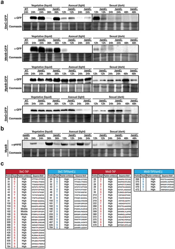
www.nature.com/scientificreports/

                           Figure 1. Discovery of the HamE scaffold protein interacting with components of the pheromone module in A.
                           nidulans. (a) TAP pulldowns of the pheromone module kinases and HamE. TAP-tagged proteins are given at the
                           top of the table and co-purified proteins are given on the left-hand side. The percentage of coverage and unique
                           peptides of each detected protein are displayed. 2 biological replicates of each strain were used. (b) Interaction
                           network of the pheromone module components based on unique peptides detected in each TAP pulldown.
                           Each black dot represents a protein detected in two independent biological replicates but not in the wild type.
                           (c) Schematic overview of the protein structure of HamE. HamE is a large, multi-domain protein that consists of
                           6 WD40 repeats at it’s N-terminus (aa residues 18–329). The red bar represents a coiled-coil domain (aa residues
                           1205–1225) and the green shaded area (1479–1570) represents a region of intrinsic protein disorder. Blue stars
                           represent phosphorylation sites detected by mass spectrometry of TAP-tagged HamE. The amino acid positions
                           and residues targeted for phosphorylation are listed in the accompanying table. S (serine), T (Threonine). (d)
                           Time course immunoblotting of HamE at various stages of development. V (vegetative), A (asexual), S (sexual).
                           For asexual and sexual induction, the HamE-GFP strain was cultured vegetatively for 24 hours in liquid GMM
                           media and transferred to GMM plates to be incubated in the light and dark respectively. SkpA is used as a
                           loading control. Full-length blots in (d) are presented in Supplementary Fig. 2. (e) Localisation of HamE-GFP
                           in vivo at 16 hours of vegetative growth. The GFP fusion protein is dispersed throughout the cytoplasm and
                           localises at the hyphal tips, cell membrane and nuclear envelope, indicated by white arrows.

                           were isolated from vegetative cultures grown for 24 hours. It was found that TAP of SteC recruited MkkB, HamE
                           and the adaptor protein SteD (Supplementary Table S1), but not MpkB. TAP of MkkB recruited SteC, SteD, MpkB
                           and HamE (Supplementary Table S2). TAP of MpkB recruited MkkB and HamE (Supplementary Table S3) and
                           TAP of HamE recruited MkkB and MpkB (Supplementary Table S4). These interactome data propose a pen-
                           tameric complex and suggest that HamE physically interacts with the kinases MkkB and MpkB but may only
                           transiently interact with SteC and SteD.

                           HamE and the pheromone module proteins contribute to the regulation of asexual and sexual
                           development. To determine the influence of the pheromone module and HamE in fungal development, sin-
                           gle and double deletion strains were constructed and asexual and sexual development was monitored (Fig. 2). The
                           hamE gene was deleted in each pheromone module mutant to generate double deletion strains (Supplementary
                           Fig S1). hamEΔ, steCΔ, mkkBΔ, mpkBΔ and steDΔ double mutants were created either by replacing the gene
                           open reading frames with the pyridoxine gene (pyroA) or the pyrithiamine resistance gene (ptrA). Mutants were

SCIentIfIC ReporTS |   (2018) 8:16588 | DOI:10.1038/s41598-018-34895-6                                                                     3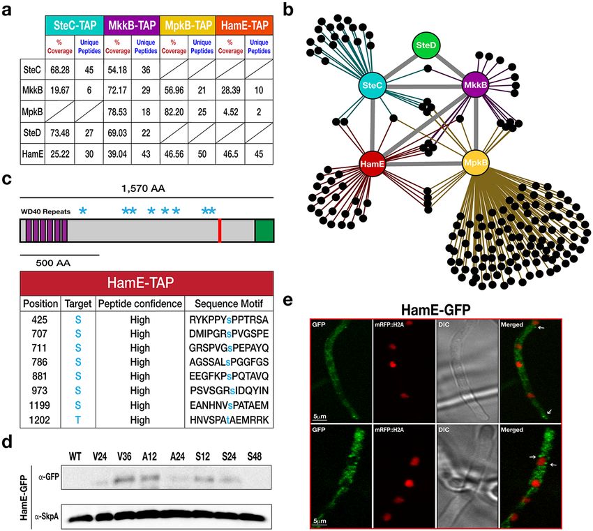
www.nature.com/scientificreports/

                           Figure 2. The pheromone module components and HamE are required for asexual conidiation and sexual
                           cleistothecia production in A. nidulans. (a) Vegetative, asexual and sexual phenotypes of deletion strains.
                           Strains were spot inoculated (5 × 10³ spores) in triplicate on GMM plates containing supplements and induced
                           asexually (4 days in the light at 37 °C) and sexually (5 days in the dark at 37 °C). (b) Close-up stereomicroscopic
                           images of the strains from (a). Images were taken at 5x magnification. (c) Stereomicroscopic images of
                           sexually induced strains taken at 8x magnification. (d) Graphical representation of the colony diameters of
                           each asexually induced strain from (a) with respect to the AGB551 wild type strain. Measurements were taken
                           from three independent biological replicates for each strain and the averages were plotted ± s.d. P-values
                           were calculated by performing unpaired Student’s t-tests (**P < 0.01; ***P < 0.001). (e) Quantification of
                           cleistothecia production in each strain. L = light, D = dark. Three 2x magnification images of each asexual and
                           sexual replicate from (a) were taken and the cleistothecia were counted (N = 9). The averages were plotted ±
                           s.d. as a percentage of the sexually-induced wild type average. (f) Quantification of asexual conidiation in each
                           asexually and sexually-induced replicate (N = 3) from (a). Mean values were plotted ± s.d. as a percentage of the
                           asexually-induced wild type average. P-values were calculated as described above (*P < 0.05; **P < 0.01), with
                           light and dark-induced colonies being compared to the respective light and dark-induced wild type colonies.

SCIentIfIC ReporTS |   (2018) 8:16588 | DOI:10.1038/s41598-018-34895-6                                                                      4
www.nature.com/scientificreports/

                           spot inoculated on GMM agar plates and incubated in the presence and absence of light (continuous illumination
                           under white fluorescent light) for 4 and 5 days respectively (Fig. 2a). hamE::gfp and hamE::tap fusions, introduced
                           into a hamE deletion strain, were also spot-inoculated to show that these fusions are functional.
                               The colony diameters of all asexually-induced strains (Fig. 2a, upper panel) were measured and the averages
                           of three independent replicates for each strain were plotted as a percentage of the respective wild type average. It
                           was observed that steC, mkkB, mpkB and steD mutants all exhibited 20–30% reductions in colony size (Fig. 2d).
                           However, the hamE mutant did not show any defects in colony growth and was comparable to the wild type. The
                           quantities of sexual fruiting bodies known as cleistothecia produced by each mutant were determined in compar-
                           ison to the wild type strain. It was found that all single and double mutants exhibited a pale phenotype (Fig. 2a,
                           lower panel) and showed complete abolishment of sexual development, with no fruiting bodies being produced
                           and only premature aggregates of Hulle cells known as nests being formed (Fig. 2b,c,e). Lastly, asexual conidia-
                           tion was quantified for each mutant strain, in comparison to the wild type strain. All single and double mutants
                           exhibited a 50–60% reduction in asexual conidiation (Fig. 2a,b upper panels and 2f). The phenotypes of the hamE
                           deletion were fully restored by its GFP and TAP fusions (Fig. 2a).
                               These results show that SteC, MkkB, MpkB and SteD are all required for regulation of colony growth rate,
                           asexual conidiation and sexual cleistothecia development. These data also suggest that HamE may function in a
                           similar manner to the pheromone module components to regulate both asexual and sexual development.

                           Expression of various sexual development and SM genes is dependent on the pheromone
                           module proteins and HamE. Given that the pheromone module mutants and the HamE mutant exhibited
                           defects in sexual development, we decided to show the influence of these proteins on the regulation of various
                           sexual development and SM genes. In A. nidulans, sexual reproduction is co-ordinated with SM production by
                           the heterotrimeric velvet complex (VeA-VelB-LaeA)32. A. nidulans is capable of producing over 40 SMs, that can
                           exhibit beneficial as well as deleterious effects. Examples of SMs produced by A. nidulans include the carcinogenic
                           Sterigmatocystin (ST), antibiotic Penicillin (PN) and the anti-tumour agent Terrequinone A (TQ)51.
                               Real-time/quantitative Polymerase Chain Reaction (qPCR) analysis was performed to determine the relative
                           expression levels of the velvet complex genes veA, velB and laeA in each mutant strain. Reductions in expression
                           of each gene was evident in each mutant (40–70% reduction) in comparison to the wild type (Fig. 3a).
                               By utilising Reversed-Phase High Performance Liquid Chromatography (RP-HPLC), the levels of ST in each
                           mutant were measured and compared to the wild type (Fig. 3b). It was found that each mutant produced signifi-
                           cantly reduced levels of ST (less than 15% of wild type) when cultured in liquid GMM for 48 hours. This coincides
                           with the relative gene expression values detected via qPCR analysis which show reductions in expression (60–90%
                           reduction) of the transcriptional activator gene aflR and the two structural genes stcQ and stcE of the ST gene
                           cluster in all mutants (Fig. 3c).
                               The relative expression values of genes belonging to the PN and TQ clusters were also tested. Expression of
                           the PN genes acvA, aatA and ipnA was significantly reduced (55–85% reduction) in all mutants in comparison to
                           wild type (Fig. 3d). Expression values of the TQ genes tdiA and tdiB showed the most dramatic reductions in all
                           mutants (90–95% reduction) in comparison to wild type (Fig. 3e). Taken together, these data suggest that HamE
                           and the pheromone module proteins all contribute in a similar manner to regulate the expression of the velvet
                           complex genes, allowing for subsequent regulation of sexual development and production of various SMs.

                           HamE influences expression and phosphorylation of the pheromone module components. To
                           investigate the influence of HamE on the expression of the pheromone module proteins, SteC, MkkB, MpkB and
                           SteD were fused to GFP epitope tags and expressed in a strain lacking the hamE gene (Supplementary Fig S1). A
                           time course immunoblotting was performed to compare the levels of expression of each protein in HamE(+) and
                           HamE(−) backgrounds throughout different stages of development (Fig. 4a). This revealed dynamic expression
                           profiles for each protein, with SteC, MkkB, MpkB and SteD all exhibiting differences to one another. This suggests
                           that these proteins may play unique roles separate to those they perform as part of the pheromone module.
                               SteC was found to be upregulated at 24 hours of vegetative growth in both HamE(+) and HamE(−) back-
                           grounds. However, at 36 hours of vegetative growth, it is evident that SteC is degraded in the wild type but expres-
                           sion is maintained in a HamE(−) background. Slight upregulation of SteC also occurs at 12 and 24 hours of
                           asexual growth in the hamE mutant. Interestingly, at 12 hours of sexual induction, SteC is expressed in abun-
                           dance in the wild type but expression is reduced in the HamE(−) background. MkkB was found to be strongly
                           expressed during the early stages of vegetative growth (24 and 36 hours) with increased expression at 36 hours of
                           growth in the hamE mutant. MkkB was also found to be readily degraded during the early stages of asexual and
                           sexual development. MpkB was found to be expressed at all stages of development and deletion of hamE did not
                           have any dramatic effects on MpkB expression. It can be noted that MpkB showed slightly reduced expression at
                           48 hours of sexual growth in the hamE mutant. SteD showed consistent expression during vegetative growth and
                           early asexual development (12 hours), with the HamE(−) background exhibiting no effect on expression at these
                           time points. Slight reductions can be observed in the hamE mutant at 24 hours of asexual growth and 48 hours of
                           sexual growth.
                               Activation of the pheromone module is determined by it’s protein-protein interactions and phosphorylation
                           states. Since HamE stably interacts with MkkB and MpkB, which are the two central kinases of the pheromone
                           module, we decided to look at the phosphorylation states of MkkB and MpkB and also the MAP3K SteC. SteD
                           was not used as it is the adaptor protein of the module. We first tested the phosphorylation of MpkB in HamE(+)
                           and HamE(−) backgrounds during each stage of development (Fig. 4b). MpkB becomes dually phosphorylated
                           at residues Threonine 182 and Tyrosine 184, allowing for it’s activation. By using an α-phospho-p44/42 MAPK
                           antibody which detects phosphorylation at these residues, it was found that MpkB phosphorylation is signifi-
                           cantly reduced in the hamE mutant at all stages of development. We then assessed whether HamE plays a role in

SCIentIfIC ReporTS |   (2018) 8:16588 | DOI:10.1038/s41598-018-34895-6                                                                       5
www.nature.com/scientificreports/
Figure 3. The regulation of various sexual development and secondary metabolism genes is dependent on the
pheromone module components and HamE. (a) Expression levels of the veA, velB and laeA genes belonging
to the velvet complex. Strains were inoculated (5 × 10⁶ spores/ml) in 40 ml of GMM media and incubated
for 48 hours at 37 °C on a shaker. For each qPCR experiment (a,c–e), 2 independent biological replicates and
3 technical replicates were used (N = 6) for each strain. The average expression level values were plotted ±
s.d. as a percentage of the wild type average. (b) HPLC detection of secondary metabolite sterigmatocystin
(ST) levels in deletion strains. Strains were inoculated in triplicate and cultured according to the parameters
described in (a). Average peak area values were plotted as a percentage of the wild type ± s.d. P-values were
calculated by performing unpaired Student’s t-tests (*P < 0.05). (c) Expression levels of the aflR, stcQ and stcE
genes belonging to the ST gene cluster. (d) Expression levels of the acvA, aatA and ipnA genes belonging to the
penicillin gene cluster. (e) Expression levels of the tdiA and tdiB genes belonging to the terrequinone A gene
cluster.
SCIentIfIC ReporTS | (2018) 8:16588 | DOI:10.1038/s41598-018-34895-6 6www.nature.com/scientificreports/
Figure 4. Expression levels and phosphorylation states of the pheromone module components in the presence
and absence of hamE. (a) The expression levels of sGFP-tagged SteC, MkkB, MpkB and SteD fusion proteins
were determined at various stages of development in the presence and absence of hamE. Vegetative cultures
were grown in liquid GMM media. For asexual and sexual cultures, strains were initially grown for 24 hours
vegetatively in liquid GMM media and then mycelia was transferred to GMM plates to be incubated in the light
and dark respectively. 80 μg of each protein sample was loaded on 10% acrylamide gels and for loading controls,
these gels were stained in 0.1% Coomassie Brilliant Blue R-250 dye and exposed using the G: BOX Chemi XRQ
(Syngene). Sexual development samples (dark) were run in different gels separated from vegetative and asexual
samples by a vertical black line. (b) Determination of the phosphorylation status of MpkB in the presence
and absence of hamE using an anti-phospho-p44/42 antibody (Thr182/Tyr184). Sexual samples were run in
different blots. SteC samples from part (a) were used for these blots. As a result, coomassie staining controls
are the same as those for SteC samples. (c) Comparison of the phosphorylated residues of SteC and MkkB in
SCIentIfIC ReporTS | (2018) 8:16588 | DOI:10.1038/s41598-018-34895-6 7www.nature.com/scientificreports/
the presence and absence of hamE. The tables represent the total phosphorylated residues and their amino acid
positions detected by mass spectrometry using 4 independent TAP-tagged biological replicates of each strain. S
(Serine), T (Threonine), Y (Tyrosine). Full-length blots in (a,b) are presented in Supplementary Fig. 3.
the phosphorylation of SteC and MkkB. By performing TAP pulldowns of 4 biological replicates for TAP-tagged
SteC and MkkB in HamE(+) and HamE(−) backgrounds, the total phosphorylation sites were combined in each
strain for comparison (Fig. 4c, Supplementary Tables S5–10). A total of 26 phosphorylation sites were detected
for wild type SteC and 19 sites were detected in the hamE mutant. 18 phosphorylation sites were detected for wild
type MkkB but only 5 sites were detected in the hamE mutant. These data together underline complex modes of
regulation for the pheromone module proteins and highlight potential roles of HamE in the regulation of kinase
expression and phosphorylation, allowing for subsequent modulation of sexual development and secondary
metabolism.
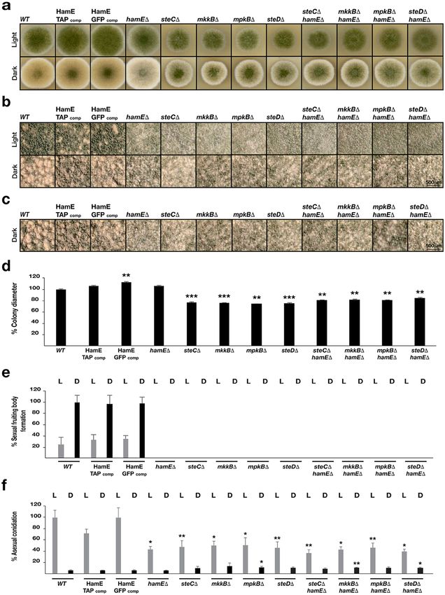
Assembly and localisation of the pheromone module is not HamE-dependent. To assess whether
HamE influences sub-cellular localisations of the pheromone module proteins, GFP-fused kinases in HamE(+)
and HamE(−) backgrounds were visualised via confocal microscopy (Fig. 5a). All members of the pheromone
module, except for MpkB, showed cytoplasmic localization without nuclear enrichment. MpkB was nucleocyto-
plasmic as evident from colocalization of GFP and nuclear mRFP signals as published previously36. It is appar-
ent from Fig. 5a that deletion of hamE does not influence localisation of these proteins in comparison to the
HamE(+) strains. We then decided to test whether HamE is required for pheromone complex assembly by per-
forming TAP pulldowns of kinases in hamEΔ backgrounds. Interestingly, it was shown that in a hamE mutant,
the entire tetrameric complex is capable of assembling (Fig. 5c, Supplementary Tables S11–S13) and MpkB is
capable of binding to transcription factor SteA (Supplementary Table S13). This signifies that MpkB is capable of
translocating into the nucleus in a HamE-independent manner.
Considering that HamE was detectable at the hyphal tips, plasma membrane and nuclear envelope (Fig. 1e),
BIFC was performed to test whether HamE co-localises with the kinase SteC. It was observed that HamE inter-
acts with SteC at the hyphal tip, plasma membrane, septa and nuclear envelope (Fig. 5b). It has been previously
shown36, via BIFC, that SteC also co-localises with MkkB, MpkB and SteD at the same sites, suggesting that HamE
may also co-localise with the entire tetrameric complex at these sites.
Discussion
MAP kinase modules are conserved signal transduction pathways in eukaryotes that regulate a range of processes.
To regulate signalling of these MAP kinase modules, large multi-domain proteins known as scaffolds are utilised.
Scaffolds often have binding domains for protein-protein interactions. As a result, scaffolds are capable of binding
and promoting the interactions of at least two signalling proteins and have been shown to exhibit active regula-
tory roles52. The yeast Fus3 module and Ste5 scaffold represent a mechanistic paradigm for MAP kinase module
signalling and regulation. However, in filamentous fungi, the lack of Ste5 homologs suggests a unique method of
signal regulation in these pathways. Identification of the Ham5 scaffold in the N. crassa MAK-2 module45,48 has
led to the hypothesis that Ham5 homologs may function as scaffolds in other filamentous fungi.
The A. nidulans pheromone module consists of the MAP3K SteC, MAP2K MkkB, MAPK MpkB and adaptor
protein SteD36. In this study, we have identified the A. nidulans Ham5 homolog (HamE) and have provided evi-
dence of scaffolding and regulatory roles for HamE in the pheromone module. HamE has been shown to physi-
cally interact with the kinases MkkB and MpkB (Fig. 1a, Supplementary Tables S2–S4) and is essential for efficient
phosphorylation of each kinase, allowing for signal propagation to the nucleus. TAP pulldowns coupled to MS
initially detected the uncharacterised HamE protein (AN2701) in purifications of SteC, MkkB and MpkB (Fig. 1a,
Supplementary Tables S1–S3). Reciprocal BLAST searches of AN2701 confirmed that HamE is a homolog of N.
crassa Ham5 and is a large protein with 6 WD40 repeats (Fig. 1c) which serve as a scaffolding domain allowing
for protein-protein interactions53. TAP purifications of HamE resulted in detection of MkkB and MpkB (Fig. 1a,
Supplementary Table S4). Although HamE was absent in purifications of SteC, HamE and SteC have been shown
to co-localise at the hyphal tips, septa, cell membrane and nuclear envelope (Fig. 5b), suggesting that HamE and
SteC may transiently interact and also that HamE may co-localise with the entire tetrameric complex at these
sites.
This study has shown that the pheromone module proteins and HamE are crucial for the regulation of asex-
ual sporulation, sexual cleistothecia formation and SM production in Aspergillus nidulans (Figs 2 and 3). In A.
nidulans, the presence and absence of light induces asexual and sexual development respectively29,54. The velvet
complex (VeA-VelB-LaeA) is required for the regulation of asexual and sexual development in response to light
signals and is critical for the co-ordination of sexual development with secondary metabolism32. It has been
shown that activation of MpkB in the pheromone module results in MpkB translocation into the nucleus where
it activates SteA and VeA, promoting velvet complex assembly and coordination of sexual development and
SM production36. The pheromone module mutants and HamE mutant exhibited 50–60% reduced sporulation
(Fig. 2f) and were unable to produce cleisthothecia, forming only premature nests of Hulle cells (Fig. 2b,e). These
data complement results observed for ham5 mutants in N. crassa, which were shown to produce reduced levels
of sexual reproductive structures known as protoperithecia45. In the A. nidulans mutants in this study, reduced
expression of velvet complex genes (Fig. 3a), or presumably reduced phosphorylation of SteA may result in altered
signalling dynamics in response to light signals, resulting in the reductions in SM production and sterile phe-
notypes observed. All mutants exhibited dramatic reductions in production of sterigmatocystin, penicillin and
terrequinone A (Fig. 3), which are all, in turn, regulated by laeA55,56.
SCIentIfIC ReporTS | (2018) 8:16588 | DOI:10.1038/s41598-018-34895-6 8www.nature.com/scientificreports/
Figure 5. HamE co-localises with SteC but does not influence the sub-cellular localisations of kinases or
complex assembly, (a) Sub-cellular localisations of the GFP-tagged pheromone module proteins at 16 hours
of vegetative growth in the presence and absence of HamE. (b) BIFC showing interaction of C-YFP-HamE
and n-YFP-SteC. White arrows indicate co-localisation of these two proteins at the hyphal tips, septa, plasma
membrane and nuclear envelope. (c) TAP pulldowns of kinases in hamEΔ backgrounds at 24 hours of
vegetative growth. (d) Schematic model of the pheromone module and the regulatory roles of HamE in kinase
signalling, fungal development and secondary metabolism. HamE binds the kinases MkkB and MpkB and
co-localises with the tetrameric complex of SteC-MkkB-MpkB-SteD at the plasma membrane, hyphal tips
and nuclear envelope. HamE is required for efficient kinase phosphorylation, specifically MpkB. ‘P’ represents
phosphate groups and inefficient kinase phosphorylation in the hamE mutant is represented by transparent
phosphate groups. Efficient MpkB phosphorylation allows for MpkB to activate SteA and VeA to regulate both
development and secondary metabolism, respectively.
Phosphoproteomics analysis revealed that HamE contains at least 8 phosphorylation sites (Fig. 1c,
Supplementary Table S5). The maximum protein coverage detected for HamE by MS was 46.5%, and so, it is likely
that more phosphorylation sites exist. This data, coupled to the expression levels of HamE at different stages of
development (Fig. 1d) suggest that HamE is a direct target of regulation. This could implicate HamE in higher order
regulatory processes such as positive and negative feedback loops in the pheromone module as is the case for yeast
Ste557,58. In a hamE mutant, expression levels of the pheromone module proteins, particularly SteC, show dramatic
changes (Fig. 4a). It was found that the intensity of MpkB phosphorylation is significantly reduced at all stages of
development in the absence of hamE (Fig. 4b), which complements the reduced levels of MAK-2 phosphorylation
SCIentIfIC ReporTS | (2018) 8:16588 | DOI:10.1038/s41598-018-34895-6 9www.nature.com/scientificreports/
observed in a N. crassa ham5 mutant45. MS analysis also showed reductions in SteC and MkkB phosphorylation
levels in the hamE mutant (Fig. 4c, Supplementary Tables S6–S9). These data suggest that HamE exhibits an active
regulatory role and it is possible that HamE is required to catalytically unlock MpkB for phosphorylation by MkkB,
which is evident for Ste5 in yeast17. This could explain why the tetrameric complex can assemble in a hamE mutant
(Fig. 5c) but MpkB phosphorylation is inefficient and transcription factor activation does not occur.
In conclusion, this study has presented new insight on the organisation and signalling dynamics of the A.
nidulans pheromone module. We propose the Ham5 homolog (HamE) as a regulatory scaffold for the pheromone
module and provide evidence that HamE modulates the expression levels and phosphorylation states of each
kinase, specifically MpkB. As a result, HamE is critical for signal transduction to the nucleus and regulation of
both asexual and sexual development as well as production of various SMs (Fig. 5d). Understanding the molecu-
lar basis of signalling mechanisms in A. nidulans will allow for application of this knowledge to other filamentous
fungi with biotechnological and medical relevance such as Aspergillus flavus and Aspergillus fumigatus since most
filamentous fungal genomes encode orthologs of HamE, rather than Ste5. Characterisation of HamE homologs in
these species may provide insight on the regulatory mechanisms involved in development as well as the produc-
tion of clinically important SMs like aflatoxins and gliotoxin.
Methods
Strains, growth media and culturing conditions. Fungal strains used in this study are listed in
Supplementary Table S14. The Aspergillus nidulans AGB551 (veA+) strain served as a wild type host for all dele-
tions and epitope taggings. Various plasmids used for the knock-out and epitope tagging experiments are listed
in Supplementary Table S15. Plasmids were cloned into Stellar (Clontech) and MACH-1 (Invitrogen) competent
Escherichia coli cells and these cells were cultured in LB media (supplemented with 100 μg/ml ampicillin) and
SOC media. For the growth of fungal strains, Glucose Minimal Media (GMM) was used. For asexual and sexual
induction, fungal strains were cultured in liquid GMM for 24 hours and the mycelia was filtered through mira-
cloth and transferred to GMM agar plates to be incubated in the light and dark respectively. For TAP experiments,
fungal strains were cultured in complete medium.
Phenotypic assays. Strains were point inoculated (5 × 10³ spores) in triplicate on GMM agar plates con-
taining appropriate supplements. Plates were incubated in the presence of light for 4 days and the absence of
light for 5 days to induce asexual and sexual development respectively. All incubations were performed at 37 °C.
Stereomicroscopic images were captured using the Olympus szx16 microscope with Olympus sc30 camera.
Digital pictures were taken and processed with the Cell Sens Standard software (Olympus). Quantifications of
colony diameter, asexual conidiation and cleistothecia production were performed using three independent bio-
logical replicates. Bar charts represent the mean values ± s.d. P-values were calculated by performing unpaired
Student’s t-tests (*P < 0.05; **P < 0.01; ***P < 0.001), using the Graphpad Prism Version 6.
Tandem Affinity Purification (TAP), GFP-Trap and sample preparation for LC-MS protein identi-
fication. Isolation and preparation of TAP and GFP fusion proteins for mass spectrometry analysis was per-
formed as explained in detail32. Detailed descriptions of methods used are given in supplementary information.
Immunoblotting. For GFP-tagged proteins, mouse α-GFP antibody (SC-9996, SantaCruz) was used at
1:1,000 dilution in blocking solution (TBST with 5% milk). Secondary goat α-mouse (170–6516, Biorad) was
used at 1:2,000 dilution in blocking solution. For the detection of SkpA, custom made rabbit α-SkpA was used at
1:1,000 dilution in blocking solution. For the detection of phosphorylated MpkB, rabbit α-phospho p44/42 (Cell
Signalling Technology) was used at 1:1,000 dilution in TBST with 5% BSA. Goat α-rabbit (Biorad) was used as a
secondary antibody for both SkpA and phosphorylated MpkB detection at 1:2,000 dilution in blocking solution.
RNA extraction and quantitative real time PCR analysis. 100 mg of mycelia was collected and mRNA
was isolated according to the ‘RNeasy Plant Mini Kit’ protocol (Qiagen). mRNA was quantified according to
the ‘Qubit RNA BR Assay Kit’ Protocol (Thermo Fisher). cDNA was synthesised from 1 μg of mRNA per strain
using the ‘Transcriptor First Strand cDNA Synthesis Kit’ (Roche). qPCR reaction mixtures were prepared using
LightCycler 480 SYBR Green I Master mix and a LightCycler 480 qPCR machine (Roche) was used to determine
gene expression levels, using a Beta-tubulin (benA) control gene as a reference. Bar charts represent the mean data
of two combined biological replicates and 6 combined technical replicates per strain, ± s.d.
RP-HPLC analysis of Sterigmatocystin levels. Detailed information on culturing conditions and prepa-
ration of samples for RP-HPLC analysis is provided in supplementary information. 3 biological replicates were
prepared per strain and data is presented as a bar chart, with the bars representing the mean ± s.d. P-values were
calculated by performing unpaired Student’s t-tests (*P < 0.05), using the Graphpad Prism Version 6.
Confocal microscopy. GFP and mRFP-tagged strains were inoculated (5 × 10³ spores) in 500 μl liquid
GMM media with supplements and cultured in Lab-Tek Chambered Coverglass W/CVT (Thermo Scientific) for
16 hours at 30 °C. Localisations of the proteins were captured using the Zeiss LSM 510 META inverted confocal
microscope.
References
1. Elion, E. A. Pheromone response, mating and cell biology. Curr Opin Microbiol 3, 573–581 (2000).
2. Dhanasekaran, D. N., Kashef, K., Lee, C. M., Xu, H. & Reddy, E. P. Scaffold proteins of MAP-kinase modules. Oncogene 26,
3185–3202, https://doi.org/10.1038/sj.onc.1210411 (2007).
3. Schaeffer, H. J. & Weber, M. J. Mitogen-activated protein kinases: specific messages from ubiquitous messengers. Mol Cell Biol 19,
2435–2444 (1999).
SCIentIfIC ReporTS | (2018) 8:16588 | DOI:10.1038/s41598-018-34895-6 10www.nature.com/scientificreports/
4. Widmann, C., Gibson, S., Jarpe, M. B. & Johnson, G. L. Mitogen-activated protein kinase: conservation of a three-kinase module
from yeast to human. Physiol Rev 79, 143–180, https://doi.org/10.1152/physrev.1999.79.1.143 (1999).
5. Yoshioka, K. Scaffold proteins in mammalian MAP kinase cascades. J Biochem 135, 657–661, https://doi.org/10.1093/jb/mvh079
(2004).
6. Saito, H. Regulation of cross-talk in yeast MAPK signaling pathways. Curr Opin Microbiol 13, 677–683, https://doi.org/10.1016/j.
mib.2010.09.001 (2010).
7. Shaul, Y. D. & Seger, R. The MEK/ERK cascade: from signaling specificity to diverse functions. Biochim Biophys Acta 1773,
1213–1226, https://doi.org/10.1016/j.bbamcr.2006.10.005 (2007).
8. Huang, G., Shi, L. Z. & Chi, H. Regulation of JNK and p38 MAPK in the immune system: signal integration, propagation and
termination. Cytokine 48, 161–169, https://doi.org/10.1016/j.cyto.2009.08.002 (2009).
9. Rincon, M. & Davis, R. J. Regulation of the immune response by stress-activated protein kinases. Immunol Rev 228, 212–224, https://
doi.org/10.1111/j.1600-065X.2008.00744.x (2009).
10. Cuenda, A. & Rousseau, S. p38 MAP-kinases pathway regulation, function and role in human diseases. Biochim Biophys Acta 1773,
1358–1375, https://doi.org/10.1016/j.bbamcr.2007.03.010 (2007).
11. Bardwell, L. A walk-through of the yeast mating pheromone response pathway. Peptides 26, 339–350 (2005).
12. Ma, D., Cook, J. G. & Thorner, J. Phosphorylation and localization of Kss1, a MAP kinase of the Saccharomyces cerevisiae
pheromone response pathway. Mol Biol Cell 6, 889–909 (1995).
13. Brewster, J. L., de Valoir, T., Dwyer, N. D., Winter, E. & Gustin, M. C. An osmosensing signal transduction pathway in yeast. Science
259, 1760–1763 (1993).
14. Mazzoni, C., Zarov, P., Rambourg, A. & Mann, C. The SLT2 (MPK1) MAP kinase homolog is involved in polarized cell growth in
Saccharomyces cerevisiae. J Cell Biol 123, 1821–1833 (1993).
15. Krisak, L. et al. SMK1, a developmentally regulated MAP kinase, is required for spore wall assembly in Saccharomyces cerevisiae.
Genes Dev 8, 2151–2161 (1994).
16. Pryciak, P. M. & Huntress, F. A. Membrane recruitment of the kinase cascade scaffold protein Ste5 by the Gbetagamma complex
underlies activation of the yeast pheromone response pathway. Genes Dev 12, 2684–2697 (1998).
17. Good, M., Tang, G., Singleton, J., Remenyi, A. & Lim, W. A. The Ste5 scaffold directs mating signaling by catalytically unlocking the
Fus3 MAP kinase for activation. Cell 136, 1085–1097, https://doi.org/10.1016/j.cell.2009.01.049 (2009).
18. Hao, N. et al. Regulation of cell signaling dynamics by the protein kinase-scaffold Ste5. Mol Cell 30, 649–656, https://doi.
org/10.1016/j.molcel.2008.04.016 (2008).
19. Kranz, J. E., Satterberg, B. & Elion, E. A. The MAP kinase Fus3 associates with and phosphorylates the upstream signaling
component Ste5. Genes Dev 8, 313–327 (1994).
20. Wu, C., Leberer, E., Thomas, D. Y. & Whiteway, M. Functional characterization of the interaction of Ste50p with Ste11p MAPKKK
in Saccharomyces cerevisiae. Mol Biol Cell 10, 2425–2440, https://doi.org/10.1091/mbc.10.7.2425 (1999).
21. Xu, G., Jansen, G., Thomas, D. Y., Hollenberg, C. P. & Ramezani Rad, M. Ste50p sustains mating pheromone-induced signal
transduction in the yeast Saccharomyces cerevisiae. Mol Microbiol 20, 773–783 (1996).
22. van Drogen, F., Stucke, V. M., Jorritsma, G. & Peter, M. MAP kinase dynamics in response to pheromones in budding yeast. Nat Cell
Biol 3, 1051–1059, https://doi.org/10.1038/ncb1201-1051 (2001).
23. W S Hoi, J. & Dumas, B. Ste12 and Ste12-like proteins, fungal transcription factors regulating development and pathogenicity.
Eukaryot Cell 9, 480–485, https://doi.org/10.1128/ec.00333-09 (2010).
24. Li, D., Bobrowicz, P., Wilkinson, H. H. & Ebbole, D. J. A mitogen-activated protein kinase pathway essential for mating and
contributing to vegetative growth in Neurospora crassa. Genetics 170, 1091–1104, https://doi.org/10.1534/genetics.104.036772
(2005).
25. Lev, S., Sharon, A., Hadar, R., Ma, H. & Horwitz, B. A. A mitogen-activated protein kinase of the corn leaf pathogen Cochliobolus
heterostrophus is involved in conidiation, appressorium formation, and pathogenicity: diverse roles for mitogen-activated protein
kinase homologs in foliar pathogens. Proc Natl Acad Sci USA 96, 13542–13547 (1999).
26. Zhao, X., Kim, Y., Park, G. & Xu, J. R. A mitogen-activated protein kinase cascade regulating infection-related morphogenesis in
Magnaporthe grisea. Plant Cell 17, 1317–1329, https://doi.org/10.1105/tpc.104.029116 (2005).
27. Dasgupta, A., Fuller, K. K., Dunlap, J. C. & Loros, J. J. Seeing the world differently: variability in the photosensory mechanisms of two
model fungi. Environ Microbiol 18, 5–20, https://doi.org/10.1111/1462-2920.13055 (2016).
28. Morris, N. R. & Enos, A. P. Mitotic gold in a mold: Aspergillus genetics and the biology of mitosis. Trends Genet 8, 32–37 (1992).
29. Adams, T. H., Wieser, J. K. & Yu, J. H. Asexual sporulation in Aspergillus nidulans. Microbiol Mol Biol Rev 62, 35–54 (1998).
30. Harris, S. D. The duplication cycle in Aspergillus nidulans. Fungal Genet Biol 22, 1–12, https://doi.org/10.1006/fgbi.1997.0990
(1997).
31. Dyer, P. S. & O’Gorman, C. M. Sexual development and cryptic sexuality in fungi: insights from Aspergillus species. FEMS Microbiol
Rev 36, 165–192, https://doi.org/10.1111/j.1574-6976.2011.00308.x (2012).
32. Bayram, O. et al. VelB/VeA/LaeA complex coordinates light signal with fungal development and secondary metabolism. Science 320,
1504–1506, https://doi.org/10.1126/science.1155888 (2008).
33. Wei, H., Requena, N. & Fischer, R. The MAPKK kinase SteC regulates conidiophore morphology and is essential for heterokaryon
formation and sexual development in the homothallic fungus Aspergillus nidulans. Mol Microbiol 47, 1577–1588 (2003).
34. Vallim, M. A., Miller, K. Y. & Miller, B. L. Aspergillus SteA (sterile12-like) is a homeodomain-C2/H2-Zn + 2 finger transcription
factor required for sexual reproduction. Mol Microbiol 36, 290–301 (2000).
35. Teague, M. A., Chaleff, D. T. & Errede, B. Nucleotide sequence of the yeast regulatory gene STE7 predicts a protein homologous to
protein kinases. Proc Natl Acad Sci USA 83, 7371–7375 (1986).
36. Bayram, O. et al. The Aspergillus nidulans MAPK module AnSte11-Ste50-Ste7-Fus3 controls development and secondary
metabolism. PLoS Genet 8, e1002816, https://doi.org/10.1371/journal.pgen.1002816 (2012).
37. Paoletti, M. et al. Mating type and the genetic basis of self-fertility in the model fungus Aspergillus nidulans. Curr Biol 17,
1384–1389, https://doi.org/10.1016/j.cub.2007.07.012 (2007).
38. Atoui, A., Bao, D., Kaur, N., Grayburn, W. S. & Calvo, A. M. Aspergillus nidulans natural product biosynthesis is regulated by mpkB,
a putative pheromone response mitogen-activated protein kinase. Appl Environ Microbiol 74, 3596–3600, https://doi.org/10.1128/
aem.02842-07 (2008).
39. Sarikaya Bayram, O. et al. LaeA control of velvet family regulatory proteins for light-dependent development and fungal cell-type
specificity. PLoS Genet 6, e1001226, https://doi.org/10.1371/journal.pgen.1001226 (2010).
40. Rispail, N. et al. Comparative genomics of MAP kinase and calcium-calcineurin signalling components in plant and human
pathogenic fungi. Fungal Genet Biol 46, 287–298, https://doi.org/10.1016/j.fgb.2009.01.002 (2009).
41. Roche, C. M., Loros, J. J., McCluskey, K. & Glass, N. L. Neurospora crassa: looking back and looking forward at a model microbe.
Am J Bot 101, 2022–2035, https://doi.org/10.3732/ajb.1400377 (2014).
42. Berlin, V. & Yanofsky, C. Isolation and characterization of genes differentially expressed during conidiation of Neurospora crassa.
Mol Cell Biol 5, 849–855 (1985).
43. Pandey, A., Roca, M. G., Read, N. D. & Glass, N. L. Role of a mitogen-activated protein kinase pathway during conidial germination
and hyphal fusion in Neurospora crassa. Eukaryot Cell 3, 348–358 (2004).
SCIentIfIC ReporTS | (2018) 8:16588 | DOI:10.1038/s41598-018-34895-6 11www.nature.com/scientificreports/
44. Read, N. D., Lichius, A., Shoji, J. Y. & Goryachev, A. B. Self-signalling and self-fusion in filamentous fungi. Curr Opin Microbiol 12,
608–615, https://doi.org/10.1016/j.mib.2009.09.008 (2009).
45. Dettmann, A., Heilig, Y., Valerius, O., Ludwig, S. & Seiler, S. Fungal communication requires the MAK-2 pathway elements STE-20
and RAS-2, the NRC-1 adapter STE-50 and the MAP kinase scaffold HAM-5. PLoS Genet 10, e1004762, https://doi.org/10.1371/
journal.pgen.1004762 (2014).
46. Aldabbous, M. S. et al. The ham-5, rcm-1 and rco-1 genes regulate hyphal fusion in Neurospora crassa. Microbiology 156, 2621–2629,
https://doi.org/10.1099/mic.0.040147-0 (2010).
47. Fu, C. et al. Identification and characterization of genes required for cell-to-cell fusion in Neurospora crassa. Eukaryot Cell 10,
1100–1109, https://doi.org/10.1128/ec.05003-11 (2011).
48. Jonkers, W. et al. HAM-5 functions as a MAP kinase scaffold during cell fusion in Neurospora crassa. PLoS Genet 10, e1004783,
https://doi.org/10.1371/journal.pgen.1004783 (2014).
49. Leeder, A. C., Jonkers, W., Li, J. & Glass, N. L. Early colony establishment in Neurospora crassa requires a MAP kinase regulatory
network. Genetics 195, 883–898, https://doi.org/10.1534/genetics.113.156984 (2013).
50. Jamet-Vierny, C., Debuchy, R., Prigent, M. & Silar, P. IDC1, a pezizomycotina-specific gene that belongs to the PaMpk1 MAP kinase
transduction cascade of the filamentous fungus Podospora anserina. Fungal Genet Biol 44, 1219–1230, https://doi.org/10.1016/j.
fgb.2007.04.005 (2007).
51. Inglis, D. O. et al. Comprehensive annotation of secondary metabolite biosynthetic genes and gene clusters of Aspergillus nidulans,
A. fumigatus, A. niger and A. oryzae. BMC Microbiol 13, 91, https://doi.org/10.1186/1471-2180-13-91 (2013).
52. Buday, L. & Tompa, P. Functional classification of scaffold proteins and related molecules. Febs j 277, 4348–4355, https://doi.
org/10.1111/j.1742-4658.2010.07864.x (2010).
53. Xu, C. & Min, J. Structure and function of WD40 domain proteins. Protein Cell 2, 202–214, https://doi.org/10.1007/s13238-011-
1018-1 (2011).
54. Purschwitz, J. et al. Functional and physical interaction of blue- and red-light sensors in Aspergillus nidulans. Curr Biol 18, 255–259,
https://doi.org/10.1016/j.cub.2008.01.061 (2008).
55. Bok, J. W. & Keller, N. P. LaeA, a regulator of secondary metabolism in Aspergillus spp. Eukaryot Cell 3, 527–535 (2004).
56. Bouhired, S., Weber, M., Kempf-Sontag, A., Keller, N. P. & Hoffmeister, D. Accurate prediction of the Aspergillus nidulans
terrequinone gene cluster boundaries using the transcriptional regulator LaeA. Fungal Genet Biol 44, 1134–1145, https://doi.
org/10.1016/j.fgb.2006.12.010 (2007).
57. Strickfaden, S. C. et al. A mechanism for cell-cycle regulation of MAP kinase signaling in a yeast differentiation pathway. Cell 128,
519–531, https://doi.org/10.1016/j.cell.2006.12.032 (2007).
58. Bhattacharyya, R. P. et al. The Ste5 scaffold allosterically modulates signaling output of the yeast mating pathway. Science 311,
822–826, https://doi.org/10.1126/science.1120941 (2006).
Acknowledgements
This study was funded by a Maynooth University John and Pat Hume Scholarship, an IRC postgraduate
scholarship (GOIPG/2018/35) to DF, Science Foundation Ireland (Grant No: 13/CDA/2142) to OB and IRC
Postdoctoral Fellowship (GOIPD/2014/178) to OSB. Quantitative PCR instrumentation and MS facility were
funded by SFI Grant No: (SFI/07/RFP/GEN/F571/ECO7) and 12/RI/2346(3) respectively.
Author Contributions
O.B. designed and supervised the project. D.F. performed the majority of the experiments, including
transformations, growth tests, immunoblotting, Mass Spectrometry and confocal microscopy; O. S. B. generated
the initial recipient strains used by D. F., B.K.; D.F., B.K., and O. B. analysed the results; D. F. wrote and O.B. edited
the manuscript.
Additional Information
Supplementary information accompanies this paper at https://doi.org/10.1038/s41598-018-34895-6.
Competing Interests: The authors declare no competing interests.
Publisher’s note: Springer Nature remains neutral with regard to jurisdictional claims in published maps and
institutional affiliations.
Open Access This article is licensed under a Creative Commons Attribution 4.0 International
License, which permits use, sharing, adaptation, distribution and reproduction in any medium or
format, as long as you give appropriate credit to the original author(s) and the source, provide a link to the Cre-
ative Commons license, and indicate if changes were made. The images or other third party material in this
article are included in the article’s Creative Commons license, unless indicated otherwise in a credit line to the
material. If material is not included in the article’s Creative Commons license and your intended use is not per-
mitted by statutory regulation or exceeds the permitted use, you will need to obtain permission directly from the
copyright holder. To view a copy of this license, visit http://creativecommons.org/licenses/by/4.0/.
© The Author(s) 2018
SCIentIfIC ReporTS | (2018) 8:16588 | DOI:10.1038/s41598-018-34895-6 12You can also read