Yme2, a putative RNA recognition motif and AAA+ domain containing protein, genetically interacts with the mitochondrial protein export machinery
←
→
Page content transcription
If your browser does not render page correctly, please read the page content below
Biol. Chem. 2022; aop

Nupur Sharma and Christof Osman*

Yme2, a putative RNA recognition motif and AAA+
domain containing protein, genetically interacts
with the mitochondrial protein export machinery
https://doi.org/10.1515/hsz-2021-0398                                      five respiratory complexes of the electron transport chain
Received October 21, 2021; accepted January 19, 2022;                      (ETC) that are responsible for energy production by the
published online January 31, 2022
                                                                           process of oxidative phosphorylation (Acín-Pérez et al.
                                                                           2008; Cogliati et al. 2018). The assembly of the ETC com-
Abstract: The mitochondrial respiratory chain is composed
                                                                           plexes is an intricate process that requires coordinated
of nuclear as well as mitochondrial-encoded subunits. A
                                                                           assembly of proteins from a bi-genomic origin. The mito-
variety of factors mediate co-translational integration of
                                                                           chondrial DNA (mtDNA) encodes for seven subunits of the
mtDNA-encoded proteins into the inner membrane. In
                                                                           ETC (in Saccharomyces cerevisiae), while the remaining
Saccharomyces cerevisiae, Mdm38 and Mba1 are ribosome
                                                                           subunits are encoded by the nuclear DNA (Turk et al. 2013).
acceptors that recruit the mitochondrial ribosome to the
                                                                           Mitochondria have developed sophisticated import path-
inner membrane, where the insertase Oxa1, facilitates
                                                                           ways to facilitate import of nuclear DNA-encoded proteins
membrane integration of client proteins. The protein Yme2
                                                                           and export of mtDNA-encoded proteins into the MIM, in
has previously been shown to be localized in the inner
                                                                           order to form functional respiratory complexes (Wiede-
mitochondrial membrane and has been implicated in
                                                                           mann and Pfanner 2017). For the export of mtDNA-encoded
mitochondrial protein biogenesis, but its mode of action
                                                                           proteins, multiple ribosome interactors, including Mdm38,
remains unclear. Here, we show that multiple copies of
                                                                           Mba1 and Mrx15, have been proposed to recruit the
Yme2 assemble into a high molecular weight complex.
                                                                           mitoribosome to the MIM (Bauerschmitt et al. 2010; Frazier
Using a combination of bioinformatics and mutational
                                                                           et al. 2006; Möller-Hergt et al. 2018; Ott et al. 2006). Protein
analyses, we find that Yme2 possesses an RNA recognition
                                                                           insertion into the MIM then occurs co-translationally via
motif (RRM), which faces the mitochondrial matrix and a
                                                                           the Oxa1 insertase (Hell et al. 2001; Szyrach et al. 2003).
AAA+ domain that is located in the intermembrane space.
                                                                           Despite the presence of dedicated and efficient import
We further show that YME2 genetically interacts with
                                                                           machineries, a number of protein quality control pathways
MDM38, MBA1 and OXA1, which links the function of Yme2
                                                                           exist that remove misfolded and superfluous proteins to
to the mitochondrial protein biogenesis machinery.
                                                                           prevent faulty complex assembly (Böttinger and Becker
Keywords: MBA1; MDM38; mitoribosome; OXA1; RRM;                            2012; Tatsuta 2009). An inefficient complex assembly may
Walker motifs.                                                             lead to ROS production and have deleterious effects on
                                                                           mitochondrial integrity, which in turn can cause various
                                                                           muscular and neurodegenerative disorders in humans
Introduction                                                               (Nunnari and Suomalainen 2012).
                                                                                A pivotal role in mitochondrial protein quality con-
The Mitochondrial Inner Membrane (MIM) is one of the                       trol, is played by members of the AAA+ (ATPases Asso-
most protein-rich membranes in the cell, as it is home to the              ciated with diverse cellular Activities) protein family, that
                                                                           perform ATP-driven unfolding, extraction and degrada-
                                                                           tion of damaged and dysfunctional proteins (Gates and
                                                                           Martin 2020; Song et al. 2021; Steele and Glynn 2019). The
*Corresponding author: Christof Osman, Faculty of Biology, Ludwig
Maximilian University Munich, D-82152 Planegg-Martinsried,                 AAA+ proteins are hetero- or homo-oligomeric complexes,
Germany; and Graduate School of Life Sciences, Ludwig Maximilian           that assemble to form ring-like structures (Gerdes et al.
University Munich, D-82152 Planegg-Martinsried, Germany,                   2012; Miller and Enemark 2016; Opalińska and Jańska
E-mail: osman@bio.lmu.de. https://orcid.org/0000-0002-1892-0222            2018). AAA+ proteins belong to the superfamily of P-loop
Nupur Sharma, Faculty of Biology, Ludwig Maximilian University
                                                                           nucleoside triphosphate binding proteins (Snider et al.
Munich, D-82152 Planegg-Martinsried, Germany; and Graduate
School of Life Sciences, Ludwig Maximilian University Munich,
                                                                           2008). The hallmark of this superfamily are the ATP bind-
D-82152 Planegg-Martinsried, Germany, E-mail: sharma@bio.lmu.de.           ing and hydrolysis domains, namely the Walker A and B
https://orcid.org/0000-0002-8602-6951                                      motifs, respectively (Walker et al. 1982). The Walker A motif
  Open Access. © 2022 Nupur Sharma and Christof Osman, published by De Gruyter.          This work is licensed under the Creative Commons
Attribution 4.0 International License.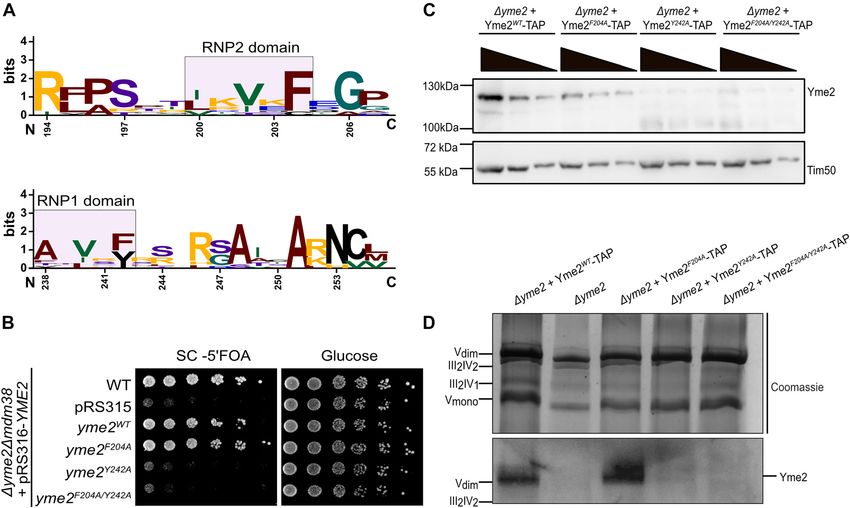
2 N. Sharma and C. Osman: Genetic link of Yme2 to the mitochondrial export machinery
(G-x(4)-GK-[TS]) is a conserved P-loop (Phosphate loop) 2017). Additionally, the AAA+ proteins Msp1 and Cdc48,
motif that lines the axial channel of the ring assembly and are involved in extraction of proteins from translocation
co-ordinates the beta and gamma phosphates of the pores of the outer membrane (Basch et al. 2020; Mår-
nucleotide during ATP hydrolysis (Gates and Martin 2020; tensson et al. 2019; Song et al. 2021; Weidberg and Amon
Wendler et al. 2012). The Walker B motif (hhhhDE, ‘h’ 2018).
denoting a hydrophobic amino acid), on the other hand, YME2 (Yeast Mitochondrial Escape protein 2) was first
contributes to ATP hydrolysis by coordinating the water- discovered in a genetic screen conducted in S.cerevisiae,
activating magnesium ion with the help of acidic residues wherein the loss of YME2 led to escape of mitochondrial
(Hanson and Whiteheart 2005). Within the P-loop super- DNA from mitochondria to the nucleus (Hanekamp and
family, AAA+ proteins belong to the ASCE (Additional Thorsness 1996; Thorsness and Fox 1993). Yme2 is a
Strand Catalytic “E”) class that is characterized by an single-spanning trans-membrane protein of the inner
additional β-strand that separates the P-loop and the mitochondrial membrane, which exposes its N- and
Walker B strands in a central five-strand containing β-sheet C-termini to the matrix and intermembrane space,
of an α–β–α sandwich (Erzberger and Berger 2006; Sera- respectively (Figure 1A) (Hanekamp and Thorsness 1996;
phim and Houry 2020). A feature that sets AAA+ proteins Leonhard et al. 2000). More recently, Yme2 was found to
apart from other members of the ASCE class is the absence co-localize with mtDNA nucleoids (Murley et al. 2013) and
of additional β-strands adjacent to the central β-sheet to be associated with the MIOREX complexes, which are
(Erzberger and Berger 2006; Miller and Enemark 2016; large expressosome-like assemblies comprising factors
Puchades et al. 2020). that are bound to mitoribosomes and are involved in
Mitochondrial protein quality control depends on a mitochondrial gene expression (Kehrein et al. 2015). Here,
network of AAA+ proteins. Two important complexes we find that YME2 exhibits negative genetic interactions
are the i-AAA and the m-AAA protease, which are with MDM38, MBA1 and OXA1, which links YME2 to
AAA-proteases that function to degrade misfolded pro- mitochondrial protein biogenesis. Furthermore, we
teins from the IMS and the matrix, respectively (Arlt et al. analyze the domain organization of Yme2 and thereby
1996; Glynn 2017; Leonhard et al. 1996; Levytskyy et al. provide insights into the possible function of Yme2.
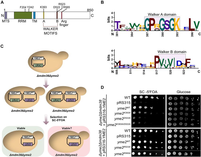
Figure 1: Genetic interactions of YME2 with components of the protein biogenesis machinery.
(A) A schematic showing the presence of Yme2 in the MIOREX complex (Kehrein et al. 2015), consisting of the mitochondrial ribosome and its
interactome. Yme2, a MIM protein of 96 kDa, has been proposed to have an N-terminal matrix facing domain (32 kDa) and a C-terminal domain
(60 kDa) facing the IMS. (B) Growth test analysis showing the genetic interactions of YME2 with MDM38, MBA1, and OXA1. The indicated strains
were grown to logarithmic phase and the serial dilutions were spotted on fermentable glucose medium and non-fermentable glycerol medium
and incubated at 30 °C for 2 days (in case of Δmdm38, Δyme2Δmdm38) and 1 day (in case of Δmba1, Δoxa1, Δyme2Δmba1, Δyme2Δoxa1). (C)
Growth test analysis of the indicated strains showing serially diluted cells spotted on glucose medium and glycerol medium without and with
2 µM Nigericin. The cells were incubated at 30 °C for 2 days.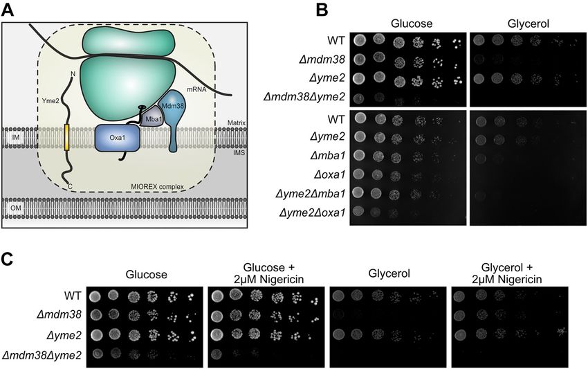
N. Sharma and C. Osman: Genetic link of Yme2 to the mitochondrial export machinery      3

Results                                                         genetic interaction with proteins of the mitochondrial export
                                                                machinery and is therefore not only linked to protein
YME2 displays a negative genetic interaction                    biogenesis through its association with the MIOREX
                                                                complex (Kehrein et al. 2015), but also through its genetic
with components of the mitochondrial
                                                                interactions. Of note, the strongest negative genetic inter-
protein export machinery                                        action is observed between YME2 and MDM38.

The presence of Yme2 in the MIOREX complex led us to
examine potential genetic interactions of YME2 with             Yme2 contains putative Walker motifs that
components required for mitochondrial protein biogen-
                                                                are important for Yme2 function
esis. First, we focused on a negative genetic interaction
between YME2 and MDM38, which has been reported
                                                                Next, we used bioinformatics tools to obtain insight into a
previously in a systematic large scale screen (Usaj et al.
                                                                possible function of Yme2. Prediction of the putative
2017). Mdm38 is a protein of the inner mitochondrial
                                                                structure of Yme2 was made using AlphaFold (Jumper et al.
membrane that acts as a receptor to recruit the mito-
                                                                2021). The resulting model revealed distinct N-terminal
chondrial ribosome to the MIM (Bauerschmitt et al. 2010;
                                                                (average per residue confidence score, or pLDDT > 90) and
Frazier et al. 2006). Moreover, Mdm38 has been proposed
                                                                C-terminal (average pLDDT > 70) domains that are sepa-
to play a role in mitochondrial K+/H+ homeostasis (Now-
                                                                rated by an alpha helix that corresponds to a predicted
ikovsky et al. 2004). We generated Δyme2, Δmdm38 and
                                                                transmembrane domain between residues 287 and 305
Δyme2Δmdm38 strains and performed growth test ana-
                                                                (average pLDDT > 70) (Figure S2A).
lyses. In agreement with previous findings, deletion of
                                                                     A careful analysis of the Yme2 amino acid sequence
MDM38 caused a growth defect on non-fermentable me-
                                                                using a homology search based on 3D structure prediction
dium, while deletion of YME2 did not result in obvious
                                                                (Meier and Söding 2015; Söding et al. 2005; Zimmermann
growth defects on either fermentable or non-fermentable
                                                                et al. 2018), revealed similarities of the Yme2’s IMS domain
medium (Frazier et al. 2006; Hanekamp and Thorsness
                                                                with AAA+ proteins and the presence of putative Walker A
1996). In contrast, Δyme2Δmdm38 cells exhibited a strong
                                                                and B motifs characteristic of P-loop ATPases (Figure 2A)
growth defect even on fermentable medium and were
                                                                (Miller and Enemark 2016; Puchades et al. 2020). The
incapable of respiratory growth at 30 and 37 °C (Figures 1B
                                                                strongest similarities in the homology search were identi-
and S1). To examine whether the Δyme2Δmdm38 pheno-
                                                                fied among members of the DNA-binding initiator clade of
type was caused due to a defect in ion transport or ribo-
                                                                AAA+ proteins, which include origin recognition proteins
some binding, a growth test analysis was performed on
                                                                and helicase-loading proteins, such as Cdc6. This finding is
media containing Nigericin. Nigericin, a K+/H+ ionophore,
                                                                rather surprising given the localization of this domain of
was previously shown to rescue the phenotype of
                                                                Yme2 in the IMS, which supposedly lacks DNA or RNA.
Δmdm38 (Nowikovsky et al. 2007). Conforming to this
                                                                Inspection of the predicted AlphaFold structure of Yme2’s
analysis, we observed that growth of Δmdm38 cells is
                                                                IMS domain supported the presence of a AAA+ fold,
restored on non-fermentable medium in the presence of
                                                                because it revealed features characteristic of AAA+ pro-
Nigericin (Figure 1C). In comparison, the severe pheno-
                                                                teins, which include a central β-sheet consisting of five
type of Δyme2Δmdm38 cells could not be rescued by
                                                                β-strands with a β5–β1–β4–β3–β2 order, a second region of
Nigericin. This result suggests that the strong negative
                                                                homology connecting β4 and β5 (including a hydrophilic
genetic interaction between YME2 and MDM38 is rather
                                                                glutamate at position 558 that may serve as a putative
linked to Mdm38’s function as a ribosome receptor than
                                                                Sensor I and a putative Arginine finger at position 565)
its role in K+/H+ homeostasis.
                                                                and a helical bundle C-terminal to the α–β–α sandwich
     Mdm38 has been linked to Oxa1-mediated insertion of
                                                                (Figure S2B) (Miller and Enemark 2016; Puchades et al.
mtDNA-encoded proteins and has also been reported to            2020). The Walker A motif is typically found in a loop
physically interact with Mba1 (Bauerschmitt et al. 2010). In    connecting β1 and β2 and contains an invariant lysine
this respect, we examined whether YME2 also genetically         residue which, when mutated, has been observed to
interacts with MBA1 and/or OXA1. Indeed, we observed            abolish ATP binding (Wendler et al. 2012). In case of Yme2
slightly reduced growth of Δyme2Δmba1 cells on non-             in S. cerevisiae, this lysine is at position 393 (Figures 2B and
fermentable medium and of Δyme2Δoxa1 cells on ferment-          S2C). The Walker B motif that typically contains two
able media at 30 and 37 °C compared to the respective single    conserved acidic residues (commonly an Aspartate and
mutants (Figures 1B and S1). Thus, YME2 exhibits a negative     Glutamate), has an unusual replacement of the Glutamate
4 N. Sharma and C. Osman: Genetic link of Yme2 to the mitochondrial export machinery
with an Arginine residue in the Yme2 sequence (Figure 2B the TAP-tag did not interfere with Yme2 function, which
and S2C) (consensus: hhhhDE; Yme2: hhhhDR) (Hanson was evident by WT-like growth of a Δmdm38 YME2-TAP
and Whiteheart 2005; Wendler et al. 2012). To examine the strain (Figure S3A). The steady state protein levels of all
importance of the Walker A and B motifs, the unusual Yme2 variants were examined in isolated mitochondria
Arginine in the Walker B motif and the putative Arginine (Figure 3A). We observed that strains expressing TAP-tagged
finger for Yme2 function, we generated YME2 variants with WT Yme2, Yme2K393A or Yme2D522A displayed comparable
Walker A ( yme2K393A), Walker B ( yme2D522A), Walker A/B Yme2 protein levels, indicating that these individual muta-
( yme2K393A/D522A), Walker B-Arginine ( yme2R523A) and Argi- tions do not cause protein instability, similar to our obser-
nine finger (yme2R565A) mutations and assessed their ability vation of untagged Yme2. In contrast, however, Yme2K393A/
to rescue the Δyme2Δmdm38 growth phenotype in a D522A
-TAP levels were slightly lower. We interpret this
plasmid shuffle experiment (Figure 2C). Δyme2Δmdm38 decrease in protein levels of Yme2K393A/D522A-TAP to reflect
cells expressing YME2 from a centromeric plasmid con- a slight destabilization of the protein, which becomes
taining a URA3 marker were transformed with a plasmid apparent upon isolation of mitochondria. Blue Native
containing the LEU2 marker, which either harbored the WT PAGE and Western Blot analysis of mitochondria isolated
or mutated forms of YME2. The cells were subsequently from strains expressing TAP-tagged Yme2 revealed a
grown on SC medium supplemented with 5′FOA, which sharp Yme2-specific band, which ran at a high molecular
only allows growth of cells that have lost the URA3-marked weight size similar to dimeric complex V (∼1250 kDa)
plasmid. In cells that have retained the URA3 plasmid, (Figure 3B). This Yme2-complex was also apparent in
5′FOA gets converted to a toxic product by the Ura3 protein, mitochondria isolated from cells expressing the Walker A
which kills the cell. In this assay, we observed that the mutant form Yme2K393A. In contrast, complex formation of
yme2R523A and yme2R565A variants containing the mutations Yme2 was compromised in the presence of the Walker B
of the Walker B-Arginine or the putative Arginine finger, mutation and virtually absent in cells expressing the
respectively, rescued growth of Δyme2Δmdm38, indicating Yme2K393A/D522A double mutant form harbouring Walker A
that these residues are not essential in the absence of and B mutations.
Mdm38. Most interestingly, the Walker mutant variants Next we asked, whether the Yme2-complex contains
yme2K393A, yme2D522A and yme2K393A/D522A failed to efficiently multiple copies of the Yme2 protein. We generated three
rescue the growth defect associated with Δyme2Δmdm38 diploid strains containing either a 9Myc- and an
cells (Figures 2D and S2D). Of note, we observed a weak 6HA-tagged YME2 allele (Yme2-9Myc/Yme2-6HA), an un-
rescuing effect of the yme2K393A variant, indicating that this tagged and an HA-tagged YME2 allele (Yme2/Yme2-6HA),
mutation does not entirely abolish Yme2 function. To test if or an untagged and a Myc-tagged YME2 allele (Yme2/
all mutant Yme2 forms are expressed and to avoid that the Yme2-9Myc). Yme2 complexes in these strains were ana-
strong Δyme2Δmdm38 phenotype may affect protein levels, lysed by BN PAGE. Interestingly, the size of the Yme2
we transformed Δyme2 cells with plasmids encoding mutant complex displayed a clear shift in Yme2-9Myc/Yme2-6HA
variants and checked expression levels in cell lysates by mitochondria compared to the strains where only one
Western bloting (Figure S2E). These analyses revealed that allele was tagged (Figure 3C). This size shift most likely
all mutant forms are expressed to levels comparable to indicates that both Yme2 variants are present in the same
wildtype Yme2. Taken together, we conclude that the Walker complex. To test this further, isolated mitochondria from
motifs are critical for Yme2 function. Yme2-9Myc/Yme2-6HA and Yme2/Yme2-6HA strains were
subjected to Myc-immunopurification. Strikingly, Yme2-
6HA efficiently co-purified with Yme2-9Myc in this exper-
Yme2 forms high molecular weight iment, while it did not purify when Yme2/Yme2-6HA
complexes mitochondria were subjected to Myc-purification (Figure 3D).
Similarly, Yme2-9Myc could be successfully co-purified with
Given the predicted AAA+ fold of Yme2 and the importance Yme2-6HA (Figure S3B). Taken together, these results
of the Walker motifs for Yme2 function, we asked whether suggest that Yme2 forms a high molecular weight complex
Yme2 forms oligomeric complexes, which is a character- that contains multiple copies of Yme2. Furthermore,
istic feature of AAA+ proteins (Puchades et al. 2020). To replacement of the lysine residue in the Walker A motif
facilitate detection of Yme2 in BN-PAGE analysis, WT or does not interfere with Yme2 complex formation, while
mutant Walker A or B TAP-tagged YME2 variants were re- mutation of the aspartate within the Walker B motif
inserted into the LEU2 locus of a Δyme2 strain. Importantly, partially impairs complex formation. Mutation of Walker AN. Sharma and C. Osman: Genetic link of Yme2 to the mitochondrial export machinery 5
Figure 2: Yme2 contains a putative AAA+ domain.
(A) Schematic showing the predicted domain organization of Yme2. The sequence of Yme2 harbours a mitochondrial targeting signal followed
by a putative N-terminal RNA binding domain (RRM), a putative C-terminal AAA+ domain with the predicted Walker A and B motifs, separated
by a predicted transmembrane (TM) domain. It also possesses an additional putative Arginine finger residue. The indicated residues were
mutated in this study. (B) Sequence logo generated from https://weblogo.berkeley.edu/ depicting the conservation of the sequence of Walker
A and B motifs of the putative AAA+ domain of Yme2 across 10 different fungal species. The numbers marking the residues refer to the Yme2
sequence in S. cerevisiae. (C) Schematic of the experimental procedure for the plasmid shuffle experiment. (D) Growth test analysis showing
the plasmid shuffle experiment. The indicated strains were grown to logarithmic phase and the serial dilutions were spotted on fermentable
glucose medium and on SC+ 5 FOA medium (to counter select for the pRS316-URA3 plasmid) and incubated at 30 °C for 2 days.
and Walker B residues strongly compromises complex Yme2 has a putative N-terminal RNA binding
formation. domain
Given the genetic link between YME2 and components
of the mitochondrial protein export machinery, we also We next focused on the domain of Yme2, which is located
assessed Yme2-TAP complex formation in Δmdm38 and in the mitochondrial matrix. Analysis of the amino acid
Δmba1 strains (Figure 3E). No alteration of the Yme2 com- sequence revealed the presence of a putative N-terminal
plex could be observed in the absence of Mdm38 or Mba1 RNA binding domain (Gabler et al. 2020; Meier and Söding
indicating that the Yme2 complex revealed in the BN-PAGE 2015; Nowacka et al. 2019; Söding et al. 2005; Zimmermann
analysis does not contain either of these proteins and et al. 2018). The RNA recognition motif (RRM) domain has
absence of these proteins does not compromise complex two conserved motifs, namely RNP1 and RNP2, wherein
formation through secondary effects. each motif contains an invariant aromatic residue that6 N. Sharma and C. Osman: Genetic link of Yme2 to the mitochondrial export machinery Figure 3: Yme2 forms high molecular weight complexes. (A) Western blot of the isolated mitochondria of the respective strains showing the steady state protein levels upon immunodetection with the Yme2-antibody. For each strain, 50, 25 and 12.5 µg mitochondria were loaded on the SDS-PAGE. Tim23 is the loading control. (B, C) Western Blot of the isolated mitochondria loaded on a Blue-Native PAGE. For each sample, 100 µg of mitochondria were solubilized and loaded on a 3–13% BN-PAGE. The western blot was probed with the indicated antibodies. The Coomassie staining shows the mitochondrial respiratory supercomplexes, as indicated. (D) Western blot showing the Myc-immunoprecipitation experiment performed with the indicated diploid strains. For each strain, 1% of the input and flowthrough (FT), and 50% of the bound fractions were loaded. The blot was decorated with Anti-HA and Anti-Myc antibody. (E) Western Blot of the isolated mitochondria from the indicated strains loaded on a Blue-Native PAGE. For each sample, 100 µg of mitochondria were solubilized and loaded on a 3–13% BN-PAGE. The western blot was probed with an anti-Yme2 antibody. The Coomassie staining shows the mitochondrial respiratory supercomplexes, as indicated. The deletion strains Δmba1 and Δmdm38 show reduced levels of supercomplexes, due to an absence of the components of the mitochondrial protein biogenesis machinery.
N. Sharma and C. Osman: Genetic link of Yme2 to the mitochondrial export machinery                  7

interacts with the nucleotide bases of a client RNA or DNA                    We next determined the stability of Yme2 variants
molecule (Figures 4A and S4A) (Maris et al. 2005). The RNP               containing RRM mutations and transformed Δyme2 cells
motifs of Yme2 were observed to be conserved among                       with plasmids encoding either WT or mutant forms of
various fungal species (Figure S4B). To test whether motifs              YME2. While the Yme2F204A variant accumulated to WT
are important for the function of Yme2, we examined if                   levels, amounts of Yme2Y242A and Yme2F204A/Y242A were not
mutant YME2 variants containing RNP1 (yme2Y242A) and                     detectable in cell lysates (Figure S4D). We also examined
RNP2 (yme2F204A) mutations would rescue growth of a                      levels of Yme2 variants containing mutations of the RRM
Δyme2Δmdm38 strain in our plasmid shuffle experiment                      domain in mitochondria isolated from strains containing
(Figure 2C). In this assay, we observed that the RNP2 mu-                TAP tagged versions integrated into the LEU2 locus. In
tation (yme2F204A) did not interfere with Yme2 function                  agreement with our results obtained from cell lysates, the
because a plasmid containing this variant restored growth                Yme2F204A-TAP levels were comparable to WT levels
of Δyme2Δmdm38 cells (Figures 4B and S4C). Thus, muta-                   (Figure 4C). Yme2Y242A-TAP and Yme2F204A/Y242A-TAP levels,
tion of the phenylalanine does not impair the function of                in contrast, were strongly reduced and only residual
the RNP2 motif. In contrast, the RNP1 mutation (yme2Y242A)               amounts were detectable. To examine complex formation
or the RNP1/RNP2 double mutation (yme2F204A/Y242A)                       of Yme2 variants containing RRM mutations, we performed
rendered Yme2 non-functional in the plasmid shuffle                       BN-PAGE analysis. In line with our findings that mutation
experiment.                                                              of the RNP2 motif does not interfere with the function of

Figure 4: Yme2 contains a putative RRM domain.
(A) Sequence logo generated from https://weblogo.berkeley.edu/ depicting the conservation of the sequence of RNP2 and RNP1 domains
of the putative RRM domain of Yme2 across 10 different fungal species. The numbers marking the residues refer to the Yme2 sequence in
S. cerevisiae. (B) Growth test analysis showing the plasmid shuffle experiment performed with the RNP mutant forms of YME2. The indicated
strains were grown to logarithmic phase and the serial dilutions were spotted on fermentable glucose medium and on SC+ 5 FOA medium (to
counter select for the pRS316-Ura3 plasmid) and incubated at 30 °C for 2 days. (C) Western Blot of the isolated mitochondria of the respective
strains showing the steady state protein levels upon decoration with the Yme2-antibody. For each strain, 50, 25 and 12.5 µg mitochondria were
loaded on the SDS-PAGE. Tim50 is the loading control. (D) Western Blot of the isolated mitochondria loaded on a Blue-Native PAGE. For each
sample, 100 µg of mitochondria were solubilized and loaded on a 3–13% BN-PAGE. The western blot was decorated with an antibody against
Yme2. The Coomassie staining shows the mitochondrial respiratory supercomplexes, as indicated.
8        N. Sharma and C. Osman: Genetic link of Yme2 to the mitochondrial export machinery

Yme2 and does not affect its abundance, we observed that           sufficient for this function. In Δyme2Δmdm38 cells, how-
the Yme2F204A variant formed WT-like high molecular                ever, tethering and precise positioning of the mitoribosome
weight complexes. In contrast, no complexes were                   could be compromised to an extent that severely hampers
observed for the Yme2Y242A and Yme2F204A/Y242A mutants             protein insertion and results in the observed growth de-
(Figure 4D). We conclude that the RNP1 motif is essential          fects. An alternative hypothesis could be that Yme2 en-
for Yme2 stability and complex formation. However, it is           gages in DNA interactions through its RRM domain and
indistinguishable at this point, whether the integrity of          facilitate mtDNA recruitment to the MIM, which could
RNP1 is required for folding of Yme2 or whether RNA                spatially link transcription and translation. Such a function
binding plays an important role in stabilizing or promoting        would be in accordance with the previously observed co-
complex formation and protein stability.                           localization of Yme2 and mtDNA (Murley et al. 2013).
                                                                        It has to be pointed out that the strong growth defect of
                                                                   Δyme2Δmdm38 cells even on fermentable medium is sur-
Discussion                                                         prising, if the function of Yme2 and/or Mdm38 lie solely in
                                                                   the membrane integration of mtDNA-encoded proteins. It
In this study, we demonstrate that YME2 negatively in-             will be interesting to examine potential functions of Yme2
teracts with MDM38, MBA1 and OXA1, which all have                  and Mdm38 beyond export of mtDNA-encoded proteins.
proposed roles in mitochondrial protein biogenesis. The                 The AlphaFold-based structural prediction suggests
strongest negative genetic interaction of YME2 is apparent         that Yme2 adopts a AAA+ fold in the IMS and our results
in Δyme2Δmdm38 cells, where a severe growth defect is              demonstrate that the Walker motifs are indispensable for
observed even on fermentable medium, which cannot be               Yme2’s function. Furthermore, the presence of Yme2 in a
rescued by the addition of Nigericin. These results suggest        high molecular weight complex containing multiple copies
a partially overlapping role of Yme2 and Mdm38, which is           of Yme2 is reminiscent of the known homo- or hetero-
crucial for mitochondrial and cellular function. Our bio-          oligomeric organizations of AAA+ proteins (Miller and
informatic and mutational analyses shed light on the               Enemark 2016). However, the Walker B motif of Yme2
domain organization of Yme2. Yme2 is predicted to possess          (hhhhDR) differs from the consensus sequence of the
a putative RNA Recognition Motif (RRM), that faces the             Walker B motif (hhhhDE) (Hanson and Whiteheart 2005)
mitochondrial matrix and an AAA+ domain, that faces the            and mutation of the putative Arginine finger (Arg 565)
IMS. Based on our mutational analyses, both domains are            appears dispensable for Yme2 function. These points raise
important for Yme2 function.                                       the question if Yme2 indeed has the ability to hydrolyze
     The genetic interaction of Yme2 with components of            nucleoside triphosphates and to function as a motor in the
the mitochondrial export machinery combined with the               IMS akin to other AAA+ proteins. In any case, identification
previous observation that Yme2 co-purifies with the mito-          of molecules that interact with its AAA+ domain will be
chondrial ribosome (Kehrein et al. 2015) suggests a func-          critical to unravel the function of Yme2.
tion for Yme2 in mitochondrial protein biogenesis. Yme2’s               In summary, our analyses link Yme2 to the mitochon-
RRM domain would be well-positioned in the mitochon-               drial protein biogenesis machinery. The identification of
drial matrix to assist in this process. RRM domains are            the RRM and AAA+ domains within Yme2 further provide
structurally versatile and have been shown to engage in            a strong foothold into mechanistic analyses of Yme2’s
RNA, DNA or protein interactions (Maris et al. 2005).              function.
Therefore, it remains to be determined which of these
molecules Yme2 interacts with through its RRM domain.
Given the strong genetic interaction between YME2 and              Materials and methods
MDM38 and the proposed function of Mdm38 as a ribosome
acceptor at the MIM (Frazier et al. 2006), an interesting          Construction of yeast strains and plasmids
hypothesis would be that Yme2 assists in protein trans-
lation by binding to either mRNAs or rRNAs through its             All the yeast strains used in the study were generated in the W303
RRM domain to tether and position components of the                background. Single deletion mutants and strains with C-terminally
translation machinery at the MIM. According to this idea,          tagged genes were constructed as previously described (Janke et al.
                                                                   2004). Double deletion mutant strains were constructed by mating
Yme2 and Mdm38, may serve partially redundant roles in
                                                                   the respective single deletions followed by tetrad dissection analysis.
tethering the translation apparatus to the MIM to facili-          The list of all yeast strains used is given in Supplementary Table S1.
tate efficient protein insertion. In absence of either YME2         The strain used for the plasmid shuffle was constructed by first de-
or MDM38, the respective other protein would still be              leting YME2 in WT background, followed by transformation with
N. Sharma and C. Osman: Genetic link of Yme2 to the mitochondrial export machinery 9
pRS316-YME2. This strain was further used for deletion of MDM38. The Multiple sequence alignment and structure prediction
plasmid shuffle experiment was performed with centromeric yeast
vectors pRS315 and pRS316. All the plasmids used in the study are
Yme2 sequence from 10 different fungal species was aligned using
listed in Supplementary Table S2. For the SDS-PAGE and BN-PAGE
MUSCLE (Madeira et al. 2019), and analyzed using Jalview (Water-
analysis, TAP-tagged YME2 strains were generated, by integration of
house et al. 2009). The sequence logo for the Walker A and B motifs,
WT or mutated forms of YME2-TAP into the LEU2 locus of a Δyme2
and the RNP 1 and 2 motifs was further created using the Weblogo tool
strain. Primer sequences can be made available on request.
(Crooks et al. 2004). For the prediction of the structure of Yme2, the
AI-based system AlphaFold (Jumper et al. 2021) was used.
Growth test analysis
Acknowledgments: We thank Katherine Madden, Alicia
Growth test analysis was performed using log-phase growing cells in Gassauer and Dr. Ina Aretz for helping with the experi-
YPD, and taking equal number of cells for the respective strains fol-
mental work, and thank Nadja Lebedeva and Tanja Kaut-
lowed by spotting them on fermentable (YPD) and non-fermentable
zleben for technical assistance. We also extend our thanks
(YPG) media, at 30 and 37 °C. Pictures were taken after 24 and 48 h of
growth at the respective temperatures. For the plasmid shuffle, the to Dr. Peter Thorsness for the Yme2 antibody and Dr.
cells were additionally spotted on SC medium supplemented with Dejana Mokranjac for Tim23 and Tim50 antibodies. We also
5′-fluoroorotic acid (FOA), to select for cells that have lost the pRS316- thank Simon Schrott for providing us with the Leucine
URA3 plasmid. integrative plasmid backbone.
Author contributions: All the authors have accepted
Mitochondria isolation responsibility for the entire content of this submitted
manuscript and approved submission.
The protocol for isolating the mitochondria was adapted from (Basch Research funding: We appreciate the support and input of
et al. 2020). The final mitochondrial pellet was resuspended in SEM the members of the Osman and Mokranjac laboratories and
buffer (250 mM Sucrose, 1 mM EDTA, 10 mM MOPS-KOH pH 7.2), and attendees of the “Mito Club” throughout the project. C.O.
further subjected to sucrose step gradient centrifugation over SEM500 and N.S. are supported by a grant from the European
buffer (500 mM sucrose, 1 mM EDTA, 10 mM MOPS-KOH pH 7.2) at
Research Council (ERCStG-714739 IlluMitoDNA).
13,000 rpm for 10 min (Morgenstern et al. 2017). The pellet was
further resuspended in SEM buffer, aliquoted and shock frozen in Conflict of interest statement: The authors declare no
liquid N2. conflicts of interest regarding this article.
Immunoprecipitation experiments
References
Mitochondria were lysed in 2% Digitonin, 50 mM NaCl, 10 mM Tris-HCl Acín-Pérez, R., Fernández-Silva, P., Peleato, M.L., Pérez-Martos, A.,
(pH 7.4) and 1x Complete protease inhibitor (Roche) for 30 min at 4 °C. and Enriquez, J.A. (2008). Respiratory active mitochondrial
The lysate was cleared by centrifugation at 13,000g for 10 min at 4 °C. supercomplexes. Mol. Cell 32: 529–539.
The lysate was incubated for 2 h with Anti-c-Myc magnetic beads Arlt, H., Tauer, R., Feldmann, H., Neupert, W., and Langer, T. (1996).
(Pierce™) or Anti-HA beads (Pierce™) at 4 °C. Bound proteins were The YTA10–12 complex, an AAA protease with chaperone-like
eluted with Laemmli buffer and analysed by Western blotting. activity in the inner membrane of mitochondria. Cell 85:
875–885.
Basch, M., Wagner, M., Rolland, S., Carbonell, A., Zeng, R., Khosravi, S.,
PAGE analysis Schmidt, A., Aftab, W., Imhof, A., Wagener, J., et al. (2020). Msp1
cooperates with the proteasome for extraction of arrested
For the SDS-PAGE, 50, 25 and 12.5 µg were centrifuged at 13,000 rpm mitochondrial import intermediates. Mol. Biol. Cell 31: 753–767.
for 10 min at 4 °C, and the pellet was resuspended in Laemmli buffer, Bauerschmitt, H., Mick, D.U., Deckers, M., Vollmer, C., Funes, S.,
and analyzed by SDS-PAGE and immunoblotting. Kehrein, K., Ott, M., Rehling, P., and Herrmann, J.M. (2010).
The protocol for BN-PAGE was adapted from (Wittig et al. 2006). Ribosome-binding proteins Mdm38 and Mba1 display
100 µg mitochondria were pelleted at 13,000 rpm for 10 min at 4 °C. The overlapping functions for regulation of mitochondrial
pellet was resuspended in solubilization buffer (50 mM NaCl, 5 mM translation. Mol. Biol. Cell 21: 1937–1944.
6-aminohexanoic acid, 50 mM imidazole/HCl pH 7.0, 50 mM K2PO4 pH Böttinger, L. and Becker, T. (2012). Protein quality control in the
7.4, 10% (v/v) glycerol) with 1.875% digitonin, and incubated for intermembrane space of mitochondria. J. Mol. Biol. 424:
20 min at 4 °C. Subsequently, samples were centrifuged at 20,000g for 225–226.
20 min at 4 °C. The supernatant was mixed with 2 µL solubilization Cogliati, S., Lorenzi, I., Rigoni, G., Caicci, F., and Soriano, M.E. (2018).
buffer containing 2% Coomassie Brilliant Blue G-250, loaded on a Regulation of mitochondrial electron transport chain assembly.
3–13% BN-PAGE, and analyzed by immunoblotting. J. Mol. Biol. 430: 4849–4873.10 N. Sharma and C. Osman: Genetic link of Yme2 to the mitochondrial export machinery
Crooks, G.E., Hon, G., Chandonia, J.-M., and Brenner, S.E. (2004). The EMBL-EBI search and sequence analysis tools APIs in 2019.
WebLogo: a sequence logo generator. Genome Res. 14: Nucleic Acids Res. 47: W636–W641.
1188–1190. Maris, C., Dominguez, C., and Allain, F.H.-T. (2005). The RNA
Erzberger, J.P. and Berger, J.M. (2006). Evolutionary relationships and recognition motif, a plastic RNA-binding platform to regulate
structural mechanisms of AAA+ proteins. Annu. Rev. Biophys. post-transcriptional gene expression. FEBS J. 272: 2118–2131.
Biomol. Struct. 35: 93–114. Mårtensson, C.U., Priesnitz, C., Song, J., Ellenrieder, L., Doan, K.N.,
Frazier, A.E., Taylor, R.D., Mick, D.U., Warscheid, B., Stoepel, N., Boos, F., Floerchinger, A., Zufall, N., Oeljeklaus, S., Warscheid,
Meyer, H.E., Ryan, M.T., Guiard, B., and Rehling, P. (2006). B., et al. (2019). Mitochondrial protein translocation-associated
Mdm38 interacts with ribosomes and is a component of the degradation. Nature 569: 679–683.
mitochondrial protein export machinery. JCB J. Cell Biol. 172: Meier, A. and Söding, J. (2015). Automatic prediction of protein 3D
553–564. structures by probabilistic multi-template homology modeling.
Gabler, F., Nam, S.-Z., Till, S., Mirdita, M., Steinegger, M., Söding, J., PLoS Comput. Biol. 11: e1004343.
Lupas, A.N., and Alva, V. (2020). Protein sequence analysis Miller, J.M. and Enemark, E.J. (2016). Fundamental characteristics of
using the MPI bioinformatics toolkit. Curr. Prot. Bioinform. AAA+ protein family structure and function. Archaea 2016:
72: e108. 9294307.
Gates, S.N. and Martin, A. (2020). Stairway to translocation: AAA+ Möller-Hergt, B.V., Carlström, A., Stephan, K., Imhof, A., and Ott, M.
motor structures reveal the mechanisms of ATP-dependent (2018). The ribosome receptors Mrx15 and Mba1 jointly organize
substrate translocation. Protein Sci. 29: 407–419. cotranslational insertion and protein biogenesis in
Gerdes, F., Tatsuta, T., and Langer, T. (2012). Mitochondrial AAA mitochondria. Mol. Biol. Cell 29: 2386–2396.
proteases—towards a molecular understanding of membrane- Morgenstern, M., Stiller, S.B., Lübbert, P., Peikert, C.D., Dannenmaier, S.,
bound proteolytic machines. Biochim. Biophys. Acta 1823: Drepper, F., Weill, U., Höß, P., Feuerstein, R., Gebert, M., et al.
49–55. (2017). Definition of a high-confidence mitochondrial proteome at
Glynn, S.E. (2017). Multifunctional mitochondrial AAA proteases. quantitative scale. Cell Rep. 19: 2836–2852.
Front. Mol. Biosci. 4: 34. Murley, A., Lackner, L.L., Osman, C., West, M., Voeltz, G.K., Walter, P.,
Hanekamp, T. and Thorsness, P.E. (1996). Inactivation of YME2/ and Nunnari, J. (2013). ER-associated mitochondrial division
RNA12, which encodes an integral inner mitochondrial links the distribution of mitochondria and mitochondrial DNA in
membrane protein, causes increased escape of DNA from yeast. Elife 2: e00422.
mitochondria to the nucleus in Saccharomyces cerevisiae. Mol. Nowacka, M., Boccaletto, P., Jankowska, E., Jarzynka, T.,
Cell Biol. 16: 2764–2771. Bujnicki, J.M., and Dunin-Horkawicz, S. (2019). RRMdb-an
Hanson, P.I. and Whiteheart, S.W. (2005). AAA+ proteins: have engine, evolutionary-oriented database of RNA recognition motif
will work. Nat. Rev. Mol. Cell Biol. 6: 519–529. sequences. Database 2019, https://doi.org/10.1093/database/
Hell, K., Neupert, W., and Stuart, R.A. (2001). Oxa1p acts as a general bay148.
membrane insertion machinery for proteins encoded by Nowikovsky, K., Froschauer, E.M., Zsurka, G., Samaj, J., Reipert, S.,
mitochondrial DNA. EMBO J. 20: 1281–1288. Kolisek, M., Wiesenberger, G., and Schweyen, R.J. (2004). The
Janke, C., Magiera, M.M., Rathfelder, N., Taxis, C., Reber, S., LETM1/YOL027 gene family encodes a factor of the mitochondrial
Maekawa, H., Moreno-Borchart, A., Doenges, G., Schwob, E., K+ homeostasis with a potential role in the Wolf-Hirschhorn
Schiebel, E., et al. (2004). A versatile toolbox for PCR-based syndrome. J. Biol. Chem. 279: 30307–30315.
tagging of yeast genes: new fluorescent proteins, more markers Nowikovsky, K., Reipert, S., Devenish, R.J., and Schweyen, R.J. (2007).
and promoter substitution cassettes. Yeast 21: 947–962. Mdm38 protein depletion causes loss of mitochondrial K+/H+
Jumper, J., Evans, R., Pritzel, A., Green, T., Figurnov, M., Ronneberger, O., exchange activity, osmotic swelling and mitophagy. Cell Death
Tunyasuvunakool, K., Bates, R., Žídek, A., Potapenko, A., et al. Differ. 14: 1647–1656.
(2021). Highly accurate protein structure prediction with Nunnari, J. and Suomalainen, A. (2012). Mitochondria: in sickness and
AlphaFold. Nature 596: 583–589. in health. Cell 148: 1145–1159.
Kehrein, K., Schilling, R., Möller-Hergt, B.V., Wurm, C.A., Jakobs, S., Opalińska, M. and Jańska, H. (2018). AAA proteases: guardians of
Lamkemeyer, T., Langer, T., and Ott, M. (2015). Organization of mitochondrial function and homeostasis. Cells 7: 163.
mitochondrial gene expression in two distinct ribosome- Ott, M., Prestele, M., Bauerschmitt, H., Funes, S., Bonnefoy, N., and
containing assemblies. Cell Rep. 10: 843–853. Herrmann, J.M. (2006). Mba1, a membrane-associated ribosome
Leonhard, K., Guiard, B., Pellecchia, G., Tzagoloff, A., Neupert, W., and receptor in mitochondria. EMBO J. 25: 1603–1610.
Langer, T. (2000). Membrane protein degradation by AAA Puchades, C., Sandate, C.R., and Lander, G.C. (2020). The molecular
proteases in mitochondria. Mol. Cell 5: 629–638. principles governing the activity and functional diversity of AAA+
Leonhard, K., Herrmann, J.M., Stuart, R.A., Mannhaupt, G., Neupert, W., proteins. Nat. Rev. Mol. Cell Biol. 21: 43–58.
and Langer, T. (1996). AAA proteases with catalytic sites on Seraphim, T.V. and Houry, W.A. (2020). AAA+ proteins. Curr. Biol. 30:
opposite membrane surfaces comprise a proteolytic system for R251–R257.
the ATP-dependent degradation of inner membrane proteins in Snider, J., Thibault, G., and Houry, W.A. (2008). The AAA+ superfamily
mitochondria. EMBO J. 15: 4218–4229. of functionally diverse proteins. Genome Biol. 9: 216.
Levytskyy, R.M., Bohovych, I., and Khalimonchuk, O. (2017). Söding, J., Biegert, A., and Lupas, A.N. (2005). The HHpred interactive
Metalloproteases of the inner mitochondrial membrane. server for protein homology detection and structure prediction.
Biochemistry 56: 4737–4746. Nucleic Acids Res. 33: W244–W248.
Madeira, F., Park, Y., Lee, J., Buso, N., Gur, T., Madhusoodanan, N., Song, J., Herrmann, J.M., and Becker, T. (2021). Quality control of the
Basutkar, P., Tivey, A.R.N., Potter, S.C., Finn, R.D., et al. (2019). mitochondrial proteome. Nat. Rev. Mol. Cell Biol. 22: 54–70.N. Sharma and C. Osman: Genetic link of Yme2 to the mitochondrial export machinery 11
Steele, T.E. and Glynn, S.E. (2019). Mitochondrial AAA proteases: a Waterhouse, A.M., Procter, J.B., Martin, D.M.A., Clamp, M., and
stairway to degradation. Mitochondrion 49: 121–127. Barton, G.J. (2009). Jalview Version 2—a multiple sequence
Szyrach, G., Ott, M., Bonnefoy, N., Neupert, W., and Herrmann, J.M. alignment editor and analysis workbench. Bioinformatics 25:
(2003). Ribosome binding to the Oxa1 complex facilitates 1189–1191.
co-translational protein insertion in mitochondria. EMBO J. 22: Weidberg, H. and Amon, A. (2018). MitoCPR-A surveillance pathway
6448–6457. that protects mitochondria in response to protein import stress.
Tatsuta, T. (2009). Protein quality control in mitochondria. J. Biochem. Science 360: eaan4146.
146: 455–461. Wendler, P., Ciniawsky, S., Kock, M., and Kube, S. (2012). Structure
Thorsness, P.E. and Fox, T.D. (1993). Nuclear mutations in and function of the AAA+ nucleotide binding pocket. Biochim.
Saccharomyces cerevisiae that affect the escape of DNA from Biophys. Acta 1823: 2–14.
mitochondria to the nucleus. Genetics 134: 21–28. Wiedemann, N. and Pfanner, N. (2017). Mitochondrial machineries
Turk, E.M., Das, V., Seibert, R.D., and Andrulis, E.D. (2013). The for protein import and assembly. Annu. Rev. Biochem. 86:
mitochondrial RNA landscape of Saccharomyces cerevisiae. 685–714.
PLoS One 8: e78105. Wittig, I., Braun, H.-P., and Schägger, H. (2006). Blue native PAGE. Nat.
Usaj, M., Tan, Y., Wang, W., VanderSluis, B., Zou, A., Myers, C.L., Protoc. 1: 418–428.
Costanzo, M., Andrews, B., and Boone, C. (2017). Zimmermann, L., Stephens, A., Nam, S.-Z., Rau, D., Kübler, J., Lozajic, M.,
TheCellMap.org: a web-accessible database for visualizing and Gabler, F., Söding, J., Lupas, A.N., and Alva, V. (2018). A completely
mining the global yeast genetic interaction network. G3 reimplemented MPI bioinformatics toolkit with a new HHpred
(Bethesda) 7: 1539–1549. server at its core. J. Mol. Biol. 430: 2237–2243.
Walker, J.E., Saraste, M., Runswick, M.J., and Gay, N.J. (1982).
Distantly related sequences in the alpha- and beta-subunits of
ATP synthase, myosin, kinases and other ATP-requiring enzymes Supplementary Material: The online version of this article offers
and a common nucleotide binding fold. EMBO J. 1: 945–951. supplementary material (https://doi.org/10.1515/hsz-2021-0398).You can also read