Extracellular Vesicles Derived From Human Umbilical Cord Mesenchymal Stem Cells Protect Against DOX-Induced Heart Failure Through the ...
←
→
Page content transcription
If your browser does not render page correctly, please read the page content below
ORIGINAL RESEARCH
                                                                                                                                           published: 25 August 2021
                                                                                                                                     doi: 10.3389/fbioe.2021.703241

                                           Extracellular Vesicles Derived From
                                           Human Umbilical Cord Mesenchymal
                                           Stem Cells Protect Against
                                           DOX-Induced Heart Failure Through
                                           the miR-100-5p/NOX4 Pathway
                                           Zhenglong Zhong†, Yuqing Tian†, Xiaoming Luo, Jianjie Zou, Lin Wu * and Julong Tian *
                                           Department of Cardiology, Affiliated Hospital of Panzhihua University, Panzhihua, China

                                           The end result of a variety of cardiovascular diseases is heart failure. Heart failure patients’
                                           morbidity and mortality rates are increasing year after year. Extracellular vesicles (EVs)
                           Edited by:
                                           derived from human umbilical cord mesenchymal stem cells (HucMSC-EVs) have recently
                        Enrico Ragni,
Galeazzi Orthopedic Institute (IRCCS),     been discovered to be an alternative treatment for heart failure, according to recent
                                  Italy    research. In this study, we aimed to explore the underlying mechanisms in which HucMSC-
                        Reviewed by:       EVs inhibited doxorubicin (DOX)-induced heart failure in AC16 cells. An miR-100-5p
                           Monica Reis,
               University of Edinburgh,    inhibitor and an miR-100-5p mimic were used to transfect HucMSCs using
                      United Kingdom       Lipofectamine 2000. HucMSC-EVs were isolated and purified using the
                          Andrea Mohr,
                                           ultracentrifugation method. AC16 cells were treated with DOX combined with
 University of Essex, United Kingdom
                                           HucMSC-EVs or an EV miR-100-5-p inhibitor or EV miR-100-5-p mimic. ROS levels
                     *Correspondence:
                                Lin Wu     were measured by a flow cytometer. The levels of LDH, SOD, and MDA were measured by
                 linwu71ch@yahoo.com       biochemical methods. Apoptotic cells were assessed by a flow cytometer. Cleaved-
                            Julong Tian
                    551333202@qq.com       caspase-3 and NOX4 protein expression were determined by Western blot. The
     †
         These authors have contributed    experiment results showed that HucMSC-EVs inhibited DOX-induced increased levels
                    equally to this work   of ROS, LDH, and MDA, and decreased levels of SOD which were reversed by an EV miR-
              Received: 30 April 2021      100-5-p inhibitor, while EV miR-100-5-p mimic had a similar effect to HucMSC-EVs. At the
              Accepted: 12 July 2021       same time, HucMSC-EV-inhibited DOX induced the increases of apoptotic cells as well as
            Published: 25 August 2021
                                           NOX4 and cleaved-caspase-3 protein expression, which were reversed by an EV miR-
                              Citation:
                                           100-5-p inhibitor. Furthermore, the NOX4 expression was negatively regulated by miR-
Zhong Z, Tian Y, Luo X, Zou J, Wu L
and Tian J (2021) Extracellular Vesicles   100-5p. Overexpression of NOX4 abolished the effects in which HucMSC-EVs inhibited
 Derived From Human Umbilical Cord         DOX-induced ROS, oxidative stress, and apoptosis increases. In conclusion, these results
    Mesenchymal Stem Cells Protect
   Against DOX-Induced Heart Failure       indicate that HucMSC-EVs inhibit DOX-induced heart failure through the miR-100-5p/
             Through the miR-100-5p/       NOX4 pathway.
                      NOX4 Pathway.
 Front. Bioeng. Biotechnol. 9:703241.      Keywords: oxidative stress, miR-100-5p, NOX4, heart failure, human umbilical cord mesenchymal stem cells,
     doi: 10.3389/fbioe.2021.703241        extracellular vesicles

Frontiers in Bioengineering and Biotechnology | www.frontiersin.org               1                                          August 2021 | Volume 9 | Article 703241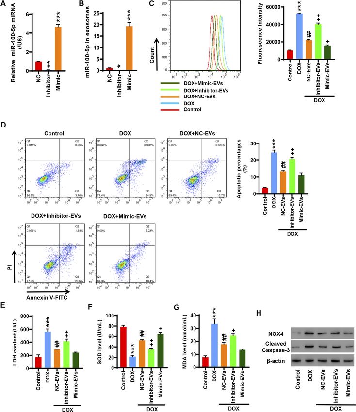
Zhong et al.                                                                                        HucMSC-EVs Inhibit DOX-Induced Heart Failure

INTRODUCTION                                                              MATERIALS AND METHODS
Heart failure is the final result of various cardiovascular                Cell Culture
diseases, among which the most common causes include                      Human AC16 cells were obtained from ATCC (Manassas, VA,
coronary heart disease, hypertension, cardiomyopathy, and                 United States) and cultured in a DMEM medium (Hyclone,
valvular heart disease. Its morbidity and mortality are                   SH30243.01, Logan, UT, United States) supplemented with
gradually increasing year by year, posing a serious threat                10% fetal bovine serum (Gibco, 16000e044, Carlsbad, CA,
to human health. Mesenchymal stem cells (MSCs) are a type                 United States) and 1% penicillin–streptomycin (Solarbio,
of pluripotent stem cells with multiple differentiation                   P1400, Beijing, China) and incubated at 37°C with 5% CO2.
potentials that implant in the mesoderm (Jaquet et al.,                   AC16 cells were treated with 2 µmol/L DOX (Sigma-Aldrich,
2005). MSCs can be derived from the bone marrow,                          25316-40-9, Shanghai, China) and HucMSC-EVs in different
placenta, adipose tissue, umbilical cord blood, etc. Human                concentrations for 24 h.
umbilical cord mesenchymal stem cells (HucMSCs) are
easier     to    obtain      and    have    proliferation      and
immunosuppressive effects. In addition, there are no                      Human Umbilical Cord Mesenchymal Stem
ethical issues for HucMSCs in clinical applications (Li                   Cell Characterization
et al., 2018; Guan et al., 2019; Xie et al., 2020). HucMSCs               HucMSCs were provided by Stem Cell Bank, Chinese
are used to increase the number of cardiomyocytes with                    Academy of Sciences and cultured in serum-free MSC
contractile function, thereby improving heart failure
caused by various reasons (Bartolucci et al., 2017; Mao
                                                                          NutriStem  ®  XF Medium (Biological Industries, Beit
                                                                          HaEmek, Israel). When the cells grew to 80–90%
et al., 2017; Kobayashi et al., 2018; Matsushita, 2020).                  confluence, they were digested with 0.25% trypsin
    Paracrine function is the main way for mesenchymal stem cells         containing 0.01% EDTA. The cells were resuspended in
to exert therapeutic effects. As one of the main components of            PBS and adjusted to 1 × 10 6 cells/ml. The mouse
paracrine, EVs include mRNA, miRNA, circRNA, protein, and                 antihuman CD34, CD45, CD44, and CD105 were added
other functional molecules, which not only participate in the             and incubated at 4 °C in dark for 15–30 min. Surface
regulation of cell proliferation and survival but also play an            antigens of MSCs were characterized by using a Beckman
important role in signal transmission and cell communication              CytoFLEX flow cytometer (Beckman Coulter Life Sciences,
(Keerthikumar et al., 2016; Tkach and Théry, 2016). EVs are               Tokyo, Japan) and the Human MSC Analysis Kit (BD
formed by eukaryotic cells through endocytosis of some signal             Biosciences, San Jose, CA, United States).
molecules to form multivesicular endosomes, and then fused with
the cell membrane and released into nanovesicles in the
extracellular environment. EVs contain different kinds of                 Isolation and Characterization of
lipids, proteins, RNAs, and other biologically active molecules,          Extracellular Vesicles
which play a variety of biological functions. EVs have been               HucMSCs were cultured in EV-free production medium for
proven to be an important medium for MSCs to exert                        24–48 h before conditioned medium was collected. EVs were
therapeutic effects. They can enter the cytoplasm through                 isolated and purified by ultracentrifugation according to the
endocytosis or direct fusion with the cell membrane, or                   protocol (Théry et al., 2006). The morphology of isolated
through receptor–ligand interactions, to transmit biological              HucMSC-EVs was observed by transmission electron
information to target cells, thereby exerting biological                  microscopy (FEI Tecnai G2 Spirit Twin, Philips, NL). The
functions and regulating abnormal microenvironment                        TSG101 and CD81 protein markers of EVs were detected by
(Davidson and Yellon, 2018; Balbi and Vassalli, 2020). Recent             Western blot.
studies have shown that MSC-derived EVs (MSC-EVs) are
expected to become a new alternative to stem cell therapy for             Extracellular Vesicle Uptake Assay
heart failure (Fang et al., 2016).                                        PKH67 EV green fluorescent dye (UR52303, Umibio, China) was
    DOX is an antitumor drug widely used clinically. It has a good        used to trace EVs being endocytosed by AC16 cells. In brief, EVs
effect on many tumors. However, DOX has very serious                      were stained with PKH67 dying working solution, in which the
cardiotoxicity and finally leads to heart failure. DOX-induced             PKH liker was mixed with diluent C at a ratio of 1:9 in dark at
cardiotoxicity can be used as an in vitro model to summarize the          room temperature. Then they were mixed well and incubated for
mechanism of heart failure. The application of the in vitro               10 min in dark. The labeled EVs were incubated on AC16 cells for
cardiotoxicity test system can greatly help understand the                24 h at 37°C, and cells were washed with 1 × PBS. Cells were
development of heart failure (Sachinidis, 2020). In this study,           mounted in a mounting medium containing DAPI (4′,6-
the effects of HucMSC-EVs on DOX-induced heart failure were               diamidino-2-phenylindole         dihydrochloride)       (DAPI-
studied. The aim of this study was to explore the molecular               Fluoromount-GTM, Yeasen Biotechnology, Shanghai, China).
mechanism of HucMSC-EVs inhibiting DOX-induced heart                      A laser-scanning confocal microscope was applied to take all
failure.                                                                  of the images (Youn et al., 2019).

Frontiers in Bioengineering and Biotechnology | www.frontiersin.org   2                                   August 2021 | Volume 9 | Article 703241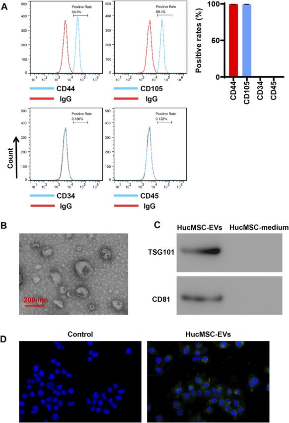
Zhong et al.                                                                                                HucMSC-EVs Inhibit DOX-Induced Heart Failure

Cell Transfection                                                               reactive oxygen species (ROS) levels. The reactive oxygen species in
The coding region of NOX4 (NM_001143837.2) was cloned into                      the cell can oxidize nonfluorescent DCFH to produce fluorescent
pCDNA3.1 (+) plasmids (Clontech, Mountain View, CA,                             DCF. By detecting the fluorescence of DCF, the level of reactive
United States) at Hind III and EcoR1 sites. It was designated                   oxygen species in the cell can be known. According to the
to oeNOX4. oeNOX4 was transfected to AC16 cells using                           production of red fluorescence in living cells, the amount and
Lipofectamine 2000 Kit (Invitrogen, Carlsbad, CA,                               change of the cell ROS content can be judged. Briefly, AC16 cells
United States). The negative control included cells with blank                  were resuspended in 1x PBS, and the density was adjusted to 5 ×
vector pCDNA3.1 (+) transfection. The primers used for                          105 cells/ml. AC16 cells were then incubated with 10 μM DCFH-DA
amplification of the coding sequence of NOX4 were as follows:                    for 20 min in dark at 37°C and subsequently subjected to the flow
   NOX4-F:5′-                                                                   cytometric analysis (Shi et al., 2016).
CCCAAGCTTATGAATGTCCTGCTTTTCTGGAAAAC-3’
(Hind III)                                                                      Cell Apoptosis
   NOX4-R:      5′-CGGAATTCTCAGCTGAAAGACTCTTTA                                  AC16 cells were seeded in 6-well plates with 1 × 10⁵ per well and
TTGTATTC-3’ (EcoR I)                                                            cultured for 12–24 h before use. AC16 cells were harvested 24 h after
                                                                                being treated with DOX combined with HucMSC-EVs or EV miR-100-
miRNA Transfection                                                              5-p inhibitor or oeNOX4. Cells were prepared with the Annexin V-
miR-100-5p mimic (5′-AACCCGUAGAUCCGAACUUGUG-3′),                                FITC Apoptosis Detection Kit (Beyotime, C1062s, Beijing, China)
miR-100-5p inhibitor (5′-CACAAGUUCGGAUCUACGGGUU-                                according to the manufacturer’s recommendations. Briefly, AC16
3′), and negative control (NC, 5′-CAGUACUUUUGUGUAGUA                            cells were centrifuged at 1,000 g for 5 min and resuspended to a
CAA-3′) were synthesized by Beyotime (Beijing, China) and                       concentration of 1 × 10⁶ cells/ml. 1 × 10⁶ resuspended cells were
transfected to cells with Lipofectamine 2000 Kit individually.                  centrifuged at 1,000 g for 5 min. The supernatant was discarded, and
                                                                                195 μL of Annexin V-FITC binding solution was added to gently
Luciferase Reporter Assay                                                       resuspend the cells. And 10 μL of Annexin V-FITC was added and
The NOX4 reporter gene plasmid is constructed by gene synthesis.                mixed gently. Then 5 μL of propidium iodide staining solution was
The NOX4 (NM_001143837.2) sequence was found in NCBI.                           added to the mix gently. The cell suspension was gently vortexed and
According to the NOX4 3′-UTR sequence, wild-type and mutant                     incubated in dark at room temperature for 15 min, and then placed in
NOX4 3′-UTR sequences with Sac I and Xho I sticky ends were                     an ice bath. At the same time, a tube without Annexin V-FITC and PI
synthesized. The mutation site was based on the binding site of hsa-            was used as a negative control. Flow cytometry was performed within
miR-100-5p and NOX4 3′-UTR sequence. The NOX4 3′-UTR                            1 h. The following method was used: The Annexin V-negative/PI-
binding site sequence containing mutation was inserted into the                 negative part represented viable cells. The Annexin V-positive/PI-
vector pGL3-promoter through Sac I and Xho I restriction sites to               negative part represented early apoptotic cells, and the Annexin
construct pGL3-Promoter-mutNOX4 3′-UTR. Wild-type NOX4 3′-                      V-positive/PI-positive part represented late apoptotic and dead cells.
UTR was inserted into the vector pGL3-Promoter through Sac I and
Xho I restriction sites to construct pGL3-Promoter-wtNOX4-3′-UTR.               Biochemical Detection
PGL3-Promoter vector had firefly fluorescent gene (luc2) and pRL-TK                The levels of lactate dehydrogenase (LDH), superoxide dismutase
with Renilla fluorescent gene (hRluc). A map of the predicted binding            (SOD), and malondialdehyde (MDA) in cells were measured,
site of hsa-miR-100-5p to NOX4 and mutant was as following.                     respectively, using the LDH (A020-2), SOD (A001-3), and MDA
                                                                                (A003-1) kits (Jiancheng Biotechnology Research Institute,
hsa-miR-100-5p                                      5′ GCCUAGAUGCCCAA 3′        Nanjing, Jiangsu, China) according to the manufacturer’s
                                                                                recommendations. Assays were performed in triplicate, and
—                                                          |||||||              the mean values of each sample were calculated manually.
wtNOX4-3′-UTR                                      5′ TATTGATACGGGTACT 3′
mutNOX4-3′-UTR                                     5′ TATTGAGCATTAGCCT 3
                                                                                QRT-PCR
                                                                                Total RNA was extracted using Trizol reagent (Invitrogen,
   293T cells were then co-transfected with miR-100-5p inhibitor,               Carlsbad, CA, United States) according to the manufacturer’s
miR-100-5p mimic and pGL3-NOX4-WT or miR-100-5p inhibitor,                      protocol, and reverse-transcribed with a RevertAid First Strand
miR-100-5p mimic, and pGL3-NOX4-MUT plasmids. After                             cDNA Synthesis Kit (Thermo Fisher Scientific Inc., Waltham,
transfection, the cells were treated with a Dual-Luciferase                     MA, United States). qRT-PCR was done using an SYBR green
Reporter Gene Detection System Test Kit. The firefly luciferase                   PCR Master Mix (Thermo Fisher Scientific Inc., Waltham, MA,
activity and Renilla luciferase activity were detected by a microplate          United States) on the ABI 7300 system. The relative abundance of
reader. Luciferase activity ratio in this study was the ratio of the            genes was quantified by using the comparative 2-ΔΔCt with
firefly luciferase activity to Renilla luciferase activity.                       β-actin or U6 as an internal control. The sequences of primers
                                                                                used in the study were as follows. Human NOX4, Primer F:
ROS Detection                                                                      5′-TTTAGATACCCACCCTCCCG-3′, Primer R:
A dichlorodihydrofluorescein diacetate (DCFH-DA) fluorescent                         5′-GGCACAGTACAGGCACAAAGG-3′.                         Human
probe (Sigma-Aldrich, D6883, Shanghai, China) combined with                     cytochrome b-245 beta chain (CYBB), Primer F: 5′-CTAAGA
the flow cytometric analysis was used to detect the changes of                   TAGCGGTTGATGGGC-3′, Primer R:

Frontiers in Bioengineering and Biotechnology | www.frontiersin.org         3                                     August 2021 | Volume 9 | Article 703241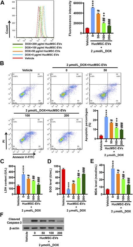
Zhong et al.                                                                                             HucMSC-EVs Inhibit DOX-Induced Heart Failure

   5′-CTTGAGAATGGATGCGAAGG-3′. Human β-actin,                                 HucMSC-EVs in the HucMSC medium (Figure 1C). HucMSC-EVs
Primer F:                                                                     were labeled by PKH67 and incubated with AC16 cells for 24 h. Under
   5′-CGTGGACATCCGCAAAGAC-3′, Primer R: 5′-TGC                                a laser scanning microscope, the green EVs were located in the
TGGGAGCCAGAGCAG-3’.                                                           cytoplasm (Figure 1D). These results suggest that HucMSC-EVs
   hsa-miR-100-5p, RT Primer:                                                 were successfully isolated.
   5′-GTCGTATCCAGTGCAGGGTCCGAGGTATTCGCA
CTGGATACGACCACAA                                                              Human Umbilical Cord Mesenchymal Stem
   G-3′, Primer F: 5′-GCGAACCCGTAGATCCGAA-3′,
Primer R:
                                                                              Cell-Extracellular Vesicles Inhibit
   5′-AGTGCAGGGTCCGAGGTATT-3′.      Human    U6,                              Doxorubicin-Induced Oxidative Stress and
Primer F:                                                                     Apoptosis
   5′-CTCGCTTCGGCAGCACA, Primer R: 5′-AACGCTTCA                               AC16 cells were treated with 2 µmol/L DOX and HucMSC at
CGAATTTGCGT-3′.                                                               concentrations of 0 μg/ml, 50 μg/ml, 100 μg/ml, and 200 μg/ml for
                                                                              24 h. Flow cytometry showed that 2 µmol/L DOX obviously induced
Western Blot                                                                  ROS levels to increase, while HucMSC-EV treatment decreased ROS
AC16 cells were lysed by the addition of RIPA lysis buffer supplemented       levels that were increased by DOX. HucMSC-EVs significantly
with a protease and phosphatase inhibitor cocktail (p8340 and p8250,          reduced ROS levels at the concentrations of 50 μg/ml, 100 μg/ml,
Sigma, St Louis, MO, United States). 25 µg of total protein was               and 200 μg/ml. The effect of HucMSC-EV treatment was
separated by SDS-PAGE and transferred onto a nitrocellulose                   concentration dependent (Figure 2A). Meanwhile, 2 µmol/L DOX
membrane. Membranes were further blocked with 5% skim milk                    increased LDH release and MDA levels, and decreased SOD levels.
and immersed into antibody solutions against TSG101 (1:2000,                  However, HucMSC-EV treatment inhibited the increases of LDH
ab120511, Abcam, Cambridge, MA, United States), CD81 (1:2000,                 release and MDA levels, and the decreases of SOD levels which were
ab109201, Abcam, Cambridge, MA, United States), NOX2 (1:5000,                 induced by DOX. The functions of HucMSC-EVs on LDH release,
ab129068, Abcam, Cambridge, MA, United States), cleaved-caspase-3             SOD levels, and MDA levels were in a concentration-dependent
(1:500, ab13847, Abcam, Cambridge, MA, United States), β-actin (1:            manner (Figures 2C–E). Furthermore, 2 µmol/L DOX induced
2000, ab8226, Abcam, Cambridge, MA, United States), and NOX4 (1:              AC16 cell apoptosis. Flow cytometry displayed that HucMSC-EVs
2000, 14347-1-AP, Proteintech, Rosemont, IL, United States). Then the         reduced apoptotic cells that were increased by DOX with the
membranes were immersed into the secondary antibody solution linked           concentrations of 50, 100, and 200 μg/ml. The action of HucMSC-
to horseradish peroxidase (A0208 and A0216, Beyotime, Shanghai,               EVs on AC16 cell apoptosis was in a concentration-dependent manner
China). Signals were captured by a chemiluminescence system.                  (Figure 2B). Western blot exhibited that HucMSC-EVs markably
                                                                              repressed the cleaved-caspase-3 expression with the different
Statistical Analysis                                                          concentrations of 50, 100, and 200 μg/ml, which were highly
All data were expressed as mean ± standard deviation (SD). One-way            expressed by DOX (Figure 2F). These findings suggest that
analysis of variance (ANOVA) was applied to assess the statistically          HucMSC-EVs obviously inhibit cardiomyocyte oxidative stress and
significant differences between more than two groups. Statistical              apoptosis that are induced by DOX in AC16 cells.
analysis was performed by Prism 8.0.2 software (GraphPad, San
Diego, United States). p value < 0.05 was considered significant.
                                                                              Human Umbilical Cord Mesenchymal Stem
                                                                              Cell-Extracellular Vesicles Inhibit NOX4
RESULTS                                                                       Expression Induced by Doxorubicin
                                                                              AC16 cells were treated with 2 µmol/L DOX and HucMSC-EVs with
                                                                              the concentrations of 0, 50, 100, and 200 μg/ml for 24 h. qRT-PCR
Identification of Human Umbilical Cord                                         indicated that DOX extremely increased the NOX2 and NOX4 mRNA
Mesenchymal Stem Cell-Extracellular                                           expression. HucMSC-EVs could repress the mRNA expression of
Vesicles                                                                      NOX2 and NOX4 at concentrations of 50, 100, and 200 μg/ml,
HucMSCs were cultured and collected. HucMSCs were identified by a              which were increased by DOX. Compared to the 2 μmol/
flow cytometer to detect their surface markers. Flow cytometry                 L_DOX+50 μg/ml_HucMSC-EV                 group,      HucMSC-EV
exhibited that HucMSC surface markers, such as CD44 and CD105,                treatment at 100 μg/ml apparently suppressed NOX2 and
were highly expressed, while hematopoietic stem marker CD34 and               NOX4 mRNA expression, which was induced by DOX
leukocyte surface antigen CD45 exhibited low expression (Figure 1A).          (Figure 3A). Western blot displayed that HucMSC-EVs
Next, HucMSC-EVs were extracted and identified. Transmission                   suppressed protein expression of NOX2 and NOX4 with the
electron microscope showed that HucMSC-EVs were small round                   concentrations of 50, 100, and 200 μg/ml, which was increased
or elliptical membranous bi-lipid membrane vesicles. Their diameters          by DOX (Figure 3B). AC16 cells were treated with 2 µmol/L
ranged in size from 30 to 100 nm. There were low electron densities in        DOX and 100 μg/ml HucMSC-EVs for 0, 6, 12, 24, and 48 h.
the vesicles (Figure 1B). The specific markers, namely, TSG101 and             qRT-PCR showed that 2 µmol/L DOX remarkably induced
CD81, of HucMSC-EVs were detected by Western blot. While there                NOX4 mRNA expression increase. HucMSC-EVs attenuated
were no expression for the specific markers TSG101 and CD81 of                 NOX4 mRNA expression with a concentration of 100 μg/ml at

Frontiers in Bioengineering and Biotechnology | www.frontiersin.org       4                                    August 2021 | Volume 9 | Article 703241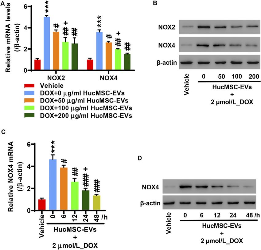
Zhong et al.                                                                                                   HucMSC-EVs Inhibit DOX-Induced Heart Failure

 FIGURE 1 | Isolation and identification of HucMSC-EVs. (A) The surface markers CD44 and CD105 of HucMSCs were identified by flow cytometry. (B) The
 morphology of purified HucMSC-EVs was observed by transmission electron microscopy: scale bar: 200 nm. (C) The markers CD81 and TSG101 of HucMSC-EVs were
 detected by Western blot. (D) HucMSC-EV endocytosis traced by PKH67 was observed by a laser scanning microscope in AC16 cells.

6, 12, 24, and 48 h, which were increased by DOX. Compared to                    Inhibition of EV miR-100-5-p Reverses
12 h, HucMSC-EV treatment markedly suppressed NOX4                               Those Effects That Human Umbilical Cord
mRNA expression with 100 μg/ml at 24 h, which was
increased by DOX (Figure 3C). Similarly, Western blot
                                                                                 Mesenchymal Stem Cell-Extracellular
displayed that the NOX4 protein expression was ameliorated                       Vesicles Inhibit Doxorubicin-Induced
at a concentration of 100 μg/ml for 6, 12, 24, and 48 h, which                   Oxidative Stress and Apoptosis
was increased by DOX (Figure 3D). Collectively, the data                         HucMSCs were transfected with miR-100-5p inhibitor
suggest that HucMSC-EVs markedly inhibit NOX4                                    (Inhibitor) or miR-100-5p mimic (Mimic). Q-PCR displayed
expression in a time- and concentration-dependent manner,                        that miR-100-5p expression was abolished by miR-100-5p
which is induced by DOX.                                                         inhibitor, while the miR-100-5p expression was aggravated by

Frontiers in Bioengineering and Biotechnology | www.frontiersin.org          5                                        August 2021 | Volume 9 | Article 703241
Zhong et al. HucMSC-EVs Inhibit DOX-Induced Heart Failure
expression in EVs (Figure 3B). AC16 cells were treated with
2 µmol/L DOX and 100 μg/ml miR-100-5p negative control in
HucMSC-EVs (NC-EVs) or miR-100-5p inhibitor in HucMSC-
EVs (Inhibitor-EVs) or miR-100-5p mimic in HucMSC-EVs
(Mimic-EVs) for 24 h. Flow cytometry demonstrated that
DOX remarkably increased ROS levels, which were decreased
with supplement of NC-EVs or Mimic-EVs. Compared to NC-
EVs, Inhibitor-EVs reversed those effects in which NC-EVs
decreased ROS production which were increased by DOX,
whereas Mimic-EVs had similar inhibitory effects to NC-EVs
for ROS production (Figure 4C). Biochemical assay exhibited
that 2 µmol/L DOX aggravated LDH release and MDA level
increases as well as SOD level decreases, which were inhibited
with supplement of NC-EVs or Mimic-EVs. In comparison to the
NC-EV group, Inhibitor-EVs reversed those effects in which NC-
EVs inhibited DOX induced the increases of LDH and MDA
levels as well as the decreases of SOD levels, while Mimic-EVs had
the similar effects to NC-EVs (Figures 4E–G). Furthermore,
apoptosis was examined with flow cytometry and Western
blot. NC-EVs and Mimic-EVs markedly reduced the
percentages of apoptotic cells, which were induced to increase
y DOX. Compared to the NC-EV group, Mimic-EVs had a
similar effect to NC-EVs for reducing the percentages of
apoptotic cells, while inhibitor-EVs reversed those effects in
which NC-EVs inhibited DOX-induced apoptotic cell increases
(Figure 4D). NOX4 and cleaved-caspase-3 protein expression
were apparently attenuated by NC-EVs or Mimic-EVs, which was
induced to increase by DOX. Compared to the NC-EV group,
Inhibitor-EVs reversed those effects which NC-EVs decreased,
NOX4 and cleaved-caspase-3 protein expression, which was
increased by DOX, while Mimic-EVs had a similar effect to
NC-EVs (Figure 4H). Taken together, these findings suggest
that downregulation of EV miR-100-5-p reverses those effects in
which HucMSC-EVs inhibit DOX-induced oxidative stress and
apoptosis.
Overexpression of NOX4 Cancels Those
Effects in Which Human Umbilical Cord
Mesenchymal Stem Cells Inhibit
FIGURE 2 | HucMSC-EVs inhibit DOX-induced oxidative stress and Doxorubicin-Induced Oxidative Stress and
apoptosis. AC16 cells were treated with 2 µmol/L DOX and HucMSC-EVs at Apoptosis
different concentrations (0, 50, 100, and 200 μg/ml) for 24 h. (A) ROS levels
were measured by flow cytometry. (B) The percentages of apoptotic
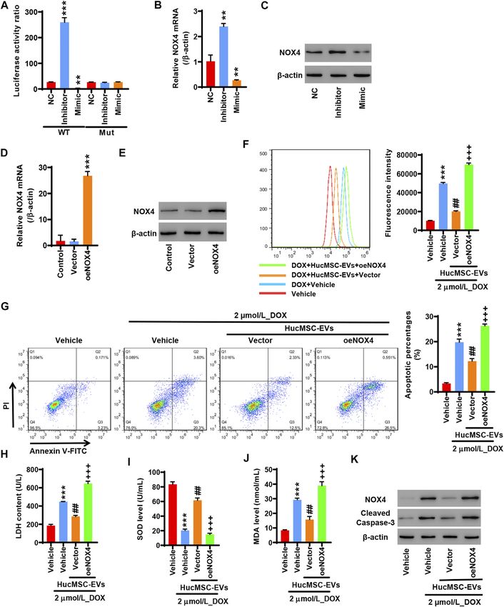
To study whether miR-100-5p targets the NOX4 protein,
cells were assessed by flow cytometry. Quadrant 1 represented dead cells. 293T cells were co-transfected with pGL3-NOX4-WT, miR-
Quadrant 2 represented late apoptotic cells. Quadrant 3 represented early 100-5p inhibitor (Inhibitor), and miR-100-5p mimic (Mimic)
apoptotic cells. Quadrant 4 represented normal cells. (C–E) The levels of LDH or pGL3-NOX4-MUT, miR-100-5p inhibitor (Inhibitor), and
(C), SOD (D), and MDA (E) were measured with biochemistry methods. (F)
miR-100-5p mimic (Mimic). The miR-100-5p inhibitor
Western blot analysis of cleaved-caspase-3. ***p < 0.001 vs. vehicle, #p <
0.05, ##p < 0.01, ###p < 0.001 vs. 2 μmol/L_DOX + 0 μg/ml_HucMSC-EVs,
markedly increased the luciferase activity of pGL3-NOX4-WT,
+
p < 0.05, ++p < 0.01 vs. 2 μmol/L_DOX + 50 μg/ml_HucMSC-EVs. while the miR-100-5p mimic obviously reduced the luciferase
activity of pGL3-NOX4-WT. There was no change in the
luciferase activity of pGL3-NOX4-MUT (Figure 5A). qRT-
miR-100-5p mimic (Figure 3A). After HucMSCs were PCR exhibited that the miR-100-5p inhibitor increased the
transfected with miR-100-5p inhibitor or miR-100-5p mimic, NOX4 mRNA expression, while the miR-100-5p mimic
HucMSC-EVs were extracted, and Q-PCR was performed. decreased the NOX4 mRNA expression (Figure 5B). Western
Q-PCR showed that miR-100-5p inhibitor abolished miR-100- blot illustrated that the NOX4 protein expression was increased
5p expression in EVs compared to the negative control, whereas by the miR-100-5p inhibitor, whereas the NOX4 protein
miR-100-5p mimic exceedingly exacerbated miR-100-5p expression was reduced by the miR-100-5p mimic
Frontiers in Bioengineering and Biotechnology | www.frontiersin.org 6 August 2021 | Volume 9 | Article 703241Zhong et al. HucMSC-EVs Inhibit DOX-Induced Heart Failure FIGURE 3 | HucMSC-EVs inhibits NOX4 expression. AC16 cells were treated with 2 µmol/L DOX and HucMSC-EVs at different concentrations (0, 50, 100, and 200 μg/ml) for 24 h. (A) qRT-PCR and (B) Western blot were performed to detect NOX2 and NOX4 expression, respectively. ***p < 0.001 vs. vehicle, #p < 0.05, ##p < 0.01 vs. 2 μmol/L_DOX + 0 μg/ml_HucMSC-EVs, +p < 0.05 vs. 2 μmol/L_DOX + 50 μg/ml_HucMSC-EVs. (C–D) AC16 cells were treated with 2 µmol/L DOX and HucMSC-EVs at a concentration of 100 μg/ml for different times (0, 6, 12, 24, and 48 h). (C) qRT-PCR and (D) Western blot were used for the detection of NOX4 expression. ***p < 0.001 vs. Vehicle, #p < 0.05, ##p < 0.01, ###p < 0.001 vs. 0 h, +p < 0.05 vs. 12 h. (Figure 5C). These results demonstrate that NOX4 is the well as decreased SOD levels which were induced by DOX; targeting protein of miR-100-5p. NOX4 expression is these effects were reversed by the overexpression of NOX4 negatively regulated by miR-100-5p. Next, NOX4 (Figures 5H–J). Moreover, DOX induced the percentages of overexpression plasmid (oe-NOX4) was constructed and apoptotic cell increase. HucMSC-EVs ameliorated the transfected to AC16 cells. oe-NOX4 markedly increased NOX4 percentages of apoptotic cells which were increased by DOX, mRNA expression (Figure 5D) and NOX4 protein expression and this was reversed by NOX4 overexpression (Figure 5G). (Figure 5E). Furthermore, AC16 cells were transfected with oe- Western blot showed that HucMSC-EVs ameliorated the NOX4 for 24 h, and then treated with 2 µmol/L DOX combined expression of NOX4 and cleaved-caspase-3 which was with 100 μg/ml HucMSC-EVs for another 24 h. Flow cytometry increased by DOX, and this was reversed by NOX4 revealed that HucMSC-EVs reduced ROS levels which were overexpression (Figure 5K). Collectively, these results indicate induced to increase by DOX, and this was reversed by the that overexpression of NOX4 abolishes those effects in which overexpression of NOX4 (Figure 5F). At the same time, HucMSC-EVs inhibit DOX-induced oxidative stress and HucMSC-EVs inhibited increased LDH and MDA levels as apoptosis. Frontiers in Bioengineering and Biotechnology | www.frontiersin.org 7 August 2021 | Volume 9 | Article 703241
Zhong et al.                                                                                                         HucMSC-EVs Inhibit DOX-Induced Heart Failure

 FIGURE 4 | Inhibition of EV miR-100-5-p reverses the effects in which HucMSC-EVs inhibit DOX-induced oxidative stress and apoptosis. (A) HucMSCs were
 transfected with miR-100-5p inhibitor (Inhibitor) or miR-100-5p mimic (Mimic). MiR-100-5p expression was determined by Q-PCR. (B) HucMSCs were transfected with
 miR-100-5p inhibitor or miR-100-5p mimic. After HucMSC-EVs were extracted, EV miR-100-5-p was determined by Q-PCR. *p < 0.05, **p < 0.01, ***p < 0.001 vs. NC.
 (C–H) AC16 cells were treated with 2 µmol/L DOX and EV miR-100-5p inhibitor (Inhibitor-EVs) or EV miR-100-5-p mimic (Mimic-EVs) at a concentration of 100 µg/
 ml. (C) ROS levels were measured by flow cytometry. (D) Apoptosis was determined by flow cytometry. Quadrant 1 represented dead cells. Quadrant 2 represented late
 apoptotic cells. Quadrant 3 represented early apoptotic cells, and Quadrant 4 represented normal cells. (E–G) LDH, SOD, and MDA levels were measured with
 biochemical assay. (H) NOX4 and cleaved-caspase-3 expression were examined by Western blot. ***p < 0.001 vs. control, ##p < 0.01 vs. 2 µmol/DOX, +p < 0.05,
 ++
    p < 0.01 vs. 2 µmol/L DOX + NC-EVs.

Frontiers in Bioengineering and Biotechnology | www.frontiersin.org              8                                          August 2021 | Volume 9 | Article 703241
Zhong et al. HucMSC-EVs Inhibit DOX-Induced Heart Failure FIGURE 5 | Overexpression of NOX4 abolishes those effects in which HucMSC-EVs inhibit DOX-induced oxidative stress and apoptosis. 293T cells were co- transfected with either pGL3-NOX4-WT, miR-100-5p inhibitor (Inhibitor) and miR-100-5p Mimic, and pGL3-NOX4-MUT, mimic (Mimic) or pGL3-NOX4-MUT, miR-100- 5p inhibitor (Inhibitor) and miR-100-5p mimic (Mimic). (A) Luciferase activity was quantified using a luminometer. **p < 0.01, ***p < 0.001 vs. WT + NC. (B) NOX4 mRNA was examined by qRT-PCR. (C) NOX4 protein expression was detected by Western blot. **p < 0.01 vs. NC. (D) AC16 cells were transfected with oeNOX4. NOX4 mRNA was examined by qRT-PCR. (E) AC16 cells were transfected with oeNOX4. NOX4 protein was detected by Western blot. ***p < 0.001 vs. vector. (F–K) AC16 cells were transfected with oeNOX4 for 24 h and then treated with 2 µmol/L DOX and 100 µg/ml HucMSC-EVs for 24 h. (F) ROS levels were measured by flow cytometry. (G) Apoptosis was measured by flow cytometry. Quadrant 1 represented dead cells. Quadrant 2 represented late apoptotic cells. Quadrant 3 represented early apoptotic cells, and Quadrant 4 represented normal cells. LDH (H), SOD (I), and MDA (J) were measured by biochemical assay. (K) NOX4 and cleaved-caspase-3 expression were determined by Western blot. ***p < 0.001 vs. vehicle, ##p < 0.01 vs. 2 μmol/L_DOX + vehicle, +++p < 0.001 vs. 2 µmol/L_DOX + HucMSC-EVs + vector. Frontiers in Bioengineering and Biotechnology | www.frontiersin.org 9 August 2021 | Volume 9 | Article 703241
Zhong et al.                                                                                              HucMSC-EVs Inhibit DOX-Induced Heart Failure

DISCUSSION                                                                     increased in heart failure rats. Whereas myocardial capillary and
                                                                               arteriolar density as well as SIRT1, FOXO3a, and MnSOD
With the aging of the population, the incidence of cardiovascular              expression are decreased. Echinacoside improves the heart
diseases such as hypertension and coronary heart disease has                   function by the SIRT1/FOXO3a/MnSOD signaling pathway in
increased significantly. The consequence of the development of                  heart failure rats (Ni et al., 2021). In DOX-induced heart failure
most cardiovascular diseases is heart failure, which has                       mice, SOD2, GPx-1, FOX3a, and SIRT3 expression are decreased,
increasingly become a major disease that seriously threatens                   apoptotic cells and cleaved-caspase-3 expression are increased, as
human health. Heart failure is manifested as insufficient                       well as inflammatory cytokines such as IL-1β, IL-6, and TNF-α
cardiac output and the inability to maintain the oxygen supply                 are increased. LongShengZhi capsule (LSZ) is a traditional
required by the body’s metabolism. It is an important reason for               Chinese medicine. After treatment of heart failure mice with
the loss of labor and death in patients with cardiovascular                    LSZ, the indicators of heart failure are significantly improved.
diseases. In recent years, MSCs are of great significance for the               LSZ decreases oxidative stress, apoptosis, and inflammatory
treatment of heart failure (Vrtovec et al., 2013; Narita and Suzuki,           cytokine levels, which are increased by DOX. LSZ increases
2015; Guijarro et al., 2016; Bartunek et al., 2017). However, MSCs             FoxO3a, SIRT3, and SOD2 expression, which are decreased by
have a short curative effect time, whereas MSC-derived EVs have                DOX (Xu et al., 2020). In our studies, HucMSC-EVs inhibited
a long curative effect time. MSC-derived EVs have a protective                 ROS production in a concentration-dependent manner, which
effect on heart failure (Chen et al., 2020b; Tan et al., 2020; Zheng           was induced by DOX. LDH and MDA levels were decreased by
et al., 2020). Chen et al. reported that EVs derived from MSCs                 HucMSC-EVs, which were increased by DOX. SOD levels were
improved cardiac hypertrophy, heart function, fibrosis, and                     increased by HucMSC-EVs, which were decreased by DOX.
myocardial apoptosis after transverse aortic constriction                      HucMSC-EV treatment was concentration dependent.
(TAC). It may be a benefit for treatment of heart failure                       Additionally, HucMSC-EV treatment decreased apoptotic cells
(Chen et al., 2020a). Gao et al. found that after myocardial                   and cleaved-caspase-3 expression in a concentration-dependent
infarction (MI), serum EVs were obtained from ischemic heart                   fashion, which were increased by DOX. These results suggest that
and kidney. Cardiorenal EV-derived miRNA-1956 promoted                         HucMSC-EVs inhibit DOX-induced heart failure in a
adipose-derived MSC-mediating angiogenesis, which is                           concentration-dependent manner in AC16 cells.
important for ischemic tissue repair (Gao et al., 2020).                          Nicotinamide adenine dinucleotide phosphate (NADPH)
Nakamura et al. demonstrated that injection of adiponectin                     oxidase is a peroxidase. There are NOX1, NOX2, NOX3,
and hMSC significantly increased hMSC-derived EV release.                       NOX4, NOX5, DUOX1, and DUOX2 in NADPH oxidase
Adiponectin accelerated hMSC-derived therapy in heart failure                  (NOX) family (Brandes et al., 2010). NOX2 and its homologue
mice (Nakamura et al., 2020). In this study, DOX induced                       NOX4 function as the core catalytic subunit of NADPH oxidase,
oxidative stress, ROS, and apoptosis increases in AC16 cells,                  which are the key to the function of the enzyme. NADPH oxidase
which were inhibited by HucMSC-EVs, and these were further                     produces ROS as a signal molecule to participate in the signal
reversed by the inhibition of EV miR-100-5-p. Meanwhile,                       transduction process that regulates cell proliferation, senescence,
HucMSC-EVs inhibited NOX4 expression which was induced                         and apoptosis (Ray et al., 2011; Schröder et al., 2012; Guo and
by DOX. Overexpression of NOX4 abolished the effects of                        Chen, 2015). When NOX family proteins are abnormally
HucMSC-EVs. NOX4 was negatively regulated by miR-100-5p.                       expressed, ROS levels increase, which cause oxidative stress
These data suggest that HucMSC-EVs have protective effects                     and participate in the formation of heart failure (Kumar et al.,
against DOX-induced heart failure in AC16 cells.                               2019). The main function of NOX family is to generate ROS
    Oxidative stress refers to the pathological process in which the           (Panday et al., 2015). Previous studies found that NOX4
balance of the oxidation system and antioxidant system causes an               knockout mice exhibited severe cardiac hypertrophy and
increase in ROS in the body and causes cell oxidative damage.                  contractile dysfunction after pressure overload, whereas NOX4
ROS is mainly produced by the mitochondria (Dietl and Maack,                   transgenic mice showed enhanced angiogenesis and increased
2017). ROS is also produced by the NADPH oxidase (Shang et al.,                expression of VEGF and Hif1α as well as less cardiac hypertrophy
2019). Oxidative stress in the myocardium causes the increase of               after pressure overload. It was suggested that NOX4 had a
ROS and causes damage of the cell membrane to release LDH.                     protection against cardiac stress by pressure overload (Zhang
MDA is the final product of lipid oxidation. SOD is the most                    et al., 2010). Cardiac-specific overexpression of NOX4 activates
important member of the antioxidant system. In heart failure,                  the nuclear transcription factor Nrf2 (Brewer et al., 2011). Nrf2 is
SOD is decreased, and LDH, MDA, and ROS are increased                          a key transcription factor in the cellular antioxidative stress
(Agostini et al., 2015; Casieri et al., 2017; Zhou and Tian,                   system. Nrf2 enters the nucleus and binds to ARE
2018; Koju et al., 2019; Ni et al., 2019). Cardiomyocyte                       (antioxidant response elements) to generate GSTs and SOD,
oxidative stress plays an important role in heart failure (Kim                 and play a role of antioxidant damage. Nrf2 shows a
et al., 2020). In heart failure, oxidative stress is markedly increased        protection against load-induced cardiac hypertrophy (Li et al.,
(Wang et al., 2019; Lubrano and Balzan, 2020). Inhibition of                   2009; Schröder et al., 2012). However, recent studies demonstrate
oxidative stress in cardiomyocytes can improve the symptoms of                 that in heart failure rats, TLR4 and NOX4 expression is
heart failure (Kumar et al., 2019; Pop et al., 2020). Heart failure is         significantly increased, autophagy and ferroptosis are
associated with oxidative stress and apoptosis. Recent studies find             enhanced, as well as the heart function is abnormal.
that apoptosis and mitochondrial oxidative stress are markedly                 Downregulation of either TLR4 or NOX4 inhibits autophagy

Frontiers in Bioengineering and Biotechnology | www.frontiersin.org       10                                    August 2021 | Volume 9 | Article 703241
Zhong et al. HucMSC-EVs Inhibit DOX-Induced Heart Failure and ferroptosis, and obviously improves cardiac function and left proliferation in HUVECs (Grundmann et al., 2011). NOX4 is ventricular remodeling (Chen et al., 2019). DOX-induced cardiac the target of miR-100-5p. Inhibition of miR-100-5p targeting and renal toxicities have been reported (Uygur et al., 2014; NOX4 leads to H2O2 release (Kriegel et al., 2015). The Tulubas et al., 2015). DOX induces apoptosis and oxidative expression of miR-100-5p is decreased by hypoxia. EVs stress in heart and kidney tissues. NOX4 is also induced to derived a human neural stem cell line that inhibits hypoxia- increase by DOX in the renal tissues. Pretreatment with induced proliferation and migration through EV miR-100-5-p omega-3 fatty acids improves the cardiac function, illustrates in pulmonary artery smooth muscle cells (Wang et al., 2020). antioxidant and antiapoptotic effects, and increases renal NOX4 Hromadnikova et al. found that downregulation of miR-100- expression (Saleh et al., 2020). In our experiments, HucMSC-EVs 5p was associated with gestational hypertension and ameliorated the expression of NOX2 mRNA and NOX4 mRNA, preeclampsia (Hromadnikova et al., 2016). Onrat et al. which was enhanced by DOX. Similarly, HucMSC-EVs compared 50 patients with dilated cardiomyopathy and 10 attenuated the expression of NOX2 and NOX4 proteins, which healthy persons. They found that miR-100-5p was was increased by DOX. HucMSC-EV treatment was overexpressed in the dilated cardiomyopathy (Onrat et al., concentration dependent. Furthermore, HucMSC-EV 2018). In this study, HucMSC-EVs inhibited DOX-induced treatment attenuated the expression of NOX4 mRNA and ROS, LDH, and MDA increases, and SOD decrease, which protein in a time–concentration–dependent fashion, which was were reversed by the EV miR-100-5-p inhibitor. There were induced to increase by DOX. Moreover, overexpression of NOX4 differences in reducing ROS levels and increasing SOD levels abolished that HucMSC-EVs inhibited DOX-induced ROS between EV miR-100-5p mimic treatment and HucMSC-EV production, increased LDH and MDA, and decreased SOD. treatment. But there were no differences in reducing LDH and Overexpression of NOX4 abolished that HucMSC-EVs MDA levels between EV miR-100-5-p mimic treatment and inhibited DOX-induced increased apoptotic cells, as well as HucMSC-EV treatment. It means that the effects of EV miR- cleaved-caspase-3 and NOX4 protein expression. These data 100-5p treatment and HucMSC-EV treatment are similar in indicate that HucMSC-EVs inhibit DOX-induced heart failure reducing ROS and oxidative stress. Furthermore, HucMSC- through targeting NOX4 in AC16 cells. EVs inhibited DOX-induced apoptotic cell increase, and Research in recent years has shown that miRNAs are cleaved-caspase-3 and NOX4 protein expression increase, important epigenetic regulatory factors. miRNAs also play which were reversed by the EV miR-100-5-p inhibitor, the important roles in EVs, which are benefit to treat whereas the effects of EV miR-100-5p treatment and cardiovascular diseases. EVs play a vital role in intercellular HucMSC-EV treatment were similar in reducing apoptosis. communication and their functions depend mainly on their Moreover, miR-100-5p inhibited NOX4 mRNA and protein internal contents (Vlassov et al., 2012). EVs act as miRNA expression, whereas the inhibition of miR-100-5p increased carriers that carry miRNAs to nearby or distant cells. EVs act NOX4 mRNA and protein expression. Taken together, these on target cells by directly releasing miRNAs to target cells data indicate that EV miR-100-5-p treatment inhibits DOX- through target cell endocytosis or membrane fusion, which is induced heart failure via targeting the NOX4 protein in AC16 considered to be an important tool for intercellular signal cells, which is similar to the effects of HucMSC-EV treatment. transduction (Montecalvo et al., 2012; Zeringer et al., 2015). EVs can deliver specific miRNAs to target cells that are as an important component of the paracrine effect of stem cells. CONCLUSION microRNAs encapsulated in EVs are the key genetic material that promotes the repair of myocardial damage. Inhibition of In summary, HucMSC-EV treatment inhibited DOX-induced miR-342-5p reduces exercise-afforded cardiac protection in oxidative stress, ROS, and apoptosis increases. HucMSC-EV myocardial ischemia/reperfusion rats. MiR-342-5p agomir treatment also inhibited DOX-induced NOX4 expression. increases the miR-342-5p levels and decreases myocardial Overexpression of NOX4 abolished those effects in which infarct size in rat hearts. Exercise-derived circulating EVs HucMSC-EVs inhibited DOX-induced oxidative stress, mediate the protective effects against myocardial ischemia/ apoptosis, and ROS production. NOX4 protein expression was reperfusion injury through EV miR-342-5p (Hou et al., 2019). negatively regulated by EV miR-100-5-p. Inhibition of EV miR- MiR-100-5p is believed to have anti-atherosclerotic effects 100-5-p reversed those effects in which HucMSC-EVs inhibited because it inhibits the proliferation of endothelial cells and DOX-induced oxidative stress, apoptosis, and ROS production, migration of blood vessels and smooth muscle cells (Shoeibi, whereas EV miR-100-5-p played a role similar to HucMSC-EVs 2020). In a mouse model of atherosclerosis, the expression of in reducing oxidative stress, apoptosis, and ROS production. It is miR-100-5p can improve endothelial function, weaken suggested that HucMSC-EVs inhibit DOX-induced heart failure atherosclerosis, and reduce plaque area (Linna-Kuosmanen through the miR-100-5p/NOX4 pathway. et al., 2020). Further studies indicate that downregulation of miR-100-5p activates the VEGFA/MYC pathway, which leads to increased endothelial cell metabolism, proliferation, and DATA AVAILABILITY STATEMENT angiogenesis, thereby promoting angiogenesis (Pankratz et al., 2018). Downregulation of miR-100 leads to the formation of The datasets presented in this study can be found in online angiogenic tubes, the increase in endothelial germination, and repositories. The names of the repository/repositories and Frontiers in Bioengineering and Biotechnology | www.frontiersin.org 11 August 2021 | Volume 9 | Article 703241
Zhong et al.                                                                                                                       HucMSC-EVs Inhibit DOX-Induced Heart Failure

accession number(s) can be found in the article/Supplementary                                  experiments and revised the manuscript. All authors read and
Material.                                                                                      approved the final manuscript.

AUTHOR CONTRIBUTIONS                                                                           FUNDING
ZZ and YT performed most of the experiments. XL performed                                      This study was supported by Affiliated Hospital of Panzhihua University
part of the experiments. JZ analyzed data. LW designed the                                     and supported by the grant from the Zhejiang Medical and Health
experiments and wrote the manuscript. JT designed the                                          Research Project, China (Grant Nos: 2018KY915 and 2019KY793).

                                                                                               Gao, L., Mei, S., Zhang, S., Qin, Q., Li, H., Liao, Y., et al. (2020). Cardio-renal
REFERENCES                                                                                        Exosomes in Myocardial Infarction Serum Regulate Proangiogenic Paracrine
                                                                                                  Signaling in Adipose Mesenchymal Stem Cells. Theranostics 10 (3), 1060–1073.
Agostini, S., Chiavacci, E., Matteucci, M., Torelli, M., Pitto, L., and Lionetti, V.              doi:10.7150/thno.37678
   (2015). Barley Beta-Glucan Promotes MnSOD Expression and Enhances                           Grundmann, S., Hans, F. P., Kinniry, S., Heinke, J., Helbing, T., Bluhm, F., et al.
   Angiogenesis under Oxidative Microenvironment. J. Cel. Mol. Med. 19 (1),                       (2011). MicroRNA-100 Regulates Neovascularization by Suppression of
   227–238. doi:10.1111/jcmm.12442                                                                Mammalian Target of Rapamycin in Endothelial and Vascular Smooth
Balbi, C., and Vassalli, G. (2020). Exosomes - beyond Stem Cells for Cardiac                      Muscle        Cells.    Circulation       123    (9),     999–1009.       doi:10.1161/
   Protection and Repair. Stem Cells 38 (11), 1387–1399. doi:10.1002/                             circulationaha.110.000323
   stem.3261                                                                                   Guan, Y. T., Xie, Y., Li, D. S., Zhu, Y. Y., Zhang, X. L., Feng, Y. L., et al. (2019).
Bartolucci, J., Verdugo, F. J., González, P. L., Larrea, R. E., Abarzua, E., Goset, C.,           Comparison of Biological Characteristics of Mesenchymal Stem Cells Derived
   et al. (2017). Safety and Efficacy of the Intravenous Infusion of Umbilical Cord                from the Human Umbilical Cord and Decidua Parietalis. Mol. Med. Rep. 20 (1),
   Mesenchymal Stem Cells in Patients with Heart Failure. Circ. Res. 121 (10),                    633–639. doi:10.3892/mmr.2019.10286
   1192–1204. doi:10.1161/circresaha.117.310712                                                Guijarro, D., Lebrin, M., Lairez, O., Bourin, P., Piriou, N., Pozzo, J., et al. (2016).
Bartunek, J., Terzic, A., Davison, B. A., Filippatos, G. S., Radovanovic, S., Beleslin,           Intramyocardial Transplantation of Mesenchymal Stromal Cells for Chronic
   B., et al. (2017). Cardiopoietic Cell Therapy for Advanced Ischaemic Heart                     Myocardial Ischemia and Impaired Left Ventricular Function: Results of the
   Failure: Results at 39 Weeks of the Prospective, Randomized, Double Blind,                     MESAMI 1 Pilot Trial. Int. J. Cardiol. 209, 258–265. doi:10.1016/
   Sham-Controlled CHART-1 Clinical Trial. Eur. Heart J. 38 (9), 648–660.                         j.ijcard.2016.02.016
   doi:10.1093/eurheartj/ehw543                                                                Guo, S., and Chen, X. (2015). The Human Nox4: Gene, Structure, Physiological
Brandes, R. P., Weissmann, N., and Schröder, K. (2010). NADPH Oxidases in                         Function and Pathological Significance. J. Drug Target. 23 (10), 888–896.
   Cardiovascular Disease. Free Radic. Biol. Med. 49 (5), 687–706. doi:10.1016/                   doi:10.3109/1061186x.2015.1036276
   j.freeradbiomed.2010.04.030                                                                 Hou, Z., Qin, X., Hu, Y., Zhang, X., Li, G., Wu, J., et al. (2019). Longterm Exercise-
Brewer, A. C., Murray, T. V. A., Arno, M., Zhang, M., Anilkumar, N. P., Mann, G.                  Derived Exosomal miR-342-5p. Circ. Res. 124 (9), 1386–1400. doi:10.1161/
   E., et al. (2011). Nox4 Regulates Nrf2 and Glutathione Redox in                                circresaha.118.314635
   Cardiomyocytes In Vivo. Free Radic. Biol. Med. 51 (1), 205–215.                             Hromadnikova, I., Kotlabova, K., Hympanova, L., and Krofta, L. (2016).
   doi:10.1016/j.freeradbiomed.2011.04.022                                                        Gestational Hypertension, Preeclampsia and Intrauterine Growth Restriction
Casieri, V., Matteucci, M., Cavallini, C., Torti, M., Torelli, M., and Lionetti,                  Induce Dysregulation of Cardiovascular and Cerebrovascular Disease
   V. (2017). Long-term Intake of Pasta Containing Barley (1-3)Beta-D-                            Associated microRNAs in Maternal Whole Peripheral Blood. Thromb. Res.
   Glucan Increases Neovascularization-Mediated Cardioprotection                                  137, 126–140. doi:10.1016/j.thromres.2015.11.032
   through Endothelial Upregulation of Vascular Endothelial Growth                             Jaquet, K., Krause, K. T., Denschel, J., Faessler, P., Nauerz, M., Geidel, S., et al.
   Factor and Parkin. Sci. Rep. 7 (1), 13424. doi:10.1038/s41598-017-                             (2005). Reduction of Myocardial Scar Size after Implantation of Mesenchymal
   13949-1                                                                                        Stem Cells in Rats: what Is the Mechanism? Stem Cell Develop. 14 (3), 299–309.
Chen, F., Li, X., Zhao, J., Geng, J., Xie, J., and Xu, B. (2020a). Bone Marrow                    doi:10.1089/scd.2005.14.299
   Mesenchymal Stem Cell-Derived Exosomes Attenuate Cardiac                                    Keerthikumar, S., Chisanga, D., Ariyaratne, D., Al Saffar, H., Anand, S., Zhao, K.,
   Hypertrophy and Fibrosis in Pressure Overload Induced Remodeling. In                           et al. (2016). ExoCarta: A Web-Based Compendium of Exosomal Cargo. J. Mol.
   Vitro Cell.Dev.Biol.-Animal 56 (7), 567–576. doi:10.1007/s11626-020-                           Biol. 428 (4), 688–692. doi:10.1016/j.jmb.2015.09.019
   00481-2                                                                                     Kim, S., Song, J., Ernst, P., Latimer, M. N., Ha, C.-M., Goh, K. Y., et al. (20202019).
Chen, F., Liang, P., Ye, F., Hou, C.-C., and Pi, L. (2020b). Mesenchymal Stem Cell                MitoQ Regulates Redox-Related Noncoding RNAs to Preserve Mitochondrial
   Therapy for Patients with Ischemic Heart Failure -past, Present, and Future.                   Network Integrity in Pressure-Overload Heart Failure. Am. J. Physiology-Heart
   Cscr 15. doi:10.2174/1574888x15666200309144906                                                 Circulatory Physiol. 318 (3), H682–h695. doi:10.1152/ajpheart.0061710.1152/
Chen, X., Xu, S., Zhao, C., and Liu, B. (2019). Role of TLR4/NADPH Oxidase 4                      ajpheart.00617.2019
   Pathway in Promoting Cell Death through Autophagy and Ferroptosis during                    Kobayashi, K., and Suzuki, K. (2018). Mesenchymal Stem/Stromal Cell-Based
   Heart Failure. Biochem. Biophysical Res. Commun. 516 (1), 37–43. doi:10.1016/                  Therapy for Heart Failure ― what Is the Best Source? ―. Circ. J. 82 (9),
   j.bbrc.2019.06.015                                                                             2222–2232. doi:10.1253/circj.CJ-18-0786
Davidson, S. M., and Yellon, D. M. (2018). Exosomes and Cardioprotection - A                   Koju, N., Taleb, A., Zhou, J., Lv, G., Yang, J., Cao, X., et al. (2019). Pharmacological
   Critical Analysis. Mol. Aspects Med. 60, 104–114. doi:10.1016/                                 Strategies to Lower Crosstalk between Nicotinamide Adenine Dinucleotide
   j.mam.2017.11.004                                                                              Phosphate (NADPH) Oxidase and Mitochondria. Biomed. Pharmacother. 111,
Dietl, A., and Maack, C. (2017). Targeting Mitochondrial Calcium Handling and                     1478–1498. doi:10.1016/j.biopha.2018.11.128
   Reactive Oxygen Species in Heart Failure. Curr. Heart Fail. Rep. 14 (4),                    Kriegel, A. J., Baker, M. A., Liu, Y., Liu, P., Cowley, A. W., Jr., and Liang, M. (2015).
   338–349. doi:10.1007/s11897-017-0347-7                                                         Endogenous MicroRNAs in Human Microvascular Endothelial Cells Regulate
Fang, Z., Yin, X., Wang, J., Tian, N., Ao, Q., Gu, Y., et al. (2016). Functional                  mRNAs Encoded by Hypertension-Related Genes. Hypertension 66 (4),
   Characterization of Human Umbilical Cord-Derived Mesenchymal Stem Cells                        793–799. doi:10.1161/hypertensionaha.115.05645
   for Treatment of Systolic Heart Failure. Exp. Ther. Med. 12 (5), 3328–3332.                 Kumar, A., Supowit, S., Potts, J. D., and DiPette, D. J. (2019). Alpha-calcitonin
   doi:10.3892/etm.2016.3748                                                                      Gene-related Peptide Prevents Pressure-overload Induced Heart Failure: Role

Frontiers in Bioengineering and Biotechnology | www.frontiersin.org                       12                                               August 2021 | Volume 9 | Article 703241
Zhong et al. HucMSC-EVs Inhibit DOX-Induced Heart Failure
of Apoptosis and Oxidative Stress. Physiol. Rep. 7 (21), e14269. doi:10.14814/ Reduces Blood Pressure In Vivo. Atvb 31 (6), 1368–1376. doi:10.1161/
phy2.14269 atvbaha.110.219238
Li, J., Ichikawa, T., Villacorta, L., Janicki, J. S., Brower, G. L., Yamamoto, M., et al. Sachinidis, A. (2020). Cardiotoxicity and Heart Failure: Lessons from Human-
(2009). Nrf2 Protects against Maladaptive Cardiac Responses to Hemodynamic Induced Pluripotent Stem Cell-Derived Cardiomyocytes and Anticancer Drugs.
Stress. Atvb 29 (11), 1843–1850. doi:10.1161/atvbaha.109.189480 Cells 9 (4), 1001. doi:10.3390/cells9041001
Li, J., Xu, S. Q., Zhao, Y. M., Yu, S., Ge, L. H., and Xu, B. H. (2018). Comparison Saleh, D., Abdelbaset, M., Hassan, A., Sharaf, O., Mahmoud, S., and Hegazy,
of the Biological Characteristics of Human Mesenchymal Stem Cells R. (2020). Omega-3 Fatty Acids Ameliorate Doxorubicin-Induced
Derived from Exfoliated Deciduous Teeth, Bone Marrow, Gingival Cardiorenal Toxicity: In-Vivo Regulation of Oxidative Stress,
Tissue, and Umbilical Cord. Mol. Med. Rep. 18 (6), 4969–4977. Apoptosis and Renal Nox4, and In-Vitro Preservation of the
doi:10.3892/mmr.2018.9501 Cytotoxic Efficacy. PLoS One 15 (11), e0242175. doi:10.1371/
Linna-Kuosmanen, S., Tomas Bosch, V., Moreau, P. R., Bouvy-Liivrand, M., journal.pone.0242175
Niskanen, H., Kansanen, E., et al. (2020). NRF2 Is a Key Regulator of Schröder, K., Zhang, M., Benkhoff, S., Mieth, A., Pliquett, R., Kosowski, J.,
Endothelial microRNA Expression under Proatherogenic Stimuli. et al. (2012). Nox4 Is a Protective Reactive Oxygen Species Generating
Cardiovasc. Res. 117, 1339–1357. doi:10.1093/cvr/cvaa219 Vascular NADPH Oxidase. Circ. Res. 110 (9), 1217–1225. doi:10.1161/
Lubrano, V., and Balzan, S. (2020). Role of Oxidative Stress-Related circresaha.112.267054
Biomarkers in Heart Failure: Galectin 3, α1-antitrypsin and LOX-1: Shang, L., Weng, X., Wang, D., Yue, W., Mernaugh, R., Amarnath, V., et al.
New Therapeutic Perspective? Mol. Cell Biochem 464 (1-2), 143–152. (2019). Isolevuglandin Scavenger Attenuates Pressure Overload-Induced
doi:10.1007/s11010-019-03656-y Cardiac Oxidative Stress, Cardiac Hypertrophy, Heart Failure and Lung
Mao, C., Hou, X., Wang, B., Chi, J., Jiang, Y., Zhang, C., et al. (2017). Intramuscular Remodeling. Free Radic. Biol. Med. 141, 291–298. doi:10.1016/
Injection of Human Umbilical Cord-Derived Mesenchymal Stem Cells j.freeradbiomed.2019.06.029
Improves Cardiac Function in Dilated Cardiomyopathy Rats. Stem Cell Res Shi, J.-x., Wang, Q.-j., Li, H., and Huang, Q. (2016). Silencing of USP22
Ther 8 (1), 18. doi:10.1186/s13287-017-0472-y Suppresses High Glucose-Induced Apoptosis, ROS Production and
Matsushita, K. (2020). Heart Failure and Adipose Mesenchymal Inflammation in Podocytes. Mol. Biosyst. 12 (5), 1445–1456.
Stem Cells. Trends Mol. Med. 26 (4), 369–379. doi:10.1016/ doi:10.1039/c5mb00722d
j.molmed.2020.01.003 Shoeibi, S. (2020). Diagnostic and Theranostic microRNAs in the
Montecalvo, A., Larregina, A. T., Shufesky, W. J., Beer Stolz, D., Sullivan, M. L. G., Pathogenesis of Atherosclerosis. Acta Physiol. 228 (1), e13353.
Karlsson, J. M., et al. (2012). Mechanism of Transfer of Functional microRNAs doi:10.1111/apha.13353
between Mouse Dendritic Cells via Exosomes. Blood 119 (3), 756–766. Théry, C., Amigorena, S., Raposo, G., and Clayton, A. (2006). Isolation and
doi:10.1182/blood-2011-02-338004 Characterization of Exosomes from Cell Culture Supernatants and
Nakamura, Y., Kita, S., Tanaka, Y., Fukuda, S., Obata, Y., Okita, T., et al. (2020). Biological Fluids. Curr. Protoc. Cell Biol. 30 (Unit 3.22). doi:10.1002/
Adiponectin Stimulates Exosome Release to Enhance Mesenchymal Stem-Cell- 0471143030.cb0322s30
Driven Therapy of Heart Failure in Mice. Mol. Ther. 28 (10), 2203–2219. Tkach, M., and Théry, C. (2016). Communication by Extracellular Vesicles: Where
doi:10.1016/j.ymthe.2020.06.026 We Are and where We Need to Go. Cell 164 (6), 1226–1232. doi:10.1016/
Narita, T., and Suzuki, K. (2015). Bone Marrow-Derived Mesenchymal Stem Cells for the j.cell.2016.01.043
Treatment of Heart Failure. Heart Fail. Rev. 20 (1), 53–68. doi:10.1007/s10741-014- Tulubas, F., Gurel, A., Oran, M., Topcu, B., Caglar, V., and Uygur, E. (2015). The
9435-x Protective Effects of ω-3 Fatty Acids on Doxorubicin-Induced Hepatotoxicity
Ni, J., Liu, X., Yin, Y., Zhang, P., Xu, Y.-W., and Liu, Z. (2019). Exosomes Derived and Nephrotoxicity in Rats. Toxicol. Ind. Health 31 (7), 638–644. doi:10.1177/
from TIMP2-Modified Human Umbilical Cord Mesenchymal Stem Cells 0748233713483203
Enhance the Repair Effect in Rat Model with Myocardial Infarction Possibly Uygur, R., Aktas, C., Tulubas, F., Alpsoy, S., Topcu, B., and Ozen, O. (2014).
by the Akt/Sfrp2 Pathway. Oxidative Med. Cell Longevity 2019, 1–19. Cardioprotective Effects of Fish omega-3 Fatty Acids on Doxorubicin-Induced
doi:10.1155/2019/1958941 Cardiotoxicity in Rats. Hum. Exp. Toxicol. 33 (4), 435–445. doi:10.1177/
Ni, Y., Deng, J., Liu, X., Li, Q., Zhang, J., Bai, H., et al. (2021). Echinacoside Reverses 0960327113493304
Myocardial Remodeling and Improves Heart Function via Regulating SIRT1/ Vlassov, A. V., Magdaleno, S., Setterquist, R., and Conrad, R. (2012).
FOXO3a/MnSOD axis in HF Rats Induced by Isoproterenol. J. Cel. Mol. Med. Exosomes: Current Knowledge of Their Composition, Biological
25 (1), 203–216. doi:10.1111/jcmm.15904 Functions, and Diagnostic and Therapeutic Potentials. Biochim. Biophys.
Onrat, S. T., Onrat, E., Ercan Onay, E., Yalım, Z., and Avşar, A. (2018). Acta (Bba) - Gen. Subjects 1820 (7), 940–948. doi:10.1016/
The Genetic Determination of the Differentiation between Ischemic j.bbagen.2012.03.017
Dilated Cardiomyopathy and Idiopathic Dilated Cardiomyopathy. Vrtovec, B., Poglajen, G., and Haddad, F. (2013). Stem Cell Therapy in Patients
Genet. Test. Mol. Biomarkers 22 (11), 644–651. doi:10.1089/ with Heart Failure. Methodist DeBakey Cardiovasc. J. 9 (1), 6–10. doi:10.14797/
gtmb.2018.0188 mdcj-9-1-6
Ozaki Tan, S. J., Floriano, J. F., Nicastro, L., Emanueli, C., and Catapano, F. (2020). Wang, J., Hu, L., Huang, H., Yu, Y., Wang, J., Yu, Y., et al. (2020).
Novel Applications of Mesenchymal Stem Cell-Derived Exosomes for CAR (CARSKNKDC) Peptide Modified ReNcell-Derived Extracellular
Myocardial Infarction Therapeutics. Biomolecules 10 (5), 707. doi:10.3390/ Vesicles as a Novel Therapeutic Agent for Targeted Pulmonary
biom10050707 Hypertension Therapy. Hypertension 76 (4), 1147–1160. doi:10.1161/
Panday, A., Sahoo, M. K., Osorio, D., and Batra, S. (2015). NADPH Oxidases: an hypertensionaha.120.15554
Overview from Structure to Innate Immunity-Associated Pathologies. Cell Mol Wang, K., Zhu, Z. F., Chi, R. F., Li, Q., Yang, Z. J., Jie, X., et al. (2019). The NADPH
Immunol 12 (1), 5–23. doi:10.1038/cmi.2014.89 Oxidase Inhibitor Apocynin Improves Cardiac Sympathetic Nerve Terminal
Pankratz, F., Hohnloser, C., Bemtgen, X., Jaenich, C., Kreuzaler, S., Hoefer, I., et al. Innervation and Function in Heart Failure. Exp. Physiol. 104 (11), 1638–1649.
(2018). MicroRNA-100 Suppresses Chronic Vascular Inflammation by doi:10.1113/ep087552
Stimulation of Endothelial Autophagy. Circ. Res. 122 (3), 417–432. Xie, Q., Liu, R., Jiang, J., Peng, J., Yang, C., Zhang, W., et al. (2020). What Is the
doi:10.1161/circresaha.117.311428 Impact of Human Umbilical Cord Mesenchymal Stem Cell Transplantation on
Pop, C., Berce, C., Ghibu, S., Scurtu, I., Soriţău, O., Login, C., et al. (2020). Clinical Treatment? Stem Cell Res. Ther 11 (1), 519. doi:10.1186/s13287-020-
Effects of Lycium Barbarum L. Polysaccharides on Inflammation and 02011-z
Oxidative Stress Markers in a Pressure Overload-Induced Heart Failure Xu, S., Wang, Y., Yu, M., Wang, D., Liang, Y., Chen, Y., et al. (2020).
Rat Model. Molecules 25 (3), 466. doi:10.3390/molecules25030466 LongShengZhi Capsule Inhibits Doxorubicin-Induced Heart Failure by
Ray, R., Murdoch, C. E., Wang, M., Santos, C. X., Zhang, M., Alom-Ruiz, S., et al. Anti-oxidative Stress. Biomed. Pharmacother. 123, 109803. doi:10.1016/
(2011). Endothelial Nox4 NADPH Oxidase Enhances Vasodilatation and j.biopha.2019.109803
Frontiers in Bioengineering and Biotechnology | www.frontiersin.org 13 August 2021 | Volume 9 | Article 703241You can also read