From Physiology to Pathology of Cortico-Thalamo-Cortical Oscillations: Astroglia as a Target for Further Research
←
→
Page content transcription
If your browser does not render page correctly, please read the page content below
REVIEW
                                                                                                                                                published: 09 June 2021
                                                                                                                                        doi: 10.3389/fneur.2021.661408

                                               From Physiology to Pathology of
                                               Cortico-Thalamo-Cortical
                                               Oscillations: Astroglia as a Target for
                                               Further Research
                                               Davide Gobbo † , Anja Scheller † and Frank Kirchhoff*†
                                               Molecular Physiology, Center for Integrative Physiology and Molecular Medicine (CIPMM), University of Saarland, Homburg,
                                               Germany

                                               The electrographic hallmark of childhood absence epilepsy (CAE) and other idiopathic
                                               forms of epilepsy are 2.5–4 Hz spike and wave discharges (SWDs) originating from
                                               abnormal electrical oscillations of the cortico-thalamo-cortical network. SWDs are
                                               generally associated with sudden and brief non-convulsive epileptic events mostly
                          Edited by:           generating impairment of consciousness and correlating with attention and learning
                         Kjell Heuser,
     Oslo University Hospital, Norway          as well as cognitive deficits. To date, SWDs are known to arise from locally restricted
                        Reviewed by:           imbalances of excitation and inhibition in the deep layers of the primary somatosensory
                     Vincenzo Crunelli,        cortex. SWDs propagate to the mostly GABAergic nucleus reticularis thalami (NRT)
   Cardiff University, United Kingdom
                                               and the somatosensory thalamic nuclei that project back to the cortex, leading to the
                       Hirokazu Oguni,
   TMG Asaka Medical Center, Japan             typical generalized spike and wave oscillations. Given their shared anatomical basis,
                   *Correspondence:            SWDs have been originally considered the pathological transition of 11–16 Hz bursts
                        Frank Kirchhoff        of neural oscillatory activity (the so-called sleep spindles) occurring during Non-Rapid
                frank.kirchhoff@uks.eu
                                               Eye Movement (NREM) sleep, but more recent research revealed fundamental functional
                           † ORCID:
                                               differences between sleep spindles and SWDs, suggesting the latter could be more
                      Davide Gobbo
     orcid.org/0000-0002-4076-2697             closely related to the slow (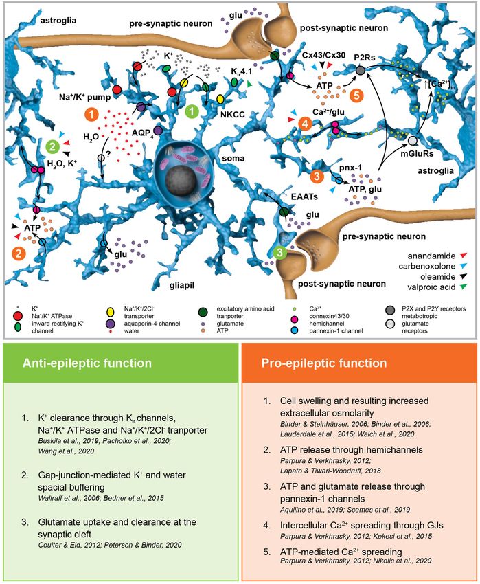
Gobbo et al. Astrocytes and Cortico-Thalamo-Cortical Oscillations
INTRODUCTION and Wistar-Albino-Glaxo rats from Rijswijk (WAG/Rij) (20,
37–42) as well as many monogenic mouse mutants (43–45).
Epilepsy is a highly heterogeneous neurological condition Although sharing most electrographic and behavioral hallmarks
characterized by enduring predisposition to unpredictable of absence seizures, animal models are characterized by higher
pathological discharge of rhythmic activity in the brain networks, SWD frequencies (5–11 Hz) (Figure 1B). Ex vivo multi-site
which is commonly referred as seizure activity (1). In virtue local field potential studies identified the peri-oral primary
of the severity and nature of the pathological alteration somatosensory cortex as initiation site of absence seizures
(abnormal, excessive, or excessively synchronous activation) in WAG/Rij (46) and GAERS rats (47–50). Notably, this
as well as the cellular and anatomical composition of the has been proven wrong for the acute pharmacological γ-
affected brain networks, seizures can cause changes in the hydroxybutyric acid (GHB) model (51–54) in mice, where
level of consciousness, behavior, memory, and emotional status. the prefrontal cortex was suggested as the initiation site of
Although the etiology of epileptiform activity is still unknown in SWDs (55). With this in mind and considering the many
half of the cases, understanding the pathological alteration at the areas contributing to the cortical pre-ictal BOLD changes of
basis of the epileptic phenotype may not only be of fundamental absence seizures, one can probably not identify a unique
therapeutical importance but also provide further insights into canonical focal onset or initiation site for absence seizures.
the functioning of the affected neural networks in the physiology Instead, the denomination cortical initiation network has been
of the healthy brain. The identification of the molecular and recently proposed (17), thereby settling the long-standing
cellular mechanisms underlying physiological oscillations is controversy about the SWDs initiation site (56–59). However,
critical for a full comprehension of their relationship to the the existence of a cortical initiation network does not imply that
respective pathological activity. In this regard, an exceptional case manipulation of the sole thalamic components of the cortico-
of study is the cortico-thalamo-cortical network, physiologically thalamo-cortical network is not sufficient to induce SWDs,
engaged during sleep and pathologically altered in the context of as it is indeed the case (60–62), or that the wide thalamo-
non-motor (absence) seizures (2). cortical innervation is not crucial for SWDs generalization, as
Absence seizures are transient non-convulsive generalized suggested by the existence of subclinical SWDs restricted to
epileptic events and are also referred as petit mal seizures (2, 3). the cortical network (48). In particular, the thalamic posterior
Phenotypically, absence seizures are coupled with sudden and nucleus plays a crucial role in the generalization of SWDs
brief impairment of consciousness and lack of responsiveness to (61, 63–66). Till recently, ex vivo studies performed in different
external stimuli as well as variable secondary clinical symptoms mammalian models identified the hyperexcitability and T-type
(e.g., automatisms, atonic, and tonic muscular components Ca2+ channel-mediated burst activity of glutamatergic thalamo-
etc.) (4, 5). Absence seizures are the sole clinical symptom cortical neurons and GABAergic neurons from the thalamic
of childhood absence epilepsy (CAE) but are also associated reticular nucleus (or nucleus reticularis thalami, NRT) as the
with several other idiopathic generalized epilepsies (4, 6–11). rhythmogenic cortico-thalamo-cortical network mechanism of
Although CAE has up to 70% remission rate (7, 12), the gold SWDs (Figures 1D,E) (24, 41, 67–71). Nevertheless, recent in
standard monotherapy, based on ethosuximide and valproic acid, vivo studies performed in rodents showed that only a small
is still ineffective in 30% of the cases (13). Moreover, clinical fraction of thalamo-cortical and cortico-thalamic neurons are
conditions displaying absence seizures are often associated with synchronously active at each SWD cycle and the cellular
severe neuropsychiatric comorbid conditions such as impaired composition of this neuronal subpopulation changes between
attention, learning, memory and cognition, which are often subsequent cycles, thus excluding the existence of distinctive
left unaltered or even worsened by common antiepileptic neuronal subpopulations (Figures 1F,G) (25, 26). This explains
drugs (14–17). why, with SWD progression, the activities of the cortico-
Although absence seizures display inter- and intraindividual thalamic and thalamo-cortical neurons undergo a phase-
variability (17, 18), they exhibit generalized bilateral 2.5– shift in time (46) since different neuronal subpopulations
4 Hz spike and wave discharges (SWDs) with no aura or participate in this excitatory feedback-loop with slightly different
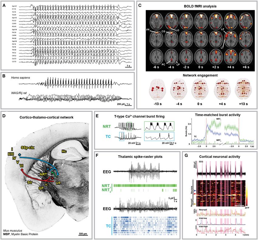
post-ictal depression (Figures 1A,B) (4, 27–29). It is widely kinetics. Moreover, this progressive phase-shift between different
accepted that the sharp spike and the slow wave component subpopulations active at the same time accounts for the
of SWDs are functionally coupled and correspond to a state of overlapping average electrical activity in the cortico-thalamic,
neuronal excitation and silence in the cortico-thalamo-cortical thalamo-cortical, and NRT neurons within any SWD cycle.
network, respectively (30). Blood oxygenation level-dependent Moreover, although interictal T-type Ca2+ channel burst activity
(BOLD) functional magnetic resonance imaging (fMRI) studies in the thalamo-cortical neurons increases right before SWD
in humans consistently showed cortical network engagement onset, overall in vivo ictal thalamic activity decreases and only
in correspondence of and even preceding the appearance cortical and NRT T-type channels are essentials for SWDs (25).
of SWDs in electrographic traces as well as an increased Interestingly, all NRT neurons fire within each SWD cycle, even
interictal synchrony in the sensorimotor cortex (Figure 1C) though a fraction of those neurons fires relatively asynchronous
(22, 23, 31–36). Most advancements on the understanding of tonic spikes rather than T-type Ca2+ channel-mediated bursts
the cellular and synaptic mechanisms underlying SWDs derive in phase with the SWDs (Figures 1E,F) (25). The enhanced
from the extensive use of genetic animal models, particularly tonic inhibition of thalamo-cortical neurons as well as the
the genetic absence epilepsy rats from Strasbourg (GAERS) increased thalamic GABA level are key aspects of absence seizures
Frontiers in Neurology | www.frontiersin.org 2 June 2021 | Volume 12 | Article 661408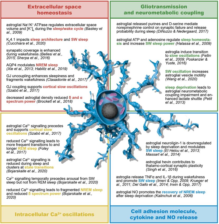
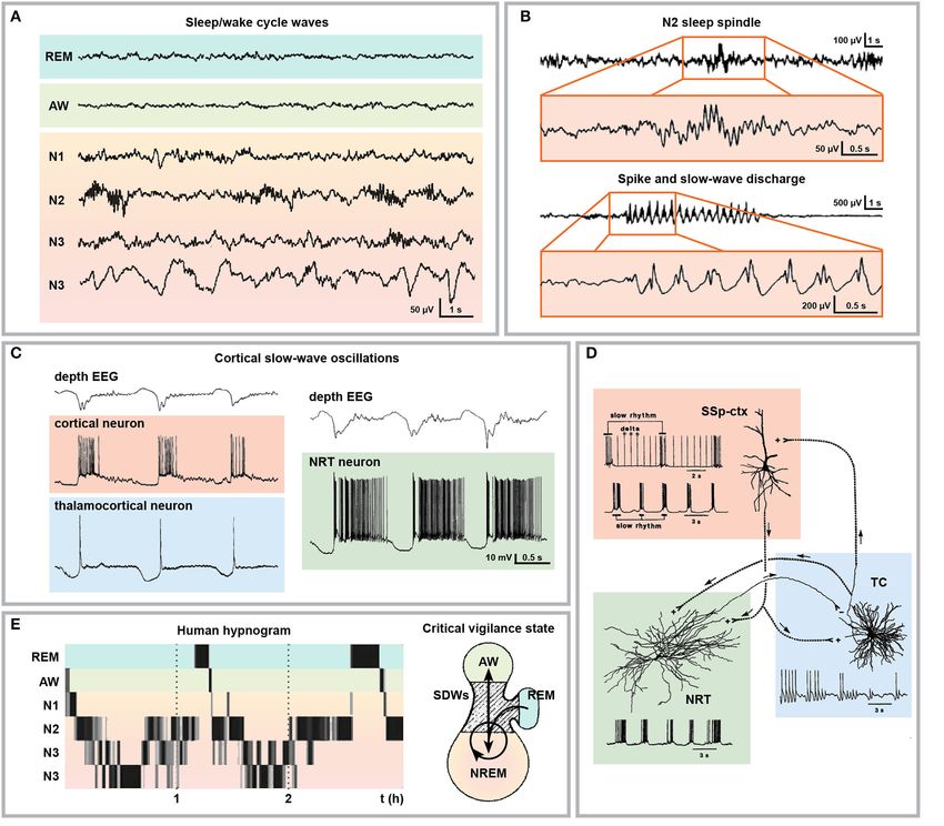
Gobbo et al.                                                                                                               Astrocytes and Cortico-Thalamo-Cortical Oscillations

 FIGURE 1 | Anatomical and electrophysiological characterization of absence seizures. (A) Human electroencephalographical recording (EEG) displaying typical 3 Hz
 spike-and-wave discharges (SWDs). (B) 3 Hz SWDs associated with Childhood Absence Epilepsy (top trace, 8-year-old boy) and 8 Hz SWDs recorded in an adult
 WAG/Rij rat (bottom trace). (C) Blood Oxygenation Level Dependent (BOLD) functional Magnetic Resonance Imaging (fMRI) changes associated with absence
 seizures before (FP, frontal polar; CG, cingulate; LO, lateral occipital; PC, precuneus; LP, lateral parietal cortex) and after (LF, lateral frontal; LT, lateral temporal cortex)
 seizure onset (top) and brain network engagement analysis around SWDs events (bottom). (D) Anatomical organization of the cortico-thalamo-cortical network in
 C57BL/6N mice. Myelin Basic Protein (MBP) immunostaining indicates the axonal fiber tracts connecting the network (own data; mouse monoclonal MBP antibody,
 BioLegend, AB_2564741, Cat. No. 808401, 1:500). Cortico-thalamic excitatory neurons from cortical layers V and VI (red) project to both NRT and thalamus;
 thalamo-cortical excitatory neurons (blue) project back to cortical layer IV and NRT; NRT GABAergic neurons (green) inhibit the thalamic nuclei. I-VI, cortical layers; GP,
 globus pallidus; Hc, hippocampus; ic, internal capsule; NRT, nucleus reticularis thalami; SSp-ctx, primary somatosensory cortex; Str, striatum; VP, ventral posterior
 thalamic nuclei. (E) Glutamatergic thalamo-cortical neurons (TC) as well as GABAergic NRT neurons display T-type Ca2+ channel-mediated burst firing during SWDs
 (left, ex vivo recording from ferret thalamic slices). (F) Spike-time raster plots of two representative NRT neurons (NRT1 , top trace; NRT2 , bottom trace) and 10 TC
 neurons with time-matched EEG in GAERS rats. The overall TC activity decreases during SWDs and only a small portion of TC neurons fire synchronously. (G)
 2-Photon laser scanning microscopy of neuronal cortical Ca2+ activity in stargazer mice during absence seizures. Heatmap of neuronal Ca2+ activity shows that only
 a subpopulation of neurons displays ictal synchronous firing. Modified from (A), (19); (B), (20, 21); (C), (22, 23); (E), (24, 25); (F), (25); (G), (26).

(25, 72–78). Moreover, the fact that SWDs can be induced                                     cortical neurons from layer VI could suggest that a balance shift
by the impairment of the cortico-thalamic glutamate release                                  toward GABAergic inhibition more than an absolute increase
due to deletion of P/Q-type Ca2+ channels in the projecting                                  of GABA levels is the key mechanism of SWD generalization

Frontiers in Neurology | www.frontiersin.org                                             3                                                June 2021 | Volume 12 | Article 661408
Gobbo et al.                                                                                      Astrocytes and Cortico-Thalamo-Cortical Oscillations

(79). Additionally, the decreased glutamate release could lead             extracellular K+ resulting from neuronal firing (100–103).
to reduced activity of cortical interneurons, thus contributing to         Astroglial K+ and glutamate uptake is altered in cultured
cortical hyperexcitability.                                                cortical astrocytes after Kir 4.1 channel downregulation (104) as
                                                                           well as in astroglial-specific Kir 4.1 knock-out mice (105, 106).
                                                                           Gain-of-function (107) as well as loss-of-function mutations
ASTROCYTES CONTRIBUTE TO                                                   (108, 109) in the human KCNJ10 gene encoding Kir 4.1
NETWORK PRIMING AND                                                        have been linked to forms of childhood epilepsies associated
SYNCHRONIZATION AS WELL AS SWD                                             with ataxia and cognitive impairment, but not to CAE.
INDUCTION, PROPAGATION, AND                                                Notably, artificially increasing extracellular K+ concentration
                                                                           ex vivo is associated with propagating epileptiform discharges
TERMINATION                                                                induced by focal optogenetic activation of parvalbumin-
After more than three decades of accumulating evidence,                    expressing interneurons (110). In vivo though, K+ clearance
nowadays it is widely established that astroglia constitute a              impairment induced by blocking GJ was not sufficient to
ubiquitous non-neuronal communication system in the brain                  induce neocortical seizures (111). Interestingly, valproic acid
involved in virtually every physiological and pathological                 (but not ethosuximide) induces Kir 4.1 overexpression in the
scenario of the central nervous system (80–82). Not only                   cortex of healthy rats (112). Nevertheless, further research
do they support synapses from a mechanical, metabolical                    is required to address the actual contribution of Kir 4.1
as well as functional point of view, but they also participate             overexpression in the anti-absence effects of valproic acid,
in synaptic transmission and plasticity, neural network                    as well as the putative role of astroglial Kir 4.1 itself in the
excitability and balance between excitation and inhibition (E/I)           development and propagation of SWDs. The combined use
as active information integrators and processors (83–86). The              of cell-specific conditional knock-out of these channels and
contribution of the astroglial network to the pathophysiology              pharmacological models of SWDs could shine new light on
of epilepsy encompasses a plethora of different molecular                  the topic.
mechanisms which currently represent one of the most fruitful                 K+ uptake is associated with cellular swelling due to
research topics in neuroscience (87–96). Pathological priming              Na /K+ -pump dependent water influx (113). Although the
                                                                              +

mechanisms of the astroglial network ultimately involve either             exact molecular mechanisms are still under debate, water
E/I imbalance or enhanced network synchronization (or                      fluxes across the astrocytic membrane are associated with K+
both simultaneously). Alternatively, astrocytes may influence              homeostasis and they influence local interstitial osmolarity
spatial and temporal propagation of seizures, thus playing                 as well as seizure generation and progression (114–120).
a key role in the phenotypical outcome of seizures and                     Given the accumulating evidence against the predominant
their severity and therefore representing a promising target               contribution of the astroglial water channel aquaporin-4 (AQP4)
for the development of new non-neurocentric drugs. Most                    in water homeostasis (113, 121), the use of AQP4 conditional
of the recent evidence focuses on astroglial contribution                  knock-out as a model of disrupted water homeostasis has
to convulsive epileptic activity. Nevertheless, we discuss                 been recently challenged. Nevertheless, water homeostasis
in the following the putative involvement of astrocytes in                 impairment and the resulting volume and osmolarity
network priming as well as seizure induction and propagation               dysregulation should affect neural network excitability. Indeed,
which could have a role in pathological epileptic scenarios                a recent structural MRI study on CAE showed significant
including SWDs, as well. We focus on the evidence that links               gray matter volume abnormalities in both frontotemporal
to observations coming from the clinics as well as genetic                 cortical region and posterior thalami compared to
and pharmacological models of SWDs, aiming to point at                     controls (122, 123).
specific topics which may be worth further research in the field              GJ coupling, particularly mediated by connexins Cx30 and
of SWDs.                                                                   Cx43, provides the astroglial network with a high level of
                                                                           intercellular structural, metabolic and functional connectivity,
                                                                           enabling the exchange of ions and small molecules (124–131).
The Astroglial Network Controls                                            In the context of epilepsy, connexins mediate ATP release (into
Extracellular Space Homeostasis Through                                    the extracellular space through hemichannels), the spreading
K+ , Water and Solute Clearance                                            of intercellular Ca2+ waves (132) and are fundamental in
By means of their close juxtaposition to synapses, their expression        the spatial buffering required for K+ and water homeostasis
of an extraordinary assortment of membrane transporters and                as well as glutamate clearance (133, 134). With respect to
receptors as well as their physical and functional coupling                absence epilepsy, most advancement in unraveling the role
through gap junctions (GJs), astroglial networks provide a perfect         of GJs has been obtained employing GJ blockers in well-
spatial buffering for neural activity (Figure 2) (97–99). Astrocytes       established genetic animal models (135). The broad-spectrum
are key regulators of the extracellular K+ concentration.                  GJ blocker carbenoxolone (CBX) decreased both amplitude and
Their high K+ permeability mediated by inwardly-rectifying                 duration of 4-aminopyridine-induced seizure-like events (SLEs)
Kir and two-pore-domain K2P channels, Na+ /K+ pumps and                    in thalamocortical slices obtained from mice with spontaneous
Na+ /K+ /Cl− transporters associated with their extensive GJ               SWDs (136, 137) as well as the duration of SWDs seen in
coupling enables them to uptake and redistribute excessive                 GAERS rats in vivo after systemic application (138). Interestingly,

Frontiers in Neurology | www.frontiersin.org                           4                                       June 2021 | Volume 12 | Article 661408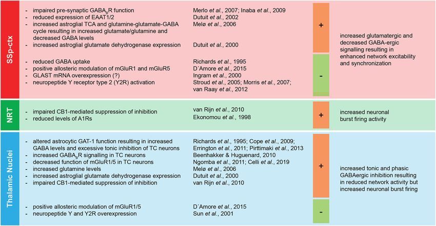
Gobbo et al.                                                                                                         Astrocytes and Cortico-Thalamo-Cortical Oscillations

 FIGURE 2 | Astroglial homeostatic control of the extracellular space has opposite effects on epileptogenesis. The astroglial network is responsible for extracellular K+
 uptake by means of inward rectifying K+ channels (Kir ), Na+ /K+ pump and Na+ /K+ /2Cl− transporter (NKCC). K+ clearance is coupled with water uptake through the
 water channel aquaporin-4 (AQP4) and possibly via yet unknown additional pathways. The excitatory amino acid transporters EAAT1 and EAAT2 are responsible for
                                                                                                                                                             (Continued)

Frontiers in Neurology | www.frontiersin.org                                        5                                             June 2021 | Volume 12 | Article 661408
Gobbo et al. Astrocytes and Cortico-Thalamo-Cortical Oscillations
FIGURE 2 | glutamate uptake. Astroglial connexins Cx43 and Cx30 enable gap-junction (GJ) coupling responsible for spatial ionic and metabolic buffering. Connexin
hemichannels as well as pannexin-1 channels (Panx1) mediate glutamate and ATP release in the extracellular space possibly activating astroglial metabotropic
glutamate receptors (mGluRs) and purinergic P2X and P2Y receptors (P2Rs), respectively. This, in term, induces intracellular Ca2+ increases in the neighboring
astroglia. The figure summarizes the pro- and anti-epileptic roles of the mechanisms described above and points to the putative targets of valproic acid and the GJ
blockers carbenoxolone, anandamide, and oleamide in this scenario.
in vivo injection of CBX in the NRT of rats with atypical synchronization. Astrocytes rely on their extensive GJ coupling
absence seizures and spontaneous SWDs decreased the duration enabling effective spatial ionic, osmotic, and functional buffering.
of SWDs (139), whereas no alteration of SWD phenotype GJ hemichannels as well as pannexin-1 channels may be
was observed if CBX was injected in the posterior thalami responsible for augmented synchronous activity through ATP and
of WAG/Rij rats and the lethargic mouse genetic model of glutamate release and following Ca2 spreading throughout the
absence epilepsy (140). Recently, intraperitoneal injection of CBX astroglial network. So far, we are still missing evidence for linking
was associated with absence seizures worsening in WAG/Rij the astroglial fine-tuning of the extracellular ion and transmitter
rats (141), hinting at non-obvious and non-trivial differences homeostasis to SWDs. However, as it is the case for other kinds
across the absence epilepsy models. The endocannabinoids of epileptiform activity, their role in regulating such network
anandamide (N-arachidonoylethanolamine, ANA) and oleamide excitability is very likely.
(cis-9,10-octadecenoamide, OLE) are specific Cx43 blockers
(142, 143). Intracerebroventricular injection of ANA decreased Astrocytes Are Actively Involved in
in a dose-dependent manner the recurrence and duration Network Dynamics and E/I Balance
of SWDs, although its mechanism of action likely involves
Through Neurometabolic Coupling,
type-1 cannabinoid (CB1) receptor activation (144) or even
direct inhibition of T-type Ca2+ channels (145). Interestingly, Neurotransmission Modulation and
although specific studies addressing the impact of OLE in Gliotransmission
absence epilepsy are still required, OLE has a sleep-inducing Astrocytes do not only contribute to neural excitability and
effect and enhances GABAA receptor-mediated responses, thus functioning by responding to neurotransmitter release and
possibly affecting the physiological, temporal-spatial pattern modification of extracellular ionic composition, they are also
of cortico-thalamo-cortical oscillations (146, 147). CBX as actively involved in neurotransmitter uptake and release, thus
well as ANA and OLE block both GJ activity and connexin having a direct control of E/I balance (Figure 3) (86, 156–158).
hemichannels regulating water and solute (notably ATP) One of the key features of absence epilepsy are altered GABA
exchanges between the intra- and extracellular space, thus levels (72, 159) and GABAergic tonic and phasic inhibition in
challenging the attribution of any observed phenotype to the the cortico-thalamo-cortical network (25, 73). In both GAERS
sole GJ coupling (131, 148). Moreover, regional differences rats and stargazer mice, astroglial GABA transporter GAT-1
in connexin isoform expression may be at the basis of malfunction leads to increased GABA levels in the thalamus
different contributions of GJ and hemichannel inhibition in resulting in altered tonic inhibition of GABAA receptors on the
different neural networks, and thus the net phenotypical thalamo-cortical neurons (72, 74, 75, 77). Notably, a number
outcome of the pharmacological manipulation (149). CBX of human mutations in SLC6A1 encoding GAT-1 leads to
is also known to block pannexin-1, which bears significant reduced GABA transport activity, and some of the mutations
topological and pharmacological similarities with the connexins are associated with CAE or clinical conditions associated
and forms single-membrane channels which have been linked with absence seizures (160–164). Moreover, GABA released
to network hyperexcitability and hypersynchronization by by astrocytes was proven to activate GABAA receptors on
mediating both ATP and glutamate release (150, 151). The the membrane of thalamocortical neurons in rodents (165)
use of antibodies or small peptides targeting specific amino and blocking astroglial GATs increased extrasynaptic GABAA
acid sequences of different connexins (152–155) could shed receptor-mediated tonic inhibition (166). On the other hand,
new light into the differential contribution of GJ coupling thalamic astrocytes express GABAA receptors themselves (167),
and hemichannel function as well as into the role of different whose specific role has not been fully resolved yet. Neuronal
connexin isoforms and pannexin-1 channels in the generation presynaptic GABAB receptor expression and function is impaired
and propagation of SWDs. Finally, ANA, but not OLE, in the neocortex of WAG/Rij rats, possibly contributing
can block Ca2+ wave propagation in astrocytes, which has to network hyperexcitability (168, 169). There is plenty of
to be taken into consideration in the interpretation of the evidence that GABAB receptors contribute to network priming
results (142, 143). in absence seizures facilitating thalamo-cortical burst firing,
as supported by the exacerbation of SWDs after baclofen
In summary, astroglial networks contribute to the imbalance or GHB treatment (170–176). Interestingly, the activation of
of neural excitation/inhibition through K+ and neurotransmitter extrasynaptic GABAB receptors require GABA spillover resulting
(glutamate but also GABA) clearance under physiological from an intense GABAergic stimulation, which is in accordance
conditions, thus counteracting network priming through with a predominant role of astrocytic GAT-1 in regulating
aberrant shifts in the E/I balance possibly leading to network SWDs, given its expression in close proximity of neuronal
Frontiers in Neurology | www.frontiersin.org 6 June 2021 | Volume 12 | Article 661408Gobbo et al. Astrocytes and Cortico-Thalamo-Cortical Oscillations FIGURE 3 | Regional specific imbalance of E/I at the basis of absence seizures. The main components of the cortico-thalamo-cortical network (SSp-ctx, primary somatosensory cortex; NRT, nucleus reticularis thalami; thalamic nuclei) display regional specific shifts toward either excitation or inhibition associated with absence seizures. The figure summarizes the pro- (+) and anti- (-) epileptic effects of specific alterations of the regional E/I balance in the pathological phenotype of absence seizures. A1R, adenosine receptor type 1; EAAT1/2, excitatory amino acid transporters 1/2; CB1, cannabinoid receptor type 1; GABA, γ-aminobutyric acid; GABAB R, metabotropic GABAB receptor; GAT-1, GABA transporter 1; mGluR1/5, metabotropic glutamate receptors 1/5; TC, thalamo-cortical; TCA, tricarboxylic acid cycle; Y2R, neuropeptide Y receptor type 2. synapses compared to a more distal location of GAT-3 (175). A astroglial GLT-1 and GLAST proteins before the development further level of complexity is given by the fact that astrocytes of absence seizures (188). Notably, GLAST is overexpressed at themselves express GABAB receptors and their activation leads the mRNA level, possibly due to a compensatory mechanism of to downstream Ca2+ signaling and possibly gliotransmission as gene transcription (189). Moreover, excessive neuronal firing is shown in the thalamus upon local ex vivo baclofen and GHB known to induce astroglial swelling and subsequent glutamate application (177). release (190). This may add a further level of complexity in As expected, the injection of the GAT inhibitor tiagabine in the already complex temporal firing dynamics of the thalamo- the thalamus enhances SWDs (178), while its injection in the cortical neurons and NRT neurons both ictally and at interictal- somatosensory cortex suppresses SWDs, as does the injection to-ictal transitions (25). Although not yet proven in the context of positive allosteric modulators of glutamate metabotropic of SWDs, astrocytes possess the extraordinary capability of receptors mGluR1 and mGluR5 in both somatosensory cortex converting intensive glutamatergic neuronal activity into tonic and thalamus (178). A line of experimental evidence suggests inhibition, by coupling the glutamate/Na+ symport with the that possibly all metabotropic glutamate receptors, including the glutamine and GABA/Na+ symport (191). Notably, the only mGluR2/3 and mGluR5 expressed on the astroglial membrane, ATP expenditure associated with this process relies on the are involved in SWDs through modulation of NMDA receptors replenishment of the intracellular GABA storage since the and GABA uptake (178–182). Indeed, a subpopulation of driving force of the glutamine and GABA release is the re- astrocytes in the thalamus expresses mGluR5 and respond to establishment of the physiological Na+ homeostasis altered cortico-thalamic glutamatergic afferents via intracellular Ca2+ by the glutamate/Na+ symport. Finally, a comprehensive oscillations (183). Therefore, it is very likely that astrocytes study of metabolic alterations in GAERS rats provides further contribute to SWD phenotype by processing glutamate signaling. insight into the cortical and thalamic astroglial contribution Astroglial glutamate transporters EAAT1 (GLutamate ASpartate to the pathology of SWDs. Most strikingly, cortical astroglial Transporter, GLAST) and EAAT2 (GLutamate Transporter-1, metabolism and glutamine-glutamate-GABA cycle are enhanced GLT-1) (184, 185) as well as astroglial glutamine-glutamate- in GAERS rats, leading to increased glutamate and glutamine GABA cycle impairment (186, 187) have already been associated levels and decreased GABA labeling (192). Interestingly, the with the development of various forms of epileptic activities. expression of astroglial glutamate dehydrogenase is increased, GAERS rats display decreased protein expression of both in the cortex before the development of absence seizures Frontiers in Neurology | www.frontiersin.org 7 June 2021 | Volume 12 | Article 661408
Gobbo et al.                                                                                        Astrocytes and Cortico-Thalamo-Cortical Oscillations

and in the thalamus before and after the development of                    (208). However, the administration of the specific A1 antagonist
absence seizures, thus possibly leading to a decreased glutamate           1,3.dipropyl-8-cyclopentylxanthine (DPCPX) in WAG/Rij rats
availability and a shift to the thalamic GABAergic inhibition              had a proepileptic effect on SWDs (209). Notably, a duplication
fundamental for the generalization of SWDs (193). In line                  in the chromosomal region containing the gene coding for
with this hypothesis, the intraperitoneal injection of branched-           the extracellular catabolic enzyme adenosine deaminase was
chain amino acids and α-ketoisocaproate pushing the chemical               associated with a case of early-onset absence epilepsy, possibly
equilibrium toward the synthesis of glutamine led to decreased             leading to an impairment in adenosine homeostasis (210, 211).
thalamic glutamate levels and the worsening of absence seizures                The neuropeptide Y (NPY) released by thalamic neurons
(194). Moreover, a gain-of-function mutation of the glutamate              promotes phase-specific long-term depression of neuronal
dehydrogenase gene leading to aberrant glutamate availability              excitability in the NRT as well as in the thalamus itself and thus
and hyperammonemia has been associated with myoclonic                      possibly contributing to thalamocortical synchronization and
absence epilepsy (195). Although further research in the field             the altered dynamics of T-type Ca2+ channel-mediated bursting
of absence epilepsy is still required, this evidence supports              activity in the thalamic nuclei (212). Interestingly, valproic acid
the role of astrocytic metabolism and glutamine-glutamate-                 treatment increases thalamic levels of NPY mRNA in GAERS
GABA cycle in providing adequate energy supply and network                 rats (213). Moreover, NPY intracerebroventricular injection as
homeostasis required for epileptic activity generation and                 well as focal administration of NPY in the somatosensory cortex
propagation (94, 196).                                                     of GAERS rats had a strong antiepileptic effect mediated by the
    In situ hybridization and Western blot analysis showed                 NPY receptor Y2 (214–216). This was confirmed by the analysis
reduced levels of CB1 receptor mRNA and protein in the                     of specific NPY receptor knock-out mice (217, 218) and injection
NRT and of the CB1 receptor in the thalamus of WAG/Rij                     of the specific Y2 receptor agonist Ac[Leu (28, 31)] NPY24-36
rats at the protein level, thereby suggesting an impaired                  and the specific Y2 receptor antagonist BIIE0246 in GAERS rats
depolarization-induced CB1-mediated suppression of inhibition              (215). Notably, viral overexpression of NPY as well as the mRNA
(197). Indeed, acute systemic injection of the synthetic CB1               of its receptor Y2, both in thalamus and somatosensory cortex
receptor agonist WIN55,212-2 resulted in a transient reduction             of GAERS rats, reduced the number of seizures and the time
in SWDs frequency, however surprisingly followed by an increase            spent in seizure activity (219). Since astrocytes produce (220)
in SWD duration in subchronic treatment (144, 197–199). Since              and release (221) NPY and express NPY receptors, including Y2
the beneficial effects of the endocannabinoid ANA, previously              receptor (222, 223), one can imagine that astrocytes may play a
described, last longer than the transient reduction in SWD                 role in NPY signaling in the pathophysiology of cortico-thalamo-
frequency induced by the synthetic CB1 agonist and since ANA               cortical networks.
does indeed shorten SWDs, its mechanism of action is likely not
                                                                               Alterations of astroglial neurometabolic coupling and
only dependent on CB1 activation but a more complex molecular
                                                                           contribution to the glutamine-glutamate-GABA cycle may be
process (144).
                                                                           at the basis of SWDs, possibly through enhanced metabolism
    The release of ATP through connexin and pannexin-1
                                                                           and glutamate presentation to cortical neurons. Moreover,
hemichannels and the resulting spread of Ca2+ waves largely
                                                                           astroglial control of extracellular neurotransmitter level, based
contribute to the astrocyte-mediated purinergic signaling in
                                                                           on the expression of glutamate and GABA transporters (EAATs
epilepsy (200). However, the net impact on the neural network
                                                                           and GATs, respectively) and receptors (both metabotropic and
is often context-dependent and may include the conversion of
                                                                           ionotropic) and direct and indirect release of glutamate and
ATP into adenosine. Adenosine levels depend on extracellular
                                                                           GABA, plays a fundamental role in maintaining the E/I balance
ectonucleotidases as well as on the astroglial adenosine kinase
                                                                           in the cortex, thalamus and NRT. Astroglial ATP release and
(ADK) and its contribution encompasses antiepileptic A1
                                                                           subsequent adenosine production seem to have context-dependent
receptor-mediated as well as proepileptic A2 and A3 receptor-
                                                                           effects on neural excitability, but generally in line with observations
mediated effects (200–203). Once again, most research results
                                                                           derived from convulsive seizures pointing at an antiepileptic and
have been derived from the analysis of convulsive seizures.
                                                                           proepileptic role of A1 and A2 receptors, respectively. Shifts in the
Nevertheless, there is a number of evidence suggesting that
                                                                           E/I toward inhibition in the thalamus (possibly through altered
purinergic signaling is altered in SWDs, too. To which extent this
                                                                           endocannabinoid signaling) and toward excitation in the NRT
is related to astroglial contribution is still elusive. With respect
                                                                           and cortex have a pro-epileptic effect on SWDs. Unexpected net
to SWDs, GAERS rats show lower expression of A1 receptors in
                                                                           outcomes of pharmacological or genetic manipulation may be
the NRT (204) and WAG/Rij rats are characterized by altered
                                                                           due to differential impact on different key nodes of the cortico-
expression of A2A receptors in the somatosensory cortex, NRT
                                                                           thalamo-cortical network and/or to astrocytic ability to both
and thalamus (205). Absence epileptic activity in WAG/Rij rats
                                                                           preserve and reverse the sign of the input signal.
increases after activation of A2A receptors directly by the specific
synthetic agonist 2-[4-(-2-carboxyethyl)-phenylamino]-5′ -N-
ethylcarboxamido-adenosine (CGS21680) (205) or indirectly
                                                                           The Classic Chicken and Egg Situation.
after intraperitoneal injection of guanosine (206) as well as of           Which Comes First: Astroglial Ca2+ Or
adenosine (207). Conversely, acute caffeine administration,                Seizures?
which is a mixed non-specific A1 and A2A receptor antagonist,              Intracellular Ca2+ oscillations are one of the most studied
reduced both amplitude and duration of SWDs in GAERS rats                  indicators of astroglial activity and information coding

Frontiers in Neurology | www.frontiersin.org                           8                                         June 2021 | Volume 12 | Article 661408
Gobbo et al. Astrocytes and Cortico-Thalamo-Cortical Oscillations
mechanism at the core of the astroglial signaling cascade contribution to the propagation and self-sustain of seizure-like
resulting, among others, in gliotransmission (86). In the context activity (244, 245).
of convulsive epilepsy, excitotoxic spilling of glutamate, GABA The role of astrocytic Ca2+ signaling in epilepsy, and
and ATP resulting from excessive network activity as well particularly in SWD-displaying epilepsies, is far from being
as dying cells induce perturbation in astroglial Ca2+ signals understood. Yet, association studies on CAE and other idiopathic
(224, 225). Conversely, spontaneous as well as induced Ca2+ epileptic forms displaying SWDs as well as the evaluation of
oscillations lead to gliotransmission thus influencing neuronal the genetic etiology of rodent absence epilepsy models point
synchrony and E/I balance (226–234). Notably, astroglial to a plethora of genes involved in voltage-gated Ca2+ channel
Ca2+ elevations precede temporally neuronal engagement and signaling and G protein-coupled receptor signaling that is worth
their attenuation results in reduction of the epileptic activity further assessment (7).
in an in vivo model of temporal lobe epilepsy (TLE) (235).
Moreover, astroglial Ca2+ activity is associated with spreading Astroglia display spontaneous Ca2+ oscillations responsible for
depolarization-mediated seizure termination (236). However, gliotransmission and homeostatic control of the E/I balance as
current research is far from understanding astroglial Ca2+ well as network synchronicity. Moreover, astrocytes respond to
contribution to seizure generation, propagation, severity, and physiological network activity and pathological neurotransmitter
termination both in mechanistic and logical (sufficiency and/or spilling and release from dying cells by Ca2+ elevations, typically
necessity) terms. In particular, research on the contribution further contributing to network priming, seizure initiation and
of astroglial Ca2+ signaling in seizure phenotype has not progression. Conversely, Ca2+ signaling-induced gliotransmitter
yet provided causative links to the SWD pathophysiology. release and modulation of astroglial neurotransmitter receptors
Nevertheless, in the following paragraph we include some and transporters may underlie putative (or potential) anti-epileptic
observations that encourage further research on the topic. roles of Ca2+ signaling. Notably, astroglial Ca2+ signaling may also
Thalamic astroglial networks display multi-cellular Ca2+ contribute to seizure suppression. To which extent this applies to
oscillations in absence of neuronal input and induce glutamate SWDs is still unclear.
release and NMDA-receptor mediated long lasting inward
currents in thalamocortical neurons as studied in acute brain slice
preparations (226, 237). Thalamic astrocytes segregate into two
Reactive Astrogliosis and the
groups: a first group with mGluR5-dependent and no voltage- Astrocyte-Derived Inflammatory Response
dependent Ca2+ oscillations in response to cortico-thalamic May Contribute to the Pathology of SWDs
activation, and a second group with no mGluR5- but voltage- Astroglial proliferation and morphological, biochemical, and
dependent Ca2+ responses (183). Moreover, thalamic astroglial functional changes associated with epilepsy as well as with other
Ca2+ responses were recorded after acute ex vivo application of neurodegenerative diseases are commonly referred to as reactive
the weak GABAB receptor agonist GHB (177), thus suggesting astrogliosis (246–248). The term is misleading since it implies
a putative role of astrocytes in the regulation of GABAergic that the pathological phenotype of astrocytes results from the
signaling in the thalamus and possible in the phenomenology epileptiform activity and oversees the possible causative role
of SWDs. Notably, sustained GABAB receptor activation led of astrocyte modifications in its genesis (249–251). In GAERS
to a decrease in glutamate release from astrocytes (177). In rats, cortical as well as thalamic astrocytes display enhanced
addition, Ca2+ signaling and GABA seem to be connected expression of the glial fibrillary acidic protein (GFAP) even before
since artificial inhibition of Ca2+ oscillation in striatal astrocytes the onset of absence seizures (193). Similarly, increased levels of
leads to GAT3 functional upregulation and increased GABA GFAP expression can be found in adult WAG/Rij rats, though
uptake (238). Further evidence suggesting an integrative role of to a lesser extent than in GAERS rats (252). Astonishingly,
thalamic astrocytes in cortico-thalamic interactions comes from the number of glial cells in the somatosensory cortex is
the observation that astroglial glutamate- and NMDA receptor- significantly decreased (253). This suggests that biochemical
mediated slow inward currents (SICs) in the thalamo-cortical and functional changes may contribute to a greater extent to
neurons are largely resistant to afferent cortico-thalamic inputs the pathology of absence seizures than morphological alteration
in their emergence but not in their frequency upon sustained or that the latter involves qualitative astroglial reorganization,
input (239, 240). Moreover, cortico-thalamic glutamatergic e.g., overlap of the astroglial processes, astroglial domain
input induced disinhibition of thalamo-cortical neurons through reorganization, structural and quantitative alteration of synaptic
astroglial mGluR2 activation, Ca2+ -dependent glutamate release contacts or blood-brain barrier dysfunction. Notably, valproic
and inhibition of presynaptic GABAergic projections from the acid diminishes the overlap of astroglial processes observed
NRT (241). In the NRT astrocytes also enhance GABAA receptor in correspondence of epileptic foci in several pathological
signaling (242). Astrocyte-induced glutamate-mediated SICs of models of convulsive seizures (254). Nevertheless, it is not
thalamo-cortical neurons seem to be dependent on extracellular clear if the same is happening in the pharmacodynamics of
glutamate levels, since exogenous exposure to the glutamate- valproic acid in the context of SWDs. The same is true for
mimetic D-aspartate increased the frequency of SICs (243). the alterations of the blood-brain barrier (BBB) which have
Although it is still unclear if abnormal or hypersynchronous been associated with many pathological scenarios, including
astroglial Ca2+ signals could promote epileptiform network epilepsy (255), but whose role in SWDs has not been extensively
activity by itself, this evidence further supports an astroglial addressed yet.
Frontiers in Neurology | www.frontiersin.org 9 June 2021 | Volume 12 | Article 661408Gobbo et al. Astrocytes and Cortico-Thalamo-Cortical Oscillations
Pathological stimulation of astrocytes during convulsive Other morphological alterations, such as astroglial overlap,
epileptiform activity leads to astrocytic upregulation and connectivity, and synaptic coverage, may play a role as well.
release of proinflammatory cytokines, with IL-1β, Il-6, and
TNFα as the most prominent ones. These factors, in turn,
can induce astroglial dysfunction leading to, among others, ABSENCE SEIZURES AND NREM SLEEP:
increased glutamate release, decreased glutamate uptake, down- TWO SIDES OF THE SAME COIN?
regulation of Kir 4.1, AQP4, connexins, and glutamine synthetase
as well as upregulation of adenosine kinase (256–258). IL- The cortico-thalamo-cortical network processes behaviourally
1β is induced in reactive astroglia in the somatosensory relevant internal and external information and determines
cortex (and not in other regions of the cortex) in adult vigilance states as well as neuronal network oscillation
GAERS rats with mature SWDs and interestingly also in some during sleep (Figure 4A), thus playing a fundamental role
young GAERS in association with immature forms of SWDs in both physiology and pathology (25, 276–281). Several
(259). Furthermore, inhibition of IL-1β biosynthesis in adult lines of evidence suggest that epilepsy and sleep are strongly
GAERS reduced both the number as well as the duration related (282). Notably, various forms of epilepsy display
of SWDs. Conversely, IL-1β intraperitoneal administration different incidences across the 24 h sleep/wake cycle and
in WAG/Rij rats induced a significant increase in SWDs among different sleep stages, possibly due to specific seizure
and worsened the proepileptic effects of the GABA reuptake susceptibility dependent on brain excitability and network
inhibitor tiagabine (260). TNFα administration also aggravates engagement (283–285).
SWDs but with kinetics incompatible with a direct effect Till recently, SWDs were considered the pathological
and therefore possibly through de novo production of IL-1β transformation of sleep spindles (also known also
itself. Moreover, before the onset of SWDs, young WAG/Rij thalamocortical spindles) occurring during stage II NREM sleep
rats showed increased TNFα blood levels, which gradually (Figure 4B) (272, 286, 287). This concept was mainly supported
decreased with age and returned to physiological levels in by studies on the temporal coincidence of sleep spindles and
adult rats displaying mature SWDs, thus possibly suggesting a SWDs (288, 289) and on the progressive transformation of
neuroprotective role of TNFα (260). The precise mechanism sleep spindles into SWDs observed after intramuscular injection
of TNFα action in this scenario is not clear, although it is of penicillin in cats (24, 290). Indeed, to some extent both
known that TNFα reduces astroglial glutamate uptake and sleep spindles and SWDs share some anatomical, cellular and
decreases neuronal GABAA receptor expression (261, 262). molecular mechanisms (291) and they are functionally correlated
Notably, IL-1β-, TNFα-, and IL-6-inducing lipopolysaccharide (292, 293). However, the identification of SWDs as pathological
(LPS) injection in WAG/Rij also promoted SWDs and the transitions from sleep spindles has been recently challenged
increase in the latter was prevented by blocking the inflammatory (294–296), in favor of a predominant role of cortical slow
response with indomethacin (263, 264) as well as blockers (Gobbo et al. Astrocytes and Cortico-Thalamo-Cortical Oscillations FIGURE 4 | Electrophysiological and cellular bases of sleep and SWD relationship. (A) Representative human electroencephalographical wave recordings during wakefulness (AW), REM sleep and different NREM sleep stages (N1, passive wakefulness or light sleep; N2, light slow-wave sleep; N3, deep slow-wave sleep). (B) Sleep spindle typically occurring during stage N2 of NREM sleep with respective magnification and comparison with SWDs. (C) Depth cortical EEG recording displaying cortical slow wave oscillations (upper traces) and time-matched intracellular recordings from cortical, thalamocortical, and NRT neurons with typical burst firing activity. (D) Schematic representation of the cellular and electrical components of cortico-thalamo-cortical oscillations. (E) Human hypnogram displaying two typical sleep cycles characterized by the succession of the NREM sleep stages followed by one episode of REM sleep (left) and schematic representation of the critical vigilance level (hatched area) promoting SWD occurrence during transitions between NREM and wakefulness, between NREM stages and from (but not to) REM sleep (right). Modified from (A), (271); (B), (272), (C), (273); (D), (274); (E), (271, 275). frequent in the beginning of the dark phase and are at their REM and deep slow-wave sleep (319), pointing at the existence minimum frequency at the onset of the light phase (316, 317). of a common circadian mechanism governing SWDs and If rats are artificially kept in dim light (thus disrupting the light slow-wave sleep. Taken together, it seems that conditions 12:12 light-dark cycle), SWDs still display 24 h cyclicity, proving associated with highly desynchronized (active wakefulness its endogenous rhythmicity, but the cycle is desynchronized and REM sleep) and highly synchronized (deep slow-wave with respect to the rhythm of the general motor activity, thus sleep) cortical activity tend to inhibit SWDs. In line with this suggesting that the mechanism governing SWDs and sleep/wake hypothesis, the anti-absence molecule uridine (320) impacts sleep cycles are different (317, 318). Interestingly, after an artificial architecture by fragmenting sleep, thus increasing the frequency shifting in the light-dark cycle, SWDs resynchronized at the of NREM-REM transitions and by inducing preferentially same speed of light slow-wave speed in comparison with both REM sleep (321). Frontiers in Neurology | www.frontiersin.org 11 June 2021 | Volume 12 | Article 661408
Gobbo et al.                                                                                          Astrocytes and Cortico-Thalamo-Cortical Oscillations

    With respect to the putative interdependency of SWDs and                   primary timekeeping center, the suprachiasmatic nucleus (SCN),
NREM sleep waves, it was reported that sleep deprivation has                   astrocyte-derived glutamate inhibits neuronal firing through
a proepileptic effect on both humans (322–325) and rodents                     presynaptic NMDA receptors specifically during the night (338).
(304, 305, 326, 327). On the other side, epilepsy is associated                Moreover, astrocytes release adenosine in a CB1 receptor-
with sleep alterations, including sleep fragmentation, day-time                and intracellular Ca2+ signaling-dependent manner and induce
drowsiness and difficulties in sleep initiation (328). To date, the            the disinhibition of SCN neurons (339). WAG/Rij rats are
field still lacks a systematic clinical study on the effect of absence         also characterized by astrogliosis and impaired GABAergic
epilepsy on sleep. Nevertheless, it was shown in WAG/Rij rats                  transmission in the thalamic intergeniculate leaflet, which
that SWDs disrupt NREM sleep and sleep architecture (329).                     coordinates inputs from the retina and outputs to the SCN
Moreover, epilepsy-induced sleep alterations depend on the                     (340). The research on the role of astrocytes on timekeeping
timing of the epileptiform activity. In a time-controlled kindling             is still at its early days, but several lines of evidence support
epileptic model in rats, seizure induction at the transition from              astroglial contribution to sleep, namely the modulation of
light to dark (zeitgeber time (ZT) 0) and from dark to light                   sleep homeostasis, sleep pressure, vigilance states and sleep-
(ZT13) altered both NREM and REM duration without affecting                    dependent cognitive function, brain energetics and network
sleep/wake cycles and the sole seizure induction at ZT13 induced               metabolic supply, network excitability and sleep-associated waste
increased levels of IL-1 and increased NREM sleep specifically                 clearance (341, 342).
(330). Interestingly, both IL-1β and TNFα increase the amount                      The sleep/wake cycle is associated with changes in interstitial
of NREM sleep (331), which could contribute to the increase                    fluid and of the ionic composition with increased extracellular
of SWD number after LPS injection (263, 264) by an increased                   space and decreased interstitial K+ concentration during sleep
state of passive awareness and slow-wave sleep. Indeed, in                     (343–345), a process that involves norepinephrine-mediated
silico meta-analysis of differentially expressed proteins from the             inhibition of the astroglial Na+ /K+ pump during wakefulness
fronto-parietal cortex and thalamus of LPS-treated WAG/Rij                     (346). This process is responsible for widespread neuronal
rats supports this scenario, given the overrepresentation of                   hyperpolarization and decreased firing rate particularly during
proteins associated with sleep regulation (332). Moreover, the                 NREM sleep (347, 348). In parallel, it was recently reported
pathological activation of the mTOR pathway involved in LPS-                   that children with an autism-associated epilepsy phenotype
induced increase in SWDs (265, 266) is responsible for the                     carrying a gain-of-function mutation in the Kir 4.1 coding
upregulation of the core clock gene product aryl hydrocarbon                   KCHJ10 gene display abnormal slow-wave sleep with a
receptor nuclear translocator-like protein 1 (ARNTL), also                     significantly longer slow-wave period (349). Norepinephrine
known as brain and muscle ARNT-Like 1 (BMAL1), as observed                     induces astroglial process elongation and astroglial synaptic
in a model of tuberous sclerosis complex, a neurological disorder              coverage during wakefulness (345, 350). Conversely, decreased
displaying epileptic activity (333). BMAL1 not only is a key                   levels of norepinephrine may be responsible for reduction
component of both circadian and sleep/wake cycles (334) as well                of direct and indirect astroglial release of ATP/adenosine
as susceptibility to seizures and epilepsy (335), but it is also at the        and D-serine, thus contributing to the overall decreased
basis of cell-autonomous circadian clock of astrocytes (336, 337).             synaptic failure and increased release probability during sleep.
                                                                               Interestingly, norepinephrine level is particularly low during
    SWD occurrence varies during the sleep/wake cycle with                     NREM sleep, due to suppression of noradrenergic neuronal
respect to both sleep and vigilance states. Notably, SWDs peak                 firing (351).
in correspondence of low and shifting-vigilance periods during                     Astroglial-dependent cerebrospinal fluid (CSF) flow is
superficial slow-wave NREM sleep and are underrepresented                      responsible for waste and interstitial fluid clearance during
during active wakefulness and REM sleep. Resynchronization                     sleep (352) and inward flow of CSF through astroglial AQP4
studies after shifting in the light-dark cycle suggest a common                occurs mainly during NREM sleep (343, 353). The CSF flow
circadian mechanism governing SWDs and NREM sleep.                             is under circadian control mediated by changes of AQP4
Moreover, even though the nature and the causative link between                polarization (354). Recently, a haplotype of AQP4 carrying
the two are far from being clearly understood, SWDs and NREM                   several single nucleotide polymorphisms (SNPs), among
sleep are similarly and consistently altered by a number of                    which some associated with reduced AQP4 expression, has
pathological and pharmacological alterations.                                  been linked to altered slow-wave NREM sleep modulation
                                                                               (355). Moreover, astroglial gap junction coupling is likely
Both Sleep Architecture and Sleep/Wake                                         to contribute to the regulation of the sleep/wake by means
Cycle Are Shaped by Astroglial Activity                                        of modulation of both CSF flow and waste clearance. To
Given the fact that both SWDs and NREM recruit the                             date, most studies addressing GJ coupling in astrocytes
cortico-thalamo-cortical network, further insights into the role               are focused on the altered metabolite trafficking (namely
of astrocytes in the pathophysiology of SWDs may derive                        glucose and lactate) resulting from GJ manipulation that
from evidence in their contribution to sleep (particularly                     impairs the fundamental role of astrocytes in synaptic energy
sleep architecture and sleep/wake cycle) (Figure 5) as well                    support and brain energy metabolism (356). Astrocyte-
as the circadian cycle. Astroglial impact on circadian clock                   specific conditional knock-out of Cx43 in mice resulted in
mechanisms generated in deep structures may contribute to                      enhanced sleepiness, fragmented wakefulness, and impaired
epilepsy in non-intuitive ways (335). In the hypothalamic                      neuromodulation of the sleep/wake cycle (357). Conversely,

Frontiers in Neurology | www.frontiersin.org                              12                                       June 2021 | Volume 12 | Article 661408
Gobbo et al. Astrocytes and Cortico-Thalamo-Cortical Oscillations FIGURE 5 | Astroglial role in sleep/wake cycle and sleep architecture. Astrocytes contribute to sleep homeostasis in terms of both sleep/wake cycle regulation and sleep architecture and dynamics. The astroglial regulation of sleep relies on their control of extracellular K+ concentration, interstitial fluid exchanges and spatial buffering through gap junctions. Moreover, astroglia support and influence neural activity by means of their neurometabolic coupling to neurons, intracellular Ca2+ oscillations, gliotransmission, and release of, among other, cell adhesion molecules, cytokines and nitric oxide. AQP4, aquaporin-4; ATP, adenosine triphosphate; GJ, gap junction; IL-1β, interleukin-1β; Kir , inward rectifying K+ channels; NO, nitric oxide; (N)REM, (non-) rapid eye movement; SW, slow wave; TNFα, tumor necrosis factor α. astroglial neurometabolic coupling impairment results from network activity also through enhanced lactate delivery to sleep deprivation that leads to astroglial upregulation of the neurons as suggested by the fact that the anticonvulsant transporters GLUT1, GLT1, the Na+ /K+ pump as well as other stiripentol is a lactate dehydrogenase inhibitor (359) in components of the astrocyte-neuron lactate shuttle (358). Sleep addition to being a positive allosteric modulator of GABAA deprivation could therefore possibly contribute to increased receptors (360). Frontiers in Neurology | www.frontiersin.org 13 June 2021 | Volume 12 | Article 661408
You can also read