Histamine H3 Receptors Inhibit Serotonin Release in Substantia Nigra Pars Reticulata
←
→
Page content transcription
If your browser does not render page correctly, please read the page content below
8704 • The Journal of Neuroscience, October 6, 2004 • 24(40):8704 – 8710

Behavioral/Systems/Cognitive

Histamine H3 Receptors Inhibit Serotonin Release in
Substantia Nigra Pars Reticulata
Sarah Threlfell,1 Stephanie J. Cragg,1 Imre Kalló,2,3 Gergely F. Turi,3 Clive W. Coen,2 and Susan A. Greenfield1
1Department of Pharmacology, University of Oxford, Oxford OX1 3QT, United Kingdom, 2School of Biomedical Sciences, King’s College London, London
SE1 1UL, United Kingdom, and 3Institute of Experimental Medicine, Hungarian Academy of Sciences, Budapest H-1450, Hungary

The substantia nigra pars reticulata (SNr) plays a key role in basal ganglia function. Projections from multiple basal ganglia nuclei
converge at the SNr to regulate nigrothalamic output. The SNr is also characterized by abundant aminergic input, including dopaminergic
dendrites and axons containing 5-hydroxytryptamine (5-HT) or histamine (HA). The functions of HA in the SNr include motor control via
HA H3 receptors (H3Rs), although the mechanism remains far from elucidated. In Parkinson’s disease, there is an increase in H3Rs and the
density of HA-immunoreactive axons in the SN.
   We explored the role of H3Rs in the regulation of 5-HT release in SNr using fast-scan cyclic voltammetry at carbon-fiber microelec-
trodes in rat midbrain slices. Immunohistochemistry identified a similar distribution for histaminergic and serotonergic processes in the
SNr: immunoreactive varicosities were observed in the vicinity of dopaminergic dendrites. Electrically evoked 5-HT release was depen-
dent on extracellular Ca 2⫹ and prevented by NaV ⫹-channel blockade. Extracellular 5-HT concentration was enhanced by inhibition of
uptake transporters for 5-HT but not dopamine. Selective H3R agonists (R)-(-)-␣-methyl-histamine or immepip inhibited evoked 5-HT
release by up to 60%. This inhibition was prevented by the H3R antagonist thioperamide but not by the 5-HT1B receptor antagonist
isamoltane. H3R inhibition of 5-HT release prevailed in the presence of GABA or glutamate receptor antagonists (ionotropic and metabo-
tropic), suggesting minimal involvement of GABA or glutamate synapses. The potent regulation of 5-HT by H3Rs reported here not only
elucidates HA function in the SNr but also raises the possibility of novel targets for basal ganglia therapies.
Key words: histamine; 5-hydroxytryptamine; basal ganglia; substantia nigra; fast-scan cyclic voltammetry; Parkinson’s disease

Introduction                                                                                                                 two, H2 and H3, are found at high densities in the basal ganglia
The substantia nigra pars reticulata (SNr) is a strategic point for                                                          (Vizuete et al., 1997; Pillot et al., 2002). H3 binding sites are
integration of inputs from the “direct” and “indirect” pathways                                                              particularly abundant in the SNr, but there is only low expression
of the basal ganglia (Alexander and Crutcher, 1990). It also pro-                                                            of the mRNA for H3Rs (Pillot et al., 2002). These findings indicate
vides a major output projection from the basal ganglia to the                                                                that H3Rs are principally expressed by afferents to and not local
thalamus, where it influences the activity of the thalamocortical                                                            projection neurons within the SN. Regions that express H3R
relay. Among the inputs to the SNr is a projection from histamine                                                            mRNA and project to the SNr include the striatum, subthalamic
(HA)-containing neurons of the tuberomammillary nucleus                                                                      nucleus, and raphe nuclei but not the dopaminergic neurons in
(Panula et al., 1989). Although there is limited information on                                                              the substantia nigra pars compacta (SNc)–ventral tegmental area
the actions of HA in the SNr, HA has been implicated in the                                                                  (Pillot et al., 2002).
modulation of GABA release at striatonigral synapses (Garcia et                                                                  The H3R is predominantly a presynaptic receptor that has
al., 1997) and in the local regulation of motor functions (Garcia-                                                           been shown to inhibit release of HA (Arrang et al., 1983), gluta-
Ramirez et al., 2004). In Parkinson’s disease (PD), there is an                                                              mate (Brown and Reymann, 1996; Brown and Haas, 1999;
increase in HA levels and in the varicosity size and density of HA                                                           Molina-Hernandez et al., 2001), GABA (Garcia et al., 1997;
fibers in the SN (Anichtchik et al., 2000a). Furthermore, H3R                                                                Yamamoto et al., 1997; Arias-Montano et al., 2001), norepineph-
binding density is elevated in the SN in PD (Ryu et al., 1994;                                                               rine (Schlicker et al., 1989, 1992, 1999), dopamine (DA)
Anichtchik et al., 2000b, 2001) and in rats after 6-OHDA lesions                                                             (Schlicker et al., 1993), acetylcholine (Arrang et al., 1995; Gior-
(Ryu et al., 1994; Anichtchik et al., 2000b, 2001).                                                                          getti et al., 1997; Prast et al., 1999), and 5-hydroxytryptamine
    Three HA receptors have been identified in the CNS, of which                                                             (5-HT) in cortex (Schlicker et al., 1988; Fink et al., 1990). The
                                                                                                                             present study was designed to elucidate the role of H3 receptors
Received July 7, 2004; revised Aug. 18, 2004; accepted Aug. 20, 2004.                                                        (H3Rs) in the SNr. We initially used immunohistochemical tech-
    This work was supported by Novartis Pharma AG, The Wellcome Trust, and Beit Memorial Research Fellowship                 niques to establish whether there is a comparable distribution of
(S.J.C.). We are grateful to Drs. Kelly Allers and Csaba Fekete for expert advice and critical reading of this manuscript.   histaminergic and serotonergic processes in the SNr. We then
    Correspondence should be addressed to Dr. Stephanie J. Cragg, Department of Pharmacology, University of
Oxford, Mansfield Road, Oxford OX1 3QT, UK. E-mail: stephanie.cragg@pharm.ox.ac.uk.
                                                                                                                             used fast-scan cyclic voltammetry (FCV) at carbon-fiber micro-
    DOI:10.1523/JNEUROSCI.2690-04.2004                                                                                       electrodes (CFMs) to monitor in real time the modulation of
Copyright © 2004 Society for Neuroscience 0270-6474/04/248704-07$15.00/0                                                     electrically evoked 5-HT release in the SNr by H3R ligands.
Threlfell et al. • Histamine H3 Receptors Regulate 5-HT in SNr J. Neurosci., October 6, 2004 • 24(40):8704 – 8710 • 8705
Materials and Methods according to the atlas of Paxinos and Watson (1982). The slices were
Tissue preparation for immunohistochemistry. Adult male Wistar rats equilibrated with the superfusion medium of the recording chamber at
(150 –180 gm; Charles River Laboratories, Isaszeg, Hungary) were main- 30 –32°C for an additional 30 min before the beginning of the experi-
tained for 1 week in a light- and temperature-controlled environment ment. The superfusion medium was a bicarbonate-buffered artificial CSF
(lights on 5:00 A.M. to 7:00 P.M.; 22 ⫾ 1°C) and allowed access to food (aCSF) maintained at 30 –32°C and contained the following (in mM): 124
and water ad libitum. To enhance detection of histamine-containing NaCl, 3.7 KCl, 26 NaHCO3, 2.4 CaCl2, 1.3 MgSO4, 1.3 KH2PO4, and 10
processes, synthesis of brain histamine was stimulated (Schwartz et al., glucose with saturated 95% O2/5% CO2. Ca 2⫹-free solutions included 1
1972; Prell et al., 1996) by L-histidine loading (6 mM, i.p.; n ⫽ 3) 1 hr mM EGTA and 2.4 mM MgCl2. Superfusion flow rate was ⬃1.5 ml/min.
before transcardial perfusion with 50 ml of phosphate buffered 4% Voltammetry and carbon fiber microelectrodes. All recordings were
made using a Millar Voltammeter (P.D. Systems, West Molesey, UK) as
1-ethyl-3(3-dimethylaminopropyl)-carbodiimide (EDCDI; Sigma,
described previously (Cragg et al., 1997a,b, 2002). The instrument was
Poole, UK) under pentobarbital anesthesia (80 mg/kg); the brains were
used as a three-electrode potentiostat, with an Ag/AgCl wire as the refer-
removed and immersed in EDCDI (4 d) followed by 2% paraformalde-
ence electrode and the stainless steel flow outlet of the bath as the auxil-
hyde (PFA) (1 d). For the immunohistochemical detection of serotoner-
iary. Voltammograms were obtained in the simple triangle wave mode
gic processes, animals (n ⫽ 3) were perfused transcardially with 4% PFA
with a voltage ramp of ⫺0.7 to ⫹1.3 V and then back to ⫺0.7 V versus
in 0.1 M PBS under pentobarbital anesthesia (80 mg/kg); the brains were
Ag/AgCl at a scan rate of 800 V/sec. The scan was switched out of circuit
removed and postfixed in 2% PFA. All brains were infiltrated in 30%
between scans. Sampling frequency was 8 Hz. Evoked extracellular con-
sucrose overnight; midbrain sections (25 m) were cut in the coronal
centrations of 5-HT ([5-HT]o) were measured using FCV with CFMs
plane using a freezing microtome (Leica, Vienna, Austria).
beveled to a point (fiber tip diameter, ⬃6 m; exposed fiber length, ⬃30
Immunohistochemistry. Pretreatment of sections included sequential
m; MPB Electrodes, London, UK). CFMs were positioned 50 –100 m
incubation in 0.5% Triton X-100 (30 min), 0.5% H2O2 (10 min), and 2%
into the tissue with the aid of a binocular microscope.
normal horse serum (30 min). The procedure used to detect histamine-
Electrode calibrations were performed after the experiment in the re-
immunoreactive processes required EDCDI fixation (Panula et al.,
cording chamber at 32°C using 0.25 M 5-HT made up in all experimen-
1984). Serotonergic processes were identified with antibodies against the
tal solutions. Calibration solutions were made immediately before use
serotonin transporter (SERT) in PFA-fixed sections. Tyrosine hydroxy-
from stock solutions of 5-HT in 0.1 M HClO4. Electrode sensitivity was
lase (TH) immunoreactivity (IR) served to identify dopamine-
determined after use, and these postslice calibration factors were used to
containing structures following either form of fixation. calculate [5-HT]o. Sensitivity varied with electrode from 10 –28 nA/M.
For double-label immunohistochemical detection of HA-IR together The minimum detection limit for 5-HT was ⬃15–20 nM (twice the am-
with TH-IR, sections were initially incubated in rabbit anti-histamine plitude of noise). Peak [5-HT]o detected in the SNr after 20 pulse, 100 Hz
(Panula et al., 1984) (1:12,000) for 72 hr at 4°C. Biotinylated donkey stimulation was typically 60 –150 nM.
anti-rabbit IgG (1:1000; Jackson Laboratories, Bar Harbor, ME) and the Electrical stimulation. Concentric bipolar stimulating electrodes (FHC,
ABC solution (1:1000; Vector Laboratories, Burlingame, CA) were used Bowdoinham, ME) were used to electrically evoke 5-HT release in the
before amplification (Adams, 1992) with biotinylated tyramide (1:1000); SN. The stimulating electrode was positioned on the tissue surface ⬃50 –
sections were then incubated overnight at 4°C in Alexa 594 streptavidin 100 m from the CFM with the aid of a binocular microscope. Stimulus
(1:500; Molecular Probes, Eugene, OR). Subsequently, mouse anti-TH pulses (0.2 msec duration; 0.6 mA) were applied in trains of 20 pulses at
[monoclonal antibody (mAb) 22941, 1:1000; Incstar, Stillwater, MN] 100 Hz at repeat intervals of 5 min; by this time, release is fully reproduc-
was applied for 48 hr at 4°C; this was visualized with Alexa 350 conju- ible and can be maintained for ⱖ2 hr. In initial experiments to charac-
gated to goat anti-mouse IgG (1:500, overnight at 4°C; Molecular terize 5-HT release, the dependence on pulse frequency and number was
Probes). verified using either trains of 20 pulses at 10 –200 Hz in random order or
For double-label immunohistochemical detection of SERT together 1–50 pulses at 100 Hz.
with TH-IR, mouse anti-SERT (mAb 1564, 1:2000; Chemicon, Te- Drugs. Drugs were dissolved in water, aqueous alkali [( S)-(⫾)-amino-
mecula, CA) was applied for 48 hr at 4°C and visualized using black 4-carboxy-methyl-phenylacetic acid (MCPG)], or acid [4-(8-methyl-
silver– gold intensified nickel diaminobenzidine (SGI-NiDAB) as de- 9H-1,3-dioxolo[4,5-h][2,3]benzodiazepin-5-yl)-benzenamine hydro-
scribed previously (Kalló et al., 2001). Subsequently, mouse anti-TH chloride (GYKI 52466)] to make stock aliquots at 1000⫻ or 10,000⫻
(mAb 22941, 1:1000; Incstar) was applied for 48 hr at 4°C and detected by final concentrations and stored at ⫺20°C until required. Stock aliquots
the brown DAB reaction product. Previous studies (Liposits et al., 1986) were diluted with aCSF to final concentration immediately before use
have demonstrated that double-label peroxidase-based immunohis- and perfused for ⬎7 min before data were included in drug analysis.
tochemistry can be successfully performed using primary antibodies D-AP-5, GYKI 52466 hydrochloride, tetrodotoxin (TTX), saclofen, ( S)-
from the same species when the first reaction product is silver– gold MCPG, citalopram hydrobromide, isamoltane hemifumarate, immepip
intensified. dihydrobromide, and thioperamide maleate were obtained from Tocris
Sections were mounted, and a coverslip was fixed with DPX (a mixture Cookson (Bristol, UK). [1-(2-(Bis-(4-fluorophenyl)methoxy)ethyl)-4-
of distyrene, tricresyl phosphate, and xylene; Fluka, Buchs, Switzerland) (3 phenylpropyl)piperazine dihydrochloride] (GBR 12909) was ob-
for the SGI-NiDAB/DAB sections or with Antifade (Molecular Probes) tained from Research Biochemicals International (Natick, MA). All other
for the immunofluorescence sections and examined using an Axiophot compounds were obtained from Sigma.
microscope (Zeiss, Göttingen, Germany) equipped with a real-time Spot Data analysis and statistics. Data were acquired and analyzed using
digital camera (Diagnostic Instruments, Sterling Heights, MI). Strathclyde Whole Cell Program (University of Strathclyde, Glasgow,
Slice preparation for FCV. Male Wistar rats (150 –220 gm) were anes- Scotland, UK). Current sampled at the 5-HT oxidation peak potential
thetized with halothane and decapitated, and their brains were removed. was measured from the baseline of each voltammogram to provide pro-
Midbrain slices were prepared according to methods described previ- files of [5-HT]o versus time. This procedure minimizes inclusion of con-
ously (Cragg et al., 1997b, 2002). A block of midbrain was mounted onto tributions from other electroactive and nonelectroactive species to the
a specimen plate with cyanoacrylate adhesive and placed in the double- neurotransmitter oxidation current (Rice and Nicholson, 1989; Venton
walled buffer tray in the Leica VT1000S vibratome. The buffer tray was et al., 2003). Illustrated data represented are means ⫾ SEM (n ⫽ number
then surrounded by ice and filled with ice-cold HEPES ringer containing of observations). Three to five animals were used in each experiment.
the following (in mM): 120 NaCl, 5 KCl, 20 NaHCO3, 6.7 HEPES acid, 3.3 Illustrated voltammograms have background current subtracted. Com-
HEPES salt, 2 CaCl2, 2 MgSO4, 1.2 KH2PO4, and 10 glucose saturated parisons for differences in means were assessed for parametric data by
with 95% O2/5% CO2. Slices 350 m thick were cut and maintained in one-way ANOVA followed by post hoc multiple comparison t tests (Dun-
individual vials in HEPES ringer for ⱖ 1 hr at room temperature before nett’s); nonparametric or normalized data were compared by unpaired
transferal to the recording chamber. The coordinates of midbrain slices Mann–Whitney U tests (M-WU) or where appropriate by Kruskal–Wal-
containing the SNr correspond to anterior 3.2– 4.2 mm of adult rat, lis variance analysis followed by Dunn’s t tests. Concentration–response8706 • J. Neurosci., October 6, 2004 • 24(40):8704 – 8710 Threlfell et al. • Histamine H3 Receptors Regulate 5-HT in SNr
Figure 2. Voltammograms for evoked 5-HT release in SNr compared with exogenous 5-HT
and DA. A, Scan waveform applied at a sampling frequency of 8 Hz versus Ag/AgCl. B, Typical
current profile, or voltammogram, of 5-HT release evoked by 20 pulses at 100 Hz in SNr. Note
that single oxidation and double-reduction current peaks with peak potentials of ⫹620, ⫺20,
and ⫺670 mV, respectively (dotted lines), that are characteristic of 5-HT ( C) in an applied
solution but not dopamine ( D). Peak [5-HT]o in B corresponds to 109 nM. C, Calibration voltam-
mogram for 5-HT (250 nM; sensitivity, 20 nA/M). D, Calibration voltammogram for DA (2 M;
sensitivity, 3.2 nA/M) shows characteristic oxidation and single reduction peaks at potentials
of ⫹620 and ⫺320 mV, respectively.
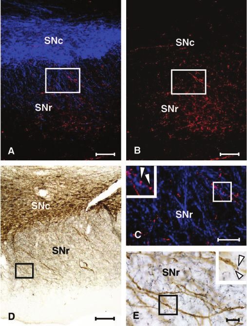
Figure 1. Photomicrographs of the SNc and SNr of rats showing immunoreactivity for hista- ble because of the different methodological conditions required
mine together with TH using fluorescence microscopy (A–C) and the serotonin transporter to reveal them.
together with TH, using bright field microscopy (D, E). A, Low-power image of histamine-IR
(with Alexa 594; red) and TH-IR (with Alexa 350; blue). B, Histamine-IR alone (in the same field Electrochemical characterization of 5-HT release in SNr
shown in A). C, An enlarged image of the boxed area (in A, B) showing histamine-IR (red) and Electrical stimulation (20 pulses, 100 Hz) in SNr evoked the re-
TH-IR (blue) in the SNr; the inset presents the boxed area (in C) at higher power (arrowheads
lease, followed by the rapid removal, of an electroactive substance
indicate varicosities with histamine-IR in the vicinity of dendritic processes containing TH-IR). D,
Low-power image of serotonin transporter-IR (black profiles) and TH-IR (brown profiles). E, An
that could be identified as 5-HT. The voltammogram of the re-
enlarged image of the boxed area (in D). Serotonin transporter-IR (black profiles) and TH-IR leased substance was readily identified as 5-HT because of an
(brown profiles) in the SNr; the boxed area (in E) is shown at higher power as an inset (arrow- oxidation peak potential at approximately ⫹600 mV (vs Ag/
heads indicate varicosities with serotonin transporter-IR in the vicinity of dendritic processes AgCl) and a dual reduction profile with peak potentials at ⬃0 and
containing TH-IR). Scale bars: A, B, D, 100 m; C, E, 50 m. ⫺680 mV (Fig. 2 A, B), identical to those obtained in calibration
with application of 5-HT (Fig. 2C). The substance can readily be
distinguished from DA (Fig. 2 D) by the characteristic double-
reduction current profile for 5-HT as noted previously in SNr
curves were generated with GraphPad Prism software (GraphPad Soft-
ware, San Diego, CA) using nonlinear regression to a sigmoidal dose– (Davidson and Stamford, 1996; Cragg et al., 1997b; Iravani and
response (variable slope) from which IC50 values were obtained and, Kruk, 1997; Bunin et al., 1998). Furthermore, it has been identi-
where appropriate, compared statistically. Statistical significance was fied previously that the primary substance detected in rat SNr
considered as p ⬍ 0.05. using this method is 5-HT and not DA (Cragg et al., 1997b;
Iravani and Kruk, 1997; Bunin et al., 1998).
Results To further confirm the identity of the electrochemical signal
Identification of HA and 5-HT immunoreactivity in SNr as 5-HT and not DA, uptake inhibitors for the SERT and the DA
In sections from rats perfused with EDCDI, abundant processes transporter (DAT) were applied. Bath application of the SERT
immunoreactive for HA or TH were observed in the SNr (Fig. inhibitor citalopram (75 nM) prolonged the extracellular lifetime
1 A–C). Varicosities immunoreactive for HA were found in the of 5-HT and significantly enhanced [5-HT]o (Fig. 3A) (M-WU;
vicinity of TH-immunoreactive processes (Fig. 1C). After PFA p ⬍ 0.001; n ⫽ 28) as shown previously with SERT inhibitors in
fixation, processes immunoreactive for SERT were found the SNr (Iravani and Kruk, 1997; Bunin et al., 1998). Conversely,
throughout the SNr (Fig. 1 D, E). As in the case of HA-IR, SERT- application of the DAT inhibitor GBR 12909 (1 M) had no effect
immunoreactive varicosities were found in the vicinity of TH- on the lifetime or peak concentration of the detected substance,
immunoreactive processes (Fig. 1 E). By reference to TH- 5-HT (Fig. 3B) (M-WU; p ⬎ 0.05; n ⫽ 14).
immunopositive processes, we conclude that within the SNr,
there is similarity in the distribution of putative terminal fields Ionic and depolarization sensitivity
containing HA or 5-HT. Analysis of the direct ultrastructural Evoked release of 5-HT was apparently abolished by inhibition of
interactions between HA-IR and 5-HT-IR is not currently feasi- voltage-gated Na ⫹ channels (Nav ⫹) by TTX (1 M) (Fig. 3C)Threlfell et al. • Histamine H3 Receptors Regulate 5-HT in SNr J. Neurosci., October 6, 2004 • 24(40):8704 – 8710 • 8707
Figure 4. Dose-dependent inhibition of 5-HT release in SNr by H3R agonists R-mHA and
immepip. A, B, Top, Mean profiles of [5-HT]o versus time evoked by 20 pulses at 100 Hz (solid
bar) expressed as a percentage of mean peak control. Bottom, Dose–response curves for mean
peak [5-HT]o expressed as a percentage of mean peak control for R-mHA ( A) or immepip ( B). A,
R-mHA dose dependently inhibited [5-HT]o (n ⫽ 14 –15, Kruskal-Wallis; Dunn’s t test vs con-
trol, **p ⬍ 0.01, ***p ⬍ 0.001) with an IC50 value of 23 nM (95% confidence interval range,
13– 43 nM; R 2 ⫽ 0.57; Hill slope, ⫺1.1). B, Immepip dose dependently inhibited [5-HT]o (n ⫽
14 –19, Kruskal-Wallis; Dunn’s t test vs control, **p ⬍ 0.01, ***p ⬍ 0.001) with an IC50 value
of 43 nM (95% confidence interval range, 20 –95 nM; R 2 ⫽ 0.65; Hill slope, ⫺0.73; not signif-
Figure 3. Release and uptake characteristics of evoked 5-HT release in SNr. A–D, Mean icantly different from 1.0).
profiles of [5-HT]o versus time evoked by 20 pulses (p) at 100 Hz (solid bar) expressed as a
percentage of mean peak control either in control or the SERT inhibitor citalopram (A; 75 nM),
the DAT inhibitor GBR 12909 (B; 1 M), the NaV ⫹ channel inhibitor TTX (C; 1 M), or Ca 2⫹-free confidence interval range, 13– 43 nM; Hill slope, ⫺1.1) and 43 nM,
aCSF ( D). A, Citalopram enhanced the extracellular lifetime of 5-HT and significantly enhanced respectively (immepip; 95% confidence interval range, 20 –95
mean peak [5-HT]o to 290 ⫾ 18% of control (M-WU; ***p ⬍ 0.001; n ⫽ 28). B, GBR 12909 did nM; Hill slope, ⫺0.73, not significantly different from 1.0). These
not modify lifetime or mean peak [5-HT]o (M-WU; p ⬎ 0.05; n ⫽ 14). C, TTX abolished evoked IC50 values were not significantly different from one another
5-HT release (M-WU; ***p ⬍ 0.001; n ⫽ 18). D, 5-HT release was reversibly abolished in ( p ⬎ 0.05).
Ca 2⫹-free aCSF (M-WU; ***p ⬍ 0.001; n ⫽ 12). E, Mean peak [5-HT]o varied significantly with To confirm that the actions of these H3R agonists were attrib-
frequency (20 pulses; one-way ANOVA, p ⬍ 0.001) in a biphasic manner (Dunnett’s t tests;
utable to selective agonism at H3Rs, their effects were assessed in
asterisks denote p values vs 10 Hz: *p ⬍ 0.05, **p ⬍ 0.01, ***p ⬍ 0.001). F, Mean peak
[5-HT]o varied significantly with pulse number (100 Hz; one-way ANOVA, p ⬍ 0.001) in a linear the presence of a selective H3R antagonist, thioperamide.
manner for pulse numbers ⬍30 (linear regression with y-intercept ⫽ 0, dotted line; R 2 ⫽ Whereas R-mHA (100 nM) reduced mean peak [5-HT]o from
0.97). 100 ⫾ 2 to 52 ⫾ 4% without thioperamide (Fig. 5A) (M-WU; p ⬍
0.001), R-mHA did not modify mean peak [5-HT]o in the pres-
ence of thioperamide (Fig. 5A) (1 M; 108 ⫾ 13% in thioperam-
(M-WU; p ⬍ 0.001; n ⫽ 18 –22) and reversibly abolished with ide only vs 94 ⫾ 6% in thioperamide plus R-mHA; n ⫽ 19 –25;
application of Ca 2⫹-free–EGTA-containing aCSF (Fig. 3D) (M- M-WU; p ⬎ 0.05). Similarly, immepip (1 M) reduced mean
WU; p ⬍ 0.001; n ⫽ 12). Mean peak-evoked [5-HT]o varied peak [5-HT]o from 100 ⫾ 3 to 52 ⫾ 4% without thioperamide
significantly with frequency across the range of 10 to 200 Hz (20 (Fig. 5B) (M-WU; p ⬍ 0.001) but did not modify [5-HT]o in the
pulses; one-way ANOVA; p ⬍ 0.001) in a biphasic manner that presence of thioperamide (Fig. 5B) (10 M; 108 ⫾ 5% in thiop-
indicated frequency dependence at ⬍100 Hz but a diminished eramide only vs 104 ⫾ 6% in thioperamide plus immepip; n ⫽
ability to follow frequencies greater than this (Fig. 3E) ( post hoc 16 –22; M-WU; p ⬎ 0.05). In contrast, the effect of R-mHA (1
Dunnett’s t tests vs 10 Hz: 20 –100 Hz, p ⬍ 0.001– 0.05; 200 Hz, M) was not prevented by the presence of a 5-HT1B receptor
p ⬎ 0.05). Mean peak-evoked [5-HT]o varied significantly with antagonist, isamoltane (Fig. 5C) (1 M; 111 ⫾ 11.0% in
the number of pulses in a train (100 Hz; one-way ANOVA; p ⬍ isamoltane only vs 54 ⫾ 7% in isamoltane plus R-mHA; M-WU;
0.001) as observed previously (Iravani and Kruk, 1997; Bunin et p ⬍ 0.001; n ⫽ 12–21) at a concentration of isamoltane previ-
al., 1998), showing a linear dependence at pulse numbers ⬍30 ously observed to block 5-HT1B agonist effects (Davidson and
(Fig. 3F ) (linear regression over 0 –30 pulses; y-intercept ⫽ 0; Stamford, 1996).
R 2 ⫽ 0.97). Trains of 20 pulses at 100 Hz were selected for all
subsequent pharmacological investigations. Direct or indirect H3R effects?
Studies were undertaken to explore whether H3R-mediated inhi-
H3R effects on 5-HT release in the SNr bition of 5-HT release might be attributable to H3Rs present on
Mean peak evoked [5-HT]o (20 pulses at 100 Hz) was signifi- GABAergic or glutamatergic inputs to 5-HT terminals rather
cantly attenuated by bath application of selective H3R agonists than on the 5-HT terminals themselves. The effects of the H3R
( R)-(-)-␣-methylhistamine (R-mHA) or immepip (Fig. 4 A, B) agonist R-mHA were investigated in the presence of mixtures of
(Kruskal-Wallis, p ⬍ 0.001; n ⫽ 14 –15). Each drug demonstrated either GABA or glutamate receptor antagonists (ionotropic and
a concentration-dependent inhibition of 5-HT release of up to metabotropic). Despite the application of the GABA receptor
49 ⫾ 5% of control by R-mHA (1 M; n ⫽ 14) and to 39 ⫾ 3% by antagonists picrotoxin (PTX; GABAA receptor antagonist, 100
immepip (5 M; n ⫽ 15) with IC50 values of 23 nM (R-mHA; 95% M) and saclofen (GABAB receptor antagonist, 50 M), R-mHA8708 • J. Neurosci., October 6, 2004 • 24(40):8704 – 8710 Threlfell et al. • Histamine H3 Receptors Regulate 5-HT in SNr
Figure 6. H3R effects on 5-HT release occur during block of GABA or glutamate synapses. A,
B, Mean profiles of [5-HT]o versus time evoked by 20 pulses, 100 Hz (solid bar), expressed as a
percentage of mean peak control in the absence of drugs (left) or in the presence of antagonists
(right) for GABA receptors ( A) or glutamate receptors ( B). A, Evoked [5-HT]o was not signifi-
cantly modified by inclusion of GABA receptor antagonists (PTX, 100 M; saclofen, 50 M; n ⫽
Figure 5. Inhibition of 5-HT release by H3R agonists is caused by selective action at H3Rs. 12). In the presence of GABA receptor block, R-mHA (1 M) significantly diminished mean peak
A–C, Mean profiles of [5-HT]o versus time evoked by 20 pulses, 100 Hz (solid bar), expressed as [5-HT]o (M-WU; ***p ⬍ 0.001; n ⫽ 14). B, Evoked [5-HT]o was not significantly modified by
a percentage of mean peak control in the absence (left) versus the presence (right) of the H3R inclusion of glutamate receptor antagonists [( S)-MCPG, 200 M; D-AP-5, 50 M; GYKI 52466, 10
antagonist thioperamide (A, B) or the 5-HT1B receptor antagonist isomoltane (C; 1 M). In A, M; n ⫽ 12]. In the presence of glutamate receptor block, R-mHA (1 M) significantly dimin-
inhibition of peak [5-HT]o by R-mHA (100 nM, M-WU; ***p ⬍ 0.001; n ⫽ 15) was eliminated in ished mean peak [5-HT]o (M-WU; ***p ⬍ 0.001; n ⫽ 16).
the presence of thioperamide (1 M; n ⫽ 19 –25). B, Inhibition of peak [5-HT]o by immepip (1
M, M-WU; ***p ⬍ 0.001; n ⫽ 15) was prevented when thioperamide was present (10 M; immunocytochemistry showed that serotonergic processes are
n ⫽ 16 –22). C, Inhibition of peak [5-HT]o by R-mHA (1 M, M-WU; ***p ⬍ 0.001; n ⫽ 12) abundant throughout the SNr. Second, the voltammograms re-
prevailed in the presence of 5-HT1B antagonist isamoltane (1 M, M-WU; ***p ⬍ 0.001; n ⫽ sulting from the evoked release indicated that the released sub-
14 –17). stance was identical in its electrochemical profile to the in-
doleamine 5-HT and not to the catecholamine DA. Third,
(1 M) significantly inhibited mean peak [5-HT]o from 106 ⫾ 6 inhibition of the SERT but not the DAT prolonged the extracel-
to 27 ⫾ 7% (Fig. 6 A) (M-WU; p ⬍ 0.001; n ⫽ 14 –21). Inhibition lular lifetime and enhanced the peak amplitude of the 5-HT-like
of 5-HT release via the H3R also prevailed when R-mHA was signal. These observations indicate that the electrochemical sig-
applied in the presence of the glutamate receptor antagonists nal was attributable to 5-HT, with negligible contribution from
D-AP-5 (NMDA receptor antagonist, 50 M), GYKI 52466 (non- DA, and confirm that the SERT participates in [5-HT]o regula-
competitive AMPA/kainate receptor antagonist, 10 M), and tion in the SNr as reported previously (Iravani and Kruk, 1997;
( S)-MCPG (Fig. 6 B) (nonselective group I/II metabotropic glu- Bunin et al., 1998).
tamate receptor antagonist, 200 M; 105 ⫾ 6% in glutamate an-
tagonists vs 30 ⫾ 4% in glutamate antagonists plus R-mHA; H3R control of 5-HT release in the SNr
M-WU; p ⬍ 0.001; n ⫽ 16 –26). Statistical comparison of R-mHA In the present study, the evoked release of 5-HT in SNr was
effects in the absence versus the presence or either GABA or potently inhibited by the H3R agonists R-mHA or immepip to
glutamate receptor antagonists (using recording sites matched levels that were ⬍50% of those seen in controls. This attenuation of
for concentration of 5-HT released in drug-free controls) does 5-HT release was dose dependent, with IC50 values for R-mHA and
not reveal a significant effect of synaptic block on R-mHA effects immepip that were consistent with previously reported values for
(M-WU; n ⫽ 14 –16). H3R efficacy in guinea pig ileum (Hew et al., 1990) and inhibition of
striatal glutamate release (Molina-Hernandez et al., 2001), respec-
Discussion tively. The inhibition of 5-HT release in SNr by activation of H3Rs
5-HT detection in SNr was confirmed further by experiments with the selective H3R antag-
The evoked neurotransmitter release detected in the SNr in this onist thioperamide. Previous application of thioperamide prevented
study was NaV ⫹ and Ca 2⫹ dependent and sensitive to depolar- the inhibition of 5-HT release by R-mHA or immepip. In contrast,
ization frequency and pulse number, as previously reported for the action of these H3R agonists was not modified by antagonism of
5-HT exocytosis in the SNr (O’Connor and Kruk, 1991; Iravani 5-HT1B receptors, a 5-HT receptor type that has been shown to
and Kruk, 1997; Bunin et al., 1998). It is well established that participate in presynaptic autoreceptor control of 5-HT release else-
5-HT is the predominant monoamine detected by FCV at CFMs where in the brain (Barnes and Sharp, 1999). This observation indi-
in the rat SNr (Cragg et al., 1997b; Iravani and Kruk, 1997; Bunin cates that the actions of the H3 agonists were not attributable to
et al., 1998); this differs from the guinea pig SNr in which 5-HT is effects on 5-HT autoreceptors.
detected far less frequently and DA predominates (Cragg et al., The potent inhibition of 5-HT signaling by H3Rs identifies a
1997b). Several lines of evidence indicate that the signal moni- powerful role for these abundant receptors in the SNr (Pillot et
tored in this study was indeed caused by the release of 5-HT. First, al., 2002). Although H3R-dependent regulation of 5-HT signal-Threlfell et al. • Histamine H3 Receptors Regulate 5-HT in SNr J. Neurosci., October 6, 2004 • 24(40):8704 – 8710 • 8709
ing has been identified previously in rat cortex (Schlicker et al., synaptic regulation of 5-HT release in the SNr. The location of the
1988; Fink et al., 1990), the current data are the first to identify a H3Rs in question does not appear to involve either GABA or gluta-
comparable mechanism within the basal ganglia. mate synapses; nevertheless, their possible presence on the terminals
of other (e.g., cholinergic) projections to the SN cannot be dis-
H3R on GABA and glutamate synapses counted. The notion that the H3Rs mediating inhibition of 5-HT
To date, there is no anatomical information available regarding release are located directly on 5-HT axons is consistent with the
the specific identity of the neurons that express H3Rs in the SNr expression of the mRNA for H3R in the raphe nuclei (Pillot et
and the ultrastructural location of these receptors. Thus, the syn- al., 2002) and the similar anatomical distribution of HA-
aptic circuitry by which H3Rs detected in the SNr by radioligand immunoreactive and 5-HT-immunoreactive varicosities in the
binding (Pillot et al., 2002) can regulate 5-HT release is uniden- SNr. Given the apparent lack of autoreceptor control of 5-HT release
tified. Because in situ hybridization for mRNA for H3Rs reveals at this site (Nissbrandt et al., 1992; Iravani and Kruk, 1997), H3R
only low expression within the SN (Pillot et al., 2002), the abun- activation may be a key mechanism for 5-HT regulation.
dant H3Rs in SN are unlikely to be expressed or localized on DA The significance of H3R-mediated control of 5-HT release
neurons in the SNc or on local or projection neurons within the depends on the function(s) of 5-HT in the SNr. Serotonergic
SNr. However, moderate H3R transcript expression has been inputs to the SNr (Corvaja et al., 1993; Moukhles et al., 1997)
identified in the raphe nuclei (Pillot et al., 2002); therefore, it is have the potential to modify motor output signals (Jacobs and
plausible that the H3Rs are synthesized by 5-HT neurons and Fornal, 1993; Rick et al., 1995). Moreover, 5-HT is a target for
located on 5-HT terminals. therapeutic intervention regarding unwanted side effects associ-
Because a similar level of H3R mRNA expression has also been ated with long-term DA replacement in PD, such as the wearing-
identified in the dorsal striatum, subthalamic nucleus, and pe- off phenomenon and L-3,4-dihydroxyphenylalanine (L-DOPA)-
dunculopontine nucleus (Pillot et al., 2002), regions that send induced dyskinesia (Nicholson and Brotchie, 2002). 5-HT2C
major non-5-HT projections (e.g., GABAergic or glutamatergic) receptors on SNr projection neurons are excitatory (Rick et al.,
to the SNr, we evaluated whether such inputs to the SNr might be 1995); their binding is increased in the SNr of PD patients with
involved in H3R-mediated inhibition of 5-HT release. Previous L-DOPA-induced dyskinesia, suggesting a possible compensa-
evidence has indicated that H3Rs in the SNr inhibit GABA release tory upregulation of receptors in response to decreased endoge-
(Garcia et al., 1997; Garcia-Ramirez et al., 2004). The possibility nous ligand (Fox and Brotchie, 2000). In this context, it should be
that H3Rs present on GABAergic or glutamatergic terminals in noted that the selective serotonin reuptake inhibitor, fluoxetine,
the SNr contribute to H3R-mediated regulation of 5-HT release reduces dyskinesias in PD patients (Durif et al., 1995).
was investigated using receptor antagonists for GABA or gluta- The capacity of HA to regulate 5-HT transmission in the SNr
mate. H3R-mediated inhibition of 5-HT release was preserved in via H3Rs may provide an attractive nondopaminergic target for
the presence of GABA– glutamate synaptic block; thus, a role for improving therapies for PD. Identifying novel approaches to ma-
H3Rs on GABA or glutamate terminals in this context seems nipulate 5-HT neurotransmission may help to enhance the effi-
unlikely. Furthermore, because in the absence of H3R agonists cacy of current treatments not only for PD but also for mood
neither GABA nor glutamate antagonists significantly modified disorders such as depression and anxiety, in which serotonergic
[5-HT]o, the control of 5-HT release in this paradigm does not manipulation is pivotal.
appear to be under direct control by glutamate or GABA syn-
apses. Whether the inhibitory action of H3R agonists on GABA References
release in the SNr (Garcia et al., 1997; Garcia-Ramirez et al., 2004) Adams JC (1992) Biotin amplification of biotin and horseradish peroxidase
is mediated via effects on 5-HT release remains to be tested. signals in histochemical stains. J Histochem Cytochem 40:1457–1463.
The present results raise the possibility that the H3R-mediated Alexander GE, Crutcher MD (1990) Functional architecture of basal
ganglia circuits: neural substrates of parallel processing. Trends Neurosci
inhibition of 5-HT release is attributable to a direct action on 13:266 –271.
5-HT terminals via H3Rs. This regulation of 5-HT release is note- Anichtchik OV, Rinne JO, Kalimo H, Panula P (2000a) An altered histamin-
worthy in light of reports indicating little, if any, regulation of ergic innervation of the substantia nigra in Parkinson’s disease. Exp Neu-
5-HT release in the SNr by 5-HT1B autoreceptors (Nissbrandt et rol 163:20 –30.
al., 1992; Iravani and Kruk, 1997). Despite a high density of Anichtchik OV, Huotari M, Peitsaro N, Haycock JW, Mannisto PT, Panula P
5-HT1B receptors in the SNr, they are observed predominantly on (2000b) Modulation of histamine H3 receptors in the brain of
6-hydroxydopamine-lesioned rats. Eur J Neurosci 12:3823–3832.
striatonigral GABAergic terminals from the striatum rather than
Anichtchik OV, Peitsaro N, Rinne JO, Kalimo H, Panula P (2001) Distribu-
on serotonergic terminals (Boschert et al., 1994; Sari et al., 1999). tion and modulation of histamine H(3) receptors in basal ganglia and
It appears that 5-HT release in the SNr is not regulated by 5-HT frontal cortex of healthy controls and patients with Parkinson’s disease.
receptors; this is not the case in the raphe nuclei where a plurality Neurobiol Dis 8:707–716.
of 5-HT receptors perform autoreceptor-like functions (Stam- Arias-Montano JA, Floran B, Garcia M, Aceves J, Young JM (2001) Histamine
ford et al., 2000; Hopwood and Stamford, 2001). H3R-mediated H(3) receptor-mediated inhibition of depolarization-induced, dopamine
control of 5-HT release in SNr may provide a substitute mecha- D(1) receptor-dependent release of [(3)H]-gamma-aminobutryic acid from
rat striatal slices. Br J Pharmacol 133:165–171.
nism for presynaptic inhibition of 5-HT release. Arrang JM, Garbarg M, Schwartz JC (1983) Auto-inhibition of brain hista-
mine release mediated by a novel class (H3) of histamine receptor. Nature
Conclusions 302:832– 837.
The present findings indicate a similar distribution of histamin- Arrang JM, Drutel G, Schwartz JC (1995) Characterization of histamine H3
ergic and serotonergic processes in the SNr. Double-label immuno- receptors regulating acetylcholine release in rat entorhinal cortex. Br J
histochemical studies at the electronmicroscopic level will be Pharmacol 114:1518 –1522.
Barnes NM, Sharp T (1999) A review of central 5-HT receptors and their
required to establish whether there are histaminergic synapses on function. Neuropharmacology 38:1083–1152.
serotonergic terminals at this site. The results of the FCV studies are Boschert U, Amara DA, Segu L, Hen R (1994) The mouse
certainly consistent with an interaction between histaminergic and 5-hydroxytryptamine1B receptor is localized predominantly on axon
serotonergic systems. Thus, the data indicate that H3Rs mediate pre- terminals. Neuroscience 58:167–182.8710 • J. Neurosci., October 6, 2004 • 24(40):8704 – 8710 Threlfell et al. • Histamine H3 Receptors Regulate 5-HT in SNr
Brown RE, Haas HL (1999) On the mechanism of histaminergic inhibition and Parkinson’s disease– opportunities for novel therapeutics to reduce
of glutamate release in the rat dentate gyrus. J Physiol (Lond) the problems of levodopa therapy. Eur J Neurol 9 [Suppl 3]:1– 6.
515:777–786. Nissbrandt H, Waters N, Hjorth S (1992) The influence of serotoninergic
Brown RE, Reymann KG (1996) Histamine H3 receptor-mediated depres- drugs on dopaminergic neurotransmission in rat substantia nigra, stria-
sion of synaptic transmission in the dentate gyrus of the rat in vitro. tum and limbic forebrain in vivo. Naunyn Schmiedebergs Arch Pharma-
J Physiol (Lond) 496:175–184. col 346:12–19.
Bunin MA, Prioleau C, Mailman RB, Wightman RM (1998) Release and O’Connor JJ, Kruk ZL (1991) Frequency dependence of 5-HT autoreceptor
uptake rates of 5-hydroxytryptamine in the dorsal raphe and substantia function in rat dorsal raphe and suprachiasmatic nuclei studied using fast
nigra reticulata of the rat brain. J Neurochem 70:1077–1087. cyclic voltammetry. Brain Res 568:123–130.
Corvaja N, Doucet G, Bolam JP (1993) Ultrastructure and synaptic targets Panula P, Yang HY, Costa E (1984) Histamine-containing neurons in the
of the raphe-nigral projection in the rat. Neuroscience 55:417– 427. rat hypothalamus. Proc Natl Acad Sci USA 81:2572–2576.
Cragg S, Rice ME, Greenfield SA (1997a) Heterogeneity of electrically Panula P, Pirvola U, Auvinen S, Airaksinen MS (1989) Histamine-
evoked dopamine release and reuptake in substantia nigra, ventral teg- immunoreactive nerve fibers in the rat brain. Neuroscience 28:585– 610.
mental area, and striatum. J Neurophysiol 77:863– 873. Paxinos G, Watson C (1982) The rat brain in stereotaxic coordinates. Syd-
Cragg SJ, Hawkey CR, Greenfield SA (1997b) Comparison of serotonin and ney: Academic.
dopamine release in substantia nigra and ventral tegmental area: region Pillot C, Heron A, Cochois V, Tardivel-Lacombe J, Ligneau X, Schwartz JC,
and species differences. J Neurochem 69:2378 –2386. Arrang JM (2002) A detailed mapping of the histamine H(3) receptor
Cragg SJ, Hille CJ, Greenfield SA (2002) Functional domains in dorsal stri- and its gene transcripts in rat brain. Neuroscience 114:173–193.
atum of the nonhuman primate are defined by the dynamic behavior of Prast H, Tran MH, Fischer H, Kraus M, Lamberti C, Grass K, Philippu A
dopamine. J Neurosci 22:5705–5712. (1999) Histaminergic neurons modulate acetylcholine release in the ven-
Davidson C, Stamford JA (1996) Serotonin efflux in the rat ventral lateral tral striatum: role of H3 histamine receptors. Naunyn Schmiedebergs
geniculate nucleus assessed by fast cyclic voltammetry is modulated by Arch Pharmacol 360:558 –564.
5-HT1B and 5-HT1D autoreceptors. Neuropharmacology 35:1627–1634. Prell GD, Hough LB, Khandelwal J, Green JP (1996) Lack of a precursor-
Durif F, Vidailhet M, Bonnet AM, Blin J, Agid Y (1995) Levodopa-induced product relationship between histamine and its metabolites in brain after
dyskinesias are improved by fluoxetine. Neurology 45:1855–1858. histidine loading. J Neurochem 67:1938 –1944.
Fink K, Schlicker E, Neise A, Gothert M (1990) Involvement of presynaptic Rice ME, Nicholson C (1989) Measurement of nanomolar dopamine diffu-
H3 receptors in the inhibitory effect of histamine on serotonin release in the sion using low-noise perfluorinated ionomer coated carbon fiber micro-
rat brain cortex. Naunyn Schmiedebergs Arch Pharmacol 342:513–519. electrodes and high-speed cyclic voltammetry. Anal Chem 77:1805–1810.
Fox SH, Brotchie JM (2000) 5-HT2C receptor binding is increased in the Rick CE, Stanford IM, Lacey MG (1995) Excitation of rat substantia nigra
substantia nigra pars reticulata in Parkinson’s disease. Mov Disord
pars reticulata neurons by 5-hydroxytryptamine in vitro: evidence for a
15:1064 –1069.
direct action mediated by 5-hydroxytryptamine2C receptors. Neuro-
Garcia M, Floran B, Arias-Montano JA, Young JM, Aceves J (1997) Hista-
science 69:903–913.
mine H3 receptor activation selectively inhibits dopamine D1 receptor-
Ryu JH, Yanai K, Watanabe T (1994) Marked increase in histamine H3
dependent [3H]GABA release from depolarization-stimulated slices of
receptors in the striatum and substantia nigra after 6-hydroxydopamine-
rat substantia nigra pars reticulata. Neuroscience 80:241–249.
induced denervation of dopaminergic neurons: an autoradiographic
Garcia-Ramirez M, Aceves J, Arias-Montano J-A (2004) Intranigral injec-
study. Neurosci Lett 178:19 –22.
tion of the H3 agonist immepip and systemic apomorphine elicit ipsilat-
Sari Y, Miquel MC, Brisorgueil MJ, Ruiz G, Doucet E, Hamon M, Verge D
eral turning behaviour in naive rats, but reduce contralateral turning in
(1999) Cellular and subcellular localization of 5-hydroxytryptamine1B
hemiparkinsonian rats. Behav Brain Res 154:409 – 415.
receptors in the rat central nervous system: immunocytochemical, auto-
Giorgetti M, Bacciottini L, Bianchi L, Giovannini MG, Cecchi M, Blandina P
radiographic and lesion studies. Neuroscience 88:899 –915.
(1997) GABAergic mechanism in histamine H3 receptor inhibition of
K(⫹)-evoked release of acetylcholine from rat cortex in vivo. Inflamm Res Schlicker E, Betz R, Gothert M (1988) Histamine H3 receptor-mediated
46 [Suppl 1]:S33–S34. inhibition of serotonin release in the rat brain cortex. Naunyn Schmiede-
Hew RW, Hodgkinson CR, Hill SJ (1990) Characterization of histamine bergs Arch Pharmacol 337:588 –590.
H3-receptors in guinea-pig ileum with H3-selective ligands. Br J Pharma- Schlicker E, Fink K, Hinterthaner M, Gothert M (1989) Inhibition of nor-
col 101:621– 624. adrenaline release in the rat brain cortex via presynaptic H3 receptors.
Hopwood SE, Stamford JA (2001) Multiple 5-HT(1) autoreceptor subtypes Naunyn Schmiedebergs Arch Pharmacol 340:633– 638.
govern serotonin release in dorsal and median raphe nuclei. Neurophar- Schlicker E, Behling A, Lummen G, Gothert M (1992) Histamine H3A
macology 40:508 –519. receptor-mediated inhibition of noradrenaline release in the mouse brain
Iravani MM, Kruk ZL (1997) Real-time measurement of stimulated cortex. Naunyn Schmiedebergs Arch Pharmacol 345:489 – 493.
5-hydroxytryptamine release in rat substantia nigra pars reticulata brain Schlicker E, Fink K, Detzner M, Gothert M (1993) Histamine inhibits do-
slices. Synapse 25:93–102. pamine release in the mouse striatum via presynaptic H3 receptors. J Neu-
Jacobs BL, Fornal CA (1993) 5-HT and motor control: a hypothesis. Trends ral Transm Gen Sect 93:1–10.
Neurosci 16:346 –352. Schlicker E, Werthwein S, Zentner J (1999) Histamine H3 receptor-
Kalló I, Butler JA, Barkovics-Kallo M, Goubillon ML, Coen CW (2001) Oestro- mediated inhibition of noradrenaline release in the human brain. Fun-
gen receptor beta-immunoreactivity in gonadotropin releasing hormone- dam Clin Pharmacol 13:120 –122.
expressing neurones: regulation by oestrogen. J Neuroendocrinol Schwartz JC, Lampart C, Rose C (1972) Histamine formation in rat brain in
13:741–748. vivo: effects of histidine loads. J Neurochem 19:801– 810.
Liposits Z, Sherman D, Phelix C, Paull WK (1986) A combined light and Stamford JA, Davidson C, McLaughlin DP, Hopwood SE (2000) Control of
electron microscopic immunocytochemical method for the simultaneous dorsal raphe 5-HT function by multiple 5-HT(1) autoreceptors: parallel
localization of multiple tissue antigens. Tyrosine hydroxylase immunore- purposes or pointless plurality? Trends Neurosci 23:459 – 465.
active innervation of corticotropin releasing factor synthesizing neurons Venton BJ, Michael DJ, Wightman RM (2003) Correlation of local changes
in the paraventricular nucleus of the rat. Histochemistry 85:95–106. in extracellular oxygen and pH that accompany dopaminergic terminal
Molina-Hernandez A, Nunez A, Sierra JJ, Arias-Montano JA (2001) Hista- activity in the rat caudate-putamen. J Neurochem 84:373–381.
mine H3 receptor activation inhibits glutamate release from rat striatal Vizuete ML, Traiffort E, Bouthenet ML, Ruat M, Souil E, Tardivel-Lacombe J,
synaptosomes. Neuropharmacology 41:928 –934. Schwartz JC (1997) Detailed mapping of the histamine H2 receptor and
Moukhles H, Bosler O, Bolam JP, Vallee A, Umbriaco D, Geffard M, Doucet its gene transcripts in guinea-pig brain. Neuroscience 80:321–343.
G (1997) Quantitative and morphometric data indicate precise cellular Yamamoto Y, Mochizuki T, Okakura-Mochizuki K, Uno A, Yamatodani A
interactions between serotonin terminals and postsynaptic targets in rat (1997) Thioperamide, a histamine H3 receptor antagonist, increases
substantia nigra. Neuroscience 76:1159 –1171. GABA release from the rat hypothalamus. Methods Find Exp Clin Phar-
Nicholson SL, Brotchie JM (2002) 5-hydroxytryptamine (5-HT, serotonin) macol 19:289 –298.You can also read