Interactions of Adenoviruses with platelets and coagulation and the vaccine-associated autoimmune thrombocytopenia thrombosis syndrome
←
→
Page content transcription
If your browser does not render page correctly, please read the page content below
Interactions of Adenoviruses with platelets and
coagulation and the vaccine-associated autoimmune
thrombocytopenia thrombosis syndrome

by Paolo Gresele, Stefania Momi, Rossella Marcucci, Francesco Ramundo, Valerio De Stefano,
and Armando Tripodi

Received: May 24, 2021.
Accepted: August 4, 2021.

Citation: Paolo Gresele, Stefania Momi, Rossella Marcucci, Francesco Ramundo, Valerio De Stefano
and Armando Tripodi. Interactions of Adenoviruses with platelets and coagulation and
the vaccine-associated autoimmune thrombocytopenia thrombosis syndrome.
Haematologica. 2021 Aug 19. doi: 10.3324/haematol.2021.279289. [Epub ahead of print]

Publisher's Disclaimer.
E-publishing ahead of print is increasingly important for the rapid dissemination of science.
Haematologica is, therefore, E-publishing PDF files of an early version of manuscripts that
have completed a regular peer review and have been accepted for publication. E-publishing
of this PDF file has been approved by the authors. After having E-published Ahead of Print,
manuscripts will then undergo technical and English editing, typesetting, proof correction and
be presented for the authors' final approval; the final version of the manuscript will then
appear in a regular issue of the journal. All legal disclaimers that apply to the
journal also pertain to this production process.
Interactions   of   adenoviruses   with   platelets   and   coagulation   and   the   vaccine-associated

autoimmune thrombocytopenia thrombosis syndrome

Paolo Gresele1, Stefania Momi1, Rossella Marcucci2, Francesco Ramundo3, Valerio De Stefano3,
Armando Tripodi4

1
 Department of Medicine and Surgery, Section of Internal and Cardiovascular Medicine, University
of Perugia, Perugia, Italy; 2Department of Experimental and Clinical Medicine, University of
Florence; Atherothrombotic Center, AOU Careggi, Florence, Italy; 3Section of Hematology,
Department of Radiological and Hematological Sciences, Catholic University, Fondazione
Policlinico A. Gemelli - IRCCS - Rome, Italy; 4Fondazione IRCCS Ca' Granda, Ospedale Maggiore
Policlinico, Angelo Bianchi Bonomi Hemophilia and Thromboses Center, Milan, Italy.

Word Count: (Introduction, Methods, Results, Discussion) 4913

Corresponding author

Paolo Gresele, MD, PhD
Department of Medicine and Surgery
Section of Internal and Cardiovascular Medicine
University of Perugia
Perugia, Italy
Tel. +39 0755783989
Fax. +39 075-5858439
Email paolo.gresele@unipg.it

                                                                                                      1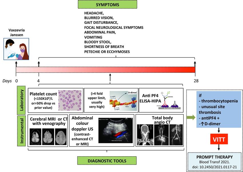
ABSTRACT

The Covid-19 pandemic has heavily impacted global health and economy and vaccination remains
the primary way of controlling the infection. During the ongoing vaccination campaign some
unexpected thrombotic events have emerged in subjects who recently received the AstraZeneca
Vaxzevria vaccine or the Johnson&Johnson (Janssen) vaccine, two adenovirus vector-based
vaccines. Epidemiological studies confirm that the observed/expected rate of these unusual
thromboses is abnormally increased, especially in women in the fertile age. The characteristics of
this complication, with venous thromboses at unusual sites, most frequently cerebral vein sinus
but also splanchnic, often multiple associated thromboses, thrombocytopenia, and sometimes
disseminated intravascular coagulation, are unique and the time course and tumultuous evolution
are suggestive of an acute immunological reaction, and indeed platelet-activating anti-PF4
antibodies have been detected in a large fraction of the affected patients.

Several data suggest that adenoviruses may interact with platelets, the endothelium and the blood
coagulation system. Here we review the interactions between adenoviral vectors and the
haemostatic    system    of   possible   relevance   for   the   vaccine      associated   thrombotic
thrombocytopenia syndrome, we analyse systematically the clinical data on the reported
thrombotic complications of adenovirus-based therapeutics and discuss all the current hypotheses
on the mechanisms triggering this novel syndrome.

Although considering current evidence the benefit of vaccination clearly outweighs the potential
risks, it is of paramount importance to fully unravel the mechanisms leading to the vaccine
associated thrombotic thrombocytopenia syndrome and to identify prognostic factors through
further research.

                                                                                                    2
INTRODUCTION
The coronavirus disease 19 (Covid-19) pandemic has prompted an unprecedented effort to
develop highly effective vaccines to prevent further spreading of the infection, the associated
mortality and the enormous strain on health care systems. Indeed, in a previously unimaginable
short time, many vaccines have been developed. Several of them underwent controlled
randomized phase III clinical trials (CRT) and globally thirteen (at 22 June 2021) have been licensed
for clinical use. At July 18th 2021 they have been administered to more than 1.9 billion subjects
worldwide (923 million fully vaccinated; 3.66 billion doses have been administered globally; 26.3%
of the world has received at least one dose of a COVID-19 vaccine). This represents the most
tremendous vaccination campaign ever undertaken
(https://www.who.int/emergencies/diseases/novel-coronavirus-2019/covid-19-vaccines;
https://ourworldindata.org/covid-vaccinations).
Although careful scrutiny of vaccine safety in CRT did not highlight significant thrombotic risks,
exceedingly rare events may have been missed and indeed during the vaccination campaign
several cases of thrombosis, and in particular thrombotic events at unusual sites associated with
thrombocytopenia, were reported. Most events occurred in subjects who received in the previous
weeks the ChAdOx1 (Vaxzevria) vaccine, but more recently several cases have also been reported
1-9
with the Ad26-CoV2S Johnson&Johnson (Janssen) vaccine . Not only the observed/expected
ratio of these unusual thromboses was abnormally high in subjects receiving the Vaxzevria
vaccine, but the clinical characteristics of these events were unique associating unusual site
venous thromboses, mainly cerebral vein sinus thrombosis (CVST), with thrombocytopenia and
sometimes disseminated intravascular coagulation (DIC). In contrast no thromboses were reported
in about 90 million subjects receiving the messenger RNA (mRNA)-based Pfizer BioNTech and only
very few in those receiving the Moderna vaccine (Spikevax), although the latter had characteristics
apparently dissimilar from those observed in Vaxzevria recipients, with one exception10 .
These findings suggest that the reported thrombotic complications (tentatively called VIPIT, VITT,
TTS or VATTS)2,11-13, are peculiar to adenoviral (Ad) vector -based vaccines and have led to
limitations and/or temporary suspensions in several countries.
3From the most recently available UK pharmacovigilance data (July 7th), CVST and other major
thromboembolic events with concurrent thrombocytopenia were reported in 147 (average age 54
years) and 258 subjects (average age 54 years), respectively, out of an estimated number of
Vaxzevria first doses administered of 24.6 million and an estimated number of second doses of
22.3 million. Thus overall incidence after first or unknown doses was 14.8 per million doses in UK
(https://www.gov.uk/government/publications/coronavirus-covid-19-vaccine-adverse-
reactions/coronavirus-vaccine-summary-of-yellow-card-reporting). Concerning Europe, as of 27
June 2021, 479 cases of suspected TTS associated with Vaxzevria were spontaneously reported to
EudraVigilance, 100 of which had a fatal outcome, out of about 51.4 million of doses of Vaxzevria
administered i.e. 19.3 per million doses (https://www.ema.europa.eu/en/documents/covid-19-
vaccine-safety-update/covid-19-vaccine-safety-update-vaxzevria-previously-covid-19-vaccine-
astrazeneca-14-july-2021_en.pdf) and 21 cases of suspected TTS associated with the COVID-19
Vaccine Janssen, 4 of which fatal, out of about 7 million of doses of COVID-19 Vaccine Janssen
administered, i.e. 3 per million doses (https://www.ema.europa.eu/en/documents/covid-19-
vaccine-safety-update/covid-19-vaccine-safety-update-covid-19-vaccine-janssen-14-july-
2021_en.pdf).
This review aims to discuss the interactions between Ad-vectors and Ad-based vaccines and the
haemostatic system and the hypotheses on the mechanisms triggering the vaccine-associated
thrombocytopenia thrombosis syndrome.
ADENOVIRUSES, PLATELETS AND THE BLOOD COAGULATION SYSTEM
Based on available data and given that VITT has been associated with Ad-vector-based vaccines,
hypotheses on a direct role of the interaction between Ad and blood components can be made.
Ad are non-enveloped DNA viruses with a nucleoprotein core encapsulated by an icosahedral
protein capsid from which proteinaceous fibers protrude. The C-terminal knob domain at the
distal end of these fibers is responsible for virus binding to its primary cellular receptor, a 46-kDa
transmembrane protein14-16 which also functions as receptor for Coxsackie B virus, hence called
coxsackie and Ad receptor (CAR)15-17. The high affinity binding of Ad to CAR starts receptor-
mediated endocytosis18. Moreover, Ad evolved other mechanisms to facilitate cell entry via the
recognition of RGD on cell surface integrins. Molecules expressed on host cell surfaces involved in
cell infection include the vitronectin-binding integrins αvβ3 and αvβ519, the fibronectin-binding
integrin α5β120 and others, such as αVβ121, all characterized by a common arginine–glycine–
4aspartate (RGD) peptide sequence which is recognized by the RGD ligand in the HI fiber knob loop
of the Ad penton base protein. Although CAR is expressed in almost all tissues, including the adult
nervous system and cerebral vasculature22,23, muscle24, heart25 and the hematopoietic system26,
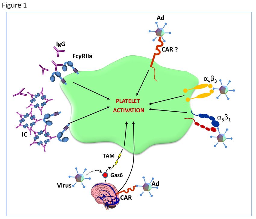
its presence in platelets is debated. Othman et al. identified CAR (by flow cytometry) and its mRNA
(by RT-PCR) in human platelets27 while Shimony et al. did not confirm its presence and proposed
that the binding of Ad to platelets is mediated by the interaction between RGD-binding motifs of
Ad and platelet αVβ328 (Figure 1). Indeed, human megakaryocytes either do not express messenger
RNA for CAR or express it at extremely low levels (J. Rowley and A.S. Weyrich, University of Utah,
personal communication). After i.v. inoculation in mice Ad rapidly bind circulating platelets,
causing their activation and the subsequent entrapment in liver sinusoids where virus-platelet
aggregates are taken up by Kupffer cells and degraded. Platelet activation is followed by blood
coagulation activation leading to DIC29. Activated platelets also release cytokines promoting
endothelial cell activation with secretion of von Willebrand factor, binding of platelets to
endothelial cells and platelet/leukocyte aggregate formation, eventually triggering the
development of microthrombi in liver sinusoids21,29. There is also a complex interplay between Ad
and the coagulation system. In fact, the distribution and activity of Ad in blood is affected by the
interaction with plasma proteins, including complement and vitamin K (VK)-dependent
coagulation factors, which act as opsonizing agents. Our knowledge of these interactions derives
mainly from in vitro observations and it is unknown whether the interplay of Ad with coagulation
proteins affects the activity of the latter. VK-dependent coagulation factors, including the
anticoagulant PC, interact with Ad-5, the most widely used Ad vector. Activated Protein C (APC) is
generated on endothelial cells via the interaction of PC with the thrombin-thrombomodulin
complex and the endothelial PC receptor (EPCR). APC requires protein S (PS) to express
anticoagulant activity30. PS circulates either free or associated with C4BP, a regulatory protein of
the complement system31. C4BP binds to activated platelets through mechanisms involving
chondroitin sulfate expressed on activated platelets32 and to membrane-associated PS on
platelets33. Interestingly, the PC anticoagulant pathway plays a peculiar pathophysiologic role in
CVST34.
Small but measurable amounts of EPCR are also found in plasma. Soluble EPCR binds both PC and
APC with an affinity similar to that of membrane-bound EPCR35 but, in contrast to the latter, it
inhibits APC anticoagulant activity thus limiting its ability to inactivate factor Va, and also binds PC
impeding its activation by the thrombin-thrombomodulin complexes32,36. In CVST increased
5soluble EPCR were observed possibly leading to a procoagulant condition and enhanced
thrombotic risk37.
Finally, Gas6 (encoded by growth arrest-specific gene 6), a VK-dependent protein with 44%
sequence homology with PS but devoid of anticoagulant activity, is widely expressed in the
cerebral nervous system where it is found on resting endothelial cells. Gas6 potentiates platelet
activation acting on TAM receptors leading to thrombus formation38 and in vitro studies have
shown that Gas6 binds to Ad enhancing their gene expression39.
The affinity of different Ad for coagulation factors is variable, with a considerable number of Ad
types unable to bind them. Ad-5, Ad-2 and Ad-16 bind strongly to FX40. Moreover, the ability of Ad
to bind coagulation factors is species-specific, e.g. Ad-5 binds human FX and mouse FX with
similar affinity, but Ad-2 binds human FX with 10-fold lower affinity than mouse FX41.
VK-dependent coagulation factors VII, IX, X, and protein C (PC) mediate the binding of Ad to
hepatocytes 42-44. For instance for Ad5 hepatotropism is critically dependent on the ability of Ad-5
hexon to bind FX. In contrast non-FX binding Ad, such as Ad-48 and Ad-26, do not show
hepatocyte tropism45. The primary reason why FX is required for Ad-5 transduction to the liver is
that it protects Ad-5 from complement attack 46.
Dissemination in the circulation of components of i.m.-injected vaccines, including the Ad vector,
has been previously ascertained47 and it is thus conceivable that some of the activatory
interactions above described between Ad and platelets, endothelium and the blood clotting
system can occur in Ad-vectored vaccine recipients. However, so far no experimental evidence
that this may have a role in VITT is available and actually it seems unlikely that sufficiently high
circulating levels of a non-replicating Ad-vector may be reached to trigger platelet activation or
blood coagulation changes. In fact, it should be considered that around 2500 billion virions/kg are
required to trigger this reactions in mice and non-human primates29,48, and even if all the
Vaxzevria viral content would spill-over in blood after i.m. administration, a concentration of 0.7
billion/kg Ad viral vector would be reached, probably insufficient to activate
platelets/coagulation49.
ANTIBODY- DEPENDENT ENHANCEMENT (ADE) AND VACCINE-ASSOCIATED ADVERSE EVENTS
(AE)
(ADE) is an immunological form of a more general phenomenon called enhanced respiratory
disease (ERD) leading to the clinical worsening of respiratory viral infections. ADE can occur either
6through the antibody-mediated enhancement of virus uptake by Fcγ receptor IIa (FcγRIIa)-
expressing phagocytic cells, thus facilitating viral infection and replication, or by boosting immune
activation through excessive Fc-mediated immunological cell effector functions or immune
complex formation with consequent increase of inflammation and immunopathology50. Both ADE
pathways can occur when non-neutralizing antibodies or antibodies at sub-neutralizing levels bind
to viral antigens without blocking or clearing the infection. ADE has been reported for SARS-Cov
and MERS-Cov vaccines in vitro and in animal models50. The cytoplasmic tail of FcγRIIa activates
51,52
the protein-tyrosine kinases Src and Syk53-55. Src-dependent signaling has been shown to be
crucial for ADE triggered by Ebola virus enhancing viral uptake into cells thus worsening the
infection56.
Circulating antibodies activating platelet IgG FcγRIIa may be key determinants of a host response
leading to uncontrolled platelet aggregation and thrombosis. Studies in transgenic mice expressing
human FcγRIIa on platelets showed that the administration of anti-CD9 antibodies caused
thrombosis accompanied by platelet consumption, a response that was absent in mice lacking the
receptor57. The clinical relevance of this pathway for thrombotic disorders in humans is confirmed
by the observation that FcγRIIa expression is higher in patients with stroke58 and that relatively
common FcγRIIa polymorphisms are associated with increased risk of thrombosis in patients with
heparin-induced thrombocytopenia (HIT)59. Immuno-complex formation, complement deposition
and local immune activation are likely mechanisms triggered by SARS-CoV-2 vaccine. Also, pre-
existing antibodies to coronavirus strains endemic in humans could mediate ADE by facilitating
cross-reactive recognition of SARS-CoV-2 in the absence of viral neutralization60.
Interestingly, chimpanzee Adenoviruses (ChAds) are much less frequently neutralized by pre-
existing antibodies present in humans than Ad5 or Ad6. Prevalence of vector-neutralizing
antibodies against Y25, now renamed ChAdOx1, the vector of the Vaxzevria vaccine, in human
sera from British and Gambian Adults was found to be 0% (n=100) and 9% (n=57), respectively61.
The presence of these antibodies in rare patients in Europe might theoretically represent one
potential mechanism triggering ADE, and possibly VITT, in vaccine recipients but no data on this
are available yet.
Despite the above hypotheses, preliminary in vitro evidence suggests that serum from
convalescent Covid-19 patients neither induced enhancement of SARS-CoV-2 infection nor innate
immunity response in human macrophages, suggesting that ADE may not be involved in the
immune-pathological processes associated with Covid-19 infection or immunization62.
7AD VECTORS USE AND THROMBOTIC EVENTS
Ad vectors for gene therapy
Ad vectors have been therapeutically used for their ability to transduce and deliver transgenes to
different cell types. However, for these indications the clinical use of Ad-vectors has been limited
to a few tens of patients and the main concern has been the development of humoral and cellular
immunity occurring upon repeated administration and/or the possible neutralization of the vector
by pre-existing immunity against the virus, while little attention was paid to the possible
interactions of Ad-vectors with platelets and the blood clotting system.
The first use of Ad-vectors for gene therapy of inherited disorders or to treat neoplasia dates back
to the 1990s . An analysis of the risks associated with the use of Ad-vectored gene therapies
among 90 individuals receiving 140 administrations for various diseases (cystic fibrosis, metastatic
colorectal cancer, cardiovascular disease), showed that 13 deaths were recorded. The authors
concluded that none were linked to the Ad-vector63. The reported hematologic abnormalities were
decreased hemoglobin, leucocytosis, thrombocytopenia, and prolongation of the aPTT, with no
cases of DIC63.
It is however puzzling that a recently EMA-licensed Ad-vectored gene therapy for spinal muscular
atrophy received a warning about the possible risk of thrombotic microangiopathy based on the
reporting of five cases in treated infants
(https://www.ema.europa.eu/en/medicines/human/EPAR/zolgensma).
Ad- vectored vaccines
Beside SARS-CoV-2, Ad vectors have been used for the preparation of other vaccines, including the
ChAdOx1-vectored vaccines for MERS-Cov, influenza, Chikungunia, etc., however with only a few
hundreds of volunteers receiving these vaccines up to June 202064 and no excess of thrombotic
events65. Even for the Ebola vaccination campaign, the largest previous example of large-scale
vaccination using an Ad-vector, a maximum of around 200,000 volunteers were treated with only
one vena cava thrombosis reported (Table 1). However, it may be extremely difficult to prove that
adverse events following immunization are caused by the vaccine itself when their occurrence is
extremely rare (https://www.nature.com/articles/d41586- 021-00880-9. Accessed on
09/04/2021).
The extent and rate of haematological AEs associated with the Ad-vectored vaccines are
summarized in Table 1. Except for common mild/moderate reactome reactions, the most
frequently recorded AEs in clinical trials were hematologic (e.g., mild haemoglobin decrease,
8thrombocytopenia, leukopenia, etc.) the majority of which recovered few days or weeks after
vaccination. Occasional abnormalities of coagulation were reported, with prolongation of the aPTT
possibly due to the development of transient antiphospholipid antibodies. Thrombotic events
were rare both for human and non-human Ad-vectored vaccines. One case of phlebitis was
observed among 114 volunteers who received a recombinant, replication-defective Ad-5-vectored
vaccine expressing HIV-1 antigenic proteins66. Another case of deep vein thrombosis among 156
treated-volunteers was observed after administration of a recombinant, replication-defective Ad-
35-vectored vaccine expressing HIV-1 antigens67. Both were considered unrelated to the vaccine.
A systematic review identified 200 clinical studies on active immunization against SARS-CoV-2. The
second most used vaccine platform, after mRNA-based vaccines, was represented by Ad-vectors
(24%)68. Concerning chimpanzee Ad-vectored vaccines (ChAdOx1 nCoV-19), neutropenia was the
most common hematologic abnormality (Table 1). Across all studies, vaccines had a good safety
profile with no difference in severe reactions between study arms69,70. In a phase III trial with a
recombinant, replication-incompetent human Ad-26 vector encoding the SARS-CoV-2 spike
protein, with 43,783 participants, 11 venous thromboembolic events were observed in the vaccine
group vs 3 in the placebo group (Table 1), however most subjects had underlying medical
conditions that might have contributed to these events. In the vaccine group there were six cases
of lower leg deep venous thrombosis and four pulmonary embolisms. Interestingly, however, also
a CVST, with cerebral haemorrhage and thrombocytopenia, occurred 21 days after vaccination in
a 25 years-old male subject who had multiple predisposing factors, including pre-existing cerebral
sigmoid sinus stenosis and infection from an unknown pathogen. Subsequent testing identified
anti-PF4 antibodies at the time of the event. The patient recovered71.
In a phase III CRT with a recombinant Ad-26-vectored and a recombinant Ad-5-vectored vaccine
(Sputnik V) among 16,501 participants, ten vascular events (0.061%) were observed including: one
DVT (0.006%), one transient ischemic attack (0.006%), one cerebral circulation failure (0.006%),
one vascular encephalopathy (0.00659) and two acute myocardial infarctions (0.012%)(four
additional events were non thrombotic)72(Table 1).
THE VACCINE-ASSOCIATED AUTOIMMUNE THROMBOCYTOPENIA THROMBOTIC SYNDROME
When the anti-SARS-CoV-2 vaccination campaign was well underway worldwide a few cases of
spontaneous, severe thromboembolic events in otherwise healthy subjects were reported, leading
to the pausing of the Vaxzevria vaccine in several European countries
9(https://www.ema.europa.eu/en/news/emas-safety-committee-continues-investigation-covid-19-
vaccine-astrazeneca-thromboembolic-events). Soon after several case reports were published,
mainly in young females, with new ones continuing to accrue, although many not been subject to
rigorous central review and anti-PF4 antibodies were measured with disparate methods, not
allowing to conclude that all are typical VITT cases.
Up to July 17th, 105 such cases with two Ad-vectored vaccines have been published (Table 2) with
some common clinical features characterizing a new syndrome, which include thrombocytopenia,
often severe, venous thrombosis at unusual sites, in particular of the cerebral sinuses but also of
the splanchnic veins, frequently associated with multiple site thromboses, both venous and
arterial, and sometimes disseminated intravascular coagulation (DIC) combined with hemorrhage.
A comparative evaluation of the clinical characteristics of the published ChAdOx1 or Ad26.CoV2.S
VITT cases suggests that while clinical symptoms are comparable, Ad26.CoV2.S-associated cases
show more thrombosis and intracerebral hemorrhage, lower D-dimer and less altered aPTT, but a
similar mortality9. In a recent, large nationwide healthcare register-based study in Denmark and
Norway involving 281,264 ChAdOx1-S-vaccinated subjects aged 18-65 and as controls the entire
age-matched populations of the two countries studied in the period 2016-2019, the standardized
morbidity ratio for CVST was 20.25 (8.14 to 41.7), an excess of 2.5 events per 100,000
vaccinations, particularly evident in women 18-44 years old73, confirming the crucial relationship
between Vaxzevria administration and VITT occurrence. The catastrophic syndrome, burdened by
a 20-50% mortality rate, has the time course and tumultuous evolution of an acute immunologic
reaction and indeed three groups of investigators identified in several of their patients circulating
antibodies to PF4/heparin complexes using an ELISA and heparin-induced platelet activation
assay2-4, proposing this disorder as a peculiar form of autoimmune heparin-induced
thrombocytopenia (aHIT).
THE AUTOIMMUNE HIT HYPOTHESIS
HIT is a rare immune mediated adverse drug reaction occurring after exposure to heparin.
Circulating heparin binds to PF4, a positively charged platelet protein released in plasma upon
activation. PF4 normally binds to negatively charged glycosaminoglycans on the endothelium,
displacing antithrombin and thus activating coagulation. However, PF4 binds with greater affinity
to heparin forming heparin/PF4 complexes which become neoantigens inducing the formation of
autoantibodies. Heparin-PF4-IgG immune complexes in turn bind to platelet FcγRIIA receptors
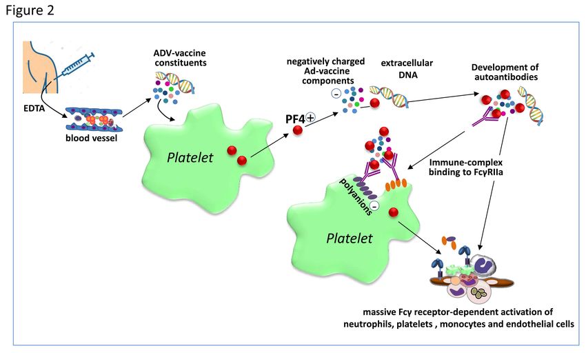
10causing activation, aggregation, and additional release of PF4, with ignition of a positive feedback loop leading to further platelet activation and consumption. Moreover, these complexes activate also monocytes, which release tissue factor, thus promoting concomitant coagulation activation. HIT is a potentially fatal condition, associated with the development of arterial or venous thrombosis74.Thrombocytopenia occurs in more than 85% of HIT patients and is usually of moderate severity, with median counts of approximately 50–60×109/L, even if values
blocking antibody prevented this phenomenon. Given the absence of previous exposure to
heparin, the authors suggested a condition resembling aHIT. More recently, in a preliminary report
published in a non-peer reviewed internet repository, the German group went on to suggest that
the Ad-vector and/or some protein components of the Vaxzevria vaccine activate platelets to
release PF4 which then forms complexes with virus proteins and other anionic constituents of the
vaccine generating neoantigens against which antibodies develop and induce strong platelet
activation via FcγRIIa stimulating granulocyte activation with NETosis and ultimately catastrophic
thrombosis47. The presence of EDTA in the vaccine would favour vascular leakage at the
inoculation site facilitating dissemination in blood of the vaccine components (Figure2).
Very recently a study using alanine scanning mutagenesis explored the binding sites on PF4 of
antibodies isolated from patients with VITT or with classical HIT. While the binding of VITT anti-
PF4 antibodies was restricted to 8 surface aminoacids, all located within the heparin-binding site
of PF4, HIT anti-PF4 antibodies bound aminoacids corresponding to two different sites on PF4;
moreover, VITT antibodies had a stronger binding response than HIT antibodies. The authors
concluded that VITT antibodies mimic the effect of heparin by binding to a similar site on PF4,
allowing PF4 tetramers to cluster with the formation of immuno-complexes which in turn cause
FcγRIIa-dependent platelet activation79.
These peculiar characteristics explain why the identification of VITT requires differential tests
compared with classical HIT 80. In suspected VITT anti-PF4 antibodies can be identified by ELISA-
based assay, but not by other rapid immunological assays typically positive in HIT, such as the
STic® Expert HIT kit, latex immunoassays and chemiluminescence-based assay4, 80, and it is better
characterized by HIPA or PIPA2,4,81 . In addition, no single ELISA method detected all
possible/probable VITT cases82.
While the aHIT hypothesis is important and provides the basis for understanding this novel
catastrophic autoimmune thrombotic syndrome, several aspects do not completely fit with VITT’s
clinical presentation and various issues remain unclarified.
First, among the reported cases in which anti-PF4 antibodies were measured, in a few they were
negative (Table 2)4,5. Second, it is expected that treatment with heparin would worsen the clinical
evolution of these patients, and indeed it is generally cautiously recommended not to use this
drug2,4,13. However, in around one fourth of the heparin-treated cases its use was successful (Table
2). Third, while VITT resembles in several respects aHIT, the latter is not as frequently associated
with CVST and rarely with DIC, and the above summarized mechanistic hypothesis does not

                                                                                                 12
explain the preferential localization of the venous thrombotic events in the cerebral and
splanchnic circulations.
Other unclear aspects are its relatively precocious onset, as early as four days after vaccination,
which seems too soon to generate high titer, class-switched high-affinity anti-PF4 antibodies.
Furthermore, there is no evidence that the anti-PF4 antibodies isolated from patients with VITT
cause thrombosis and thrombocytopenia in animal models49,83. Finally, recent observations show
that 1.2% to 8.0% of subjects receiving a first dose of Vaxzevria develop circulating anti-PF4
antibodies while the prevalence of VITT ranges from 0.0006% to 0.00125%84,85.
OTHER POSSIBLE PATHOGENIC MECHANISMS OF VITT
Very recently a preliminary report, published in a non-peer reviewed repository, provides an
interesting alternative potential pathogenic mechanism of VITT86. Covid-19 is caused by SARS-CoV-
2 which is a single strand RNA virus that is translated and replicates only in the cytosol of infected
cells in the absence of processes which are instead necessary when nuclear-encoded genes are
transcribed, and in particular of mRNA splicing. Nuclear encoded genes present intronic
sequences, thus their transcripts require splice reactions at consensus RNA sequences to eliminate
them. When an adenoviral-vectored viral RNA sequence is administered the vector infects host
cells, adenoviral DNA enters the nucleus and is then transcribed by the host transcription
machinery. However, the viral piece of DNA deriving from the SARS-CoV-2 virus is not optimized to
be transcribed into the nucleus and its open reading frame may thus be disrupted by arbitrary
splice events. These splice events would produce shorter spike protein variants, including forms
missing the C-terminal membrane anchor, thus leading to soluble circulating spike protein
molecules. The soluble spike protein may cause a strong activation of endothelial cells expressing
ACE287. Moreover, when the host immune system starts to produce antibodies against the spike
protein, endothelial cells binding soluble spike would also be decorated by these antibodies
triggering a strong inflammatory reaction through antibody-dependent or complement-dependent
cytotoxicity, thus eliciting VITT. The preferential involvement of cerebral veins with this hypothesis
could be explained by the non-undirectional blood flow in these vessels due to the lack of venous
valves, with prolonged residence time of the soluble spike protein in this district depending on
body posture or when sleeping. The immunological part of this hypothesis is also in agreement
with the apparent higher prevalence of VITT in young women, because they have stronger
13immune reactions than men and older people. To explain the rarity of VITT the authors
hypothesize that only some individuals, due to specific major histocompatibility complex (MHC)
combinations, are not able to produce neutralizing anti-spike antibodies which would instead
prevent in most of the vaccine-recipients the binding of soluble spike to endothelial ACE2 and its
ominous consequences. This hypothesis, partly validated by the identification through in silico
analysis of potential splice sites in the AstraZeneca and Johnson&Johnson codon-optimized spike
opening frames and by in vitro studies with HeLa cells showing that vaccine-transduced cells
generate transcripts smaller than the full spike protein. It would also explain why VITT has not
been reported with mRNA-vaccines, which release their cargo mRNA directly to the host cells
cytosol where it is translated into spike protein without undergoing splicing reactions. Finally, it
would account why the incidence of VITT seems to be lower with the Johnson&Johnson vaccine
compared with AstraZeneca, given that the latter carries more splice donor sequences than the
former86.
Additional hypotheses on the mechanisms triggering VITT which were raised include a genetically-
determined enhanced expression of FcγRIIa in susceptible subjects, an altered glycosylation state
of IgG produced in response to vaccination in some individuals making these antibodies more
reactive to platelet FcγRIIa49, the leakage of the Ad vector in the circulation and/or the prior
presence of cross-reactive antibodies to other coronavirus forming immune complexes activating
platelets88. However, the hypothesis that VITT develops in subjects with previous not apparent
SARS-CoV-2 infection with prior circulating IgG antibodies against the spike protein able to activate
platelet FcγRIIa89 should be excluded by the observation that most VITT subjects tested for
previous or recent Covid-19 infections were negative. Moreover, excessive transcription of the
spike protein which would then activate platelets binding to ACE290 or the vaccine-induced
expression of spike by megakaryocytes and platelets leading to a thrombo-inflammatory storm49,91
have been proposed. Another hypothesis starts from the observation that both the ChAdOx1 and
the Ad26.CoV2.S vaccines use as excipient polysorbate 80, a nonanionic surfactant crossing the
blood brain barrier which enhances microparticle uptake by endothelial cells. Therefore leakage
of Ad-vector and polysorbate in the circulation and the spike protein produced by vaccination
could preferentially localize in the brain vessel circulation triggering endothelial activation92.
However, considering that VITT develops usually at least 1 week after vaccination, it is very
unlikely that circulating Ad-vector or vaccine excipients would still be present in blood, rendering
more likely alternative explanations, and in particular an immunological reaction.
14CONCLUSIVE REMARKS
At least two Ad vector-based vaccines against SARS-CoV-2 have been associated with an excess
rate of a special form of catastrophic thrombotic syndrome associated with thrombocytopenia of
likely autoimmune origin, not observed so far with mRNA-based vaccines, suggesting that the
vectors may play a role in eliciting it. Several characteristics of the Ad vectors and/or of vaccine
composition may theoretically interact with platelets, the endothelium and the blood clotting
system precipitating this rare complication. However, the exact sequence of events leading to the
development of this syndrome and, most importantly, the reason why it evolves only in very few
subjects without apparent predisposing factors remain to be clarified.
Altogether it is clear that our understanding of the pathogenesis of VITT is far from complete and
that more mechanistic studies are required to clarify it and, hopefully, to finally identify predictive
risk factors of its development.
What is quite likely is that Ad vector-based vaccine administration triggers an immunological
reaction which, for unknown reasons, in some rare subjects involves especially blood platelets,
and possibly some peculiar vascular endothelial districts such as those of the cerebral and
splanchnic veins, precipitating the catastrophic vaccine-induced autoimmune thrombocytopenia
thrombosis syndrome.
Awareness of this condition and the prompt identification/evaluation of the affected patients may
lead to successful treatment and recovery (Figure 3)13,84.
Covid-19 continues to represent a serious global health problem and vaccination against SARS-
CoV-2 is the most effective way of limiting illness and death due to the pandemic. Based on the
current available information, and in light of the relative rarity of VITT, the benefits of vaccination
clearly outweigh the potential risks (https://www.ema.europa.eu/en/documents/dhpc/direct-
healthcare-professional-communication-dhpc-zolgensma-onasemnogene-abeparvovec-risk-
thrombotic_en.pdf). However, once the global pandemic will begin to retreat, the relative
importance of even small risks will increase65, rendering of the utmost importance the definitive
unravelling of the mechanisms leading to this ominous thrombotic syndrome, the identification of
the prognostic factors for its development and the definition of the best management strategies13.
ACKNOWLEDGEMENTS
This work was supported in part by grants from Fondazione Cassa di Risparmio di Perugia (#19663
(2020.0508) and FISR 2020 (# 1049) to PG.
15REFERENCES
1. Muir KL, Kallam A, Koepsell SA, Gundabolu K. Thrombotic Thrombocytopenia after
Ad26.COV2.S Vaccination. N Engl J Med. 2021;384(20):1964-1965
2. Greinacher A, Thiele T, Warkentin TE, Weisser K, Kyrle PA, Eichinger S. Thrombotic
Thrombocytopenia after ChAdOx1 nCov-19 Vaccination. N Engl J Med.
2021;384(22):2092-2101.
3. Schultz NH, Sørvoll IH, Michelsen AE, et al. Thrombosis and Thrombocytopenia after
ChAdOx1 nCoV-19 Vaccination. N Engl J Med. 2021;384(22):2124-2130.
4. Scully M, Singh D, Lown R, et al. Pathologic Antibodies to Platelet Factor 4 after
ChAdOx1 nCoV-19 Vaccination. N Engl J Med. 2021,384(23):2202-2211.
5. Bayas A, Menacher M, Christ M, Behrens L, Rank A, Naumann M. Bilateral superior
ophthalmic vein thrombosis, ischaemic stroke, and immune thrombocytopenia after
ChAdOx1 nCoV-19 vaccination. Lancet. 2021;397(10285):e11.
6. Muster V, Gary T, Raggam RB, WÖlfler A, Brodmann M. Pulmonary embolism and
thrombocytopenia following ChAdOx1 vaccination. Lancet. 2021;397(10287):1842.
7. Shay DK, Julianne Gee, John R Su, et al. Safety Monitoring of the Janssen (Johnson &
Johnson) COVID-19 Vaccine - United States, March-April 2021. MMWR Morb Mortal
Wkly Rep. 2021;70(18):680-684.
8. Rodeghiero F, Balduini CL. A new enemy is emerging in the fight against the SARS-CoV-
2 pandemic. Haematologica. 2021;106(8):2040-2041.
9. Hwang J, Lee SB, Lee SW. Comparison of vaccine-induced thrombotic events between
ChAdOx1 nCoV-19 and Ad26.COV.2.S vaccines. J Autoimmun. 2021;122:102681.
10. Sangli S, Virani A, Cheronis N. Thrombosis With Thrombocytopenia After the
Messenger RNA-1273 Vaccine. Ann Intern Med. 2021;L21-0244.
11. Greinacher A, Thiele T, Warkentin TE, Weisser K, Kyrle P, Eichinger S. A prothrombotic
thrombocytopenic disorder resembling heparin-induced thrombocytopenia following
coronavirus vaccination. Research Square. 2021 Apr 09. [Epub ahead of print]
12. See I, Su JR, Lale A, Woo EJ, et al. US Case Reports of Cerebral Venous Sinus
Thrombosis With Thrombocytopenia After Ad26.COV2.S Vaccination, March 2 to April
21, 2021. JAMA. 2021;325(24):2448-2456.
13. Gresele P, Marietta M, Ageno W, et al. Management of cerebral and splanchnic vein
thrombosis associated with thrombocytopenia in subjects previously vaccinated with
Vaxzevria (AstraZeneca): a position statement from the Italian Society for the Study of
Haemostasis and Thrombosis (SISET). Blood Transfus. 2021 Apr 15. [Epub ahead of
print]
14. Hsu KH, Lonberg-Holm K, Alstein B, Crowell RL. A monoclonal antibody specific for the
cellular receptor for the group B coxsackieviruses. J Virol. 1988;62(5):1647-1652.
15. Bergelson JM, Cunningham JA, Droguett G, et al. Isolation of a common receptor for
Coxsackie B viruses and ADes 2 and 5. Science. 1997;275(5304):1320-1323.
1616. Bewley MC, Springer K, Zhang YB, Freimuth P, Flanagan JM. Structural analysis of the
mechanism of AD binding to its human cellular receptor CAR. Science.
1999;286(5444):1579-1583.
17. Roelvink PW, Mi Lee G, Einfeld DA, Kovesdi I, Wicham TJ. Identification of a conserved
receptor-binding site on the fiber proteins of CAR-recognizing Adenoviridae. Science.
1999;286(5444):1568-1571.
18. Greber U, Willets M, Webster P, Helenius A. Stepwise dismantling of AD 2 during entry
into cells. Cell. 1993;75(3):477-486.
19. Wickham TJ, Mathias P, Cheresh DA, Nemerow GR. Integrins alpha v beta 3 and alpha v
beta 5 promote AD internalization but not virus attachment. Cell. 1993;73(2):309-319.
20. Davison E, Diaz RM, Hart IR, Santis G, Marshall JF. Integrin a5b1-mediated AD infection
is enhanced by the integrin-activating antibody TS2/16. J Virol. 1997;71(8):6204–6207.
21. Zhang Y, Bergelson JM. AD receptors. J Virol. 2005;79(19):12125-12131.
22. Honda T, Saitoh H, Masuko M, et al. The coxsackievirus-AD receptor protein as a cell
Adhesion molecule in the developing mouse brain. Brain Res Mol Brain Res.
2000;77(1):19-28.
23. Patzke C, Max KEA, Behlke J et al. The coxsackievirus-adenovirus receptor reveals
complex homophilic and heterophilic interactions on neural cells. J Neurosci.
2010;30(8):2897-2910.
24. Nalbantoglu J, Pari G, Karpati G, et al. Expression of the primary coxsackie and AD
receptor is downregulated during skeletal muscle maturation and limits the efficacy of
AD-mediated gene delivery to muscle cells. Hum Gene Ther. 1999;10(6):1009-1019.
25. Noutsias M, Fechner H, de Jonge H, et al. Human coxsackie-AD receptor is colocalized
with integrins alpha(v)beta(3) and alpha(v)beta(5) on the cardiomyocyte sarcolemma
and upregulated in dilated cardiomyopathy: implications for cardiotropic viral
infections. Circulation. 2001;104(3): 275-280.
26. Rebel V, Hartnett S, Denham J, et al. Maturation and lineage-specific expression of the
coxsackie and AD receptor in hematopoietic cells. Stem Cells. 2000;18(13):176-182.
27. Othman M, Labelle A, Mazzetti I, Elbatarny HS, Lillicrap D. AD-induced
thrombocytopenia: the role of von Willebrand factor and P-selectin in mediating
accelerated platelet clearance. Blood. 2007;109(7):2832-2839.
28. Shimony N, Elkin G, Kolodkin-Gal D, Krasny L, Urieli-Shoval S, Haviv YS. Analysis of
Adenoviral attachment to human platelets. Virol J. 2009;6:25.
29. Stone D, Liu Y, Shayakhmetov D, Li ZY, Ni S, Lieber A. AD-platelet interaction in blood
causes virus sequestration to the reticuloendothelial system of the liver. J Virol.
2007;81(9):4866-4871.
30. Griffin JH, Zlokovic BV, Mosnier LO. Activated protein C: biased for translation. Blood.
2015;125(19):2898-2907.
31. Esmon CT. Protein C. Prog Hemost Thromb. 1984;7:25-54.
32. Hamad OA, Nilsson PH, Lasaosa M, et al. Contribution of chondroitin sulfate A to the
binding of complement proteins to activated platelets. PLoS One. 2010;5(9):e12889.
1733. Dahlbäck B, Wiedmer T, Sims PJ. Binding of anticoagulant vitamin K-dependent
protein S to platelet-derived microparticles. Biochemistry. 1992;31(51):12769-12777.
34. Ferro JM, Canhão P, Stam J, Bousser MG, Barinagarrementeria F; ISCVT Investigators.
Prognosis of cerebral vein and dural sinus thrombosis: results of the International
Study on Cerebral Vein and Dural Sinus Thrombosis (ISCVT). Stroke. 2004;35(3):664-
670.
35. Kurosawa S, D J Stearns-Kurosawa, N Hidari, C T Esmon. Identification of functional
endothelial protein C receptor in human plasma. J Clin Invest. 1997;100(2):411-418.
36. Gandrille S. Endothelial cell protein C receptor and the risk of venous thrombosis.
Haematologica. 2008;93(6):812-816.
37. Javanmard SH, ShahsavarzAdeh T, SaAdatnia M. Soluble thrombomodulin and
endothelial cell protein C receptor levels in patients with cerebral venous and sinus
thrombosis. Eur Neurol. 2013;70(3-4):156-158.
38. Gould WR, Baxi SM, Schroeder R, et al. Gas6 receptors Axl, Sky and Mer enhance
platelet activation and regulate thrombotic responses. J Thromb Haemost.
2005;3(4):733-741.
39. Nidetz NF, Gallagher TM, Wiethoff CM. Inhibition of type I interferon responses by
Adenovirus serotype-dependent Gas6 binding. Virology. 2018;515:150-157.
40. Allen RJ, Byrnes AP. Interaction of Adenovirus with antibodies, complement, and
coagulation factors. FEBS Letters. 2019;593(24):3449-3460.
41. Lenman A, Muller S, Nygren MI, Frangsmyr L, Stehle T, Arnberg N. Coagulation factor IX
mediates serotype-specific binding of species A Adenoviruses to host cells. J Virol.
2011;85(24):13420-13431.
42. Parker, A. L., J. H. McVey, J. H. Doctor, et al. Influence of coagulation factor zymogens
on the infectivity of ADes pseudotyped with fibers from subgroup D. J Virol.
2007;81(7):3627-3631.
43. Parker, A. L., S. N. WAddington, C. G. Nicol, D. M. et al. Multiple vitamin K-dependent
coagulation zymogens promote AD-mediated gene delivery to hepatocytes. Blood.
2006;108(8):2554-2561.
44. Shayakhmetov DM, Gaggar A, Ni S, Li, Lieber A. AD binding to blood factors results in
liver cell infection and hepatotoxicity. J Virol. 2005;79(12):7478-7491.
45. Kalyuzhniy O, Di Paolo NC, Silvestry M, et al. Adenovirus serotype 5 hexon is critical for
virus infection of hepatocytes in vivo. Proc Natl Acad Sci U S A. 2008;105(14):5483-
5488.
46. Duffy MR, Doszpoly A, Turner G, Nicklin SA, Baker AH. The relevance of coagulation
factor X protection of Adenoviruses in human sera. Gene Ther. 2016;23(7):592-596.
47. Greinacher A, Selleng K, Wesche J et al. Towards Understanding ChAdOx1 nCov-19
Vaccine-induced Immune Thrombotic Thrombocytopenia (VITT). Research Square.
2021 Apr 20. [Epub ahead of print]
48. Brunetti-Pierri N, Palmer DJ, Beaudet AL, Carey KD, Finegold M, Ng P. Acute toxicity
after high-dose systemic injection of helper-dependent adenoviral vectors into
nonhuman primates. Hum Gene Ther. 2004;15(1):35-46.
49. Kadkhoda K. Post-Adenoviral-based COVID-19 vaccines thrombosis: A proposed
mechanism. J Thromb Haemost. 2021;19(7):1831-1832.
1850. Lee WS, Wheatley AK, Kent SJ, DeKosky BJ. Antibody-dependent enhancement and
SARS-CoV-2 vaccines and therapies. Nat Microbiol. 2020;5(10):1185-1191.
51. HamAda F, Aoki M, Akiyama T, Toyoshima K. Association of immunoglobulin G Fc
receptor II with Src like protein-tyrosine kinase Fgr in neutrophils. Proc Natl Acad Sci U
S A. 1993;90(13):6305-6309.
52. Ghazizadeh S, Bolen JB, Fleit HB. Physical and functional association of Src-related
protein tyrosine kinases with Fc gamma RII in monocytic THP-1 cells. J Biol Chem.
1994;269(12):8878-8884.
53. Kiener PA, Rankin BM, Burkhardt AL, et al. Cross-linking of Fc gamma receptor I (Fc
gamma RI) and receptor II (Fc gamma RII) on monocytic cells activates a signal
transduction pathway common to both Fc receptors that involves the stimulation of
p72 Syk protein tyrosine kinase. J Biol Chem. 1993;268(32):24442-24448.
54. Nimmerjahn F, Ravetch JV. Fcγ receptors as regulators of immune responses. Nat Rev
Immunol. 2008;8(1):34-47.
55. Joshi T, Butchar JP, Tridandapani S. Fcγ Receptor Signaling in Phagocytes. Int J
Hematol. 2006;84(3):210-216.
56. Furuyama W, Marzi A, Carmody AB, et al. Fcγ-receptor IIa-mediated Src Signaling
Pathway Is Essential for the Antibody-Dependent Enhancement of Ebola Virus
Infection. PLoS Pathog. 2016;12(12):e1006139.
57. Taylor SM, Reilly MP, Schreiber AD, Chien P, Tuckosh JR, McKenzie SE. Thrombosis and
shock induced by activating antiplatelet antibodies in human Fc gamma RIIA transgenic
mice: the interplay among antibody, spleen, and Fc receptor. Blood. 2000;96(13):4254-
4260.
58. Calverley DC, Brass E, Hacker MR, et al. Potential role of platelet FcgammaRIIA in
collagen-mediated platelet activation associated with atherothrombosis.
Atherosclerosis. 2002;164(2):261-267.
59. Pamela S, Anna Maria L, Elena D et al. Heparin-induced thrombocytopenia: the role of
platelets genetic polymorphisms. Platelets. 2013;24(5):362-368.
60. Tetro, J. A. Is COVID-19 receiving ADE from other coronaviruses? Microbes Infect.
2020;22(2):72-73.
61. Collocca S, Barnes E, Folgori A, et al. Vaccine vectors derived from a large collection of
simian Adenoviruses induce potent cellular immunity across multiple species. Sci
Transl Med. 2012;4(115):115ra2.
62. Obdulio GN, V'kovski V, Zettl F, Zimmer G, Thiel V, Summerfield A. No Evidence for
Human Monocyte-Derived Macrophage Infection and Antibody-Mediated
Enhancement of SARS-CoV-2 Infection. Front Cell Infect Microbiol. 2021;11:644574.
63. Crystal RG, Harvey BG, Wisnivesky JP, et al. Analysis of risk factors for local delivery of
low- and intermediate-dose Adenovirus gene transfer vectors to individuals with a
spectrum of comorbid conditions. Hum Gene Ther. 2002;13(1):65-100.
64. Folegatti PM, Bittaye M, Flaxman A, et al. Safety and immunogenicity of a candidate
Middle East respiratory syndrome coronavirus viral-vectored vaccine: a dose-
19escalation, open-label, non-randomised, uncontrolled, phase 1 trial. Lancet Infect Dis.
2020;20(7):816-826.
65. Kupferschmidt K, Vogel G. What’s the future of vaccines linked to rare clotting
disorders? Science breaks down the latest. 2021 May 3. [Epub ahead of print]
66. Jaoko W, Karita E, Kayitenkore K, et al. Safety and immunogenicity study of MulticlAde
HIV-1 Adenoviral vector vaccine alone or as boost following a multiclAde HIV-1 DNA
vaccine in Africa. PLoS One. 2010;5(9):e12873.
67. Keefer MC, Gilmour J, Hayes P, Gill D, et al. A phase I double blind, placebo-controlled,
randomized study of a multigenic HIV-1 Adenovirus subtype 35 vector vaccine in
healthy uninfected Adults. PLoS One. 2012;7(8):e41936.
68. Rego GNA, Nucci MP, Alves AH, et al. Current Clinical Trials Protocols and the Global
Effort for Immunization against SARS-CoV-2. Vaccines (Basel). 2020;8(3):474.
69. Van Doremalen N, Lambe T, Spencer A, et al. ChAdOx1 nCoV-19 vaccination prevents
SARS-CoV-2 pneumonia in rhesus macaques. Nature. 2020;586(7830):578-582.
70. Folegatti PM, Ewer KJ, Aley PK, et al. Oxford COVID Vaccine Trial Group. Safety and
immunogenicity of the ChAdOx1 nCoV-19 vaccine against SARS-CoV-2: a preliminary
report of a phase 1/2, single-blind, randomised controlled trial. Lancet.
2020;396(10249):467-478.
71. Sandoff J, Gray G, Vandebosch A, Cárdenas V, et al. Safety and Efficacy of Single-Dose
Ad26.COV2.S Vaccine against Covid-19. N Engl J Med. 2021;384(23):2187-2201.
72. Logunov DY, Dolzhikova IV, Shcheblyakov DV, et al. Safety and efficacy of an rAd26 and
rAd5 vector-based heterologous prime-boost COVID-19 vaccine: an interim analysis of
a randomized controlled phase 3 trial in Russia. Lancet. 2021;397(10275):671-681.
73. Pottegård A, Lars Christian Lund 2, Øystein KarlstAd 3, et al. Arterial events, venous
thromboembolism, thrombocytopenia, and bleeding after vaccination with Oxford-
AstraZeneca ChAdOx1-S in Denmark and Norway: population based cohort study. Brit
Med J. 2021;5;373:n1114.
74. Marcucci R, Berteotti M, Gori AM, et al. Heparin induced thrombocytopenia: position
paper from the Italian Society on Thrombosis and Haemostasis (SISET). Blood Transfus.
2021;19(1):14-23.
75. Cuker A, Arepally GM, Chong BH, et al. American Society of Hematology 2018
guidelines for management of venous thromboembolism: heparin-induced
thrombocytopenia. Blood Adv. 2018;2(22):3360-3392.
76. Greinacher A, Farner B, Kroll H, et al. Clinical features of heparin-induced
thrombocytopenia including risk factors for thrombosis. A retrospective of 408
patients. Thromb Haemost. 2005;94(1):132-135.
77. Greinacher A, Selleng K, Warkentin TE. Autoimmune heparin-induced
thrombocytopenia. J Thromb Haemost. 2017;15(11):2099-2114.
78. Warkentin TE, Greinacher A. Spontaneous HIT syndrome: Knee replacement, infection,
and parallels with vaccine-induced immune thrombotic thrombocytopenia. Thromb
Res. 2021;204:40-51.
79. Huynh A, Kelton JG, Arnold DM, Daka M, Nazy I. Antibody epitopes in vaccine-induced
immune thrombotic thrombocytopenia. Nature. 2021 Jul 7. [Epub ahead of print]
2080. Favaloro EJ. Laboratory testing for suspected COVID-19 vaccine-induced (immune)
thrombotic thrombocytopenia. Int J Lab Hematol. 2021 Jun 17. [Epub ahead of print]
81. Vayne C, Guery EA, Kizlik-Masson C, et al. Beneficial effect of exogenous platelet factor
4 for detecting pathogenic heparin-induced thrombocytopenia antibodies. Br J
Haematol. 2017;179(5):811-819.
82. Platton S, Bartlett A, MacCallum P, et al. Evaluation of laboratory assays for anti-
platelet factor 4 antibodies after ChAdOx1 nCOV-19 vaccination. J Thromb Haemost.
2021;19(8):2007-2013.
83. Cines DB, Bussel JB. SARS-CoV-2 Vaccine-Induced Immune Thrombotic
Thrombocytopenia. N Engl J Med. 2021;384(23):2254-2256.
84. Sørvoll IH, Horvei KD, Ernstsen SL, et al. An observational study to identify the
prevalence of thrombocytopenia and anti-PF4/polyanion antibodies in Norwegian
health care workers after COVID-19 vaccination. J Thromb Haemost. 2021;19(7):1813-
1818.
85. Thiele T, Ulm L, Holtfreter S, et al. Frequency of positive anti-PF4/polyanion antibody
tests after COVID-19 vaccination with ChAdOx1 nCoV-19 and BNT162b2. Blood.
2021;138(4):299-303.
86. Kowarz E, Krutzke L, Reis J, Bracharz S, Kochanek S, Marschalek R. Vaccine-induced
Covid-19 spike open reading frame resul in spike protein variant that may cause
thromboembolic events in patients immunized with vector-based vaccine. Research
Square. 2021 May 26. [Epub ahead of print]
87. Lei Y, Zhang J, Schiavon CR, et al. SARS-CoV-2 Spike Protein Impairs Endothelial
Function via Downregulation of ACE 2. Circ Res. 2021;128(9):1323-1326.
88. Chakraborty S, Gonzalez J, Edwards K. Proinflammatory IgG Fc structures in patients
with severe COVID-19. Nat Immunol. 2021;22(1):67-73.
89. Douxfils J, Favresse J, Dogné JM. Hypotheses behind the very rare cases of thrombosis
with thrombocytopenia syndrome after SARS-CoV-2 vaccination. Thromb Res.
2021;203:163-171.
90. Shen S, Zhang J, Fang Y, et al. SARS-CoV-2 interacts with platelets and megakaryocytes
via ACE2-independent mechanism. J Hematol Oncol. 2021;29;14(1):72.
91. Millington-Burgess SL, Harper MT. A double-edged sword: antibody-mediated
procoagulant platelets in COVID-19. Platelets. 2021; 32(5):579-581.
92. Choi PHI. Thrombotic Thrombocytopenia after ChAdOx1 nCoV-19 Vaccination. N Engl J
Med. 2021;385(3):e11.
21TABLE 1 - Studies with Ad-vectored vaccines reporting hematological adverse effects (AEs). The AEs reported are related only to the vaccine groups and not to
the placebo groups. CSVT: cerebral sinus venous thrombosis; DVT: deep venous thrombosis; PE: pulmonary embolism; WBC: white blood cells
Adenoviral Pathogens Study (authors - year) N° participants Thrombocytopenia (n) Venous Coagulation Other haematological Other systemic AEs
vector Thromboembolism (n) disorders complications
Fatigue, malaise,
Influenza A Phase Ia, dose-escalation (S1) 15 NR NR NR Leukopenia
headhache
Phase I, dose-escalation, non-randomized, Anemia, neutropenia,
su
ChAdOx1 MERS 24 NR NR NR Fatigue, headache,myalgia
uncontrolled (64) lymphopenia
ri Phase I/II, single-blind, randomized,
vo
SARS-CoV-2 1077 NR NR NR Neutropenia Fatigue, headache
controlled (70)
ne
aPTT prolongation
Phase I, dose-escalation, open-label (S2) 20 NR NR Leukopenia Fever
(15%)
d
A Phase I/IIa, double-blind, placebo- aPTT prolongation Anaemia, lymphopenia,
ee controlled, dose-finding (S3)
120 NR NR
(n=1) neutropenia
Fatigue, malaise, haedache
zn
ap
Ebola aPTT prolongation
Phase I, dose-escalation, open-label (S4) 60 1 NR Leukopenia, eosinophilia Fatigue, headache,myalgia
(n=4)
ChAd3
m
ih Phase II, randomized, observer-blind,
placebo-controlled (S5)
3030 7
Vena cava thrombosis
(n=1)
NR Anaemia Fever, headache
C Phase II, randomized, observer-blind,
600 5 NR NR Anaemia Fever, headache
placebo-controlled (S6)
Phase I, open-label, single-site, dose- Fatigue, headache,myalgia,
RSV 42 2 NR NR Anaemia
escalation (S7) nausea
Phase I, double-blind, randomized, Fatigue, malaise,
114 NR Phl ebitis (n=1) NR Leukopenia, anemia
placebo-controlled (66) headhache
Phase I, double-blinded, placebo-controlled Fever, malaise, myalgia,
HIV 36 NR NR NR Neutropenia
(S8) chills
Phase IIb, double-blind, randomized,
801 NR NR NR Neutropenia, anemia Headache, malaise, myalgia
controlled (S9)
su Ad5
Phase I, randomized, double-blind,
120 NR NR NR Anaemia, leukopenia Fever
irv
placebo-controlled (S10)
Ebola
on Phase I, single-site, double-blind,
aPTT prolongation Malaise, myalgia, headache,
e
randomized, placebo-controlled, dose- 32 NR NR NR
(n=2) chills
d escalation (S11)
A
na SARS-Cov2
Phase I, single-centre, dose-escalation,
double-blind, non-randomized (S12)
108 4 NR NR NR
Fever, fatigue, headache,
muscle pain
m
u
H SARS-CoV-2
Phase III, randomized, double-blind,
placebo-controlled (71)
44325 NR
DVT (n=6), PE (n=4), CSVT
(n=1)
NR NR
Fatigue, headache,myalgia,
nausea
Ad26
Phase II, randomized, double-blind, Fatigue, haedache, myalgia,
Ebola 423 NR NR NR Anaemia, neutropenia
placebo-controlled (S13) chills
Phase I, double-blind, randomized, Malaise, myalgia, headache,
Ad35 HIV
placebo-controlled (67)
58 NR DVT (n=1) NR NR
chills 22You can also read