Microchip capillary electrophoresis dairy device using fluorescence spectroscopy for detection of ciprofloxacin in milk samples - Nature
←
→
Page content transcription
If your browser does not render page correctly, please read the page content below
www.nature.com/scientificreports
OPEN Microchip capillary electrophoresis
dairy device using fluorescence
spectroscopy for detection
of ciprofloxacin in milk samples
Rick Bosma, Jasen Devasagayam, Ashutosh Singh & Christopher M. Collier*
Detecting antibiotics in the milk supply chain is crucial to protect humans from allergic reactions, as
well as preventing the build-up of antibiotic resistance. The dairy industry has controls in place at
processing facilities, but controls on dairy farms are limited to manual devices. Errors in the use of
these manual devices can result in severe financial harm to the farms. This illustrates an urgent need
for automated methods of detecting antibiotics on a dairy farm, to prevent the shipment of milk
containing antibiotics. This work introduces the microchip capillary electrophoresis dairy device, a
low-cost system that utilizes microchip capillary electrophoresis as well as fluorescence spectroscopy
for the detection of ciprofloxacin contained in milk. The microchip capillary electrophoresis dairy
device is operated under antibiotic-absent conditions, with ciprofloxacin not present in a milk sample,
and antibiotic-present conditions, with ciprofloxacin present in a milk sample. The response curve for
the microchip capillary electrophoresis dairy device is found through experimental operation with
varied concentrations of ciprofloxacin. The sensitivity and limit of detection are quantified for the
microchip capillary electrophoresis dairy device.
Of critical importance is the prevention of antibiotic contamination in the milk supply chain. This preven-
tion of antibiotic contamination protects human beings against a number of health issues including antibiotic
resistance1 and allergic reactions2. Antibiotic contamination is of particular concern for the dairy industry, where
antibiotics such as ß-lactams, cephalosporins, tetracyclines, and fluoroquinolones can contaminate milk due to
treatment of bovine ailments3,4. As such, devices that are capable of detecting antibiotics in milk are of inter-
est for the dairy industry. However, at the dairy farm level, there are only manual devices available, including
prepared incubated tests such as the Delvotest T 5 and lateral flow assays such as the Charm ROSA t est6. These
manual devices subject dairy farms to substantial human error, resulting in situations where dairy farmers will
ship milk that is contaminated with antibiotics to processing plants, suffer monetary losses, and (after multiple
offences) suffer loss of l icensure7.
The Delvotest T test is a highly sensitive detection system which is able to detect a wide range of antibiotic
residues, especially for tetracyclines and β-lactams, as it measures the inhibitory substance in the milk sample,
which is the antibiotic residue at a concentration above the limit of detection (LOD) of the t est5. Milk samples are
added to an agar medium, which contains spores of Geobacillus stearothermophilus var. calidolactis, along with
glucose (a fermentable sugar), and a pH colour indicator, bromocresol purple. The test is incubated for several
hours. If the antibiotic residue in the sample is below the LOD, the bacillus spores will germinate, producing
an acid from fermentation. Hence, antibiotic residues below the LOD will cause the indicator to change colour
from purple to yellow. Antibiotic residues above the LOD inhibits the germination of the bacillus spores and
fermentation does not occur so the colour of the indicator is left u nchanged5.
The Charm Rapid One-Step Assay (ROSA) test works similarly, where a mixture of the milk sample and buffer
solution is incubated within the sample compartment of the test strip6. It works by utilizing a lateral-flow format
with gold-bead receptors which detect and measure β-lactam and tetracycline antibiotics in a milk sample. After
adding the milk to the test strip, it is incubated for several minutes, which will rehydrate and mobilize the recep-
tors. These receptors are carefully designed to have a sensitivity which will detect antibiotics at concentrations
Applied Optics and Microsystems Laboratory, University of Guelph, Guelph, ON N1G 2W1, Canada. *
email:
ccollier@uoguelph.ca
Scientific Reports | (2020) 10:13548 | https://doi.org/10.1038/s41598-020-70566-1 1
Vol.:(0123456789)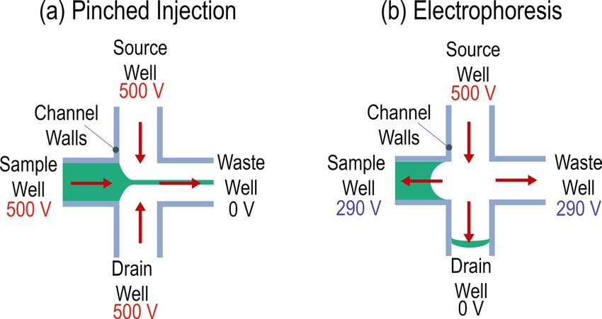
www.nature.com/scientificreports/
below the maximum residual limit (MRL). Receptors which are unreacted with the antibiotic will bind to form
a red test line. Receptors which react with the antibiotics pass across this test line and bind with the control line
of the test strip, causing the control line to darken. Hence, if the colour of the indicator line is lighter than the
control line on the strip, or even if the line is not formed, it signifies the presence of specific antibiotics in milk8.
The Delvotest T and Charm ROSA tests are both manual processes which would require well-trained personnel
to make qualitative decisions.
To reduce the manual error associated with manual devices (as discussed above), laboratory device tests
may be employed to detect the presence of antibiotics in milk. These laboratory device tests include mass
spectrometry9, liquid chromatography10, immunoassay11, potentiometric12, and surface plasmon resonance13
devices. However, these laboratory devices are slow because they require cumbersome processes including trans-
portation off-site from the dairy farm, sample preparation, and extensive incubation periods9,12. The temporal
limitations of laboratory devices are in stark contrast to the demands of on-farm antibiotic device detection,
where full antibiotic detection should ideally occur within a few minutes during the milking process for each
individual cow. It should be considered that if antibiotic detection takes place on the combined milk repository
from many cows, the entire milk repository is in jeopardy if any antibiotic contamination occurs. Additionally,
it is important to be able to develop a system that could potentially be used by untrained personnel, which
microfluidic devices can offer.
This work responds to the need for on-site detection of antibiotics, such as ciprofloxacin, in milk samples
through the development of a microchip capillary electrophoresis dairy d evice14. Actuation and sensing aspects
of microfluidic technology15 are integrated, as lab-on-a-chip microfluidic technology is a commonly used method
for sensing p urposes14,15. The microchip capillary electrophoresis dairy device combines respective microchip
capillary electrophoresis and fluorescence spectroscopy16–18 techniques. The microchip capillary electrophoresis
technique leverages capillary electrophoresis, whereby a controlled electric field is strategically used to separate
constituent components and isolate the analyte(s) within a microfluidic channel19. The fluorescence spectroscopy
technique is utilized to achieve the ultimate detection of an important antibiotic, ciprofloxacin. Ciprofloxacin
is an ideal choice of antibiotic for this study because it is a metabolite of the commonly used antibiotic, enro-
floxacin, which is currently approved for widespread use in regions of the world and there are strict regulations
on the maximum residual levels of the sum of enrofloxacin and c iprofloxacin20,21. To introduce and develop the
microchip capillary electrophoresis dairy device, this work discusses the design and fabrication of the device,
chemicals used, operation, antibiotic-absent and antibiotic-present operation, and ciprofloxacin detection.
It should be noted that there are several relevant studies to our work that involve combinations of capillary
electrophoresis and antibiotic s tudy22–34. These studies are primarily larger channels while our presented work
focuses on smaller channels and considerably shorter channel lengths. Many of these mentioned studies work
with effective lengths ranging from 500 to 960 mm with inner capillary diameters of 50–75 μm. For example, in
a study by Springer et al., the capillary used had an effective length of 525 mm and an inner diameter of 75 μm,
while applying up to 20 kV to create the electric field26. In another example, Lara et al. used a capillary with an
effective length of 960 mm and an inner diameter of 50 μm, while applying up to 25 kV to the e lectrodes24. In
comparison, our microchip capillary system utilizes microcapillaries possessing an effective length of 79 mm
with an inner diameter of 50 μm. This makes it possible to use lower voltages to achieve equivalent electric field
strengths, while also achieving faster fluid velocities as a result of the smaller channel widths. This microscale
scalability is particularly important in highly-parallel applications requiring high-throughput analyses, such as
is required on dairy farms27. Another factor to consider is the sample preparation required, where the afore-
mentioned studies require solid-phase extraction of milk samples using centrifugation to elute the antibiotic
residues. In comparison, the presented microchip capillary electrophoresis dairy device only requires filtering
before being injected into the device, as the pinched sample injection will send a minute sample for detection.
Milk-based, scalable, and accessible systems, as in the presented work, are of great interest to the large dairy
research community.
Results
Fluorescence spectroscopy for detection. To allow for future potential of mass-market adoption of the
microchip capillary electrophoresis dairy device, cost minimization is an important design factor that is consid-
ered. Therefore, the fluorescence spectroscopy technique is implemented with low-cost components, being an
ultraviolet (UV) light emitting diode (LED) for the fluorescence excitation and a photodiode for the fluorescence
detection, as in previous w ork35. As ciprofloxacin emits fluorescence at an excitation wavelength of 270 nm36, the
UV LED for fluorescence excitation (VLMU60CL00-280–125 Vishay USA) is selected for its wavelength range
of 270–290 nm and sufficient optical power of 2.4 mW. The UV LED is fixed on the UV LED printed circuit
board (PCB). As ciprofloxacin emits fluorescence at a wavelength of 440 nm36, an optical bandpass filter (86-339,
Edmund Optics, USA) is selected for its passband wavelength range of 420–460 nm. There was an initial attempt
to use an optical long pass filter (FGL435, Thorlabs USA), however, this optical long pass filter suffered from
severe self-fluorescence. The light passed through the optical bandpass filter is then detected by a photodiode
mounted on the detection PCB. While commercially available photodiodes with peak response at wavelength of
440 nm are unavailable, the SFH 2440 photodiode manufactured by Osram (Munich, Germany) has a spectral
sensitivity of 40% of its maximum at this wavelength of 440 nm, which is sufficient. The photocurrent generated
from the photodiode is amplified by a transimpedance circuit with a gain of 109. The output signal from the tran-
simpedance circuit is passed through an electrical lowpass filter and the resulting signal is recorded by a National
Instruments USB-6341 data acquisition system (controlled by a computer).
Scientific Reports | (2020) 10:13548 | https://doi.org/10.1038/s41598-020-70566-1 2
Vol:.(1234567890)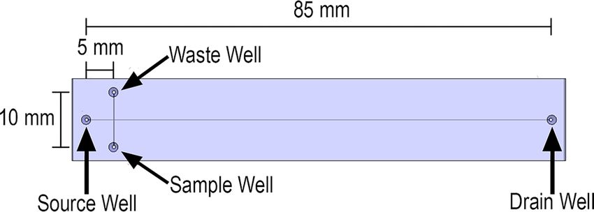
www.nature.com/scientificreports/
Figure 1. Diagram of the T8050 Glass Microchip with a double-T junction at the separation channel.
Fluid actuation by electric field application. To apply electric potential to the microfluidic chip, plati-
num electrodes (Surepure Chemetals, USA) are chosen, as platinum will not corrode under the experimen-
tal conditions of the microchip capillary electrophoresis dairy device. Platinum electrodes were formed from
1.295 mm diameter platinum wire with tapered ends to allow insertion into microfluidic wells without contact-
ing the sides of the wells. Retention plates help secure the platinum electrodes in place. Copper wire electrodes,
being a low-cost alternative, were initially used, however, severe corrosion occurred.
The voltage application to the microfluidic chip was regulated by a four channel, laboratory-built voltage
sequencer with each channel capable of supplying 0–500 V. This voltage sequencer was energized by a Keithley
2290-5 power supply.
Operational procedures. Because the developed system requires complicated design and fabrication of
multiple components, a commercially available microfluidic chip with proven functionality is chosen, being
the T8050 microfluidic chip from Micronit (Enschede, Netherlands). The name of each well is illustrated in the
schematic of the microfluidic chip is presented in Fig. 1.
Prior to experimentation, it is necessary to power the ultraviolet (UV) light emitting diode (LED) for 90 min
to allow the output of the microchip capillary electrophoresis dairy device to stabilize. (This UV LED could be
replaced to reduce this cumbersome warmup period.) In addition, the 1 M NaOH solution is injected into the
microfluidic chip under pressure from a syringe (3 mL) until all four wells are filled. The NaOH is left in the
microfluidic chip for 15 min and then washed out with Millipore water. The microfluidic chip is then drained and
filled with citrate buffer. The citrate is emptied from the sample well using a pipette and the well is subsequently
filled with filtered milk, for antibiotic-absent operation, or a solution of milk and ciprofloxacin hydrochloride
monohydrate, for antibiotic-present operation. It is envisioned that a future iteration of this system would incor-
porate automated chip preparation and sample loading in order to create a fully automated microchip capillary
electrophoresis dairy device. This would make it feasible to implement the system on dairy farms for farmers
to reliably use.
Flow of the buffer solution through the microfluidic channel is a result of electroosmotic flow and segrega-
tion of the milk sample into its composing elements within the electroosmotic flow is a result of electrophoresis.
These combined effects (electroosmotic flow and electrophoresis) on the velocity of any given element within
the buffer solution is
u = uEOF + uEP = (µEOF + µEP )E. (1)
Here the velocity of an element is u, the velocity from electroosmotic flow is uEOF , the electroosmotic flow mobil-
ity is µEOF , the velocity induced by electrophoresis is uEP , the electrophoresis mobility is µEP , and the applied
electric field is E . The electroosmotic flow mobility is typically much larger compared to the electrophoresis
mobility. As such, the electroosmotic flow velocity determines the net direction of the elements and the electro-
phoresis velocity determines how elements separate in the net flow.
During operation of the microchip capillary electrophoresis dairy device, application of 500 V to the sample,
source, and drain wells results in the sample being pulled towards the waste well. The waste well is at a potential of
0 V. This is pinched sample injection, as illustrated in Fig. 2a, with a high potential applied to the sample, source,
and drain wells and ground potential applied to the waste well. The result is electroosmotic flow from sample,
source, and drain wells to the waste well. To perform the electrophoresis, 500 V is applied to the source well,
290 V is applied to the sample and waste wells, and 0 V is applied to the drain well, as in Fig. 2b. The visualization
of this flow would reveal the sample is pinched by the flows from the source and drain while entering the waste
channel, as in Fig. 2a, and then a narrow plug travelling towards the drain well, as in Fig. 2b.
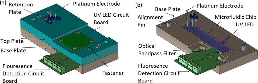
Antibiotic‑absent and antibiotic‑present operation. The microchip capillary electrophoresis dairy
device is first operated with a milk sample containing no antibiotics to establish an antibiotic-absent response.
This antibiotic-absent response is presented in Fig. 3a. When the injection begins, three of the four platinum
electrodes rapidly transition potential from 0 to 500 V. This results in a noise spike in the recorded data and
allows observation of the onset of sample loading and electrophoresis. This is noted in Fig. 3a with injection
starting at 30 s and electrophoresis starting at two minutes and 30 s. It should be observed that as the sample
approaches the UV LED for measurement at approximately 18 min, there is no discernable response, indicating
the ciprofloxacin is not present. It should be noted that through careful selection of voltages and channel lengths
Scientific Reports | (2020) 10:13548 | https://doi.org/10.1038/s41598-020-70566-1 3
Vol.:(0123456789)www.nature.com/scientificreports/
Figure 2. (a) Electric field configuration for pinched sample injection. This portion of the experiment fills the
injection channel. (b) Electric field configuration for separation. This portion of the experiment takes a pinched
sample from the injection step and sends this small sliver towards the drain well.
Figure 3. (a) Antibiotic-absent test of detection system by testing with milk without ciprofloxacin. (b)
Antibiotic-present test of detection system by testing with milk with 0.25 mM concentration of ciprofloxacin.
(with the electric field being approximately equal to the ratio between these values), it is envisioned that the
analysis time can be greatly reduced, in accordance with dairy requirements for analysis t ime37.
The microchip capillary electrophoresis dairy device is then operated with a milk sample containing 0.25 mM
ciprofloxacin to establish an antibiotic-present response (being the change of the output voltage at the temporal
region of interest). This antibiotic-present response is presented in Fig. 3b. As previously noted, the noise spikes
Scientific Reports | (2020) 10:13548 | https://doi.org/10.1038/s41598-020-70566-1 4
Vol:.(1234567890)www.nature.com/scientificreports/
Figure 4. The change in the output voltage as it responds to various concentrations of ciprofloxacin in milk
is shown. Each concentration is repeated (three trials) with the mean displayed. The linear fit R2 value is 0.99.
Error bars are standard error, being the standard deviation divided by the number of trials.
associated with sample loading and electrophoresis are observed at 30 s and two minutes and 30 s, respectively.
As the sample approaches the UV LED for measurement, at the 18 min temporal region of interest, there is a
clear electrophoretic peak, indicating the presence of ciprofloxacin in the milk sample.
The Fig. 3 results should be discussed in terms of potential comigration, where another analyte may travel
along with the ciprofloxacin and interfere with the measured signal. The antibiotic-absent experiment of Fig. 3a
is performed without ciprofloxacin in milk, represented by a flat response. The antibiotic-absent experiment of
Fig. 3b is performed with antibiotics in milk, and this is the only change from the first experiment. Since only
the ciprofloxacin was added, the electrophoretic peak shown in Fig. 3b only represents the fluorescence emitted
by the (isolated) ciprofloxacin. As such, comigration did not play a role.
Operation with varied ciprofloxacin concentrations. To determine the response curve of the micro-
chip capillary electrophoresis dairy device (being the change of the output voltage versus ciprofloxacin con-
centration), multiple concentrations of ciprofloxacin are injected (in separate experimental runs) and the cor-
responding output voltages are measured. The results are as shown in Fig. 4. It can be seen that the change in the
output voltage from the microchip capillary electrophoresis dairy device increases with increasing ciprofloxacin
concentration. It should be mentioned that at concentrations lower than 0.1 mM, the output signal could not be
distinguished from the system noise.
To determine the sensitivity of the microchip capillary electrophoresis dairy device, the slope of the response
curve is inspected. The sensitivity is found to be 10.5 mV/mM. To determine the limit of detection of the micro-
chip capillary electrophoresis dairy device, data is collected over 10 min with a blank sample. Under these blank
sample conditions, the limit of detection is found to be 0.19 mM. The method for determining the limit of detec-
tion is reported in the literature25. Here, the limit of detection is calculated as three times the standard deviation
(for 99% certainty) of the noise within the system and comparing to the peak height for a known concentration.
At high concentrations (i.e., beyond the 1 mM upper domain of Fig. 4), the ciprofloxacin does not stay
in solution. This results in the microfluidic channels (within the microfluidic chip) becoming clogged. We
determine experimentally that when mixing ciprofloxacin and milk at concentrations of 1 mM and greater, the
ciprofloxacin no longer dissolves in the milk, meaning that it becomes saturated in the solution. At a concentra-
tion of 1 mM, the solution was at the onset of no longer being homogeneous. As noted in the literature38, the
maximum concentration of ciprofloxacin in cow milk is 0.09 mM (30 mg/L). Therefore, it is unlikely that the
clogging observed at higher concentrations would be observed for typical on-farm applications of the microchip
capillary electrophoresis dairy device. The milk samples used in this study had ciprofloxacin manually added and
mixed. As such, these lab samples were made at ciprofloxacin concentrations higher than those found in nature,
to demonstrate proof-of-concept. Future work can be focused on further improvement to the MCE device to
reduce the limit of detection for exact industry dairy applications.
Operation of the microchip capillary electrophoresis dairy device as a sensor, with battery operation, can be
discussed. Although it is envisioned that in a dairy barn environment, the device would be permanently installed
in milking parlours (with main power available), the low power draw of the electronics creating the high voltage
(from associated low current) may enable battery operation. This would require an integrated boost convertor
and can be considered in the final market implementation of the microchip capillary electrophoresis dairy device.
Conclusions
The microchip capillary electrophoresis dairy device developed has been shown to be capable of distinguishing
between samples of milk without ciprofloxacin and samples of milk with ciprofloxacin, with a linear correlation
between concentrations of ciprofloxacin and output voltage. Additionally, operation parameters (i.e., the working
voltages and system warmup time) have been established. The microchip capillary electrophoresis dairy device
Scientific Reports | (2020) 10:13548 | https://doi.org/10.1038/s41598-020-70566-1 5
Vol.:(0123456789)www.nature.com/scientificreports/
Figure 5. (a) Complete microchip capillary electrophoresis dairy device. Enclosed in the fixture are the glass
microchip, detection electronics, with the platinum electrodes held by the retention plates. (b) Complete
microchip capillary electrophoresis dairy device with the top plate removed. Seen here is the microfluidic chip
with the UV LED above the chip for analyte excitation.
was found to have a limit of detection of 0.19 mM of ciprofloxacin and sensitivity of 10.5 mV/mM. Future work
could be focused on lowering the limit-of-detection and the analysis time, for use in regulatory control.
Methods
Materials and reagents. Ciprofloxacin hydrochloride monohydrate is purchased from Fischer Scientific
(Ottawa, Canada). Sodium hydroxide (NaOH), sodium citrate dihydrate, and citric acid are purchased from
Sigma Aldrich (Oakville, Canada). Two percent milk is purchased from a local vendor. Water used in this study
is purified by a Milli-Q water purification system (Millipore, USA) to form Milli-Q water.
Solution preparation. Milli-Q water is used to make 1 M NaOH solution and 0.1 M citrate buffer solution.
The buffer solution pH is adjusted to a pH of 5.5 using NaOH. The two percent milk is filtered through 0.45 µM
syringe filters prior to use. Ciprofloxacin hydrochloride monohydrate is added to two percent milk to make solu-
tion at concentrations of 1.00, 0.75, 0.50, 0.25, and 0.10 mM.
Fixture fabrication. In the microchip capillary electrophoresis dairy device, the UV LED, fluorescence
detection PCB, platinum electrodes, and the microfluidic chip all need to be accurately located relative to each
other, while blocking ambient light and preventing short circuits. Therefore, top and base plates are designed
from opaque acetal resin and machined to accommodate proper alignment. The top plate positions the UV LED
and platinum electrodes, which is located relative to the base plate using datum alignment pins. The bottom plate
positions the microfluidic chip and the fluorescence detection PCB (containing the photodiode). This design
allows for the removal of the top plate, along with the platinum electrodes and circuit boards while the sample
is pipetted into the microfluidic chip.
The top and base plates to hold the microfluidic chip are designed using SolidWorks and machined from
opaque acetal resin (DuPont, USA) using a Tormach 770 PCNC (Tormach, USA). The UV LED and fluorescence
PCBs are laboratory designed using Altium Designer (Altium, USA) and manufactured by PCB Unlimited (OR,
USA).
The complete microchip capillary electrophoresis dairy device is presented in Fig. 5a, and the microchip
capillary electrophoresis dairy device with the top plate removed is presented in Fig. 5b (to reveal the based
plate, microfluidic chip, and optical components).
Received: 16 February 2020; Accepted: 24 July 2020
References
1. Sullivan, T. J., Wedner, H. J., Shatz, G. S., Yecies, L. D. & Parker, C. W. Skin testing to detect penicillin allergy. J. Allergy Clin.
Immunol. 68, 171–180 (1981).
2. Yang, G. & Zhao, F. Electrochemical sensor for chloramphenicol based on novel multiwalled carbon nanotubes@molecularly
imprinted polymer. Biosens. Bioelectron. 64, 416–422 (2014).
3. Schenck, F. J. & Callery, P. S. Chromatographic methods of analysis of antibiotics in milk. J. Chromatogr. A https: //doi.org/10.1016/
S0021-9673(97)01291-0 (1998).
4. Neubert, H.-J. Measuring antibiotics in milk. Anal. Chem. 78, 7908–7908 (2006).
5. Bion, C. et al. Analysis of 27 antibiotic residues in raw cow’s milk and milk-based products-validation of delvotest T. Food Addit.
Contam. Part A Chem Anal. Control. Expo. Risk Assess. 33, 54–59 (2015).
6. Beltrán, M. C., Romero, T., Althaus, R. L. & Molina, M. P. Evaluation of the Charm maximum residue limit β-lactam and tetracy-
cline test for the detection of antibiotics in ewe and goat milk. J. Dairy Sci. 96, 2737–2745 (2013).
7. Mitchell, J. M., Griffiths, M. W., McEwen, S. A., McNab, W. B. & Yee, A. J. Antimicrobial drug residues in milk and meat: causes,
concerns, prevalence, regulations, tests, and test performance. J. Food Prot. 61, 742–756 (1998).
8. Salter, R. S. et al. Validation of the Charm 3 SL3 β-lactam test for screening raw milk in compliance with the U.S. pasteurized milk
ordinance. J. AOAC Int. 94, 348–357 (2011).
Scientific Reports | (2020) 10:13548 | https://doi.org/10.1038/s41598-020-70566-1 6
Vol:.(1234567890)www.nature.com/scientificreports/
9. Zhao, Y. et al. Highly integrated microfluidic chip coupled to mass spectrometry for on-line analysis of residual quinolones in
milk. Anal. Chem. 91, 13418–13426 (2019).
10. Liu, Z., Moate, P., Cocks, B. & Rochfort, S. Simple liquid chromatography-mass spectrometry method for quantification of major
free oligosaccharides in bovine milk. J. Agric. Food Chem. 62, 11568–11574 (2014).
11. Xu, F. et al. Immunoassay of chemical contaminants in milk: a review. J. Integr. Agric. 14, 2282–2295 (2015).
12. Gowers, S. A. N. et al. Development of a minimally invasive microneedle-based sensor for continuous monitoring of β-lactam
antibiotic concentrations in vivo. ACS Sens. 4, 1072–1080 (2019).
13. Fernández, F., Pinacho, D. G., Sánchez-Baeza, F. & Marco, M. P. Portable surface plasmon resonance immunosensor for the detec-
tion of fluoroquinolone antibiotic residues in milk. J. Agric. Food Chem. 59, 5036–5043 (2011).
14. DFO. Inhibitor Load Testing Program. Milk Producer (2017).
15. Nichols, J. A. et al. Optical sensing for on-chip digital microfluidics. In: Proceedings of SPIE Conference Microfluid. BioMEMS, Med.
Microsystems X 82510L (2012). https://doi.org/10.1117/12.909390.
16. Strohm, E. M. et al. Sizing biological cells using a microfluidic acoustic flow cytometer. Sci. Rep. 9, 4775 (2019).
17. Gnyawali, V., Strohm, E. M., Wang, J.-Z., Tsai, S. S. H. & Kolios, M. C. Simultaneous acoustic and photoacoustic microfluidic flow
cytometry for label-free analysis. Sci. Rep. 9, 1585 (2019).
18. Zeng, Y., Chen, H., Pang, D. W., Wang, Z. L. & Cheng, J. K. Microchip capillary electrophoresis with electrochemical detection.
Anal. Chem. 74, 2441–2445 (2002).
19. Jin, L. J., Giordano, B. C. & Landers, J. P. Dynamic labeling during capillary or microchip electrophoresis for laser-induced fluo-
rescence detection of protein-SDS complexes without pre- or postcolumn labeling. Anal. Chem. 73, 4994–4999 (2001).
20. Tagit, O. & Hildebrandt, N. Fluorescence sensing of circulating diagnostic biomarkers using molecular probes and nanoparticles.
ACS Sens. 2, 31–45 (2017).
21. Aresta, A., Cotugno, P. & Zambonin, C. Determination of ciprofloxacin, enrofloxacin, and marbofloxacin in bovine urine, serum,
and milk by microextraction by a packed sorbent coupled to ultra-high performance liquid chromatography. Anal. Lett. 52, 790–802
(2019).
22. Official Journal of the European Union L15. Commission regulation (EU) No. 37/2010 on pharmacologically active substances and
their classification regarding maximum residue limits in foodstuffs of animal origin (2010).
23. Sun, H., Zhao, W. & He, P. Effective separation and simultaneous determination of four fluoroquinolones in milk by CE with SPE.
Chromatographia 68, 425–429 (2008).
24. Lara, F. J., García-Campaña, A. M., Alés-Barrero, F., Bosque-Sendra, J. M. & García-Ayuso, L. E. Multiresidue method for the
determination of quinolone antibiotics in bovine raw milk by capillary electrophoresis–tandem mass spectrometry. Anal. Chem.
78, 7665–7673 (2006).
25. Mu, G., Liu, H., Xu, L., Tian, L. & Luan, F. Matrix solid-phase dispersion extraction and capillary electrophoresis determination
of tetracycline residues in milk. Food Anal. Methods 5, 148–153 (2012).
26. Springer, V. H. & Lista, A. G. Micellar nanotubes dispersed electrokinetic chromatography for the simultaneous determination of
antibiotics in bovine milk. Electrophoresis 33, 2049–2055 (2012).
27. Long, C., Deng, B., Sun, S. & Meng, S. Simultaneous determination of chlortetracycline, ampicillin and sarafloxacin in milk using
capillary electrophoresis with electrochemiluminescence detection. Food Addit. Contam. Part A 34, 24–31 (2017).
28. Vera-Candioti, L., Olivieri, A. C. & Goicoechea, H. C. Development of a novel strategy for preconcentration of antibiotic residues
in milk and their quantitation by capillary electrophoresis. Talanta 82, 213–221 (2010).
29. Zhou, L. et al. A label-free and universal platform for antibiotics detection based on microchip electrophoresis using aptamer
probes. Talanta 167, 544–549 (2017).
30. Zhou, L. et al. Microchip electrophoresis array-based aptasensor for multiplex antibiotic detection using functionalized magnetic
beads and polymerase chain reaction amplification. Sens. Actuators B Chem. 263, 568–574 (2018).
31. Blasco, C., Picó, Y. & Andreu, V. Analytical method for simultaneous determination of pesticide and veterinary drug residues in
milk by CE-MS. Electrophoresis 30, 1698–1707 (2009).
32. Wang, S., Yang, P. & Cheng, Y. Analysis of tetracycline residues in bovine milk by CE-MS with field-amplified sample stacking.
Electrophoresis 28, 4173–4179 (2007).
33. Moreno-González, D., Hamed, A. M., Gilbert-López, B., Gámiz-Gracia, L. & García-Campaña, A. M. Evaluation of a multiresidue
capillary electrophoresis-quadrupole-time-of-flight mass spectrometry method for the determination of antibiotics in milk samples.
J. Chromatogr. A 1510, 100–107 (2017).
34. Santos, S. M., Henriques, M., Duarte, A. C. & Esteves, V. I. Development and application of a capillary electrophoresis based
method for the simultaneous screening of six antibiotics in spiked milk samples. Talanta 71, 731–737 (2007).
35. Hall, G. H., Glerum, D. M. & Backhouse, C. J. Light emitting diode, photodiode-based fluorescence detection system for DNA
analysis with microchip electrophoresis. Electrophoresis 37, 406–413 (2016).
36. Murillo Pulgarín, J. A., Alañón Molina, A. & Muñoz Fernández, L. Determination of ciprofloxacin, the major metabolite of enro-
floxacin, in milk by isopotential fluorimetry. J. Agric. Food Chem. 56, 8838–8843 (2008).
37. Watters, R. D. et al. The effect of manual and mechanical stimulation on oxytocin release and milking characteristics in Holstein
cows milked 3 times daily. J. Dairy Sci. 98, 1721–1729 (2015).
38. Mahmood, T., Abbas, M., Ilyas, S., Afzal, N. & Nawaz, R. Quantification of fluoroquinolone (enrofloxacin, norfloxacin and cipro-
floxacin) residues in cow milk. Int. J. Chem. Biochem. Sci. 10, 10–15 (2016).
Acknowledgements
The authors acknowledge financial support from the Natural Sciences and Engineering Research Council of
Canada, Ontario Centres of Excellence (and Larry Wood of Norwell Dairy Systems), and the Canada Founda-
tion for Innovation.
Author contributions
R.B and C.M.C. conceived and designed the experiments and components; R.B. and J.D. performed the experi-
ments and analyzed the data; A.S., and C.M.C. provided faculty supervision; R.B, J.D, and C.M.C. co-wrote the
manuscript; all authors reviewed the manuscript.
Competing interests
The authors declare no competing interests.
Additional information
Correspondence and requests for materials should be addressed to C.M.C.
Reprints and permissions information is available at www.nature.com/reprints.
Scientific Reports | (2020) 10:13548 | https://doi.org/10.1038/s41598-020-70566-1 7
Vol.:(0123456789)www.nature.com/scientificreports/
Publisher’s note Springer Nature remains neutral with regard to jurisdictional claims in published maps and
institutional affiliations.
Open Access This article is licensed under a Creative Commons Attribution 4.0 International
License, which permits use, sharing, adaptation, distribution and reproduction in any medium or
format, as long as you give appropriate credit to the original author(s) and the source, provide a link to the
Creative Commons license, and indicate if changes were made. The images or other third party material in this
article are included in the article’s Creative Commons license, unless indicated otherwise in a credit line to the
material. If material is not included in the article’s Creative Commons license and your intended use is not
permitted by statutory regulation or exceeds the permitted use, you will need to obtain permission directly from
the copyright holder. To view a copy of this license, visit http://creativecommons.org/licenses/by/4.0/.
© The Author(s) 2020
Scientific Reports | (2020) 10:13548 | https://doi.org/10.1038/s41598-020-70566-1 8
Vol:.(1234567890)You can also read