Morphological structures and histochemistry of roots and shoots in Myricaria laxiflora (Tamaricaceae)
←
→
Page content transcription
If your browser does not render page correctly, please read the page content below

Open Life Sciences 2021; 16: 455–463
Research Article
Linbao Li#, Di Wu#, Qiaoling Zhen, Jun Zhang, Liwen Qiu, Guiyun Huang*, Chaodong Yang*
Morphological structures and histochemistry
of roots and shoots in Myricaria laxiflora
(Tamaricaceae)
https://doi.org/10.1515/biol-2021-0049 and a cuticle at the young stage and cork at the mature
received May 24, 2020; accepted February 17, 2021 stage. The leaves had two layers of palisade tissue, a hya-
Abstract: Myricaria laxiflora (Tamaricaceae) is an endan- line epidermis, sunken stomata, and a thick, papillose
gered plant that is narrowly distributed in the riparian cuticle. Aerenchyma presented in the roots and shoots.
zone of the Three Gorges, along the Yangtze River, China. Several Myr. laxiflora structures, including aerenchyma,
Using bright-field and epifluorescence microscopy, we apoplastic barriers in the roots and shoots, were adapted
investigated the anatomical and histochemical features to riparian habitats. In addition, shoots had typical xero-
that allow this species to tolerate both submerged and phyte features, including small leaves, bilayer palisade
terrestrial environments. The adventitious roots of Myr. tissues, sunken stomata, a thick, papillose cuticle, and a
laxiflora had an endodermis with Casparian bands and hyaline epidermis. Thus, our study identified several ana-
suberin lamellae; the cortex and hypodermal walls had tomical features that may permit Myr. laxiflora to thrive in
lignified thickenings in the primary structure. In the the riparian zone of the Three Gorges, China.
mature roots, the secondary structure had cork. The apo- Keywords: aerenchyma, apoplastic barriers, endodermis,
plastic barriers in stems consisted of a lignified fiber ring lignified cortex and hypodermis, thick papillose cuticle
# Linbao Li and Di Wu are contributed equal to this work, and as 1 Introduction
co-first authors.
Myricaria laxiflora (Tamaricaceae) is an endangered spe-
cies that prior to the construction of the Three Gorges
* Corresponding author: Guiyun Huang, Rare Plants Research
Institute of Yangtze River, China Three Gorges Corporation, Yichang, Dam (TGD) was narrowly distributed in the riparian
Hubei 443000, China, e-mail: huang_guiyun@ctg.com.cn zone along the Yangtze River, China, from Banan County,
* Corresponding author: Chaodong Yang, Engineering Research Chongqing Province, to Zhijiang County, Hubei Province
Center of Ecology and Agriculture Use of Wetland, Ministry of [1–5]. After TGD construction was completed in 2009, only
Education, Hubei Key Laboratory of Waterlogging Disaster and
a few natural populations of Myr. laxiflora remained, all
Agricultural Use of Wetland, Yangtze University, Jingzhou, Hubei
434025, China, e-mail: 546728708@qq.com
downstream of Yidu and Zhijiang counties; as Myr. laxiflora
Linbao Li: Rare Plants Research Institute of Yangtze River, China habitats upstream of the TGD were lost, some of the plants
Three Gorges Corporation, Yichang, Hubei 443000, China, from the upstream localities have been preserved ex situ
e-mail: li_linbao@ctg.com.cn [4,6–9]. In its native environment along the Yangtze River,
Di Wu: Rare Plants Research Institute of Yangtze River, China Myr. laxiflora remains dormant while completely submerged
Three Gorges Corporation, Yichang, Hubei 443000, China,
during summer flood pulses and then sprouts in the autumn
e-mail: wu_di3@ctg.com.cn
Qiaoling Zhen: Rare Plants Research Institute of Yangtze River, and winter after the floods recede [1–5]. Myr. laxiflora may
China Three Gorges Corporation, Yichang, Hubei 443000, China, represent a promising plant with which to restore the
e-mail: zhen_qiaoling@ctg.com.cn ecology of Yangtze River after the degradation associated
Jun Zhang: Rare Plants Research Institute of Yangtze River, China with TGD construction [8–14].
Three Gorges Corporation, Yichang, Hubei 443000, China,
Like many other wetland plants, Myr. laxiflora is typi-
e-mail: zhang_jun15@ctg.com.cn
Liwen Qiu: Rare Plants Research Institute of Yangtze River, China
cally subjected to anoxic submersion during summer
Three Gorges Corporation, Yichang, Hubei 443000, China, flooding [2,3,15–17]. Aquatic and amphibious plants have
e-mail: 415777682@qq.com aerenchyma and tight barriers to store and retain oxygen
Open Access. © 2021 Linbao Li et al., published by De Gruyter. This work is licensed under the Creative Commons Attribution 4.0
International License.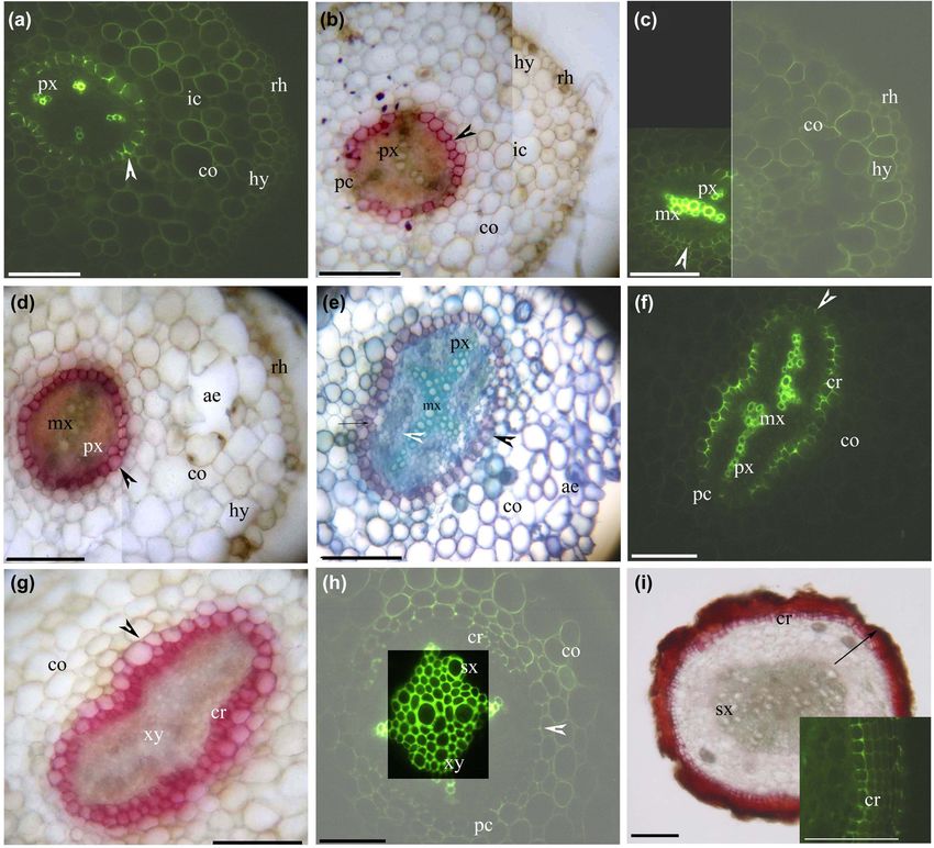
456 Linbao Li et al.
in anoxic conditions and during water–solute exchanges the Yangtze River in Yidu County, Hubei, China. Approxi-
[18–21]. In the amphibious species Cynodon dactylon, mately 50 adventitious roots and 20 shoots with leaves
Artemisia lavandulaefolia, and Alternanthera philoxer- were collected from 10 individuals.
oides, which we have studied from the Jianghan Plain Adventitious root and leaf samples were fixed in for-
down to the Three Gorges, air spaces included aer- maldehyde–alcohol–acetic acid immediately following
enchyma and pith cavities in roots and shoots, and bar- collection [51]. After fixation, the root tissues were sec-
riers included the endodermis, exodermis, and suberized tioned freehand under a stereoscope (JNOEC JSZ6, China),
peripheral ring [22–25]. Myr. laxiflora growing in the using a two-sided blade razor. Adventitious root samples
riparian zone of the Yangtze River may have aerenchyma (∼30–80 mm long) were sectioned at 10, 20, 30, 40, or
and structures similar to these amphibious species. 50 mm from the root tip. Aged tissue with attached cortex
Other species in the Tamaricaceae that are closely was sloughed off. Each distance from the root tip was repre-
related to Myr. laxiflora have diverse habitats and are widely sented by 3–6 sections from different samples per stain.
distributed in mountainous, cold, and arid regions world- Shoot bases were immersed in tap water immediately
wide as well as in those with saline-alkali soils. Plants in the following collection. Shoots (∼150–270 mm long) were
Tamaricaceae are often used for ecological restoration sectioned at 10, 20, 30, 40, and 50 mm from the shoot
[26–33]. In this family, xerophyte shoots have abundant apex. Each distance from the shoot base was represented
palisade tissues under the epidermis [34–41]; the epidermis by 3–6 sections from different samples per stain. Sections
itself has a thick, papillose cuticle [28,36,42] and sunken (10–30 µm thick) were cut in the middle of the seedling
stomata [38,43]. In addition, species that belong to Tamar- leaves. Leaves were represented by 3–6 sections from
icaceae have deep roots [32,34,44–46], which represent an different samples per stain.
adaptation to drought stress [47–49].
The structure and physiology of Myr. laxiflora seeds
and shoots may play important roles in the propagation
of this species as well as its invasion of new habitats 2.2 Histochemistry and microscopy
[8–14,29,50]. However, little is known of the anatomical
and histochemical features that allow Myr. laxiflora to tol- Sections were stained with one of three stains. SR7B was
erate submersion and exposure. To our knowledge, the only used to identify suberin in the cell walls [52], BAB was
relevant previous study of this species showed that the used to identify Casparian bands and lignin in the cell
surfaces of young branches had smooth, thin cuticles [29]. walls [53,54], and TBO was used to visualize tissue struc-
To address this knowledge gap, we aimed to investi- tures [55,56]. All specimens were examined using bright-
gate whether the anatomical and histochemical features field microscopy under a Leica DME microscope and
of Myr. laxiflora were consistent with its tolerance to sub- photographed with a digital camera (Nikon E5400, Japan).
mersion as well as to diverse terrestrial environments. Specimens stained with BAB were viewed under an
Evidence of such adaptative features might help to explain Olympus IX71 epifluorescence microscope and photo-
the ability of this plant to thrive despite summer dormancy graphed with a digital camera (RZ200C–21, China) [22].
and to grow in diverse terrestrial environments during the
spring, autumn, and winter. To study the structures of
roots and shoots, we analyzed the anatomical and histo-
chemical characters of Myr. laxiflora samples, primarily 3 Results and discussion
using berberine hemisulfate–aniline blue (BAB) to visua-
lize Casparian bands and lignified walls, Sudan red 7B
3.1 General structure
(SR7B) to visualize suberin lamellae, and toluidine blue
O (TBO) to visualize anatomical features.
Myr. laxiflora had thick adventitious roots (Figure 1), fine
adventitious roots (Figure 2), and shoots (Figures 3 and 4).
The thick adventitious roots possessed four to five layers
2 Materials and methods of cortex cells in the primary structure (Figure 1a–d); in
the secondary structure, the cortex sloughed off with the
bark (Figure 1e–i). The fine adventitious roots contained
2.1 Sample collection and processing one or two layers of cortex cells in the primary structure
and only cork in the secondary structure (Figure 2). Both
In October 2019, we collected adventitious roots, stems, thick and thin adventitious roots had diarch to tetrarch stele
and leaves specimens of Myr. laxiflora at the riparian of with differentiated proto- and metaxylem, a cortex with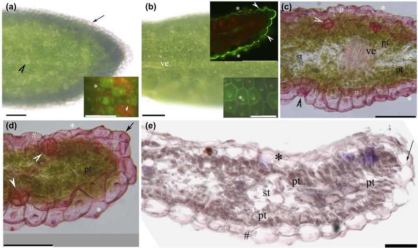
Morphological structures and histochemistry of Myr. laxiflora 457 Figure 1: Photomicrographs of thick adventitious roots (50–80 mm long) of Myricaria laxiflora, showing some of the secondary growth; scale bars = 50 µm. (a) Protoxylem, endodermis (arrowhead), lignified cortex, intercellular space, hypodermis, and rhizodermis. Staining: BAB. (b) Protoxylem, endodermis (arrowhead), passage cells, cortex, intercellular space, hypodermis, and rhizodermis. Staining: SR7B. (c) Protoxylem, metaxylem, endodermis (arrowhead), lignified cortex, hypodermis, and rhizodermis. Staining: BAB. (d) Protoxylem, meta- xylem, endodermis (arrowhead), cortex, aerenchyma, hypodermis, and rhizodermis. Staining: SR7B. (e) Protoxylem, vascular cambia (white arrowhead), divided pericycle (arrow), endodermis (black arrowhead), cortex, and aerenchyma. Staining: TBO. (f) Protoxylem, metaxylem, cork, endodermis (arrowhead), passage cells, and cortex. Staining: BAB. (g) Primary xylem, cork, endodermis (arrowhead), and cortex. Staining: SR7B. (h) Primary xylem, secondary xylem, cork, endodermis (arrowhead), passage cells, and lignified cortex. Staining: BAB. (i) Secondary xylem, cork, and bark (whole arrow). Staining: SR7B. Inset shows cork. Staining: BAB. Abbreviations used in the figure are as follows: ae – aerenchyma; BAB – berberine sulfate–aniline blue; ch – chloroplast; cr – cork; co – cortex; cu – cuticle; ep – epidermis; hy – hypodermis; ic – intercellular space; mx – metaxylem; pc – passage cells; pt – palisade tissue; f – phloem fibers; pi – pith; xy – primary xylem; px – protoxylem; rh – rhizodermis; st – spongy tissue; SR7B – Sudan red 7B; sx – secondary xylem; TBO – toluidine blue O; ve – vein. an endodermis, a hypodermis, and a rhizodermis. The The Myr. laxiflora stem possessed cork and an epidermis cortex and hypodermal walls had lignified thickenings. as well as a lignified phloem fiber ring enclosing a central Aerenchyma were present in the root cortices. cylinder of bundles internal to the cortex (Figure 3a–i).

458 Linbao Li et al. Figure 2: Photomicrographs of Myricaria laxiflora fine adventitious roots (30–50 mm long); scale bars = 50 µm. (a) Protoxylem, endodermis (arrowhead), lignified cortex, intercellular space, hypodermis, and rhizodermis. Staining: BAB. (b) Endodermis (arrowhead), passage cells, cortex, hypodermis, and rhizodermis. Staining: SR7B. (c) Protoxylem, metaxylem, endodermis (arrowhead), passage cells, lignified cortex, intercellular space. Staining: BAB. (d) Protoxylem, metaxylem, endodermis (arrowhead), cortex. Staining: SR7B. (e) Primary xylem, cork. Staining: BAB. (f) Cork. Staining: SR7B. The Myr. laxiflora leaf had palisade tissue, a hyaline mar- (mature adventitious roots), the cortex and hypodermis ginal epidermis, stomata, and a cuticle with a papillose had been sloughed off, the stele has a secondary xylem, surface (Figure 4a–f). and the cork had suberized to form bark (Figure 1i). 3.2 Structure of the thick adventitious root 3.3 Structure of the fine adventitious root At 10 mm from the root tip, the stele had diarch and At 10 mm from the root tip, Casparian bands, suberin tetrarch protoxylem poles, the endodermis had Casparian lamellae, and passage cells were present on the endo- bands and almost complete suberin lamellae (only a few dermis, the cortex and hypodermal walls had ligni- passage cells), the cortex and hypodermal walls had ligni- fied thickenings, and the stele had a diarch protoxylem fied thickenings, and an intercellular space appeared (Figure 2a and b). At 20 mm from the root tip, the endo- within the cortex (Figure 1a and b). At 20 mm from the dermis had almost complete suberin lamellae with a few root tip, the stele had metaxylem poles, the endodermis passage cells, the stele had a metaxylem, and the cortex had almost complete suberin lamellae, the cortex had irre- and hypodermis begin to slough off (Figure 2c and d). At gular lysigenous aerenchyma, and the rhizodermis was 30 mm from the root tip, the pericycle redivided to form still intact (Figure 1c and d). At 30 mm from the root tip, a phellogen and produce suberized cork, while the stele redivided pericycle also formed phellogen to begin cork had only primary xylem (Figure 2e and f). production and the endodermis had few passage cells We demonstrated that the primary structures of the (Figure 1e–g). At about 40 mm from the root tip, the peri- thick and fine adventitious roots exhibit similar anato- cycle over the protoxylem poles and the cells between mical and histochemical features of Myr. laxiflora. Myr. the primary xylem and the primary phloem had become laxiflora roots had a suberized endodermis and a lignified a vascular cambium to initiate the secondary xylem, the hypodermis, while the cortex and hypodermal walls had cork was partially undeveloped, and the cortex begin to lignified thickenings near the endodermis. The cortex of slough off (Figure 1e–h). At >50 mm from the root tip the thick adventitious roots had more cell layers than that
Morphological structures and histochemistry of Myr. laxiflora 459
Figure 3: Photomicrographs of Myricaria laxiflora stems (150–270 mm long). Scale bars = 50 µm. (a) Pith, primary xylem, phloem fibers,
cortex, intercellular space, and epidermis. Staining: TBO. (b) Pith, primary xylem, phloem fibers, cortex, chloroplast, epidermis, and cuticle.
Staining: BAB. Inset shows phloem fibers and cuticle. Staining: SR7B. (c) Secondary xylem, vascular cambia (arrowhead), phloem fibers,
cortex, divided cork, and epidermis. Staining: TBO. (d) Pith, secondary xylem, phloem fibers, cortex, and cuticle. Staining: BAB.
(e) Secondary xylem, phloem fibers, cortex, intercellular space, chloroplast, cork, and cuticle. Staining: SR7B. (f) Secondary xylem,
vascular cambia (arrowhead), phloem fibers, cortex, intercellular space, cork, and epidermis. Staining: TBO. (g) Pith, secondary xylem,
phloem fibers, cortex, cork, and cuticle. Staining: BAB. (h) Secondary xylem, phloem fibers, cortex, cork, and cuticle. Staining: SR7B.
(i) Pith, secondary xylem, phloem fibers, cortex, cork, and cuticle. Staining: BAB. Inset shows bark (arrow). Staining: SR7B.
of the fine adventitious roots. In addition, the thick this cortex lacks lignified walls); in addition, unlike Myr.
adventitious roots had a secondary structure containing laxiflora, the roots of O. javanica are surrounded by aer-
cork, as commonly observed in eudicots [57–59]. In contrast, enchyma, and the walls possess suberin lamellae [60].
the fine adventitious roots had only primary xylem. The cortices and hypodermis of the aquatic roots of Alt.
The young roots of Myr. laxiflora were similar in philoxeroides have lignified walls and aerenchyma [25],
structure to the young roots of Oenanthe javanica and as well as broccoli and Cardamine hupingshanensis [61–63].
Alt. philoxeroides [25,60]. However, the hypodermis of It is possible that the lignified thickenings we observed in
O. javanica has more cell layers than that of Myr. laxiflora the roots of Myr. laxiflora relate to the riparian habitats of
as well as a cortex with spacious aerenchyma (although the Three Gorges [25,61–63].460 Linbao Li et al.
Figure 4: Photomicrographs of Myricaria laxiflora leaves. Scale bar = 50 µm. (a) Adaxial surface, stomata (arrowhead), and hyaline
epidermal margin (arrow). Unstained. Inset shows stomata (arrowhead), and fine papillae (*). Staining: BAB. (b) Abaxial surface and vein.
Unstained. Upper inset shows lower and marginal epidermal walls with thick cuticle (arrowhead) and papillae (*). Staining: BAB. Lower
inset shows large papillae (*). Staining: BAB. (c) Middle blade, vein, upper epidermis with cuticle and fine papillae (white #), lower
epidermis with cuticle and large papillae (gray #), stomata (arrowhead), palisade tissue, aerenchyma, spongy tissue, and papillae (*).
Staining: SR7B. (d) Marginal blade, upper epidermis with cuticle and fine papillae (white #), lower epidermis with cuticle and large papillae
(gray #), stomata (arrowhead), palisade tissue, papillae (*), and marginal epidermis (arrow). Staining: SR7B. (e) Blade, upper epidermis (*),
lower epidermis (#), palisade tissue, spongy tissue, and hyaline epidermal margin (arrow). Staining: TBO.
The roots of wetland or aquatic eudicots from Jianghan a spacious pith was present in the center (Figure 3a and b).
Plain (China) or from the Amazon Basin, such as Art. At 30–40 mm from the new shoot apex, the vascular cam-
lavandulaefolia, Art. selengensis, Ranunculus trichophyllus, bium produced an internal secondary xylem; the phloem
and Tabernaemontana juruana, possess an endodermis, fibers had strengthened and lignified; and the cortical cells
a uniseriate exodermis, and a cortex that lacks lignified had redivided to form suberized cork, one cell layer thick,
walls [24,64,65]. In contrast, the roots of wetland grasses, under the epidermis (Figure 3c–e). At the new shoot base,
such as Oryza sativa, Phalaris arundinacea, Phragmites the cork had several layers of suberized cells (Figure 3f–h).
australis, and Zizania latifolia, possess an endodermis In 1-year-old shoots, the cylinder bundles had spacious
and a multiseriate exodermis [20,22,23,66,67]. The bar- secondary xylem, and the cork has suberized to form
riers of these wetland or aquatic species were stronger bark (Figure 3i). Intercellular spaces and chloroplasts were
than Myr. laxiflora with an endodermis and lignified present in the stem cortices (Figure 3a, b, e, and f).
cortex as well as hypodermis in roots. Young stems of Myr. laxiflora possessed a lignified
fiber ring, a thick cuticle, and a cortex either with chloro-
plasts and small aerenchyma or with one layer of cork
3.4 Stem structure cells. In contrast, mature stems had prominent secondary
xylem in the center of the stem and a thick bark. In con-
The stem had a lignified phloem fiber ring, enclosing a trast, Zhang et al. [29] found that young branches of Myr.
central cylinder of bundles internal to the cortex, and an laxiflora had smooth, thin cuticles. The lignified fiber ring
epidermis with a thick cuticle. At 10 mm from the new in the young stems of Myr. laxiflora was similar to the
shoot apex, the fiber ring enclosed vascular bundles, and lignified sclerenchymal ring observed in C. hupingshanensis,Morphological structures and histochemistry of Myr. laxiflora 461
P. arundinacea, and Z. latifolia [22,23,63,67]. This ring might Alt. philoxeroides aquatic roots, even though Alt. philox-
serve to increase the mechanical strength of the young stem. eroides shoots have large air spaces [25], and the leaves
Suberized cork, which is commonly observed in eudicots have lysigenous of xeromorphic New Zealand hemp [59],
[58,59], is similar to the phellem of Art. selengensis and while Myr. laxiflora shoots have narrow intercellular spaces.
Alt. philoxeroides [24,25] and to the suberized and lignified In contrast, the shoots of wetland plants, such as Pas.
peripheral mechanical ring in the Paspalum distichum, Pha. distichum, Art. lavandulaefolia, and Art. selengensis, have
arundinacea, and Z. latifolia [22,23,67]. This suggests that spacious pith cavities and cortical lacunae, which might
Myr. laxiflora’s tolerance to flooding is typical of amphibious facilitate survival when submerged over long periods
plants. The shoot cortices have chloroplasts, which are pre- [22–24,64,67].
sent in other Tamaricaceae species [28,30,68]. These obser-
vations indicate that Myr. laxiflora has features that belong
to Tamaricaceae taxology and serve as adaptations to the
environments in the Three Gorges. 4 Conclusion
We identified that Myr. laxiflora have typical amphibious
plant features, including apoplastic barriers consisting of
3.5 Leaf structure
the endodermis, lignified wall thickenings, cork, and cuticle
as well as the aerenchyma, suggesting that Myr. laxiflora is
The upper surface of the leaf has obvious stomata, small
well adapted to the riparian habitats of the Three Gorges
epidermal cells, a thin cuticle, and fine papillae. The
along the Yangtze River [16–19,24,25,54,60,63,72]. The shoots
lower surface and edge of the leaf have sunken stomata,
of Myr. laxiflora have typical xerophyte features, common
large epidermal cells, a thick cuticle, and large papillae
across the Tamaricaceae, including small leaves, bilayer pali-
(Figure 4a–d, e). The edges of the epidermal cells are
sade tissues, sunken stomata, a thick papillose cuticle, and a
hyaline (Figure 4a and e). Palisade tissue was observed
largely hyaline epidermis [26,28,30,35–37,40,42,59,68,69].
below and above the adaxial and abaxial epidermis,
Our results help to explain how the rare plant Myr. laxiflora
respectively; scant spongy mesophyll tissue was observed
survives in flooded and receded environments and may help
between the layers of palisade tissue (Figure 4c–e).
to contextualize the taxonomy, evolution, and phylogeny of
Aerenchyma was present in the middle of the leaf blade
Myr. laxiflora within Tamaricaceae.
(Figure 4c).
The leaves of Myr. laxiflora are typical of xerophytes
Funding information: This work was supported by the
adapted to arid environments: they are small and have
China Three Gorges Corporation (2019H210), and the Engi-
two layers of palisade tissue, sunken stomata, and a thick
neering Research Center of Ecology and Agriculture Use of
papillose cuticle. Bilayer palisade tissues are also found
Wetland, Ministry of Education opening fund, Yangtze
in several other xerophytes, including Myr. bracteate
University (KFT202004).
[26], Myr. germanica [70], Reaumuria soongoriea [35],
Tamarix ramosissima [30], Elaeagnus angustifolia [36],
Conflict of interest: The authors state no conflict of
Eschweilera tenuifolia [69], Populus euphratica [36,40],
interest.
Peganum nigellastrum [39], Alhagi sparsifolia [34], and
Ziziphus jujuba var. spinosa [41]. Similarly, the leaves of
Data availability statement: The datasets generated during
several xerophyte plants, including Myr. germanica [70],
and/or analyzed during the current study are available
Tam. laxa [28,42], Tam. ramosissima [30], Tam. chinensis
from the corresponding author on reasonable request.
[68], Ela. angustifolia [36], and Caragana spp. [37,38],
have sunken stomata, thick cuticles, and surface papillae
[59]. The epidermis at the abaxial margins of the leaves
of Myr. laxiflora was largely hyaline and may function
similar to the white hairs on xerophyte leaves [36,37] or
References
the hyaline tips of bryophyte leaves [71].
[1] Zhang PY, Zhang YJ. A study on the taxonomy of the genus
In plant tissues, aerenchyma help to retain oxygen
Myricaria Desv. in China. Bull Bot Res. 1984;4:67–80.
when the plant is submerged, in order to improve sur- [2] Wu JQ, Zhao ZE, Jin YX, Shen ZH. Investigation and study on the
vival [15–17,20,72,73]. The roots of Myr. laxiflora had aer- endemic plant Myricaria laxiflora in the Three-Gorge Reservoir
enchyma and histochemical features similar to those of area. J Wuhan Bot Res. 1998;16:111–6.462 Linbao Li et al.
[3] Li ZZ, Wang CH, Xu TQ, Wu JQ, Huang HW. Conservation [21] Ranathunge K, Lin J, Steudle E, Schreiber L. Stagnant deoxy-
genetics of the endemic species Myricaria laxiflora genated growth enhances root suberization and lignifications,
(Tamaricaceae) in the Three-Gorges Reservoir area, Hubei. but differentially affects water and NaCl permeabilities in rice
Biodivers Sci. 2003;11:109–17. (Oryza sativa L.) roots. Plant Cell Environ. 2011;34:1223–40.
[4] Wang Y, Wu JQ, Tao Y, Li Z, Huang H. Natural distribution and [22] Yang CD, Zhang X, Zhou CY, Seago Jr JL. Root and stem
ex situ conservation of endemic species Myricaria laxiflora in anatomy and histochemistry of four grasses from the Jianghan
water-level-fluctuation zone within Three Gorges Reservoir Floodplain along the Yangtze River. China Flora.
area of Changjiang River. J Wuhan Bot Res. 2003;21(5):415–22. 2011;206:653–61.
[5] Tao Y, Li JQ, Jiang MX, Jin X. Study on diversity of structural [23] Zhang X, Hu LJ, Yang C, Zhou C, Yuan L, Chen Z, et al. Structural
characters of Myricaria laxiflora. J Wuhan Bot Res. features of Phalaris arundinacea L. in the Jianghan Floodplain
2004;22:315–22. of the Yangtze River. China Flora. 2017;229:100–6.
[6] Bao DC, Lu ZJ, Jiang MX, Xu SD, Yao Q, Liu QF, et al. Population [24] Zhang X, Yang C, Seago Jr JL. Anatomical and histochemical
structure and dynamics of remanent Myricaria laxiflora traits of roots and stems of Artemisia lavandulaefolia and
downstream from the Three Gorges Dam. J Wuhan Bot Res. A. selengensis (Asteraceae) in the Jianghan Floodplain. China
2010;28:711–7. Flora. 2018;239:87–97.
[7] Tian H, Kang M, Liu Y, Ye Q, Yao X. High genetic diversity in [25] Yang CD, Yang XL, Zhang X, Zhou CY, Zhang F, Wang XE, et al.
remnant natural populations of Myricaria laxiflora, a species Anatomical structures of alligator weed (Alternanthera
once considered to be extinct in the wild. Aquat Bot. philoxeroides) suggest it is well adapted to the aquatic–
2012;103:48–53. terrestrial transition zone. Flora. 2019;253:27–34.
[8] Qin HW, Liu ZX, Zhong Y, Liu R, Zheng LD, Su HY. The effects of [26] Wei Y, Tan DY, Yin LK. The discussions on the anatomical
submergence and waterloggingon growth and regrowth of structure of leaf and its taxonomic relationship of
endangered species Myricaria laxiflora. Chin Agric Sci Bull. Tamaricaceae in china. Acta Bot Boreali-Occident Sin.
2014;30:284–8. 1999;19:113–8.
[9] Chen F, Guan S, Ma Y, Xie Z, Lv K, Huang Y, et al. Impact of [27] Qong M, Takamura H, Hudaberdi M. Formation and internal
regulated water level fluctuations on the sexual reproduction structure of Tamarix cones in the Taklimakan Desert. J Arid
of remnant Myricaria laxiflora populations. Glob Ecoll Conserv. Environ. 2002;50:81–97.
2019;18:e00628. [28] Zhang DY, Tan DY, Zhang J, Pan BR. Comparative anatomy of
[10] Chen FQ, Xie ZQ. Reproductive allocation, seed dispersal and young branches of 16 species of Tamarix from China with
germination of Myricaria laxiflora, an endangered species in reference to their ecological significance. Acta Bot Yunnanica.
the Three Gorges Reservoir area. Plant Ecol. 2007;191:67–75. 2003;25(6):653–62.
[11] Chen FQ, Xie ZQ. Survival and growth responses of Myricaria [29] Zhang DY, Zhang J, Tan DY, Pan BR. Anatomical observation of
laxiflora seedlings to summer flooding. Aquat Bot. young branches of 6 species of Tamaricaceae from China. Acta
2009;90:333–8. Bot Boreali-Occident Sin. 2003;23:380–8.
[12] Chen FQ, Xie ZQ. The physiological and biochemical responses [30] Gong WC, Zhuang L, Zhao WQ, Tian ZP. Anatomical structure
of endangered Myricaria laxiflora to simulated summer and ecological adaptability of two kinds of halophytes
flooding. J Trop Subtrop Bot. 2009;17:249–53. (Haloxylon ammondendron Chenopodiaceae and Tamarix
[13] Yuan WQ, Zhan HY, Chen FQ, Xia HW, Luo YC, Liu CC. Ecological ramosissima Tamaricaceae). Acta Ecol Sin. 2009;29:6764–71.
characteristics of seed germination of an endangered species [31] Liu Y, Wang Y, Huang H. Species‐level phylogeographical his-
Myricaria laxiflora. Ecol Environ. 2008;17(6):2341–5. tory of Myricaria plants in the mountain ranges of western
[14] Ma YR, Guan SP, Chen FQ, Lv K. Characteristics of seed China and the origin of Myr. laxiflora in the Three Gorges
germination and seedling growth of Myricaria laxiflora under mountain region. Mol Ecol. 2009;18:2700–12.
different groundwater table treatments. Bot Res. [32] Shan LS, Li Y, Geng DM, Dong QL. Ecological adaptation of
2018;7(02):150–7. Reaumuria soongorica root system architecture to arid
[15] Vartapetian BB, Jackson MB. Plant adaptations to anaerobic environments. Sci Cold Arid Reg. 2014;32:150–8.
stress. Ann Bot. 1997;79(Supplement A):3–20. [33] Zhang ML, Meng HH, Zhang HX, Vyacheslav BV, Sanderson SC.
[16] Jackson MB, Colmer TD. Response and adaptation by plants to Himalayan origin and evolution of Myricaria (Tamaricaeae) in
flooding stress. Ann Bot. 2005;96:501–5. the Neogene. PLoS One. 2014;9:e97582.
[17] Bailey–Serres J, Voesenek LACJ. Flooding stress: acclimations [34] Chen CC, Sun YW, Chang KL. A preliminary study on the mor-
and genetic diversity. Annu Rev Plant Biol. 2008;59:313–39. phology and anatomy of the dominant of the vegetations dis-
[18] Colmer TD, Gibberd MR, Wiengweera A, Tinh TK. The barrier to tributing along the middle and low drainage basin of the Shor-
radial oxygen loss from roots of rice (Oryza sativa L.) is ler River based on the view point of ecology. J Lanzhou Univ.
induced by growth in stagnant solutions. J Exp Bot. 1961;3:61–96.
1998;49:1431–6. [35] Wang YZ, Wang XL, Li Y. Observation on leaf structure of some
[19] Enstone DE, Peterson CA, Ma F. Root endodermis and exo- species in desert steppe. J Lanzhou Univ. 1983;19:87–96.
dermis: structure, function, and responses to the environment. [36] Luo XY, Deng YB. Anatomical observation of leaves and
J Plant Growth Regul. 2003;21:335–51. assimilative branches on several xerophytes structure in
[20] Kotula L, Ranathunge K, Schreiber L, Steudle E. Functional and Xinjiang. J Xinjiang Univ. 1986;1:77–84.
chemical comparison of apoplastic barriers to radial oxygen [37] Yang G, Wang CG. A preliminary study on the stem and leaf
loss in roots of rice (Oryza sativa L.) grown in aerated or structure of several xerophytes in Lop Nur area. Arid Zone Res.
deoxygenated solution. J Exp Bot. 2009;60:2155–67. 1984;1:57–63.Morphological structures and histochemistry of Myr. laxiflora 463
[38] Yan L, Li H, Liu Y. The anatomical ecology studies on the leaf of [57] Ginzburg C. Organization of the adventitious root apex in
13 species in Caragana genus. J Arid Land Resour Environ. Tamarix aphylla. Am J Bot. 1967;54(1):4–8.
2002;16:100–6. [58] Evert RF. Esau’s plant anatomy: meristems, cells, and tissues
[39] Zhao JH, li QF, Gao YH, Bao YL, Tian FY. Xerophytic structure of of the plant body: their structure, function, and development.
vegetative organs of Peganum nigellastrum Bunge in desert 3rd ed. Hoboken, New Jersey, USA: Wiley– Interscience; 2006.
steppe of Inner Mongolia. Inn Mong Pratacult. 2009;21:38–41. [59] Crang R, Lyons-Sobaski S, Wise R. Plant anatomy: a concept-
[40] Liu Y, Li X, Chen G, Li M, Liu M, Liu D. Epidermal micro- based approach to the structure of seed plants. 1st ed.
morphology and mesophyll structure of Populus euphratica Gewerbestrasse, Switzerland: Springer; 2018.
heteromorphic leaves at different development stages. PLoS [60] Zhang X, Hu LJ, Zhou CY, Yang CD. Studies on anatomy and
One. 2015;10:e0137701. apoplastic barrier histochemistry characters of Oenanthe
[41] Zhu GL, Wei XZ. Leaf morphological plasticity of Ziziphus javanica (Bl.) DC. adapted to wetland environment. China Veg.
jujuba var. spinosa in response to natural drought gradient 2016;1:52–8.
ecotopes. Acta Ecol Sin. 2016;19:6178–87. [61] López-Pérez L, Fernández-García N, Olmos E, Carvajal M. The
[42] Yao XL, Huang PY. Observations on morphology and anatomy phi thickening in roots of broccoli plants. An adaptation
about the seedling of Tamarix laxa. J Xinjiang Univ. mechanism to salinity. Int J Plant Sci. 2007;168:1141–9.
1998;3:77–82. [62] Fernández-García N, López-Pérez L, Hernandez M, Olmos E.
[43] Dörken VM, Parsons R. Morpho–anatomical studies on the leaf Role of phi cells and the endodermis under salt stress in
reduction in Casuarina: the ecology of xeromorphy. Trees. Brassica oleracea. N Phytol. 2009;181:347–60.
2017;31:1165–77. [63] Xiang JQ, Ming JJ, Yin HQ, Zhu YF, Li YJ, Long L, et al. Anatomy
[44] Jiang LX, Li Y. Comparison on architecture characteristics of and histochemistry of the roots and shoots in the aquatic
root systems and leaf traits for three desert shrubs adapted to Selenium hyperaccumulator Cardamine hupingshanensis
arid habitat. J Desert Res. 2008;28:1118–24. (Brassicaceae). Open Life Sci. 2019;14:318–26.
[45] Shan LS, Li Y, Ren W, Su SP, Dong QL, Geng DM. Root [64] Vecchia FD, Cuccato F, Rocca NL, Larcher W, Rascio N.
architecture of two desert plants in central Hexi Corridor of Endodermis-like sheaths in the submerged freshwater
Northwest China. Chin J Appl Ecol. 2013;24:25–31. macrophyte Ranunculus trichophyllus Chaix. Ann Bot.
[46] He GZ, Chen YN, Chen YP, Wang RZ. Adaptive strategy of 1999;83:93–7.
Tamarix spp. root architecture in arid environment. J Beijing [65] De Simone O, Haase K, Müller E, Junk WJ, Hartmann K,
Norm Univ (Nat Sci). 2016;52:277–82. Schreiber L, et al. Apoplastic barriers and oxygen transport
[47] Wahid A. Physiological significance of morpho–anatomical properties of hypodermal cell walls in roots from four
features of xerophytes. Int J Agric Biol. 2003;5:207–12. Amazonian tree species. Plant Physol. 2003;132:206–17.
[48] De Micco V, Aronne G. Morpho–anatomical traits for plant [66] Soukup A, Armstrong W, Schreiber L, Rochus F, Votrubová O.
adaptation to drought. In: Aroca R, editor. Plant responses to Apoplastic barriers to radial oxygen loss and solute penetra-
drought stress. Berlin: Springer–Verlag; 2012. p. 37–61. tion: a chemical and functional comparison of the exodermis
[49] Dörken VM, Parsons RF. The foliar change in two species of of two wetland species, Phragmites australis and Glyceria
Melaleuca (Myrtaceae): a morpho-anatomic and ontogenetic maxima. N Phytol. 2007;173:264–78.
approach. Trees. 2018;32:1013–28. [67] Yang CD, Zhang X, Li JK, Bao MZ, Ni DJ, Seago Jr JL. Anatomy
[50] Tao Y, Chen F, Wan KY, Li JQ, Meng AP, Chen SS. Study on and histochemistry of roots and shoots in wild rice
structural traits of seed of Myricaria laxiflora (Tamaricaceae). (Zizania latifolia Griseb.). J Bot. 2014;2014:1–9.
Acta Bot Yunnanica. 2008;30:190–4. [68] Wang GY, Fu YP, Yang YL, Lu JM, Yu ZM, Lu Q. Study on
[51] Jensen WA. Botanical histochemistry – principles and practice. anatomical structure of Tamarix chinensis. J Changchun Norm
CA, USA. San Francisco: W.H. Freeman; 1962. Univ. 2009;28:43–5.
[52] Brundrett MC, Kendrick B, Peterson CA. Efficient lipid staining [69] Herrera A, Escala M, Rengifo E. Leaf anatomy changes related
in plant material with Sudan red 7B or Fluorol yellow 088 in to physiological adaptations to flooding in Amazonian tree
polyethylene glycol–glycerol. Biotechnol Histochem. species. Braz J Plant Physiol. 2009;21:301–8.
1991;66:111–6. [70] Dörken VM, Parsons RF, Marshall AT. Studies on the foliage of
[53] Brundrett MC, Enstone DE, Peterson CA. A berberine–aniline Myricaria germanica (Tamaricaceae) and their evolutionary
blue fluorescent staining procedure for suberin, lignin and and ecological implications. Trees. 2017;31:997–1013.
callose in plant tissue. Protoplasma. 1988;146:133–42. [71] Gillespie LM, Volaire F. Are winter and summer dormancy
[54] Seago Jr JL, Peterson CA, Enstone DE, Scholey CA. symmetrical seasonal adaptive strategies? The case of
Development of the endodermis and hypodermis of Typha temperate herbaceous perennials. Ann Bot. 2017;119:311–23.
glauca Godr. and T. angustifolia L. roots. Can J Bot. [72] Seago Jr JL, Marsh LC, Stevens KJ, Soukup A, Votrubová O,
1999;77:122–34. Enstone DE. A re-examination of the root cortex in wetland
[55] Feder N, O’Brien TP. Plant microtechnique: some principles flowering plants with respect to aerenchyma. Ann Bot.
and new methods. Am J Bot. 1968;55:123–42. 2005;96:565–79.
[56] Peterson RL, Peterson CA, Meiville LH. Teaching plant anatomy [73] Striker GG. Flooding stress on plants: anatomical, morpho-
through creative laboratory exercise. Ontartio: NRC Press logical and physiological responses. In: Mworia JK, editor.
Ottawa; 2008. Janeza Trdine, Croatia: Botany InTech; 2012.You can also read