Normal Variants and Pitfalls of 18F-FDG PET/CT Imaging in Pediatric Oncology - Frontiers
←
→
Page content transcription
If your browser does not render page correctly, please read the page content below

REVIEW
                                                                                                                                           published: 03 March 2022
                                                                                                                                   doi: 10.3389/fnume.2022.825891

                                             Normal Variants and Pitfalls of
                                             18 F-FDG PET/CT Imaging in Pediatric
                                             Oncology
                                             Khushica Purbhoo*† and Mboyo Di-Tamba Vangu †

                                             Department of Nuclear Medicine and Molecular Imaging, Chris Hani Baragwanath Academic Hospital, University of the
                                             Witwatersrand, Johannesburg, South Africa

                                             Positron emission tomography (PET) with 2-[fluorine-18] fluoro-2- deoxy-D-glucose
                                             (FDG) is a well-established modality that is used in adult oncologic imaging. Its use in
                                             pediatric oncology has increased over time. It enables increased diagnostic accuracy
                                             due to the combination of functional and morphologic imaging, resulting in optimal patient
                                             management. However, the clinician should be aware that the normal distribution of FDG
                                             uptake in children differs from adults. Also, even though FDG is used widely in oncology,
                                             it is not tumor specific. Uptake of FDG may be seen in numerous benign conditions,
                            Edited by:
               Jasna Milos Mihailovic,
                                             including inflammation, infection, and trauma. Proper interpretation of pediatric FDG
         University of Novi Sad, Serbia      PET/CT studies requires knowledge of the normal distribution of FDG uptake in children,
                        Reviewed by:         and an insight into the physiologic variants, benign lesions, and PET/CT related artifacts.
                        Raluca Mititelu,     Understanding the potential causes of misinterpretation increases the confidence of
 Central University Emergency Military
                    Hospital, Romania        image interpretation, reduce the number of unnecessary follow-up studies, optimize
                       Lucia Leccisotti,     treatment and more importantly, reduce the radiation exposure to the patient. We review
 Agostino Gemelli University Polyclinic
                          (IRCCS), Italy
                                             and discuss the physiological distribution of FDG uptake in children, the variation in
                    Tamara Geliashvili,      distribution, lesions that are benign that could be misinterpreted as malignancy, and the
  Russian Cancer Research Center N.          various artifacts associated with PET/CT performed in pediatric oncology patients. We
                     N. Blokhin, Russia
                                             add a pictorial illustration to prompt understanding and familiarity of the above-mentioned
                 *Correspondence:
                  Khushica Purbhoo           patterns. Therefore, we believe that this review will assist in reducing possible mistakes
         purbhookhushica@gmail.com           by reading physicians and prevent incorrect interpretation.
     † These   authors have contributed      Keywords: 18 F-FDG PET/CT, pediatric nuclear medicine, oncology, physiological, normal variant
                    equally to this work

                   Specialty section:        INTRODUCTION
         This article was submitted to
                      PET and SPECT,         Positron emission tomography (PET) with 2-[fluorine-18] fluoro-2- deoxy-D-glucose (FDG) is a
                a section of the journal
                                             well-established modality that is used in adult oncologic imaging. Its use in pediatric oncology has
         Frontiers in Nuclear Medicine
                                             increased over time. It enables increased diagnostic accuracy due to the combination of functional
       Received: 30 November 2021            and morphologic imaging, resulting in optimal patient management. However, the clinician should
        Accepted: 21 January 2022
                                             be aware that the normal distribution of FDG uptake in children differs from adults (1). Also, even
         Published: 03 March 2022
                                             though FDG is used widely in oncology, it is not tumor specific. Uptake of FDG may be seen in
                              Citation:      numerous benign conditions, including inflammation, infection and trauma. Proper interpretation
   Purbhoo K and Vangu MD-T (2022)
                                             of pediatric FDG PET/CT studies requires knowledge of the normal distribution of FDG uptake
          Normal Variants and Pitfalls of
 18
    F-FDG PET/CT Imaging in Pediatric
                                             in children, and an insight into the physiologic variants, benign lesions, and PET/CT related
                             Oncology.       artifacts. Understanding the potential causes of misinterpretation increases the confidence of image
           Front. Nucl. Med. 2:825891.       interpretation, reduce the number of unnecessary follow-up studies, optimize treatment and more
     doi: 10.3389/fnume.2022.825891          importantly, reduce the radiation exposure to the patient.

Frontiers in Nuclear Medicine | www.frontiersin.org                               1                                           March 2022 | Volume 2 | Article 825891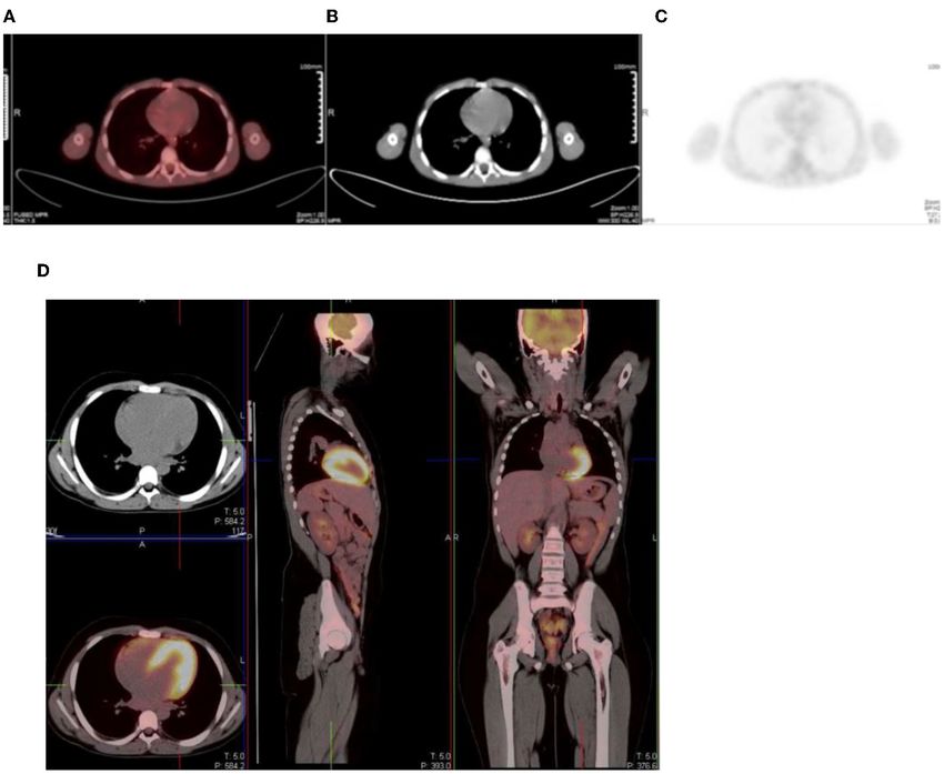
18
Purbhoo and Vangu                                                                                                   F-FDG PET/CT Imaging in Pediatrics

   We review and discuss the physiological distribution of                written instruction of the procedure should be shared with the
FDG uptake in children, the variation in distribution, lesions            patient and his/her parents/guardian (1, 2). At our institution
that are benign that could be misinterpreted as malignancy,               the patient/parent/guardian is called 24–48 h prior to the scan
and the various artifacts associated with PET/CT performed in             to discuss the preparation for the study. Written information is
pediatric oncology patients. We add a pictorial illustration to           also given at the time of booking the study. The patient should
prompt understanding and familiarity of the above-mentioned               fast for at least 4–6 h prior the study, but can drink water to
patterns. Therefore, we believe that this review will assist in           maintain good hydration, should sedation not be considered
reducing possible mistakes by reading physicians and prevent              (1, 2). Intravenous hydration post FDG injection, during the
incorrect interpretation.                                                 uptake period can be achieved with 0.9% saline solution. Soft
                                                                          drinks and sweets during the preparation phase are strongly
18 F-FDG         MOLECULE                                                 discouraged (1).
                                                                             The radiotracer injection should be timed as close as possible
FDG is an analog of glucose and is labeled with a positron-               to breast/bottle feed in infants and a feed may be given from
emitting isotope, F (fluorine)−18. It is transported into the             30 min after the injection (1, 2). Prior to the injection, the fasting
cells by glucose transporters (GLUT), is phosphorylated by                blood glucose level should be determined, with the preferred
hexokinase and remains trapped within the cell (1, 2). Cancer             level being lower than 11.1 mmol/L (200mg/dL) (1). High blood
cells preferentially use non-oxidative glucose metabolism with            glucose level result in circulating glucose that competitively
up-regulation of glucose transporter receptors and hexokinase             inhibits the cellular uptake of FDG and decreases FDG uptake
and a reduced intracellular glucose-6-phosphatase expression.             at the sites of pathology. Patients without diabetes mellitus
This results in accumulation of FDG within the tumor cells at a           may develop hyperglycemia when stressed or after starting
greater rate than in normal tissue. Active inflammatory processes         glucocorticoid therapy. The PET study should be delayed until
also cause an increase in FDG uptake. This is due to increased            these patients have better control of their glucose. Metformin
glucose use by activated granulocytes and mononuclear cells               has been shown to cause intense FDG uptake in the colon and
(3, 4). Thus, FDG is not tumor specific and accumulates in non-           in the small intestine (4). It also increases the skeletal muscle
malignant pathology, such as infection and inflammation (5–7).            and liver uptake of FDG. Therefore, if not contraindicated,
                                                                          metformin should be discontinued 24–48 h before the study (4).
                                                                          The supervising physician should be notified if the blood glucose
ONCOLOGIC INDICATIONS FOR IMAGING                                         level is >11.1 mmol/L, and a decision should be made whether to
WITH 18 F- FDG                                                            proceed with the FDG injection (1).
                                                                             To reduce the discomfort caused by the intravenous cannula,
The most common indications for FDG PET imaging in                        local anesthetic creams can be used. The duration of the uptake
pediatric oncology includes lymphoma (Hodgkin’s disease and               period should be kept constant with a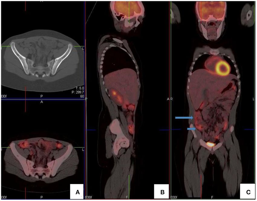
18
Purbhoo and Vangu                                                                                                                  F-FDG PET/CT Imaging in Pediatrics

 FIGURE 1 | Motion artifact of the brain on axial PET and PET/CT images (A,B). No obvious pathology is seen on the non- contrast axial CT component (C) of the PET
 study.

Irritable, uncooperative or claustrophic children will most likely                  variable between patients (Figure 2). Physiologic uptake is
need sedation (1).                                                                  normally seen with intense uptake in the brain. Uptake is
    Bladder catheterization can reduce the accumulation of tracer                   also seen in the heart, liver, spleen, gastrointestinal tract, bone
activity in the bladder. Insertion of urinary catheter is not routine               marrow, and there is excretion from the renal collecting system
practice in pediatric patients as it may increase the risk of                       into the ureters and urinary bladder.
infection and cause added stress for the child (2). Children who
are investigated for tumors in the pelvis may be considered for
catheterization to avoid lesions being obscured by tracer activity                  HEAD AND NECK
in the urinary bladder. This will also reduce the radiation dose to
the child (2). Furosemide (0.5–1 mg/kg; maximum dose, 20 mg)                        The brain is dependent on glycolytic metabolism as a source of
can be given at the time of injection or 15 min post injection to                   energy and physiological uptake is seen in the normal cerebral
enhance diuresis and minimize activity in the urinary tract (2).                    cortex and basal ganglia (Figure 2). During the fasted state, the
The child should be encouraged to void prior to the start of                        brain metabolism accounts for 20% of whole-body metabolism
acquisition. In infants who are not toilet trained, the nappy                       (1). The injected dose of FDG represents 6% of the total brain
should be changed before acquisition (1).                                           uptake (2). In infants, the glucose uptake differs from older
                                                                                    children (4). FDG uptake is most intense in the gray matter
                                                                                    and basal ganglia in older children and neuronal activation
ACQUISITION                                                                         increases FDG uptake (4). Uptake in malignant primary tumors
                                                                                    may be obscured by the intense FDG uptake in brain and
The patient should be comfortably immobilized during the
                                                                                    metastases in the brain parenchyma adjoining scalp and skull
study acquisition with Velcro straps, tapes, or cushions to avoid
                                                                                    bones may be missed. It is essential to window the gray scale
movement artifacts (1). A field of view extending from the skull
                                                                                    appropriately in order to not miss a lesion in the skull and
base to the mid-thigh is routinely used. The added radiation
                                                                                    the brain.
exposure to the pediatric patient from the CT component of the
                                                                                        Uptake in the visual cortex in the occipital lobe due to visual
study has been of concern (1, 2). In neuroblastoma and sarcoma
                                                                                    activation will occur if a child is kept in a room with bright lights
patients and in patients with lymphoma who have suspected bone
                                                                                    during the uptake period. Patients should be kept in a dimly
or bone marrow disease, the entire upper limb and lower limbs
                                                                                    lit room with minimal sensory stimulation during the uptake
should be included in the field of view (1).
                                                                                    phase to minimize this effect. Muscle activity can be seen at the
   Premedication to minimize brown adipose tissue or muscle
                                                                                    convergence of the extraocular muscles and along the length of
uptake is used. Oral contrast increases the attenuation artifact
                                                                                    these muscles (Figure 3).
that may result from mis-registration between the CT and PET
                                                                                        The Waldeyer’s ring is consists of lymphoid tissue in
acquisition. This risk is greatest with barium containing oral
                                                                                    the nasopharynx and oropharynx. It consists of adenoids
contrast agents and is lower with negative contrast agents, such
                                                                                    (nasopharyngeal tonsils), palatine tonsils faucial tonsils and
as water (4).
                                                                                    lingual tonsils at the base of the tongue and the lateral aspect
                                                                                    of the oropharynx. It shows intense FDG uptake in 6–8 years
NORMAL BIO-DISTRIBUTION OF FDG                                                      of age and diminishes with age (10). Underlying pathology
                                                                                    may be masked by the intense FDG uptake in the Waldeyer’s
The normal bio-distribution and physiologic variants of FDG                         ring. Symmetrical uptake is likely physiologic and asymmetrical
uptake in children differ from adults, and it is important to                       uptake may be pathological (1, 4). Mild to moderate uptake can
recognize these to avoid misinterpretation. The physiologic                         be seen in the adenoids, tonsils, and at the base of the tongue due
accumulation of FDG mimics glucose metabolism, which is                             to the physiologic activity of lymphatic tissue (Figure 4). Infants

Frontiers in Nuclear Medicine | www.frontiersin.org                             3                                            March 2022 | Volume 2 | Article 825891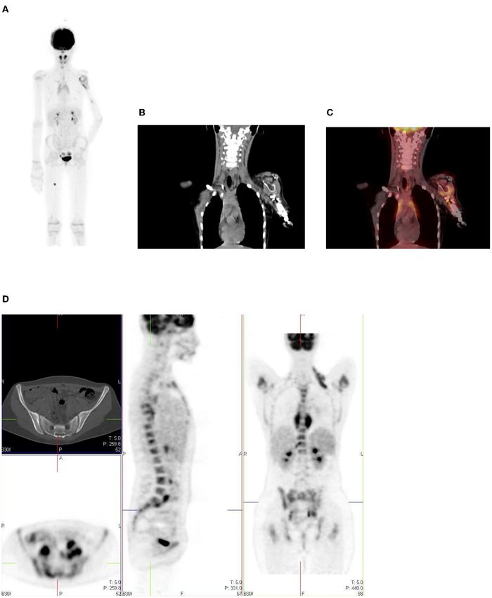
18
Purbhoo and Vangu                                                                                                                     F-FDG PET/CT Imaging in Pediatrics

 FIGURE 2 | Physiologic uptake of 18 F-FDG. Axial, sagittal and coronal FDG PET/CT (left) and PET images (right) show physiologic uptake in a 13 year old boy.
 Normal FDG uptake is seen in the brain, heart, liver, spleen, colon, urinary bladder and the bone marrow. Note the marked intense activity in the brain compared with
 the activity in the rest of the body.

 FIGURE 3 | Axial CT, fused PET/CT and PET only images showing physiologic uptake in the medial and lateral ocular muscles.

may show increased uptake in the oral muscles due to sucking of                            Homogeneous uptake is seen in the thymus in healthy
a pacifier during the uptake phase (2, 3).                                             children (1, 2). The thymus is a lymphoid organ, is bilobed
   The larynx and vocal cords show either no uptake or mild                            lymphoid organ and is located in the anterior and superior
symmetrical uptake, which may have an inverted U shape.                                mediastinum and has an inverted ‘V’ shape on the coronal
Prominent laryngeal uptake that is symmetrical is often observed                       plane (Figure 5). The shape and size of thymus is age-related.
in smaller children if they cry after tracer injection (1). It is                      Neonates have a large thymus, and can increase in size up to
important to remind children not to talk during the uptake                             the age of two (11). During adolescence there is involution of
phase, as excessive talking may cause prominence of activity in                        the thymus and it is replaced with fat, resulting in a decrease
the laryngeal structures. Asymmetric uptake in the vocal cord is                       of the physiologic uptake. It can cover the left and right aspects
suggestive of pathology such as malignancy, post-therapy change,                       of the mediastinum. Prominence of thymic uptake is seen
or unilateral vocal cord paralysis. Diffusely increased uptake in                      following chemotherapy (2, 12). Homogeneous thymus uptake
the salivary gland can be seen after chemotherapy or radiation                         at post-therapy FDG PET imaging and in the absence of uptake
therapy (1).                                                                           at pre-therapy FDG PET likely indicates post-therapy thymic
   FDG uptake in the thyroid gland is rare in children. Diffuse                        hyperplasia. Thymic hyperplasia occurs in children during
thyroid uptake may represent Graves’ disease or thyroiditis. Focal                     severe stress or chronic disease named “thymic rebound,” when
uptake in the thyroid gland is seen in benign thyroid nodules                          seen after chemotherapy (11). Intense or heterogeneous uptake
or thyroid malignancy, and further work-up is suggested in                             may raise suspicion for thymus or other anterior mediastinal
such scenario.                                                                         pathology. If the activity in the thymus equals the uptake in

Frontiers in Nuclear Medicine | www.frontiersin.org                                4                                            March 2022 | Volume 2 | Article 825891
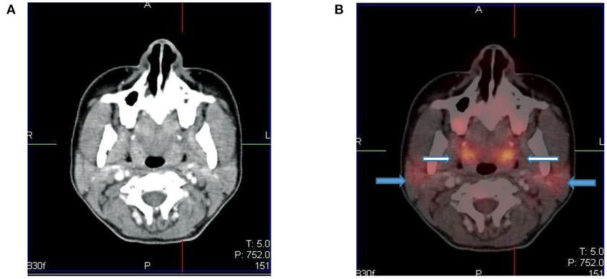
18
Purbhoo and Vangu                                                                                                                   F-FDG PET/CT Imaging in Pediatrics

 FIGURE 4 | Axial CT (A) and fused PET/CT (B) images showing symmetric increased uptake in the tonsils (white arrows) and bilateral mild uptake in the parotid
 glands (blue arrows).

 FIGURE 5 | 12 year old male referred for the evaluation of active vasculitis. FDG PET study showing diffuse symmetrical increased uptake in the thymus seen on the
 axial, sagittal and coronal views (inverted ‘’V”).

the bladder or cerebellum, it is suggestive of malignancy (12).                      CHEST
In a study by Brink et al., increased FDG accumulation in the
thymus was seen in 73% of children with a malignancy prior                           Breast
to chemotherapy and in 75% of children post chemotherapy                             The proliferative glandular tissue of the breast results in diffuse
(12). Thus, in children, when evaluating the thymus awareness                        physiological FDG uptake (1). Higher uptake may be seen in
of the patterns of uptake must be noted. It is helpful to                            adolescence due to dense breast tissue. The nipples normally
use the CT component of the study as a guide to assist                               have uptake, and this is identified on non-attenuation-corrected
interpretation (13).                                                                 images (Figure 6).

Frontiers in Nuclear Medicine | www.frontiersin.org                              5                                            March 2022 | Volume 2 | Article 825891
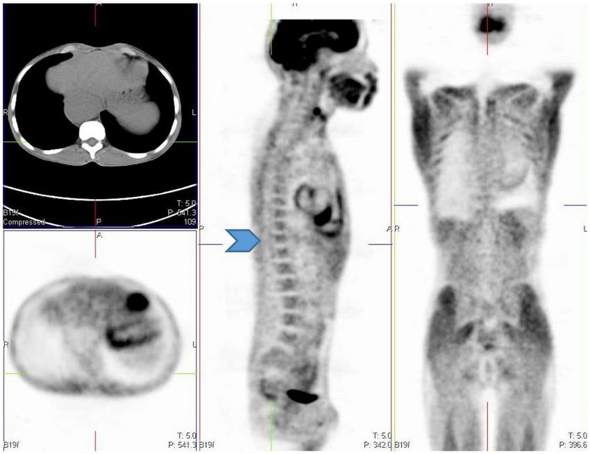
18
Purbhoo and Vangu                                                                                                                  F-FDG PET/CT Imaging in Pediatrics

 FIGURE 6 | Diffuse uptake in the breast tissue in a 17-year-old female with embryonal rhabdomyosarcoma referred for interim PET study. CT (A) PET (B) and fused
 PET/CT (C) showing mild low grade uptake in bilateral breast tissue.

 FIGURE 7 | (A–C) Axial fused FDG PET/CT (A), CT (B) and PET (C) images showing a 16 year old male who was well fasted and shows no/minimal cardiac uptake.
 (D) Transverse, sagittal and coronal PET/CT in a 13 year old boy with intense physiological FDG uptake in the left ventricle of the myocardium.

Myocardium                                                                          serum glucose and insulin levels are high (Figures 7D–F). Thus,
Myocardial uptake is variable and can range from no uptake to                       in oncology, myocardial activity is minimized by fasting for 4–6 h
intense uptake in the left ventricular myocardium (1). Cardiac                      prior to the FDG injection.
uptake depends on substrate availability (2, 14). FDG uptake is
low during fasting, as the predominant source of substrate is fatty                 Esophagus
acids, and the serum insulin level is low (Figures 7A–C). The                       Activity in the esophagus is seen as mild linear uptake anterior
uptake can be variable or inhomogenous even in the fasting state,                   to the spine and is best seen in the sagittal plane (Figure 8).
and findings may be misinterpreted as a mediastinal abnormality                     Marked uptake along the esophagus is seen in patients with
(1). Myocardial uptake can be intense in the fed state, when the                    esophagitis from reflux or after radiation therapy. Diffuse and

Frontiers in Nuclear Medicine | www.frontiersin.org                             6                                            March 2022 | Volume 2 | Article 825891
18
Purbhoo and Vangu                                                                                                               F-FDG PET/CT Imaging in Pediatrics

                                                                                      seen anywhere from the cervical region up to the lumbar spine
                                                                                      on the sagittal and coronal planes (15, 16). It is imperative that all
                                                                                      the reconstructed views are viewed to conclude this physiological
                                                                                      uptake pattern of FDG (Figure 13).

                                                                                      Genitourinary System
                                                                                      FDG is unlike glucose and is excreted through the urinary
                                                                                      system. This results in large amount of activity in the urinary
                                                                                      tract from the kidneys and includes the bladder (Figure 14).
                                                                                      Reconstruction artifact may occur if there is significant retention
                                                                                      in the renal collecting system resulting in interference with
                                                                                      the visualization of the upper abdomen (1). The activity can
                                                                                      minimized by ensuring the patient is well-hydrated and/or after
                                                                                      the administration of diuretics (1, 2). The patient should void
                                                                                      prior to imaging if toilet trained, as this will assist with the
                                                                                      clearance from the urinary tract. Tracer activity in the ureters can
                                                                                      be identified by the anatomical contours of the ureters and with
                                                                                      correlation with the CT component of the study. Catheterisation
 FIGURE 8 | A 15-year-old male known with Hodgkin’s lymphoma, referred for            may be necessary in a few cases where the accumulation of
 a restaging PET/CT post chemotherapy. Sagittal PET (A) showing linear                tracer in the urinary tract interferes with the evaluation of known
 uptake anterior to the thoracic spine corresponding to uptake in the                 lesions close to the genitourinary tract (1). Focal activity within
 esophagus (blue arrow), likely attributed to reflux or esophagitis. The CT (B)       the ureter can mimic malignant lymphadenopathy (1). Urine
 and fused PET/CT (C) will assist in this interpretation too.
                                                                                      activity in bladder diverticula may resemble lymphadenopathy
                                                                                      or ovarian tumors (1). Other causes of false-positive findings
low grade uptake is seen in the gastro-esophageal junction,                           include congenital variants such as ectopic kidney, horseshoe
which is physiological and should not be confused with disease                        kidney and anatomical distortion due to urinary diversion (1).
involvement (Figure 9).                                                                   Testicular uptake in males show a diffuse symmetric pattern
                                                                                      with moderate intensity that decreases with age (1) (Figure 15).
                                                                                      Males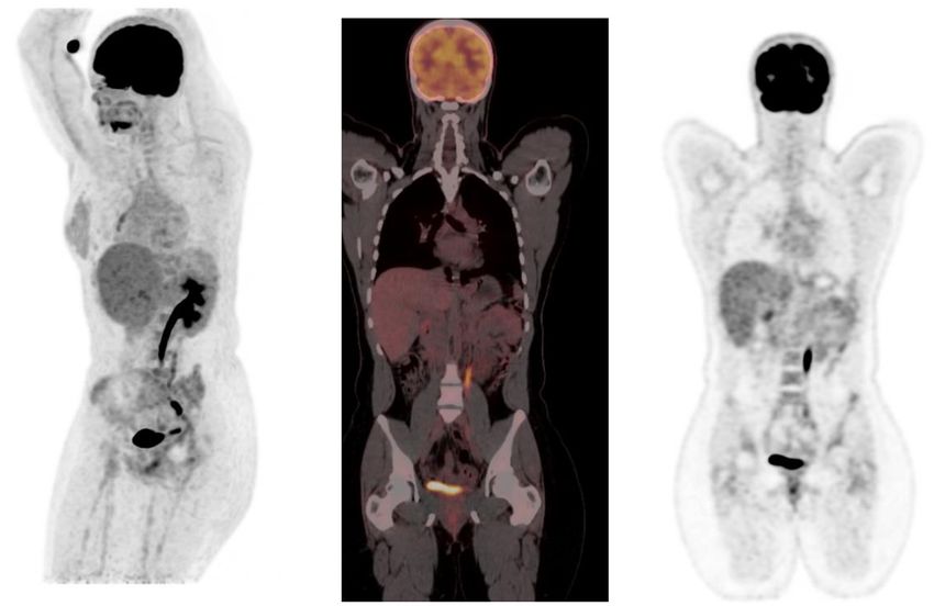
18
Purbhoo and Vangu                                                                                                                    F-FDG PET/CT Imaging in Pediatrics

 FIGURE 9 | 6-year-old female with Ewing’s sarcoma of the left proximal tibia. Axial PET (A), CT (B) and fused PET/CT (C) showing mild diffuse uptake in the
 gastroesophageal junction (white arrow and crosshairs) that is physiological.

 FIGURE 10 | Axial PET, fused FDG PET/CT and CT showing increased FDG uptake along the wall of a contracted stomach, which is physiological.

 FIGURE 11 | FDG PET/CT (A) axial, (B) sagittal and (C) coronal images show markedly increased FDG uptake in the normal right colon (long arrow). Note the more
 intense uptake in the cecum due to the higher amount of lymphoid tissue (short arrow).

Frontiers in Nuclear Medicine | www.frontiersin.org                               8                                            March 2022 | Volume 2 | Article 825891
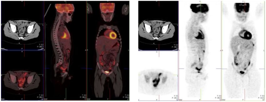
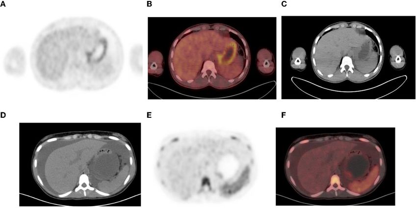
18
Purbhoo and Vangu                                                                                                                          F-FDG PET/CT Imaging in Pediatrics

 FIGURE 12 | (A–C) Normal physiological liver to spleen ratio. Physiological liver uptake is always more than the spleen uptake, except in certain pathologies like
 lymphomatous involvement of the spleen, HIV or in patients who are post chemotherapy or after the administration of granulocyte colony stimulating factor. Note the
 physiological the uptake in the gastric wall in a contracted stomach. Note that in (A) and (C), non-contrast CT has a limited sensitivity in showing splenic lesions,
 however the uptake in the spleen is greater than the physiological liver uptake and the presence of splenomegaly is compatible with pathologic involvement. (D–F)
 Diffuse increased uptake in the spleen, greater than the physiological liver uptake in a 10-year-old female with diffuse large B cell lymphoma (DLBCL) prior to
 chemotherapy. Staging PET/CT showed stage 4 disease with nodal disease above and below the diaphragm, bone marrow and splenic involvement. (B) PET and (C)
 fused PET/CT showing increased uptake in the spleen and bone marrow compared to the physiological uptake in the liver. Note that in (F), a non-contrast CT has a
 limited sensitivity in showing splenic lesions, however the uptake in the spleen is greater than the physiological liver uptake and the presence of splenomegaly is
 compatible with pathologic involvement.

 FIGURE 13 | 12 year old female referred for evaluation of active vasculitis. Incidental finding of linear uptake in the conus medullaris at T11-L1 level is physiological
 (Blue arrowhead). Also note the diffuse uptake in the muscles of the chest and lower limb due to playing sport the day before the injection. Other causes of diffuse
 muscle uptake may include high glucose level prior to injection of FDG.

Frontiers in Nuclear Medicine | www.frontiersin.org                                   9                                              March 2022 | Volume 2 | Article 825891
18
Purbhoo and Vangu F-FDG PET/CT Imaging in Pediatrics
FIGURE 14 | MIP and coronal FDG PET/CT and PET images showing physiological activity in the left renal collecting system, left ureter and urinary bladder. The MIP
image is helpful in differentiating tracer excretion in the ureter from nodal pathology. Identifying the uptake to the ureter on CT also helps in differentiating from nodal
pathology.
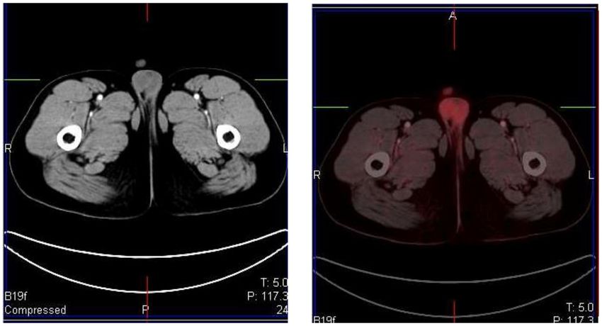
FIGURE 15 | A 16 year old male with Hodgkin’s lymphoma referred to evaluate the end of treatment response to chemotherapy. Pubertal boys may have diffuse,
increased and symmetrical uptake in the testes. No abnormality was seen at CT.
relaxants such as benzodiazepines can be used to decrease muscle is usually seen in winter. Physiological and intense uptake can be
uptake (1). Patients should rest comfortably during the uptake found in various sites, including the neck, supraclavicular area,
phase to avoid marked muscle uptake. axillae, mediastinum, paravertebral and perinephric regions (1, 2)
(Figure 16). FDG uptake in brown fat is normally bilateral and
Brown Adipose Tissue symmetric, however, focal and/or asymmetrical uptake can occur,
Brown fat is a sub-type of adipose tissue that is rich in that may result in false-positive findings and misinterpreted as
mitochondria and helps to regulate the body temperature by non- pathology (18). It is helpful to identify this pitfall by localization
shivering thermogenesis (17). Sympathetic stimulation helps to on CT to a fat density and the lack of any corresponding soft-
generate heat in the body. It is activated in cold temperature, and tissue mass (11). As brown fat is sympathetically innervated, an
Frontiers in Nuclear Medicine | www.frontiersin.org 10 March 2022 | Volume 2 | Article 82589118
Purbhoo and Vangu F-FDG PET/CT Imaging in Pediatrics
FIGURE 16 | 15 year old male with stage 3 Hodgkin’s lymphoma referred for follow up FDG PET post chemotherapy. Coronal PET (B), and PET/CT (C) showing
bilateral symmetrical uptake in the neck and paravertebral region of the cervical and thoracic spine. Crosshairs localized to fat density on CT (A). FDG uptake in brown
adipose tissue is commonly seen in children and adolescents. Many pediatric patients have mild brown fat uptake in the neck or supraclavicular regions. However,
intense uptake may include pericardiac and perirenal brown adipose tissue. 18 F-FDG uptake in activated brown fat may obscure sites of pathologic FDG uptake and
decrease confidence in the interpretation of the study.
anxious patient at the time of the FDG injection may contribute regeneration, and usually resolves within a month (1, 2)
to its visualization (11). Data suggests that FDG uptake in (Figure 16). Hyperplasia and hematopoietic stimulation from
adipose tissue occurs more often due to an acute response to anemia may also cause an increase in uptake in the marrow (1, 2).
cold weather instead of the prolonged period of cold weather Treatment with cytokines such as granulocyte colony-stimulating
(17). Keeping the patient in a warm environment prior to factor (CSF), hematopoietic growth factor, or erythropoietin
the injection and post injection is a simple approach that is can produce diffuse skeletal FDG uptake. Increased uptake can
routinely used. FDG uptake due to activated brown fat can also persist for 3 weeks after the discontinuation of granulocyte CSF
be avoided by the administration of premedication including, treatment, therefore, it is advisable to postpone the study until
diazepam, fentanyl, or propanolol prior to injection (1, 2, 16, about 4 weeks after the treatment (1). Metastases originating in
17). bone marrow can be distinguished by focal and increased uptake
with non-uniform distribution of FDG (14). The red marrow
Growth Plates in the pediatric population is metabolically active compared to
Physiological FDG uptake is seen in the growth plates of pediatric the yellow marrow as seen in adults. This shows an increase in
patients (2). This uptake may differ during different stages of the uptake in the proximal humeri, proximal and distal femurs
development and corresponds to the age and the site of the and proximal tibias, which are sites of red marrow in a growing
growth plate (2). The highest uptake is seen in the distal femur child (2) (Figures 17A–C). This should not be mistaken as bone
and looks like a horizontal band of increased FDG uptake in marrow involvement by disease. Reduced bone marrow FDG
the physes and apophyses (2). The uptake in the growth plates uptake can be seen several months after external beam radiation
is usually bilateral and usually symmetrical. therapy and is due to the replacement of bone marrow by fatty
tissue (1).
Bone Marrow In our institution, interim FDG-PET/CT is performed before
Homogenous and low grade uptake is seen in the bone marrow the next chemotherapy cycle, and at least 10 days from the
and is less intense than the physiological liver activity. Bone previous cycle. The end of treatment PET following completion
marrow uptake that is more intense than the physiological of treatment is carried out at 6–8 weeks. It is common to visualize
liver activity is considered abnormal. Increased bone marrow a diffuse increased splenic uptake due to recent chemotherapy
uptake can be seen following chemotherapy, due to physiologic or treatment with a colony-stimulating factor, and in such
Frontiers in Nuclear Medicine | www.frontiersin.org 11 March 2022 | Volume 2 | Article 82589118 Purbhoo and Vangu F-FDG PET/CT Imaging in Pediatrics FIGURE 17 | (A–C) 9 year old female with high grade osteosarcoma. Coronal PET image (A) shows normal pediatric red marrow distribution. There is homogenous FDG uptake in the proximal humeri, proximal and distal femurs, and proximal tibias reflecting normal FDG uptake in red bone marrow. This uptake is usually minimal or absent in adults due to the conversion of red marrow to yellow marrow, which is less metabolically active. Pathology in the left shoulder with increased FDG uptake in the left humeral head and proximal humerus is compatible with orthopedic hardware (B). CT image (B) showing the prosthesis and fused PET/CT image (C) showing the peri-prosthetic FDG uptake. Note however, active disease cannot entirely be excluded. Due to pain in the left arm, the patient was unable to extend and rotate the left hand for correct imaging position. (D) 16 year old female with newly diagnosed Hodgkin’s lymphoma. Staging PET shows increased uptake in the marrow, greater than the liver with areas of inhomogeneous and focal uptake, indicating bone marrow infiltration. The CT did not show anatomical lesions (Top left). Note the left neck (site of biopsy confirmed disease) and intrathoracic nodal disease. scenarios, there is also an increase in bone marrow uptake. Focal corresponding to an anatomical lesion on CT is in keeping with increased and inhomogenous uptake in the bone marrow and/or marrow/osseous involvement (19, 20) (Figure 17D). Frontiers in Nuclear Medicine | www.frontiersin.org 12 March 2022 | Volume 2 | Article 825891
18
Purbhoo and Vangu                                                                                                                       F-FDG PET/CT Imaging in Pediatrics

 FIGURE 18 | (A–C) 2 year old male with neuroblastoma who had a staging PET/CT. Artifact in the abdomen due to metal component of nasogastric tube in-situ
 resulting in streak artifact on CT (A) and fused PET/CT (B). Note the PET only image shows that there is normal excretion in the renal calyces with physiological low
 grade uptake in the liver. Coronal CT scannogram (D) shows the tube extending into the abdomen (Blue arrow) with streak artifact on coronal CT (E).

Infection and Inflammation                                                              Technical Artifacts
FDG is not specific for malignancy and is seen in infection,                            Hybrid PET/CT imaging can create artifacts associated with
inflammation and benign pathology (1). In inflammation, there                           the CT data rather than PET data for attenuation correction.
is upregulation of lymphocytes, macrophages and granulocytes,                           Increased uptake due to metallic objects such as prostheses,
which have increased glucose transporter proteins, GLUT                                 pacemakers, or chemotherapy catheters may cause false-positive
1 and GLUT 3 and thus higher affinity to FDG. Focal                                     findings (1) (Figure 18). The high CT attenuation values results
FDG accumulation in the gluteal muscles are injection site                              in a false increase in PET attenuation coefficients, creating
granulomas, especially in patients with recent intramuscular                            an overestimation of the PET activity corresponding to the
injection. Patients receiving subcutaneous injection of low                             metallic object on the attenuation-corrected image (1). In the
molecular-weight heparin can develop FDG-avid subcutaneous                              same scenario, increased concentration of intravenous and
areas of soft-tissue attenuation at the injection sites. The                            oral contrast leads to the overcorrection of activity and false-
accumulation of inflammatory cells and macrophages within                               positive results at PET if contrast-enhanced CT data are
these lesions explains the FDG accumulation in the lesions. These                       used for attenuation correction. The non-attenuated corrected
nodules can mimic metastatic tumor deposits and the clinical                            PET images should be viewed to distinguish this attenuation
information is imperative to avoid misinterpretation. Soft tissue                       correction artifact from true increased uptake, as the increased
stranding will guide the inflammatory nature of these lesions.                          activity will not be seen on the non- corrected image (1).
Active vascular inflammation may demonstrate FDG uptake.                                   The ideal manner for determining the SUV in children
FDG PET/ CT can help detect large vessel vasculitis and reflect                         may be different from that used in adults because of the
the distribution of active inflammatory activity in the wall of the                     growth that occurs in childhood. Therefore, in pediatric patients,
vessel (1).                                                                             SUV calculated by body surface area is a better metabolic

Frontiers in Nuclear Medicine | www.frontiersin.org                                13                                            March 2022 | Volume 2 | Article 825891
18
Purbhoo and Vangu                                                                                                                      F-FDG PET/CT Imaging in Pediatrics

marker of activity rather than that calculated based on body                         in the metaphysis around the knee-joints, and are usually oval,
weight (1).                                                                          radiolucent lesions with a thin sclerotic rim and lie parallel to the
    Respiratory motion causes mis-registration of PET and CT                         long axis of the bone (2).
data that can result in attenuation correction artifact that is seen
near the diaphragm and base of lung (1).                                             Radiation Safety
    Injection of radiotracer into central lines should be avoided as                 The main focus of radiation safety is dose reduction, which
the retention of tracer in the line or at its tip can mimic FDG-avid                 implies a lower potential risk of cancer (21). Radiosensitivity
pathology and may be misinterpreted as a false positive finding.                     is increased in children due to actively developing organs
The line should be flushed with an ample volume of normal saline                     at the time of the scan and their longer post exposure life
if a central line is used for tracer injection due to difficult venous               expectancy. Children have a higher relative risk of leukemia,
access (1).                                                                          brain, breast, skin, and thyroid cancers compared with adults
    Negative scan findings cannot exclude small or microscopic                       exposed to radiation post-adolescence (21). To address the
malignant involvement. Tumors with low metabolic activity or                         concern of radiation burden in children, adult PET/CT
poor cellular composition may show minimal uptake of FDG                             acquisition protocols are modified in order to minimize the
(1). The standardized uptake values do not help to differentiate                     radiation dose delivered; however, it is imperative to ensure
benign vs. malignant etiology.                                                       that such modifications do not compromise the diagnostic
    Pitfalls related to therapy should be remembered, such                           information required.
as drug-induced pulmonary toxicity. Usage of cytotoxic and
immunotherapy drugs may cause lung toxicity with features of                         CONCLUSION
alveolar-interstitial infiltrates and alveolar damage, which may
have increased FDG uptake (2). Knowledge of the adverse-effects                      FDG PET/CT is now increasing used in the oncologic evaluation
of the cytotoxic drugs and their imaging features will be helpful in                 of pediatric patients. The physiological distribution of FDG
the proper interpretation of the images. Post-treatment radiation                    uptake in children differs from adults and it is important to
inflammatory changes are FDG avid areas of consolidation and                         recognize the normal bio-distribution of FDG in children and
are differentiated from pathology due to the sharp demarcation                       take cognisance of the artifacts, and potential pitfalls. Knowing
from the normal lung, limited to the radiation field (2).                            these potential causes of misinterpretation can increase the
    Benign bone lesions show variable FDG uptake in the growing                      accuracy of interpretation, decrease the number of unnecessary
skeleton. Examples are fibro-osseous defects like non-ossifying                      follow-up studies and improve treatment outcome.
fibromas and fibrous cortical defects and they appear as focal
bone lesions with moderate to intense FDG uptake in first and                        AUTHOR CONTRIBUTIONS
second decade of life (2). They are asymptomatic lesions and
are mostly incidental findings. These lesions are benign, and do                     All authors listed have made a substantial, direct, and intellectual
not require further investigation or treatment. They are located                     contribution to the work and approved it for publication.

REFERENCES                                                                                 tomography/computed tomography (PET/CT): accumulated data from four
                                                                                           years of experience with PET/CT. Semin Nucl Med. (2007) 37:206–
 1. Stauss J, Franzius C, Pfluger T, Juergens KU, Biassoni L, Begent                       22. doi: 10.1053/j.semnuclmed.2007.01.001
    J, et al. Guidelines for 18F-FDG PET and PET-CT imaging                           9.   Edwards KW. Preparation and logistic considerations in performing PET and
    in paediatric oncology. Eur J Nucl Med Mol Imaging. (2008)                             PET/computed tomography in pediatric patients. PET Clin. (2020) 15:285–
    35:1581–8. doi: 10.1007/s00259-008-0826-x                                              92. doi: 10.1016/j.cpet.2020.03.001
 2. Shammas A, Lim R, Charron M. Pediatric FDG PET/ CT: physiologic uptake,          10.   Lucignani G, De Palma D. PET/CT in paediatric oncology: clinical usefulness
    normal variants, and benign conditions. Radiographics. (2009) 29:1467–                 and dosimetric concerns. Eur J Nucl Med Mol Imaging. (2011) 38:179–
    86. doi: 10.1148/rg.295085247                                                          84. doi: 10.1007/s00259-010-1661-4
 3. Zhuang H, Alavi A. Evolving role of PET in pediatric disorders. PET Clin.        11.   Viglianti B, Wong K, Gross M, Wale D. Common pitfalls in oncologic FDG
    (2020) 15:xv–xvii. doi: 10.1016/j.cpet.2020.05.001                                     PET/CT Imaging. J Am Osteopat Coll Radiol. (2018) 7:5–17.
 4. Grant FD. Normal variations and benign findings in pediatric 18F-                12.   Brink I, Reinhardt MJ, Hoegerle S, Altehoefer C, Moser E, Nitzsche EU.
    FDG-PET/ CT. PET Clin. (2014) 9:195–208. doi: 10.1016/j.cpet.2013.                     Increased metabolic activity in the thymus gland studied with 18F-FDG
    12.002                                                                                 PET: age dependency and frequency after chemotherapy. J Nucl Med.
 5. Chamroonrat W. PET/computed tomography in the evaluation of fever of                   (2001) 42:591–5.
    unknown origin and infectious/inflammatory disease in pediatric patients.        13.   Smith CS, Schöder H, Yeung HWD. Thymic extension in the superior
    PET Clin. (2020) 15:361–9. doi: 10.1016/j.cpet.2020.03.002                             mediastinum in patients with thymic hyperplasia: potential cause of false-
 6. Reavey HE, Alazraki AL, Simoneaux SF. Normal patterns of 18F-                          positive findings on 18F-FDG PET/CT. Am J Roentgenol. (2007) 188:1716–
    FDG appendiceal uptake in children. Pediatr Radiol. (2014) 44:398–                     21. doi: 10.2214/AJR.06.0552
    402. doi: 10.1007/s00247-013-2835-6                                              14.   Shreve PD, Anzai Y, Wahl RL. Pitfalls in oncologic diagnosis with FDG
 7. Corrigan AJG, Schleyer PJ, Cook GJ. Pitfalls and artifacts in the use                  PET imaging: Physiologic and benign variants. Radiographics. (1999) 19:61–
    of PET/CT in oncology imaging. Semin Nucl Med. (2015) 45:481–                          77. doi: 10.1148/radiographics.19.1.g99ja0761
    99. doi: 10.1053/j.semnuclmed.2015.02.006                                        15.   Taralli S, Leccisotti L, Mattoli MV, Castaldi P, de Waure C, Mancuso A, et al.
 8. Metser U, Even-Sapir E. Increased 18F-Fluorodeoxyglucose uptake in                     physiological activity of spinal cord in children: an 18F-FDG PET-CT study.
    benign, nonphysiologic lesions found on whole-body positron emission                   Spine. (2015) 40:E647–52. doi: 10.1097/BRS.0000000000000895

Frontiers in Nuclear Medicine | www.frontiersin.org                             14                                              March 2022 | Volume 2 | Article 825891
18
Purbhoo and Vangu F-FDG PET/CT Imaging in Pediatrics
16. Gillman J, States LJ, Servaes S. PET in pediatric lymphoma. PET Clin. (2020) Conflict of Interest: The authors declare that the research was conducted in the
15:299–307. doi: 10.1016/j.cpet.2020.03.007 absence of any commercial or financial relationships that could be construed as a
17. Basu S, Alavi A. Optimizing interventions for preventing uptake in the potential conflict of interest.
brown adipose tissue in FDG-PET. Eur J Nucl Med Mol Imaging. (2008)
35:1421–3. doi: 10.1007/s00259-008-0720-6 Publisher’s Note: All claims expressed in this article are solely those of the authors
18. Truong MT, Erasmus JJ, Munden RF, Marom EM, Sabloff BS, Gladish GW, and do not necessarily represent those of their affiliated organizations, or those of
et al. Focal FDG uptake in mediastinal brown fat mimicking malignancy: the publisher, the editors and the reviewers. Any product that may be evaluated in
a potential pitfall resolved on PET/CT. Am J Roentgenol. (2004) 183:1127–
this article, or claim that may be made by its manufacturer, is not guaranteed or
32. doi: 10.2214/ajr.183.4.1831127
endorsed by the publisher.
19. Wen Z, Zhang L, Zhuang H. Roles of PET/computed tomography
in the evaluation of neuroblastoma. PET Clin. (2020) 15:321–
31. doi: 10.1016/j.cpet.2020.03.003 Copyright © 2022 Purbhoo and Vangu. This is an open-access article distributed
20. Harrison DJ, Parisi MT, Khalatbari H, Shulkin BL. PET with 18F- under the terms of the Creative Commons Attribution License (CC BY). The
fluorodeoxyglucose/computed tomography in the management of pediatric use, distribution or reproduction in other forums is permitted, provided the
sarcoma. PET Clin. (2020) 15:333–47. doi: 10.1016/j.cpet.2020.03.008 original author(s) and the copyright owner(s) are credited and that the original
21. Magill D, Alavi A. Radiation safety concerns related to PET/computed publication in this journal is cited, in accordance with accepted academic practice.
tomography imaging for assessing pediatric diseases and disorders. PET Clin. No use, distribution or reproduction is permitted which does not comply with these
(2020) 15:293–8. doi: 10.1016/j.cpet.2020.03.012 terms.
Frontiers in Nuclear Medicine | www.frontiersin.org 15 March 2022 | Volume 2 | Article 825891You can also read