On the injectability of free-standing magnetic nanofilms - Unitn
←
→
Page content transcription
If your browser does not render page correctly, please read the page content below
Biomed Microdevices (2017) 19:51
DOI 10.1007/s10544-017-0192-1

On the injectability of free-standing magnetic nanofilms
Silvia Taccola 1 & Virginia Pensabene 1,2,3 & Toshinori Fujie 4,5 & Shinji Takeoka 6 &
Nicola M. Pugno 7,8,9 & Virgilio Mattoli 1

# Springer Science+Business Media New York 2017

Abstract Free-standing films with sub-micrometric thick-                Keywords Nanofilm . Nanopatch . Injectability . Magnetic
ness, composed of soft polymers and functional nanostruc-               nanocomposite . SIEBIMM . Minimally invasive surgery
tures are promising candidates for many potential applications
in the biomedical field, such as reduced port abdominal sur-
gery. In this work, freely suspended poly(L-lactic acid)
nanofilms with controlled morphology embedding                          1 Introduction
superparamagnetic iron oxide nanoparticles were fabricated
by spin-coating deposition. The mechanical properties of                Recent developments in the field of nanotechnology have led
magnetic nanofilms were investigated by Strain-Induced                  to the realization of free-standing nanostructured ultrathin
Elastic Buckling Instability for Mechanical Measurements                polymeric films (also called Bnanofilms^), which are charac-
(SIEBIMM) test. Our results show that these freely suspended            terized by an aspect ratio of size and thickness greater than 106
nanocomposite nanofilms are highly flexible and deformable,             (size in the order of centimeter and tens-of-nanometers thick-
with Young’s moduli of few GPa. Since they can be handled in            ness) (Jiang and Tsukruk 2006, Ono and Decher 2006, Tang
liquid with syringes, a quantitative description of the                 et al. 2006, Mamedov and Kotov 2000). The combination of
nanofilms behavior during the manipulation with clinically              nanometer thickness and macroscopic size imparts to these
applicable needles has been also provided. These magnetic               quasi-two-dimensional (2-D) structures unique physical prop-
nanofilms, remotely controllable by external electromagnetic            erties, such as high flexibility, transparency and noncovalent
fields, have potential applications in minimally invasive sur-          adhesiveness. Moreover, the integration of functional nano-
gery as injectable nanopatches on inner organs wall.                    structures, such as magnetic nanoparticles, gold nanoparticles,

* Silvia Taccola                                                        5
                                                                            Japan Science and Technology Agency, PRESTO, 4-1-8 Honcho,
  silvia.taccola@iit.it                                                     Kawaguchi, Saitama 332-0012, Japan
* Virgilio Mattoli                                                      6
                                                                            Department of Life Science and Medical Bioscience, Faculty of
  virgilio.mattoli@iit.it                                                   Science and Engineering, Waseda University, TWIns, 2-2
                                                                            Wakamtsu-cho, Shinjuku-ku, Tokyo 162-8480, Japan
1
    Center for MicroBioRobotics IIT@SSSA, Istituto Italiano di          7
    Tecnologia, Viale Rinaldo Piaggio 34, 56025 Pontedera, Italy            Laboratory of Bio-Inspired & Graphene Nanomechanics,
2
                                                                            Department of Civil, Environmental and Mechanical Engineering,
    Present address: School of Electronic and Electrical Engineering,       University of Trento, Via Mesiano 77, 38123 Trento, Italy
    University of Leeds, Woodhouse Lane, Leeds LS2 9JT, UK
                                                                        8
3
    School of Medicine, Leeds Institute of Biomedical and Clinical          School of Engineering and Materials Science, Queen Mary
    Sciences, University of Leeds, Woodhouse Lane, Leeds LS2 9JT, UK        University of London, Mile End Road, E1 4NS, London, UK
4                                                                       9
    Waseda Institute for Advanced Study, Waseda University, TWIns,          Ket Lab, Edoardo Amaldi Foundation, Italian Space Agency, Via del
    2-2 Wakamatsu-cho, Shinjuku-ku, Tokyo 162-8480, Japan                   Politecnico snc, 00133 Rome, Italy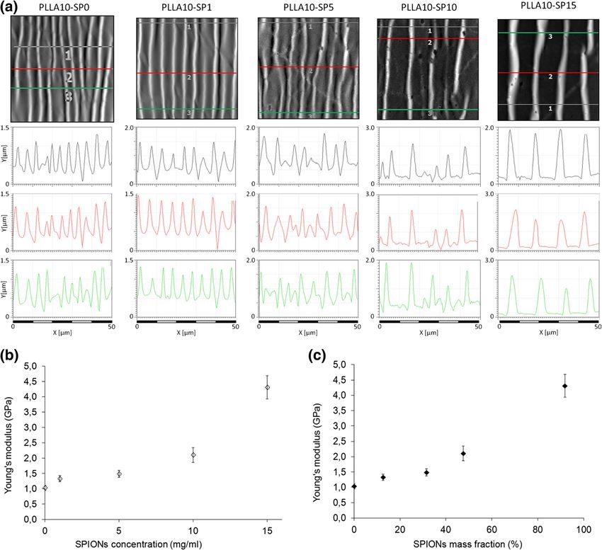
51     Page 2 of 9                                                                      Biomed Microdevices (2017) 19:51

and carbon nanotubes, into the polymeric matrix, as well as           the final aim to anchor permanent magnets onto the abdominal
the use of functional polymers, can bestow high-performance           and intestinal serial surfaces to enable magnetic tissue retrac-
nanofilms of new magnetic, optical, mechanical, or electronic         tion during reduced port surgery (Pensabene et al. 2011). This
properties (Fujie 2016, Jiang et al. 2004, Mamedov et al.             approach enabled long term anchoring of surgical assistive
2002, Redolfi Riva et al. 2014, Greco et al. 2011). Free-             tools in an endoscopic procedure, but could not guarantee
standing polymeric nanofilms have been fabricated using dif-          stable adhesion on wet tissues during multidirectional mag-
ferent approaches, including Langmuir-Blodgett (Endo et al.           netic retraction.
2006), layer-by-layer (LbL) (Decher 1997), dip-coating (Tang             Herein, we envisaged to develop a biocompatible polymer-
et al. 2006), sol-gel (Vendamme et al. 2006) and spin-coating         ic nanofilm, which could be remotely manipulated and posi-
method (Okamura et al. 2009). Up to date, nanofilms have              tioned onto the stomach incision site precisely by using
been investigated for applications in nano-electronics, nano/         minimal-invasive external tools. The integration of magnetic
μ sensing and actuation devices, electrochemical devices,             components into PLLA nanofilms represents the first step for
nanoscale chemical and biological reactors, drug-delivery sys-        the development of magnetic nanofilms with the potential of a
tems and as ultra-conformable electrodes (Kang et al. 2008;           remote-controlled manipulation by permanent and gradient
Redolfi Riva et al. 2013; Taccola et al. 2011, 2013; Zucca et al.     magnetic fields, as already theoretically and experimentally
2015).                                                                demonstrated (Mattoli et al. 2010, Taccola et al. 2011). The
   Recently, biodegradable and biocompatible materials (e.g.          use of magnetic fields to remotely control microdevices in
polysaccharides or polyesters) have been proposed as soft             narrow and delicate districts and apparata of the human body
patches for cosmetic use, as innovative alternative to tradition-     is a well-accepted approach nowadays in surgical and diag-
al surgical sutures, as nanopatches on gastrointestinal wall or       nostic procedures, where remote-magnetic navigation cathe-
as flexible cell growth supports (Okamura et al. 2009, Fujie          ters or magnetic robotic capsules are finely controlled by cou-
et al. 2007, Fujie et al. 2009, Pensabene et al. 2009, Ricotti        pling with permanent magnetic fields (Ciuti et al. 2010). Free-
et al. 2010, Fujie et al. 2011, Fujie et al. 2012, Ventrelli et al.   standing polymeric nanofilms composed of PLLA, embed-
2014, Fujie et al. 2014). In particular, it has been demonstrated     ding superparamagnetic iron oxide nanoparticles (SPIONs),
that free-standing poly(L-lactic acid) (PLLA) nanofilms can           were successfully fabricated in our laboratory and their mor-
adhere tightly to skin or organs by physical adhesion (i.e. van       phological and magnetic properties have been characterized
der Waals interaction) and have an excellent sealing effect on        (Taccola et al. 2011). In this study, we focused on the biomed-
the closure of gastric incisions as a wound dressing that re-         ical application of magnetic nanofilms as injectable
quires no adhesive agents (Okamura et al. 2009; Pensabene             nanopatches, and evaluated their mechanical properties and
et al. 2009).                                                         Binjectability^ through clinically applicable syringes and
   In this framework, the concept of nanofilms as Bnano-ad-           needles (See Fig. 1). In particular, the Binjectability^ was de-
hesive plasters^ for wound repair can become extremely ap-            fined as the ability of the magnetic nanofilm to pass through a
pealing in the field of minimally-invasive surgery, combining         needle without distortion, and was experimentally evaluated
the adhesive properties of PLLA nanofilms with the ability of         by varying the lateral size of the nanofilms and SPIONs con-
these flexible structures to be easily manipulated with syringes      centrations with respect to syringe needle of different inner
and pipettes, injecting and ejecting multiple times without           diameters. Finally we successfully introduced an analytical
distortion. Thus, the PLLA nanofilms could be injected inside         model able to predict the injectability of nanofilm at given
the human body in different fluids and spaces employing a             syringe needle diameter.
small catheter or a plastic cannula, or directly through the
working channel of an endoscope and then used as
nanopatches on inner organ walls. For example, nanofilms              2 Materials and methods
could be employed in endoscopic or laparoscopic surgery
for localized thermal ablation procedures (Baker et al. 2006),        Materials Silicon wafers (400 μm thick, p type, boron doped,
as a new method for surface ferromagnetisation of tissues             , Si-Mat Silicon Materials, Kaufering, Germany), used
(Wang et al. 2014), as surface marking of localized small             as substrates for film deposition, were cut (2 cm × 2 cm) and
lesions discovered during endoscopic examinations (Wang               treated using an acid washing solution (SPM: 96%:30%
et al. 2016).                                                         H2SO4/H2O2 = 4:1 (v/v)) at 120 °C for 10 min and then thor-
   Recently polymeric biocompatible glues have been loaded            oughly rinsed with deionized (DI) water (18 MΩ cm) in order
with ferromagnetic particles to be used as novel tools for mag-       to remove dust and impurities. Poly(vinyl alcohol) (PVA, av-
netic grasping (Wang et al. 2008), as distinct from pull retrac-      erage Mw 13,000–23,000, 98% hydrolyzed) was purchased
tion, in conjunction with an adjustable magnetic force system         from Kanto Chemical Co., Inc. (Tokyo, Japan). Poly(L-lactic
deployed inside the abdominal cavity or outside the body.             acid) (PLLA, Mw 80,000–100,000) was obtained from
Polymeric mucoadhesive films were instead synthetized with            Polysciences Inc. (Warrington, PA). Commercially available
Biomed Microdevices (2017) 19:51                                                                                 Page 3 of 9 51

Fig. 1 Overview of the
experimental framework used for
the evaluation of nanofilms
injectability vs. geometrical and
composition features

superparamagnetic magnetite/maghemite nanoparticles with a          drops of silanizing agent (chlorotrimethylsilane, Sigma–
polymeric coating layer (EMG1300), having a nominal diame-          Aldrich). The PDMS substrate was prepared at a 10:1 ratio
ter of 10 nm, were purchased from FerroTec Co. (San Jose, CA).      by weight of base elastomer to curing agent. The mixture, after
                                                                    the release of entrapped air bubbles by a vacuum bell desic-
                                                                    cator was spin-coated onto the silanized Si substrates for 60 s
2.1 Fabrication of single layer PLLA and PLLA-SPIONs
                                                                    at a speed of 200 rpm and then cured at T = 95 °C for 60 min in
nanofilms
                                                                    an oven. The cured PDMS was cut into slabs (4 × 2 cm2).
                                                                    Nanofilms were released into water, and collected on a
Free-standing magnetic PLLA nanofilms and unloaded PLLA
                                                                    prestretched (~5% strain of the original size) PDMS substrate.
nanofilms (used as a control for characterization) were pre-
                                                                    The samples were dried overnight prior to the Strain-Induced
pared by spin coated assisted deposition following the proce-
                                                                    Elastic Buckling Instability for Mechanical Measurements
dure described in details elsewhere (Taccola et al. 2011).
                                                                    (SIEBIMM) test. The strain of the PDMS substrate was then
Briefly, a PVA aqueous solution (1 wt%) was deposited by
                                                                    relaxed, producing the buckling of the nanofilm. The buckling
spin-coating on a silicon wafer (at 4000 rpm for 20 s) forming
                                                                    wavelength of the nanofilm was measured by Atomic Force
a water-soluble sacrificial layer. A stable colloidal solution of
                                                                    Microscope (AFM) imaging, using a Veeco Innova Scanning
SPIONs and PLLA in chloroform (PLLA 1 wt%) was then
                                                                    Probe Microscope (Veeco Instuments Inc., Santa Barbara,
spin-coated on the sacrificial layer by using the same spinning
                                                                    CA) operating in tapping mode, with a RTESPA Al-coated
parameters. After each step, the sample was held at 80 °C on a
                                                                    silicon probe (Veeco Instruments Inc.). The formula used to
hot plate for 1 min to remove the excess solvent. Finally, the
                                                                    calculate the Young’s modulus of the nanofilm is reported as
polymer-coated wafer was immersed in water: the PVA sacri-
                                                                    Eq. 1 in this paper.
ficial layer was dissolved, thus releasing a freely suspended
insoluble PLLA nanofilm. All routines for PLLA nanofilms
fabrication were conducted in a clean room (class 10,000) to        2.3 Injectability test
avoid contamination. In this study, different PLLA nanofilms
were prepared by varying the number of nanoparticles added          The capability of nanofilms to be manipulated without break-
to the solution. The samples were referred as PL10-SPx,             ing through syringes equipped with clinically applicable
denoting films prepared using 10 mg mL−1 PLLA and x mg              needles (Terumo Medical Corporation, Elkton, MD, USA,
mL−1 SPIONs colloidal solutions (x = 0, 1, 5, 10, 15).              needles inlet diameter 1.1 mm, 0.9 mm, 0.6 mm, 0.45 mm)
                                                                    has been experimentally investigated and quantified by the
2.2 Characterization of mechanical properties                       parameter I (Binjectability^). Due to their hydrophobicity,
                                                                    the manipulation of free-standing nanofilms in water was pos-
Silicon wafers (400 μm thick, p-type, boron doped, ,           sible only after the addition of a PVA solution (0.1 wt%) in
Si-Mat Silicon Materials) were silanized by placing them in a       suspension medium, in which PVA was acting as a surfactant.
desiccator for 30 min along with a vial that contained a few        Nanofilms were then inserted into the syringe filled with this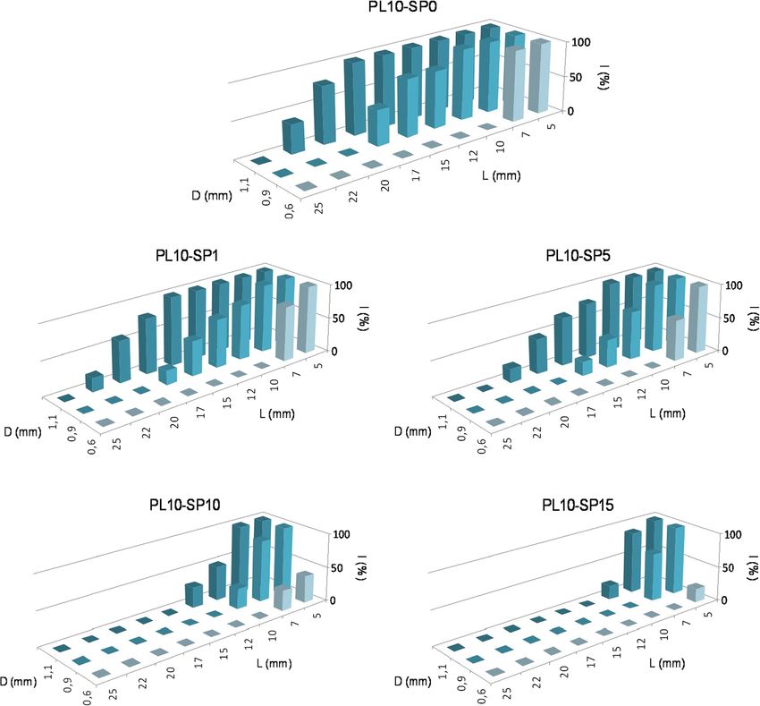
51     Page 4 of 9                                                                    Biomed Microdevices (2017) 19:51

solution and consequently forced through the needle by a            (Pensabene et al. 2009). The physical properties of nanofilms
constant pressure of 2.5 psi. A syringe pump was used in order      obtained from a dispersion containing 10 mg mL−1 PLLA and
to apply the constant pressure from the syringe piston.             different concentrations of SPIONs (0, 1, 5, 10, 15 mg mL−1)
   For each SPIONs concentration, the injectability test was        were summarized in Table 1.
performed on square nanofilms having different lateral size (5,        The thickness of the nanofilms, determined from AFM
7, 10, 12, 15, 17, 20, 22 and 25 mm). Each test was indepen-        thickness measurements, increased with the number of
dently repeated 10 times using new samples, new needles and         SPIONs in the composite and varied in a range from 87 nm
new solutions. The injectability test determined if a nanofilm      for PL10-SP0 to 205 nm for PL10-SP15. The magnetic be-
can be successfully injected through a specified needle.            havior of nanofilms, could be described by superparamagnetic
Therefore, each trial had only two possible outcomes: if the        modeling, with the saturation magnetization depending only
nanofilm could pass through the needle without distortion, the      on the nanoparticles number density. The magnetization eval-
injection succeeded; if the nanofilm caused the clogging of the     uation has been used to estimate the mass magnetic suscepti-
needle or was damaged during the passage through the needle,        bility of the nanofilms and the SPIONs mass fraction in the
the injection failed. Then, the Binjectability^ parameter (I) was   nanocomposite that are key factors in describing the magnetic
measured as the number of success among 10 trials, expressed        guidance of the freely suspended nanofilms in liquid environ-
as a percentage.                                                    ment (Mattoli et al. 2010; Taccola et al. 2011).
   The morphology of the nanofilms after the injection was             As reported in Taccola et al. 2011, a uniform distribution of
evaluated on samples collected from the suspended state and         SPIONs inside the polymeric matrix was evidenced via both
dried on clean silicon wafers using optical microscopy, while       AFM and transmission electron microscopy (TEM). It is note-
scanning electron microscope (SEM) and atomic force mi-             worthy that, as the SPIONs concentration increased, the pres-
croscopy (AFM) have been employed for identifying cracks,           ence of particle clusters emerging from the surface of the
wrinkles or other discontinuities caused by the induced stress-     samples was evident.
es. Optical images of the film surface were taken by using a
Hirox KH7700 digital microscope (Hirox Co Ltd., Tokyo,              3.2 Mechanical properties of magnetic nanofilms.
Japan). AFM measurements were carried out on a Veeco
Innova Scanning Probe Microscope (Veeco Instuments Inc.,            Mechanical properties free-standing nanofilms were evaluated
Santa Barbara, CA) operating in tapping mode, using an              SIEBIMM, a technique used for the determination of the
RTESPA Al-coated silicon probe (Veeco Instruments Inc.).            Young’s modulus of polymer ultrathin films (Stafford et al.
SEM images were obtained using a Zeiss EVO/MA 10 field              2004). This technique is based on measuring the wavelength λ
emission microscope (Carl Zeiss SMT, Oberkochen,                    of the periodic wrinkles formed on the buckled surface of
Germany) at an acceleration voltage of 10 keV. Specimens            polymer thin films coating a relatively soft, thick elastic sub-
for the SEM experiments were sputtered with a thin layer of         strate such as PDMS. If the elastomer substrate is pre-
gold before the observation.                                        stretched, the relaxation of strain induces the buckling of the
                                                                    film. By applying buckling mechanics, the Young’s modulus
                                                                    (En) is obtained by using the following formula (Stafford et al.
3 Results and discussions                                           2004):
                                                                                          3
3.1 Preparation and characterization of magnetic                           3 E s 1−ν 2n      λ
                                                                    En ¼                                                        ð1Þ
nanofilms                                                                      1−ν 2s       2πt

In our previous work, we developed free-standing and flexible       where E is the Young’s modulus, ν is the Poisson’s ratio and n,
PLLA nanofilms loaded with superparamagnetic nanoparti-             s subscripts refer to nanofilm and substrate (PDMS), respec-
cles in a simple, fast single-step deposition process (Taccola      tively, whereas λ is the wavelength of the wrinkles and t the
et al. 2011). The effect of each production parameters (i.e.        nanofilm thickness. In Eq. 1, we employed Young’s modulus
PLLA and SPIONs concentration in the deposited dispersion)          value of PDMS Es = 1.8 MPa, the Poisson’s ratios of the
on the morphological properties and magnetic behavior of            nanofilm νn = 0.33 and of the PDMS νs = 0.50, by following
nanofilms has been completely characterized (Taccola et al.         the Rubner’s report (Stafford et al. 2004). The wavelength was
2011). From this study, magnetic nanofilms fabricated from          estimated by AFM analysis of wrinkled nanofilms, as showed
the spin coating of 10 mg mL−1 PLLA solutions appeared to           in Fig. 2a. By incorporating the measured wavelength into
be particularly suitable for the proposed application, because      Eq. 1 the Young’s modulus of the nanofilms En was evaluated
of the good homogeneity of SPIONs dispersion and the                (Fig. 2b, c).
~100 nm thickness, which was previously reported as key                The result for purely polymeric nanofilms confirmed what
feature for an efficient adhesion on the mucosal tissue             previously reported for PLLA nanofilms with comparable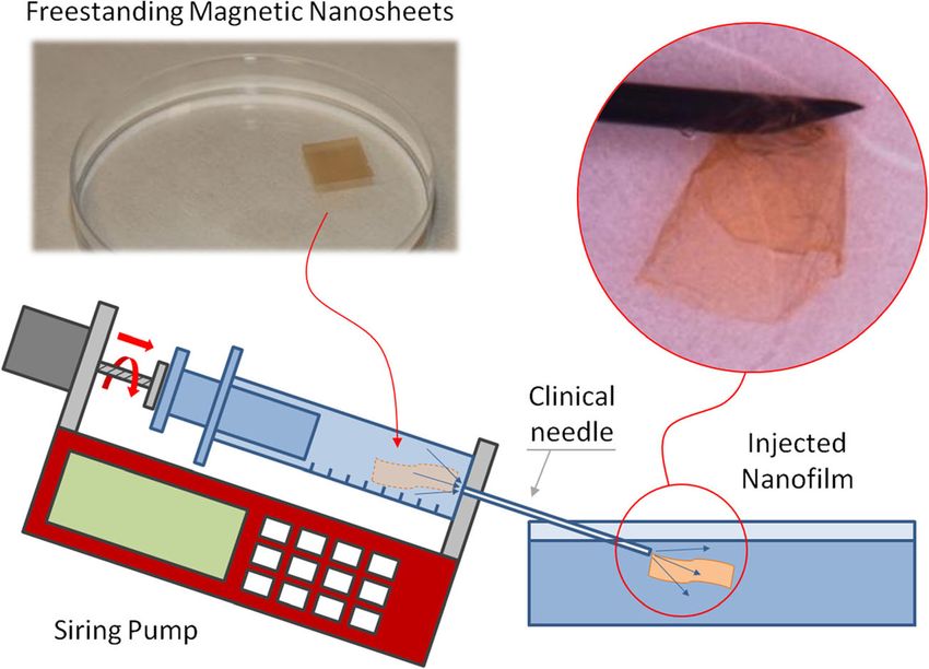
Biomed Microdevices (2017) 19:51                                                                                               Page 5 of 9 51

Table 1      Morphological and magnetic properties of 10 mg mL−1 PLLA and PLLA/SPIONs nanofilms (data from Taccola et al. 2011)

Sample            Thickness (nm)      Roughness (nm)   Nanoparticles mass fraction in the composite (%)     Mass magnetic susceptibility (cm3 g−1)

PL10-SP0          87 ± 4              2.3              -                                                    -
PL10-SP1          141 ± 4             3.7              12.1                                                 0.012
PL10-SP5          139 ± 2             5                31.5                                                 0.032
PL10-SP10         154 ± 3             17.6 (118)a      47.5                                                 0.048
PL10-SP15         205 ± 8             31.2 (132.3)a    91.7                                                 0.093
a
    Clusters’ average equivalent disk radius (nm)

thickness, whose Young’s modulus was estimated to be                       nanoparticles in the structure resulted in a more rigid behavior
around 1–2 GPa (Fujie et al. 2013). The addition of magnetic               of the nanofilms. These results are in good agreement with

Fig. 2 Mechanical properties of the PLLA nanofilms with different          estimate the wavelength (defined as the distance between two consecutive
SPIONs concentrations evaluated by the SIEBIMM measurement. a              ripple maxima). b The Young’s modulus of PLLA nanofilms in term of
AFM images of wrinkled nanofilms and relative surface profiles used to     SPIONs concentration and (c) mass fraction
51 Page 6 of 9 Biomed Microdevices (2017) 19:51
those reported in the literature concerning freely suspended of 100% using 1.1 mm diameter needles, while PL10-SP15
layer-by-layer nanomembranes whose mechanical properties nanofilms (thickness 205 nm), with the same lateral size,
can be significantly enhanced by the introduction of well dis- showed a lower injectability of only 20%. The incorporation
persed inorganic nanoparticles (e.g. gold nanoparticles) (Jiang of SPIONs affected not only the volume of the nanofilms but
et al. 2004). also their elastic properties making nanocomposite nanofilms
less elastic deformable than purely polymeric ones and con-
3.3 Injectability of magnetic nanofilms tributing to decrease the injectability. Consequently, for the
same diameter of the needle and lateral size of the nanofilm,
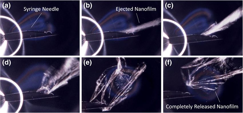
In general, free-standing magnetic nanofilms can be manipu- the injectability of the nanofilms decreased with increasing
lated with syringes, injecting and ejecting multiple times with- SPIONs concentration.
out distortion (Fig. 3a-e). Even after manipulation, the
nanofilms spread in the suspending medium and were unfold- 3.4 Film injectability design
ed (Fig. 3f).
In particular, the influence of three parameters on According to experimental data, we have developed a simple
nanofilms injectability was investigated: lateral dimension of model for the prediction of the injectability of films and thus
the nanofilms, SPIONs content and the needles diameter. For for their preliminary design. Crumpling a film of lateral di-
each SPIONs concentration, injectability of the magnetic mensions L1 and L2 and thickness h would lead to a ball of
nanofilms was plotted against lateral dimension of the radius r. According to fractal laws (Carpinteri and Pugno
nanofilms (L) and needles diameter (D) (Fig. 4). None of the 2005) we expect a scaling in the form of rd = k ∙ L1 ∙ L2 ∙ h
nanofilms could be injected through the needle with an inlet where 2 ≤ d ≤ 3 is the fractal dimension of the crumpled ball
diameter of 0.45 mm. Then, 0.6 mm represented the lower and k is a constant (with not integer physical units).
limit of needle diameter in the selected range. Considering L1 = L2 = Lmax, h for each type of nanofilm
The maximum lateral size (Lmax) that allowed the injection (Table 1), 2r = D as condition of injectability (note that also
of the nanofilms without risk of rupture (I = 100%) in the 2r equal to a fraction of D would lead to identical results, with
function of SPIONs concentration and needle diameters, sum- different values of k), and deducing k from the single case of
marized in Table 2, is an important parameter for the practical D = 0.6 mm and PL10-SP0, we can compare the predictions of
application. the model in the limiting cases of d = 2 and d = 3 with the
The size of the nanofilms (lateral size and thickness) played experimental observations. The comparison is reported in
a primary role during the injection because the nanofilms with Table 2 and shows a close agreement.
larger dimensions could cause the clogging of the needle.
Therefore, the increase in the SPIONs concentration, and sub- 3.5 Morphology of injected films
sequent increase in the thickness of the nanofilms, reduced the
injectability. For example, PL10-SP5 samples (thickness From a morphological point of view, observing the shape of
140 nm) with a lateral size of 10 mm showed an injectability the nanofilms immediately after the injection, nanofilms with
Fig. 3 Manipulation of a PL10-SP0 nanofilm in water by a plastic syringe equipped with a needle with an inlet diameter of 1.1 mm (a): free-standing
nanofilm can be ejected through the hole of the syringe (b-e) spreading in the water without damaging the structure (f)Biomed Microdevices (2017) 19:51 Page 7 of 9 51
Fig. 4 Nanofilms injectability (I) as a function of the nanofilms lateral size (L) and needle diameters (D), for different SPIONs concentration
high injectability (I > 70%) showed the ability to recover their tissues. For this reason, after the release, the stretch and the
shape and spread in the water (Fig. 5a) more easily than low- spread of the nanofilms could be further promoted alternating
injectable nanofilms (I < 70%), which tended to remain in a flux of water and air directly through the working channel of
Bcrumpled state^ (Fig. 5b). The ability to recover the spread- the endoscope or with the aid of the external magnetic field.
ing shape is an important factor for the intended application The surface of the nanofilms after the injection has been
because the crumpled state could prevent the adhesion to the analyzed by digital optical microscopy on nanofilms collected
and dried on clean silicon wafers. Crumpled nanofilms have
Table 2 Maximum lateral size L max which corresponds to an been unfolded with the aid of tweezers and fluxes of water
injectability of 100%. Comparison between experimental observations through the syringe and collected on silicon wafers after they
and model predictions (in brackets)
have assumed the spread shape. Nanofilms with high
Sample Maximum lateral size Lmax (mm) injectability did not show showed any cracks, wrinkles or other
discontinuities caused by the induced stresses (Fig. 5c). When
D = 0.6 mm D = 0.9 mm D = 1.1 mm
I > 70%, the unaltered integrity of the structure was demonstrat-
PL10-SP0 7 (=7) 10 (10–13) 17 (13–17) ed also at the micro-scale observing the surface of the nanofilms
PL10-SP1 5 (4) 7 (6–8) 15 (8–11) by AFM before and after the injection (Fig. 6a, b).
PL10-SP5 5 (4) 7 (7–8) 10 (8–11) On the other hand, low-injectable nanofilms showed not
PL10-SP10 - (4) 5 (6–7) 7 (7–10) only macroscopic ruptures but also the presence of disconti-
PL10-SP15 - (3) 5 (4–5) 5 (5–7) nuities on their surface (Fig. 5d). In particular, irreversible
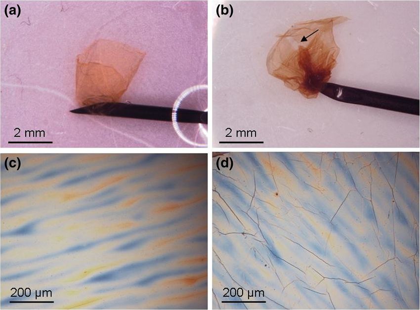
wrinkles were formed on nanofilms’ surface due to the51 Page 8 of 9 Biomed Microdevices (2017) 19:51
Fig. 5 Digital optical microscope
images of magnetic nanofilms
during and after the injection (on
nanofilms collected and dried on
clean silicon wafer) through a
1.1 mm diameter needle: (a) and
(c) a spread PL10-SP10 nanofilm
(L = 7 mm, I = 100%); (b) and (d)
a crumpled PL10-SP15 nanofilm
(L = 10 mm, I = 20%). The arrow
indicates a rupture site in the low-
injectable nanofilm
compressive stresses that occur during the injection. The wrin-
kling phenomenon was observed for the samples with low
injectability (I < 70%), independently from the content of
SPIONs in the polymeric matrix.
As mentioned before, the incorporation of SPIONs makes
the nanofilms thicker and less elastically deformable: in the
same range of needle diameters and lateral size of the
nanofilms, the injectability of the nanofilms decreased with
increasing SPIONs concentration. Consequently, at higher
SPIONs concentration (i.e. SP10–10, SP10–15), the wrin-
kling phenomenon occurs more frequently. Also in this case,
choosing the right conditions for the injection (diameter of the
needle and lateral size of the nanofilm) the wrinkling phenom-
enon can be avoided.
4 Conclusions
The present study revealed that magnetic PLLA-SPIONs
nanofilms are extremely flexible and deformable. The inclu-
sion of polymer-coated SPIONs in a PLLA matrix, although
leading to an increase in the mechanical properties of the
nanofilms, kept their elastic modulus at a considerably low
value. The ability of these flexible nanofilms to be manipulat-
ed with syringes without distortion has been experimentally
studied and was quantified by the injectability parameter I. An
analytical model to predict the injectability threshold vs. geo-
Fig. 6 AFM surface topography of a PL10-SP10 nanofilm (L = 7 mm, metrical parameters was also presented. Integrity of the
D = 0.9 mm, I = 90%) before (a) and after (b) the injection nanofilm structure and ability to recover the spread shape afterBiomed Microdevices (2017) 19:51 Page 9 of 9 51
the ejection can be guaranteed by choosing the right combi- C. Jiang, V.V. Tsukruk, Adv. Mater. 18, 829 (2006)
C. Jiang, S. Markutsya, Y. Pikus, V.V. Tsukruk, Nature Mater. 3, 721
nation of needle size, nanofilm lateral dimension and SPIONs
(2004)
content. As foreseen in previous papers these nanofilms can T.J. Kang, M. Cha, E.Y. Jang, J. Shin, H.U. Im, Y. Kim, J. Lee, Y.H. Kim,
be manipulated and precisely positioned within the working Adv. Mater. 20, 3131 (2008)
environment by using an external magnetic field and could A.A. Mamedov, N.A. Kotov, Langmuir 16, 5530 (2000)
thus provide a novel controllable injectable support in bio- A.A. Mamedov, N.A. Kotov, M. Prato, D. Guldi, J. Wicksted, A. Hirsch,
Nature Mater. 1, 190 (2002)
medical applications.
V. Mattoli, S. Sinibaldi, V. Pensabene, S. Taccola, A. Menciassi, P. Dario,
Proceedings of ICRA 2010–2010 I.E. International Conference on
Acknowledgement This work was supported in part by JFE (The Robotics and Automation, Anchorage (Alaska, USA), (2010)
Japanese Foundation for Research and Promotion of Endoscopy) Grant Y. Okamura, K. Kabata, M. Kinoshita, D. Saitoh, S. Takeoka, Adv. Mater.
(T.F.). JSPS KAKENHI (grant number 15H05355 for T.F., 16K14009 for 21, 4388 (2009)
S.T.) from MEXT, Japan, and the Precursory Research for Embryonic S.S. Ono, G. Decher, Nano Lett. 6, 592 (2006)
Science and Technology (PRESTO) from the Japan Science and
V. Pensabene, V. Mattoli, T. Fujie, A. Menciassi, S. Takeoka, P. Dario,
Technology Agency (JST) (grant number JPMJPR152A for T.F.).
Proceedings of the 9th nanotechnology conference IEEE Nano;
Nicola M. Pugno is supported by the European Research Council PoC
Genova (2009)
2015 BSilkene^ No. 693670, by the European Commission H2020 under
V. Pensabene, P. Valdastri, S. Tognarelli, A. Menciassi, A. Arezzo, P.
the Graphene Flagship Core 1 No. 696656 (WP14 BPolymer
Dario, Surg. Endosc. 25, 3071 (2011)
Nanocomposites^) and under the FET Proactive BNeurofibres^ No.
E. Redolfi Riva, A. Desii, S. Sartini, C. La Motta, B. Mazzolai, V. Mattoli,
732344.
Langmuir 29, 13190 (2013)
E. Redolfi Riva, A. Desii, E. Sinibaldi, G. Ciofani, V. Piazza, B.
Mazzolai, V. Mattoli, ACS Nano 8, 5552 (2014)
References L. Ricotti, S. Taccola, V. Pensabene, V. Mattoli, T. Fujie, S. Takeoka, A.
Menciassi, P. Dario, Biomed. Microdev 12, 809 (2010)
C.M. Stafford, C. Harrison, K.L. Beers, A. Karim, E.J. Amis, M.R.
I. Baker, Q. Zeng, W. Li, C. Sullivan, J. Appl. Phys 99, 08H106 (2006)
Vanlandingham, H. Kim, W. Volksen, R.D. Miller, E.E. Simonyi,
A. Carpinteri, N. Pugno, Nature Mater. 4, 421 (2005)
Nature Mater. 3, 545 (2004)
G. Ciuti, R. Donlin, P. Valdastri, A. Arezzo, A. Menciassi, M. Morino, P.
S. Taccola, A. Desii, V. Pensabene, T. Fujie, A. Saito, S. Takeoka, P.
Dario, Endoscopy 42, 148 (2010)
Dario, A. Menciassi, V. Mattoli, Langmuir 27, 5589 (2011)
G. Decher, Science 277, 1232 (1997)
H. Endo, Y. Kado, M. Mitsuishi, T. Miyashita, Macromolecules 39, 5559 S. Taccola, F. Greco, A. Zucca, C. Innocenti, C. de Julián Fernández, G.
(2006) Campo, C. Sangregorio, B. Mazzolai, V. Mattoli, ACS Appl. Mater.
T. Fujie, Polym. J. 48, 773 (2016) Interfaces 5, 6324 (2013)
T. Fujie, Y. Okamura, S. Takeoka, Adv. Mater. 19, 3549 (2007) Z. Tang, Y. Wang, P. Podsiadlo, N.A. Kotov, Adv. Mater. 18, 3203 (2006)
T. Fujie, N. Matsutani, M. Kinoshita, Y. Okamura, A. Saito, S. Takeoka, R. Vendamme, S. Onoue, A. Nakao, T. Kunitake, Nat. Mater. 5, 494
Adv. Funct. Mater. 19, 2560 (2009) (2006)
T. Fujie, L. Ricotti, A. Desii, A. Menciassi, P. Dario, V. Mattoli, Langmuir L. Ventrelli, T. Fujie, S. Del Turco, G. Basta, B. Mazzolai, V. Mattoli, J.
27, 13173 (2011) Biomed, Mater. Res. Part A 102, 2652 (2014)
T. Fujie, A. Desii, L. Ventrelli, B. Mazzolai, V. Mattoli, Biomed. Z. Wang, L. Wang, B. Tang, T. Frank, S. Brown, A. Cuschieri, Surg.
Microdevices 14, 1069 (2012) Endosc. 22, 1838 (2008)
T. Fujie, Y. Kawamoto, H. Haniuda, A. Saito, K. Kabata, Y. Honda, E. Z. Wang, P. André, D. McLean, S.I. Brown, G.J. Florence, A. Cuschieri,
Ohmori, T. Asahi, S. Takeoka, Macromolecules 46, 395 (2013) Med. Eng. Phys. 36, 1521 (2014)
T. Fujie, Y. Mori, S. Ito, M. Nishizawa, H. Bae, N. Nagai, H. Onami, T. Q. Wang, E. Chen, Y. Cai, C. Chen, W. Jin, Z. Zheng, X. Jin, Y. Chen, X.
Abe, A. Khademhosseini, H. Kaji, Adv. Mater. 26, 1699 (2014) Zhang, Q. Li, World J. Surg. Oncol. 14, 231 (2016)
F. Greco, A. Zucca, S. Taccola, A. Menciassi, T. Fujie, H. Haniuda, S. A. Zucca, C. Cipriani, S. Sudha, S. Tarantino, D. Ricci, V. Mattoli, F.
Takeoka, P. Dario, V. Mattoli, Soft Matter 7, 10642 (2011) Greco, Adv Healthc. Mater. 4, 983 (2015)You can also read