Pca postclassicalarchaeologies - european journal of - European Journal of Post ...
←
→
Page content transcription
If your browser does not render page correctly, please read the page content below
pca            european journal of
postclassicalarchaeologies
                                     Volume 11
                                     May 2021

SAP
Società
Archeologica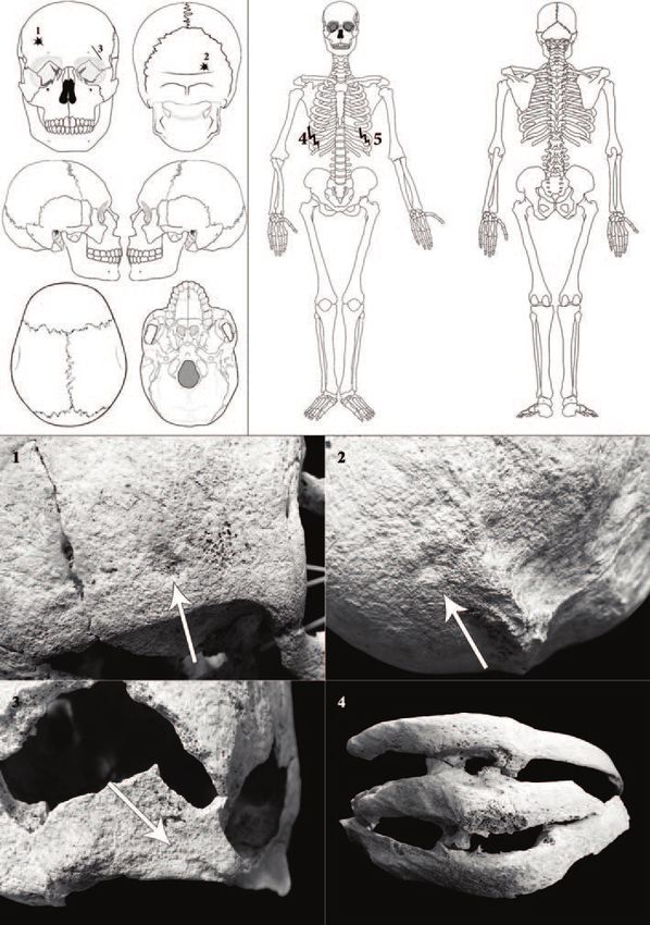
pca                   european journal of
              postclassicalarchaeologies

                volume 11/2021

SAP Società Archeologica s.r.l.

        Mantova 2021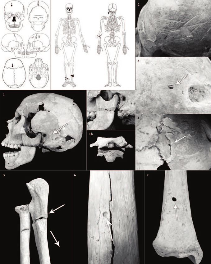
pca
EDITORS EDITORIAL BOARD
Gian Pietro Brogiolo (chief editor) Paul Arthur (Università del Salento)
Alexandra Chavarría (executive editor) Alicia Castillo Mena (Universidad Complutense de Madrid)
Margarita Díaz-Andreu (ICREA - Universitat de Barcelona)
Martin Carver (University of York)
José M. Martín Civantos (Universidad de Granada)
Girolamo Fiorentino (Università del Salento)
Caterina Giostra (Università Cattolica del Sacro Cuore - Milano)
Susanne Hakenbeck (University of Cambridge)
Matthew H. Johnson (Northwestern University of Chicago)
Vasco La Salvia (Università degli Studi G. D’Annunzio di Chieti e Pescara)
Bastien Lefebvre (Université Toulouse - Jean Jaurès)
Alberto León (Universidad de Córdoba)
Tamara Lewit (University of Melbourne)
Yuri Marano (Università Ca' Foscari Venezia)
Federico Marazzi (Università degli Studi Suor Orsola Benincasa di Napoli)
Andrew Reynolds (University College London)
Mauro Rottoli (Laboratorio di archeobiologia dei Musei Civici di Como)
Colin Rynne (University College Cork)
Marco Valenti (Università degli Studi di Siena)
Giuliano Volpe (Università degli Studi di Foggia)
Post-Classical Archaeologies (PCA) is an independent, international, peer-reviewed journal devoted to the commu-
nication of post-classical research. PCA publishes a variety of manuscript types, including original research, discus-
sions and review articles. Topics of interest include all subjects that relate to the science and practice of archaeology,
particularly multidisciplinary research which use specialist methodologies, such as zooarchaeology, paleobotany, ar-
chaeometallurgy, archaeometry, spatial analysis, as well as other experimental methodologies applied to the archae-
ology of post-classical Europe.
Submission of a manuscript implies that the work has not been published before, that it is not under consideration
for publication elsewhere and that it has been approved by all co-authors. Authors must clear reproduction rights
for any photos or illustration, credited to a third party that they wishe to use (including content found on the Internet).
For more information about ethics (including plagiarism), copyright practices and guidelines please visit the website
www.postclassical.it.
PCA is published once a year in May. Manuscripts should be submitted to editor@postclassical.it in accordance
to the guidelines for contributors in the webpage http://www.postclassical.it.
Post-Classical Archaeologies’ manuscript review process is rigorous and is intended to identify the strengths and
weaknesses in each submitted manuscript, to determine which manuscripts are suitable for publication, and to work
with the authors to improve their manuscript prior to publication.
This journal has the option to publish in open access. For more information on our open access policy please visit
the website www.postclassical.it.
How to quote: please use “PCA” as abbreviation and “European Journal of Post-Classical Archaeologies” as full title.
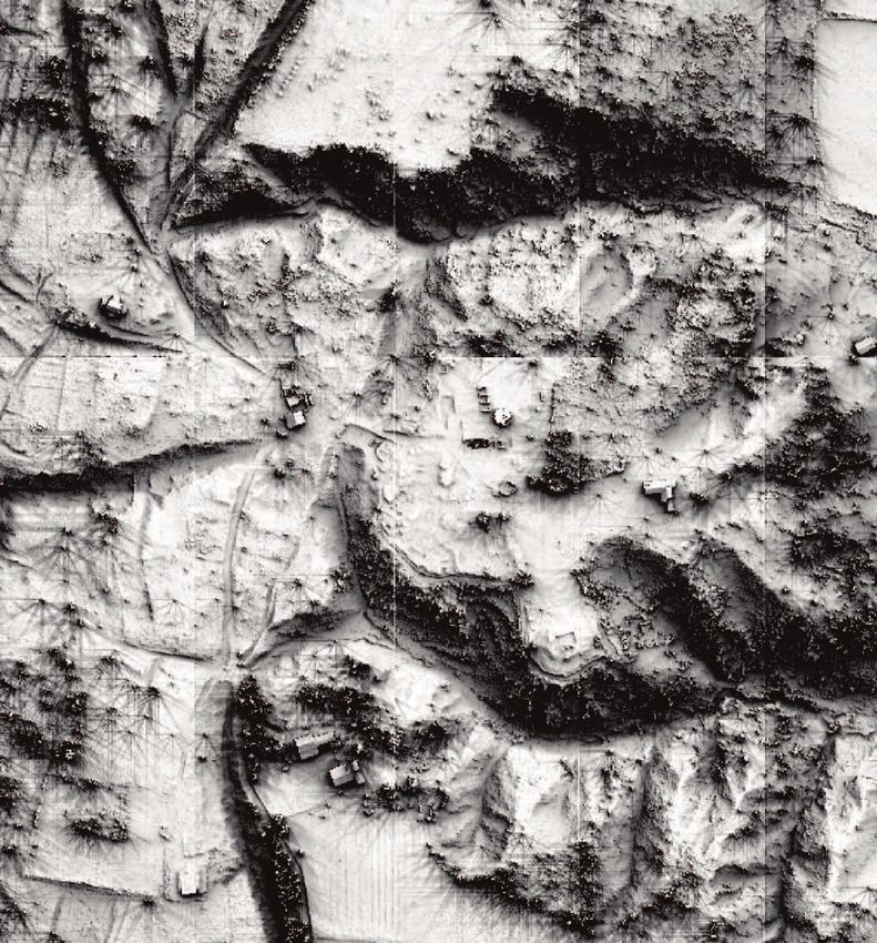
Cover image: LiDAR survey of Castelseprio (north Italy) (project funded by Varese Province).
“Post-Classical Archaeologies” is indexed in Scopus. It was approved on 2015-05-13 according to ERIH PLUS criteria
for inclusion and indexed in Carhus+2018. Classified A by ANVUR (Agenzia Nazionale di Valutazione del sistema Uni-
versitario e della Ricerca).
DESIGN:
Paolo Vedovetto
PUBLISHER:
SAP Società Archeologica s.r.l.
Strada Fienili 39/a, 46020 Quingentole, Mantua, Italy
www.saplibri.it
Authorised by Mantua court no. 4/2011 of April 8, 2011
ISSN 2039-7895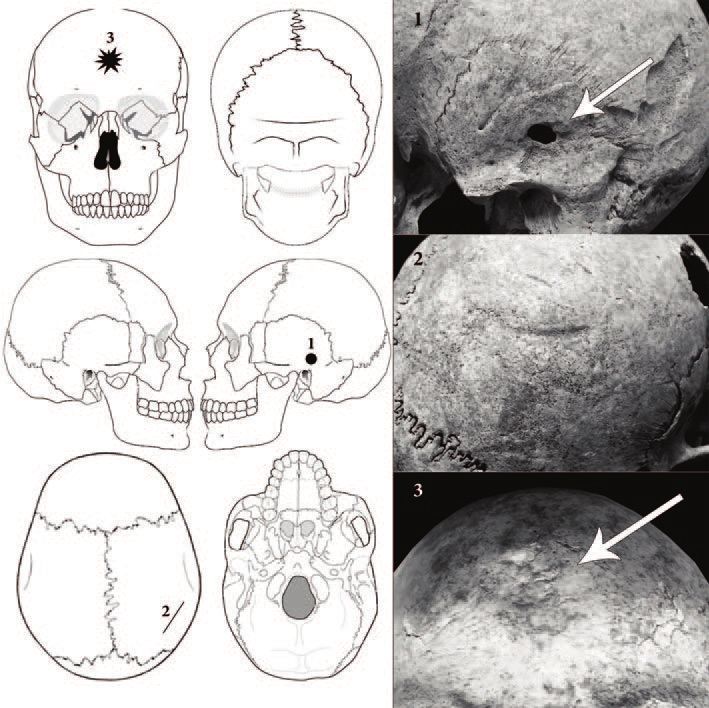
pca                 european journal of
                                                     postclassicalarchaeologies
                                                                        volume 11/2021

                                                               CONTENTS PAGES

EDITORIAL                                                                        5

RESEARCH - COMMONS: AN ARCHAEOLOGICAL PERSPECTIVE
P.G. Gould      Community in archaeology: an assessment                          7
S. Rippon       Communities, continuity, and change: territorial identities in   25
                early medieval southern Britain
G.P. Brogiolo Comunità rurali e beni collettivi tra fonti scritte e paesaggi     59
              stratificati
A.M. Stagno, J. Narbarte Hernández, C. Tejerizo García The social dimen-         81
                sion of commons between practices and jurisdiction. Case
                studies from southern Europe (17th-21st c.)

BEYOND THE THEME
J. Benedito-Nuez, J.J. Ferrer-Maestro, J.M. Melchor-Monserrat Perviven-          111
              cia y transformación: testimonios arqueológicos de la diná-
              mica urbana de la ciudad romana de Saguntum entre los
              siglos III y VII
E. Zanini       Cost, value and wealth redistribution: micro- and macro-         137
                economy in Early Byzantine evergetism
S. Bortolotto, N. Cattaneo, A. Garzulino, S. Massa, S.M. Masseroli,              163
               R.M. Rombolà Castelseprio archaeological site: LiDAR
               and GIS multiscale dataset supporting on-field investiga-
               tion and enhancing landscape understanding
F. Andriani, F. Armenise, G.A. Panzarino, S. Sublimi Saponetti Signs of          189
                interpersonal violence and war: paleotraumatology in Apu-
                lia during the Late Antiquity and the Middle Ages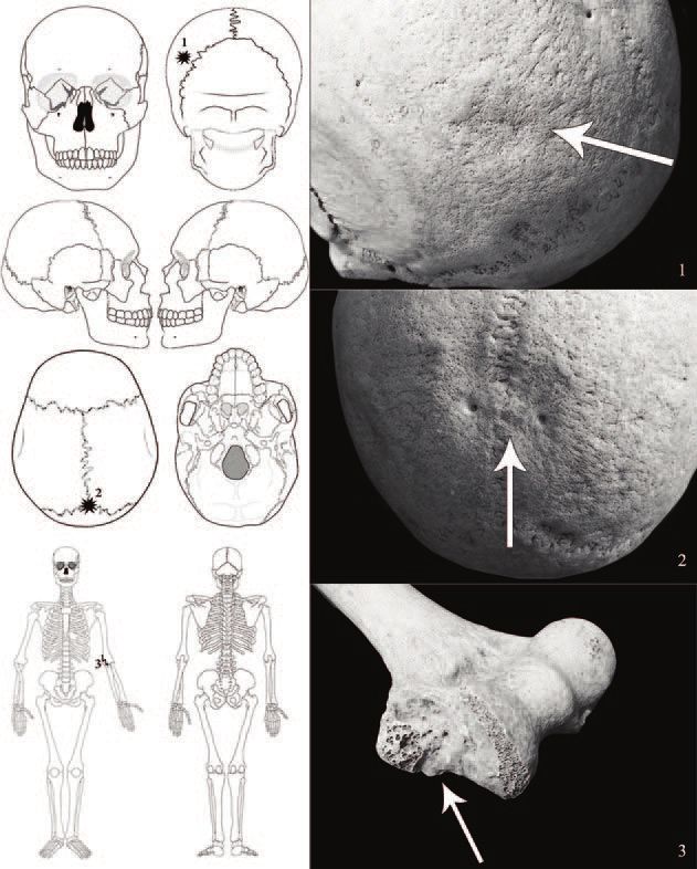
E. Dorado, J. Herrerín, I. Ramírez, L. Parro Klippel-Feil Syndrome in a                        253
              Mudejar population: a sign of endogamy in a social minority
G. Marra           Studio del paesaggio storico urbano di Ascoli Piceno nel                    267
                   Basso Medioevo: dalla ricostruzione alla comunicazione
                   digitale con Google Earth
C. Bonacchi, M. Lorenzon Assessing the transforming social values of                           303
              cities in the longue durée: analysis of a Florence neighbour-
              hood from the Middle Ages to the present
G. De Felice       Novecento. Apulia at war. A project of archaeology of the                   327
                   contemporary past between research, education and par-
                   ticipation

DOSSIER - ARCHAEOLOGY AND SCHOOL
S. Schivo          L’archeologia nei manuali di storia: il caso di Padova                      349
J.C. González Vargas, R. Fabregat, A. Carrillo-Ramos, T. Jové                                  387
             Motiv-ARCHE: an augmented reality application to co-cre-
             ate cultural heritage resources with teenagers

REVIEWS                                                                                        439
T. Ingold, Making. Antropologia, archeologia, arte e architettura - by E. Giannichedda
G.P. Brogiolo, P.M. De Marchi (eds), I Longobardi a nord di Milano. Centri di potere
        tra Adda e Ticino - by E. Salvatori
Y. van den Hurk, On the Hunt for Medieval Whales. Zooarchaeological, historical and
        social perspectives on cetacean exploitation in medieval northern and west-
        ern Europe - by M. Fecchio
R. Fantoni, R. Cerri, P. de Vingo (eds), La Pietra Ollare nelle Alpi. Coltivazione e utiliz-
        zo nelle zone di provenienza - by P. Vedovetto
J. Darlington, Fake Heritage: Why we rebuild monuments - by F. Benetti

                                                      PCA volume 11/2021 ISSN: 2039-7895
                                                      Post-Classical Archaeologies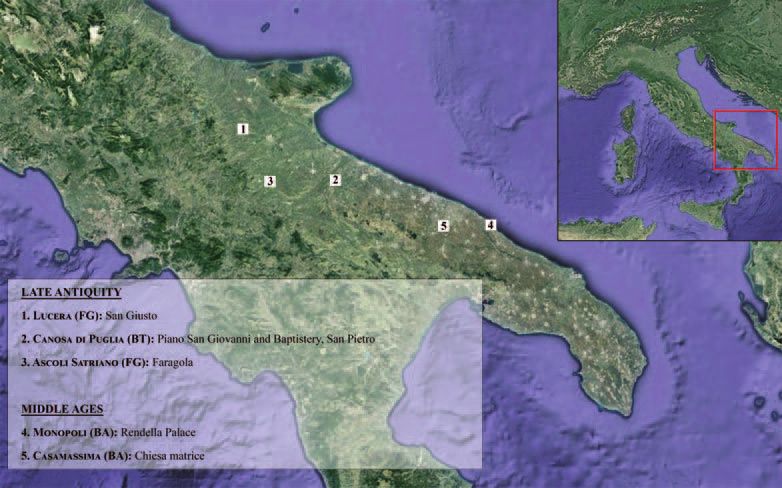
beyond the
                                                                                             theme
                              Fabrizia Andriani, Fabio Armenise,
               Ginevra Anna Panzarino, Sandro Sublimi Saponetti*

     Signs of interpersonal violence
and war: paleotraumatology in Apulia
            during the Late Antiquity
                 and the Middle Ages

    1. Introduction

    Since the 1990s, many archaeological excavations have been carried out in
cemeteries in Apulia (Southern Italy) dated from Late Antiquity to the Early Mid-
dle Ages (400-1000 AD). Numerous skeletal remains have been discovered and
studied over the years by the Anthropology Laboratory of the Department of Bi-
ology at the University of Bari Aldo Moro, and among them a number of individ-
uals with signs of violence. The purpose of this study is to determine the nature
of these lesions based on historical, archaeological, anthropological and pale-
opathological sources. Through statistical analysis of the injuries, it is possible to
identify distribution according to age, sex and skeletal region, hypothesize the
weapons used, reconstruct how violent acts were committed and acknowledge
cultural influences (if any). After the section on ‘Materials and Methods’, the
‘Case Studies’ section describes the archaeological context and samples in
chronological order. For each individual, there will be a description of all the le-
sions detected, the identification of the weapons used and a plausible recon-

* Fabrizia Andriani: external collaborator of the Department of Biology, University of Bari Aldo Moro,
Bari, Italy, fabrizia.andriani@gmail.com; Fabio Armenise: president of the cultural association Com-
pagnia d’Arme Stratos. Ricerca - Ricostruzione - Divulgazione, fabio.armenise@alice.it; Ginevra Anna
Panzarino, University of Valencia, Spain, ginevrapanzarino@gmail.com; Sandro Sublimi Saponetti:
Department of Biology, University of Bari Aldo Moro, Bari, Italy, and corresponding author: sandro.su-
blimisaponetti@uniba.it.
Sandro Sublimi Saponetti supervised the study and dealt with the anthropological and paleopatho-
logical aspects of skeletal remains; Fabrizia Andriani has carried out the statistical analysis and made
the charts; Fabio Armenise studied the lesions, recognized or hypothesized the weapons, and recon-
structed the actions causing the traumas; Ginevra Anna Panzarino has been involved in the analysis
of the historical and archaeological context, organised the photographic apparatus and coordinated
the final draft of the paper.

              PCA 11 (2021) ISSN: 2039-7895 (pp. 189-252) Post - Classical Archaeologies
                     Received: 26-03-2020 - Accepted: 10-09-2020 - Revised: 18-12-2020            189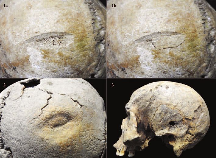
Fabrizia Andriani, Fabio Armenise, Ginevra Anna Panzarino, Sandro Sublimi Saponetti

                Fig. 1. Italy, Apulia: map of the sites mentioned in the paper.

struction of the violent action. The lesions will be described and analysed provid-
ing photographs and charts, while the violent action will be hypothesized. In the
last part of the study (‘Results and discussion’) the statistical data will be dis-
cussed, according to traumatological elements (injury type, distribution, weapon
typology, reconstruction of the action, type of violence) and geo-cultural factors
(context, history, distribution of sites, possible cultural influences).

   2. Materials and methods

    The material will be presented in chronological order, from the Baptistery of
San Giovanni in Canosa (BT), San Giusto in Lucera (FG), Piano San Giovanni in
Canosa (BT), Faragola in Ascoli Satriano (FG) San Pietro in Canosa (BT), Palaz-
zo Rendella in Monopoli (BA) and Chiesa Madre of Casamassima (BA) (fig. 1,
tab. 1). The oldest, dating back to the middle of the 5th century AD, is the Bap-
tistery of San Giovanni in Canosa (BT). The most recent one, which dates back
to the 11th century AD, is Palazzo Rendella in Monopoli (BA). These sites are lo-
cated in the northern and central parts of the region: Canosa, Lucera and Ascoli
Satriano in the North; Casamassima and Monopoli in the center. Canosa (BT)

190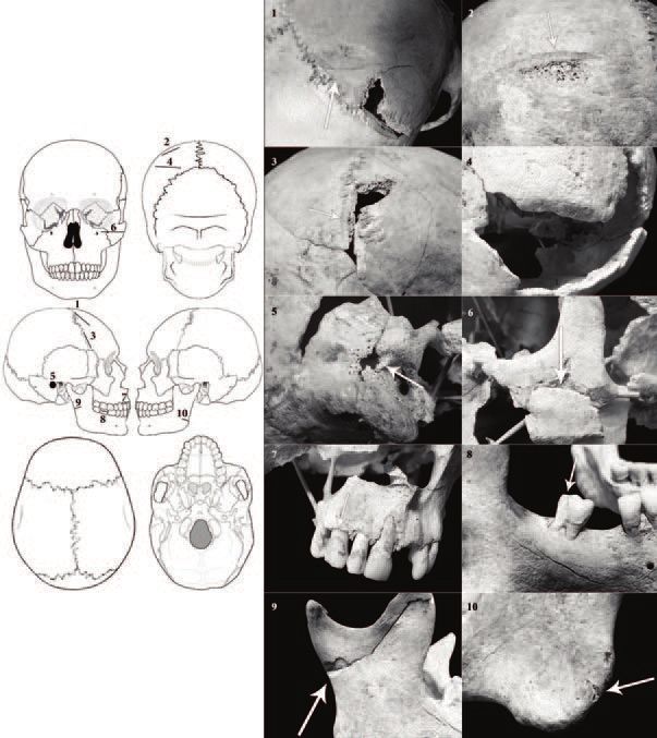
Signs of interpersonal violence and war: paleotraumatology in Apulia...

City                          Site                                 Dating                N.
Canosa di Puglia (BT)         Baptistery of San Giovanni           5th century AD        1
Lucera (FG)                   San Giusto                           6th century AD        6
Canosa (BT)                   Piano San Giovanni                   7th century AD        1
Ascoli Satriano (FG)          Faragola                             7th century AD        1
Canosa (BT)                   San Pietro                           7th century AD        6
Monopoli (BA)                 Palace Rendella                      8th-11th century AD   14
Casamassima (BA)              Chiesa matrice                       10th century AD       9

                Tab. 1. List of archaeological sites and number of individuals.

has been the focal point of most research, as several excavations have been
carried out by universities. The skeletal samples are of 38 individuals, not always
fully preserved, with single or multiple lesions. The most numerous samples are
those of Palazzo Rendella in Monopoli (BA) and of Chiesa Madre of Casamassi-
ma (BA).
    The biological and paleopathological profile of individuals have been recon-
structed according to the following aspects: identification of metric and morpho-
metric features (Martin, Saller 1956-59); determination of age and sex (Ferem-
bach et al. 1980; Lovejoy et al. 1985); identification of nutritional and/or stress
diseases, periodontal disease and caries (Brothwell 1981; Larsen 1999), tartar
build-up (Dobney, Brothwell 1987) and degree of dental wear (Molnar 1971),
enamel hypoplasia lines (Goodman, Rose 1990), markers of skeletal biomechan-
ical stress – syndesmosis lesions, enthesopathies, new articular surfaces and
degenerative joint diseases – (Rogers et al. 1987; Kennedy 1989; Palfi 1992; Lai,
Lovell 1992; Robb 1994; Robb, Mallegni 1994; Mariotti et al. 2007); degenerative
changes of the spine (Borgognini Tarli, Repetto 1986); estimation of body mass
(Ruff et al. 1997); analysis of the cross sectional geometry of the humerus and
femur (Tracey et al. 1994; Larsen 1999; Capasso et al. 1999; Ledger et al. 2000;
Stock, Pfeiffer 2001) and the human typologies (Thoma 1988). Statistical analy-
ses are based on the incidence and percentage frequencies of individual data
(Cavalli-Sforza 1983; Cohen 1993).
    A datasheet was designed to record all the useful aspects to study traumas
and their interpretation, with this structure:

NAME: City, archaeological site (chronology)
          eg. Casamassima, Chiesa Matrice (10th sec. AC);
GRAVE: Initials
          eg. CAS28us180
AGE: “infant”, “juvenile” “adult”, “elderly” (age range)
          eg. Adult (40-44);

                                                                                          191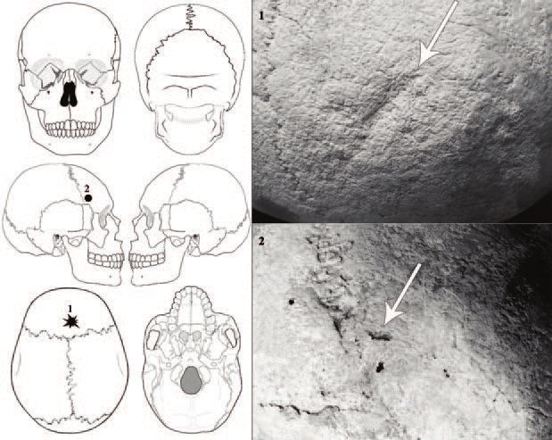
Fabrizia Andriani, Fabio Armenise, Ginevra Anna Panzarino, Sandro Sublimi Saponetti

HEALTH STATUS: clinical picture
           eg. Button-shaped osteoma on the skull;
BONES FOUND: Percentage value1
           eg. 26,4%;
CRANIAL/POSTCRANIAL WOUND2: BONE, position and side, measure and angle, type
(“blunt”, “bullet”, “blade”, “point”, “unknown”)
           eg. 1) Cranial wound: Right PARIETAL, sagittal suture 12 mm away from the lambda.
           Blade, penetrating (9° from the bottom, transverse diameter 51 mm, supero-inferior
           diameter 32 mm). Peri mortem;
           2) Cranial wound: MANDIBLE, left branch, underneath the coronoid process. Blade
           (1,5 mm deep; antero-posterior diameter 16,5 mm). Ante mortem.
No. of TOTAL WOUNDS: quantity
           eg. 2;
No. of CRANIAL WOUNDS: quantity
           eg. 2;
No. of POSTCRANIAL WOUND: quantity
           eg. 0;
SKULL: Type (Lesion identification no.)
           eg. Blade (1 Cranial wound; 2 Cranial wound);
UPPER LIMBS: Type (Lesion identification no.)
           g. 0;
TORSO: Type (Lesion identification no.)
           eg. 0;
LOWER LIMBS: Type (Lesion identification no.)
           eg. 0;
WEAPON: Lesion identification no.: type and identification
           eg. 1 Cranial wound: blade, long (sword);
           2 Cranial wound: blade, long (sword);
DYNAMICS OF ACTION: Lesion identification no.: position and number of aggressor/s, type of
shot (“downward”, “middle”, “upward”), direction of the weapon (“from below”, “from above”,
“from right”, “from left”, “unknow”)
           eg. 1 Cranial wound: posterior aggressor, blow from left to right;
           2 Cranial wound: posterior aggressor, blow from left to right;
EPISODE OF VIOLENCE: “ambush”, “battle”, “skirmish”, “unknown”
           eg. unknow.

    The determination of the type of injury and weapon was performed by com-
parison with other data known in literature (De Vita 1983; Lovell 1997; Shaw
2001; Mitchell et al. 2006; Stefan et al. 2006; Novak 2007; Roksandic et al. 2007;
Fornaciari, Giuffra 2009; Slaus et al. 2010; Milner et al. 2015; Constantinescu et
al. 2017). In the classification of the blows in the italian version of this study we

1   Calculated on the total number of human bones (around 203).
2   Each lesion has a progressive number.

192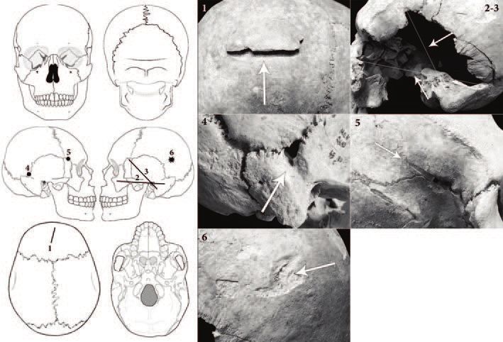
Signs of interpersonal violence and war: paleotraumatology in Apulia...

adopted a terminology drawn from medieval defensive treaties (cf. Rapisardi
1998): fendente meaning “downwards blow”, mezzano “middle blow” and sot-
tano “upward blow”; mandritto “from right to the left”, manrovescio “from left to
right”. As for weapon identification, this was determined by the joint examination
of the morphology of the injury and the historical context. The reconstruction of
the blow and the dynamics of the act of violence has been reenacted with a fenc-
ing master (Fabio Armenise). We used several weapons and reenacted the
blows on the bones from different positions, to understand the direction and type
of the blows but especially the position of the two fighters. In the case of multi-
traumatized individuals with blows interpreted as ‘overkill’ (Grey Friars Research
Team 2015, p. 68; Solarino et al. 2019) this was not always possible. In fact, anal-
ysis of the direction in which blows were inflicted varied depending on the posi-
tion of the victim in relation to the aggressor(s). In these cases, the victim was
often lying on the ground, unconscious or dead, and the enemy kept on striking
on the body. The enemy was often enraged and did so to damage with his fury
the victim’s body or to reclaim a sort of personal glory.

   3. Case studies

   3.1. Canosa, Baptistery of San Giovanni (mid 5th century AD)

   In Canosa (BT) in Piano di San Giovanni (section 3.3) several excavations
were carried out in the 70s by the then Soprintendenza Archeologica della Puglia
and from 2002 to 2010 in collaboration with the Universities of Foggia and Bari
(Giuliani et al. 2012). The Early Christian worship building, commissioned by
Bishop Sabino (514-566), is a baptistery with a central plan. Here the remains of
some individuals with injuries dating back to the pre-Roman period (Sublimi
Saponetti et al. 2010) and the mid-5th century (Sublimi Saponetti et al. 2008) were
found (tab. 2).

    BSG1 is an adult male (40-44 years) 172 cm tall and weighing approximately
80 kg. Two hypoplasia lines and some caries were detected, with moderate den-
tal wear and tartar build-up; muscles are well-shaped and there are signs of
moderate arthrosis especially in the lower limbs; traces of non-specific infection,
periostitis of the tibia and a button-shaped excrescence on the left orbit are also
visible. This man presents multiple traumas, both cranial (fig. 2a) and postcranial
(fig. 2b), two with scarring and ten showing that no healing had occurred. The
first perimortem injury (1 Cranial wound) is on the right squama of the frontal
bone and the back end touches the pars bregmatica of the right coronal suture.
It has a v-shape, with anterolateral/posteromedial oblique orientation. The lateral
half has been cut by a blade, while the medial half for this reason has a wrinkled

                                                                                        193
Fabrizia Andriani, Fabio Armenise, Ginevra Anna Panzarino, Sandro Sublimi Saponetti
Acronym Sex, age Bone, district Wound
Sharp
Sharp
Sharp
Skull
Sharp
Puncture
BSG1 M Mat Drilling?
Sharp
Jaw Sharp
Sharp
L. Humerus Sharp
L. Rib 11-12; L2-L4 L.; Sacred; L. Iliac wing Sharp
Tab. 2. Canosa (BT), Baptistery of St. John: list of individuals with injuries.
Fig. 2a. BSG 1. Injuries on the skull.
194Signs of interpersonal violence and war: paleotraumatology in Apulia...
surface. The second injury (2 Cranial wound), has also a v-shape and is located
on the left parietal bone; it has the same characteristics of 1 Cranial Wound, but
with a transverse orientation. In both cases these lesions are caused by a bladed
weapon used in a semi-tangential direction. In this case the weapon, while being
pulled back, chipped the bones. Particularly in the second case, the recovered
fragment of external diploë chipped off by the blade confirmed our idea. The
third wound (3 Cranial wound) involves the frontal bone, the right coronary suture
and the right parietal bone: it has a slightly oblique and transverse angle. The
blade perforated the bone in an orthogonal direction and the signs produced by
the passage of the blade are visible in the outer part of the diploë. An ante-
mortem lesion (4 Cranial wound) is at the back of the left parietal bone almost
near the lambdoid suture. Due to the poor preservation of the cranial area, it is
possible to observe solely the upper edge, that appears sclerotic and smoothed
with an oblique antero-lateral and postero-medial orientation. For this reason, the
injury might be the result of an old healed trauma probably followed by a craniec-
tomy and a wound cleaning. On the right temporal bone, a few millimeters above
the outer acoustic meatus, the bone is completely perforated (5 Cranial wound),
and the lesion is star-shaped and surrounded by signs of an inflammatory reac-
tion (cribrosity). This may have been caused by a pointed object that penetrated
orthogonally the bone. There are five unhealed injuries caused by a sharp object
on the facial bone and the jaw. The left zygomatic bone displays across its width
a horizontal semi-tangential injury (6 Cranial wound) caused by a bladed
weapon with a blow from top to bottom. Underneath, while removing the weapon,
a bone fragment was partially cut off. The maxillary right canine crown (7 Cranial
wound) and the mandibular second right molar (8 Cranial wound) appear bro-
ken. In both cases, the enamel of the crown on the vestibular side is chipped.
The upper part of the right mandibular branch (9 Cranial wound) is crossed by
an oblique fracture line that starts with a blade lesion located on the posterior rim
of the branch and completely cuts the coronary apophysis. The blade lesion it-
self shows a small semilunar cut section at the lower level with postero-inferior
and anterior-superior oblique orientation, while the upper part of the wound
points out the consequences resulting from removal of the weapon. At the top of
the gonion of the left mandibular angle, there is a minute, barely visible lesion (10
Cranial wound) with no signs of healing. The lesions involving the zygomatic
bone, teeth and gonion may be the result of four blows.
The last lesion could be due to both a downward blow or a middle one with
a slight inclination from the bottom up. Two traumatic events are visible on the
postcranial skeleton. An oblique triangular smooth cut is present on the left
humerus, particularly on the lateral edge of the distal surface (11 Postcranial
wound). The lesion direction is lateral-superior and medium-inferior and displays
a damaged area caused by the removal of the weapon. The injury may have
been caused by the victim’s attempt to parry the blow. The left side of the trunk
195Fabrizia Andriani, Fabio Armenise, Ginevra Anna Panzarino, Sandro Sublimi Saponetti
displays extensive bone loss, involving the XI
and XII thoracic ribs, all the lumbar vertebrae,
the sacrum and the back of the iliac wing (12
Postcranial wound). The thoracic ribs XI and XII
have a clean cut. The bone loss is located in the
lateral margin and in the transverse processes
of the left side of the lumbar vertebrae and the
first sacral vertebrae, along a vertical oblique
plane. The wing of ilium shows a destroyed
bone matrix.
The morphology of the coastal lesions indi-
cates the direction of the blow, a violent down-
ward slash from an aggressor standing on the
left side behind the victim’s back. We can hy-
pothesize a correspondence of the rib, verte-
brae and coxal lesions, if we imagine that the
victim had the trunk in a twisted and bent pos-
ture to the right. Likely, the weapon used for the
deadly blow may have been a bladed weapon
with high penetration power, such as an axe.
The individual may have been the victim of an
ambush by several aggressors wounding con-
tinuously without cease, perhaps even while
lying on the ground.
3.2. Lucera, San Giusto (6th century AD)
During the excavations carried out by Foggia
University in 1999, a large cemetery was discov-
ered in San Giusto (Lucera, FG), between the fu- Fig. 2b. BSG 1. Injuries on the post-
nerary basilica and the productive area of the cranial skeleton.
villa (Volpe et al. 2001, 2013). Dating back to the
6th century AD, the cemetery can be divided into
five areas: the funeral basilica, the narthex with the rooms attached to the basil-
ica, the pastophories in the area behind the apse, the area between the baptistry
and the baths; and finally the production area of the complex. In 103 graves, 120
individuals have been divided by sex and social class: 29 are subadults and 85
adults, including 55 males, 15 females, and 15 of unknown sex. The bones
showed many diseases and some skulls had Mongolian features and cranial de-
formation (Sublimi Saponetti et al. 2005). In the San Giusto samples, six individ-
uals presented some injuries (tab. 3).
196Signs of interpersonal violence and war: paleotraumatology in Apulia...
Acronym Sex, age Bone/district Wound
SG98 M Ad Tibia Puncture
SG124 M Ad Skull, parietal Puncture
Skull, frontal Sharp
SG130 M Young
Skull, frontal Puncture
Blunt
SG us32 M Ad Skull, frontal
Puncture
SG us45 M Ad Skull, parietal Puncture
Blunt
Skull, frontal
Sharp
SG us292 M Ad Skull, occipital Blunt
No. 3 R. Ribs Fracture
No. 3 R. Ribs Fracture
Tab. 3. Lucera (FG), San Giusto: list of individuals with injuries.
SG98 is an adult male that shows on the skull features of the Mongol human
type. These features are hyperbrachicrany, stenometopy, numerous wormian
bones, a flattened midface with the maxillar ascending apophysis on an almost
frontal plane, as well as the upper medial “shovel incisors”. A perimortem punc-
ture wound is present on the left tibia, on the antero-lateral surface of the distal
epiphysis (1 Postcranial wound) (fig. 3). The wound, showing no healing pro-
cess, is a full-thickness cut and v-shaped. The weapon used was a projectile
(arrow), thrown from far away by an enemy in front of the victim. The scenario
may have been an ambush, perhaps linked to a war episode.
Fig. 3. SG98.
197Fabrizia Andriani, Fabio Armenise, Ginevra Anna Panzarino, Sandro Sublimi Saponetti
Fig. 4. SG124.
SG124 is an adult male with a sub-circular puncture wound on the left parietal
bone of the skull (1 Cranial wound) (fig. 4). It is an unhealed postmortem wound,
caused by a pointed weapon, such as an arrow, shot by an enemy placed in
front of the victim and from far away with a trajectory from top to bottom. In this
case the wound may be the result of a war ambush as well.
SG130 is a juvenile male (16–17 years old), 170 cm tall and weighing approx-
imately 70 kg. His health and nutritional status cannot be defined as either poor
or very good, since the percentage of medium-diaphyseal cortical section of the
humerus (%CA) is 68%. The stress markers reveal the use of muscles for manual
work. The frontal bone of the skull shows two traumatic injuries, one of which was
fatal (fig. 5). The first (1 Cranial wound) consists of a perforation roughly quad-
rangular in shape. From the outside, the right antero-lateral margin and the pos-
terior part of the injury has an irregular and raised edge. The left margin has a
very well-defined and oblique edge. The endocranial view of injury reveals the
opposite effect, with a well-defined edge on the right and an irregular one on the
left. On the right side of the lesion there are two fracture lines that completely cir-
cumscribe a raised bone area. The lesion was perimortem because the bone re-
acted to the blow in a way that required a tissue hydration. The absence of bone
regrowth suggests that the wound was fatal. The victim was probably violently
stabbed with a pointed weapon with a quadrangular section. The second lesion
(2 Cranial wound) is on the anterolateral portion of the right frontal bone, consist-
ing of a loss of cortical surface of a circular shape. The bone is cut off tangen-
tially toward the convex part of the frontal squama, showing the typical blade
marks. This injury does not show signs of healing. He may have been hit with two
198Signs of interpersonal violence and war: paleotraumatology in Apulia...
Fig. 5. SG130.
different weapons or by a single weapon with a pointed head e and an opposite
sharp side: a tool that presents both features is an occasional weapon like a
pickaxe or dolabra.
SG us32 is an adult male with two injuries on the frontal bone (fig. 6). The first
one (1 Cranial wound) is an antemortem triangular blunt force trauma on the me-
dial portion. The second one (2 Cranial wound) is a perimortem star-shaped
puncture on the right side in front of the pars complicata of the coronal suture. In
the first case, the wound was caused by a downward blow from right to left by a
frontal assault with a club. The puncture was, instead, caused by an arrow shot
by an enemy standing on the right side of the victim. The context may have been
an ambush, perhaps linked to a war episode.
SG us45 is an adult male with a perimortem quadrangular puncture lesion (1
cranial wound) on the right parietal bone (fig. 7). The wound was caused by an
assault from the right side of the victim with a downward blow from right to left.
The weapon and the violent act cannot be precisely determined.
SG us292 is an adult male with five ante mortem injuries, three on the skull
and two postcranial (fig. 8). The first one (1 Cranial wound) is a sub-circular
blunt force trauma located on the right part of the frontal bone. The second one
(2 Cranial wound), also a blunt trauma, is sub-circular and located on the right
portion of the occipital bone. An old blade wound (3 Cranial wound) can also
be seen on the left part of the frontal bone. The VIII, IX and X ribs on the right
side (4 Postcranial wound) and three unidentified ones on the left are fractured.
199Fabrizia Andriani, Fabio Armenise, Ginevra Anna Panzarino, Sandro Sublimi Saponetti
Fig. 6. SG us32.
Fig. 7. SG us45.
200Signs of interpersonal violence and war: paleotraumatology in Apulia...
Fig. 8. SG us292.
201Fabrizia Andriani, Fabio Armenise, Ginevra Anna Panzarino, Sandro Sublimi Saponetti
While the rib injuries cannot be traced back to a specific episode, even though
it may have been an extremely violent one, blunt injuries may have been
caused by enemies inflicting blunt objects standing both in front of and behind
the victim. The blade injury resulted from an enemy standing on the front and
attacking with a long-bladed weapon like a spatha inflicting a downward blow
from right to left.
3.3. Canosa, Piano San Giovanni (7th century AD)
Beside the homonymous baptistery commissioned by Bishop Sabino (514-
566) (section 3.1) there is the monumental basilica of Santa Maria with three
naves, preceded by a columned portico paved with mosaics. By the end of the
6th century, the basilica was used to house tombs, and this habit went on in the
Early Middle Ages. In the 7th century, the atrium in the area opposite the baptis-
tery collapsed and the church of Salvatore was built in its place. In the 11th cen-
tury a new building for worship was built in the area of Basilica Santa Maria and
some parts of the church were transformed into residential or storage areas. One
of the recovered individuals (Lavermicocca, Sublimi Saponetti 1990) was found
among the remains of the left nave and suffered a cranial lesion (Sublimi
Saponetti 1991) (tab. 4).
Acronym Sex, age Bone/ district Wound
PSG3 M Ad Skull, parietal Blunt
Tab. 4. Canosa (BT), Piano San Giovanni: list of individuals with injuries.
Fig. 9. PSG3.
202Signs of interpersonal violence and war: paleotraumatology in Apulia...
PSG3 is an elderly male (48 years) about 173 cm tall and with gracile bones.
His skull shows similarities with Mongolian ethnicity (hyperbrachicrany, hyper-
stenometopy, platopia, roundness and wormian bones). A large elliptical-shaped
injury is present on the right parietal bone (1 Cranial wound) (fig. 9). The injury
has a concavity that develops from an antero-posterior angle, with a small
sunken area at the bottom perhaps due to a perforation. The wound may have
been caused by a blunt object – a club or a stick. The enemy was behind the
victim’s back hitting with a downward blow from right to left. The scenario may
have been a skirmish or an ambush where the victim was running away.
3.4. Canosa, San Pietro (7th century AC)
Since 2001 the Universities of Foggia and Bari and then the Soprintendenza
Archeologica della Puglia examined the archaeological site located on the hill of
San Pietro in Canosa (BT) near the aqueduct of Herod Attic (Volpe et al. 2002,
2003, 2007). The Bishop Sabino (514-566) commissioned the building of an early
suburban Christian complex. The church has a large narthex and an atrium with
porticoed wings, paved with mosaic. This part of the church was later occupied
by burials, while other graves were located in an area between the atrium and
the south residential building. There were different burial typologies with single
and multiple burials, generally without grave goods. The sample is composed of
60 individuals out of 23 graves: 31 males, 18 females and 11 undefined sex, of
different ages (34% infants). The site may have been considered a healing place
Acronym Sex, age Bone/district Wound
CSP4 M Mat Nasal bones Fracture
L. Rib II Fracture
CSP12 M Ad L. Humerus, distal epiphysis Crushing fracture
R. Clavicle Fracture
R. Ulna Osteomyelitis
CSP26 M Sen Skull, parietal Sharp
Skull, frontal Blunt (2)
CSP38A M Ad Skull, acustic meato Fracture
CSP38B M Ad Skull, frontal Puncture
CSP53 M Young Skull, vertebrae Sharp
L. Radiu, ulna Sharp
Skull, frontal Puncture (2)
L. Tibia Puncture
R. Tibia Puncture
Tab. 5. Canosa (BT), San Pietro: list of individuals with injuries.
203Fabrizia Andriani, Fabio Armenise, Ginevra Anna Panzarino, Sandro Sublimi Saponetti
Fig. 10. CSP4.
in the past (Sublimi Saponetti et al. 2011), as suggested by the concentration of
diseases and traumatized individuals (77%) – particularly high incidence of tu-
berculosis and a case of bone fluorosis. In the San Pietro sample, 12% of indi-
viduals have traumas, of which 6 are examined here (tab. 5).
CSP4 is an elderly adult, 175 cm tall and weighing 82 kg. The skeleton is
strong and vigorously shaped by tendons and ligaments. The subject shows two
injuries (fig. 10). The first (1 Cranial wound) is the result of repeated nasal bones
fractures which may have been caused by punches received from the front. The
second one (2 Postcranial wound) is on the second left rib and was caused by
a downward blow. Moreover, the individual shows other features. First the liga-
ment of the left knee joint is broken. Secondly, the zygomatic processes reveal
a cribriform and coarse area due to a cortical inflammation, which may have
been caused by a mechanical irritation from a very adherent headgear. Finally,
the jaw front teeth is marked by an atypical wear that may have been due to the
habit of pulling leather strings.
CSP12 presents multiple traumas, in particular three lesions can be seen on the
postcranial skeleton (fig. 11). The first one (1 Postcranial wound) is a lateral third
left clavicle fracture, whose callus caused a shortening of the diaphysis. The sec-
ond (2 Postcranial wound) is a compression fracture of the left humerus trochlea
(elbow) showing remodelling during the healing process. The third lesion (3
Postcranial wound) is a fracture callus with a hole perhaps related to an os-
teomyelitis inflammatory process on the distal epiphysis of the right ulna. The indi-
vidual may have had frequent accidents or been subjected to violence (beatings).
204Signs of interpersonal violence and war: paleotraumatology in Apulia...
Fig. 11. CSP12.
CSP26 is an elderly male. The incomplete nature of the skeletal remains is
due to deposition phenomena (bones in a secondary burial outside the grave).
The individual has three cranial lesions, two antemortem and one perimortem
(fig. 12). The first two lesions (1 and 2 Cranial wounds) are blunt force traumas
and sub-circular shaped, located on the frontal bone, one on the central portion,
and the other on the left temporal ridge. The perimortem lesion (3 Cranial
wound) is a blade cut. It is located on the right part of the frontal bone in an an-
tero-posterior direction. From this wound two lines of fracture start and surround
a large crushed area. The blunt injury may have been inflicted by an enemy in
front of the victim. The weapon used may have been a rounded object that hit
the victim with downward blows from right to left. The cut injury, on the other
hand, was allegedly inflicted by an enemy behind the victim and armed with a
short blade weapon, perhaps an axe – which would justify both the perforation
profile and the crushed area. The individual had already suffered cranial lesions
in the past, and in this last scenario he may have been attacked in an ambush
or in a battle.
CSP38A is an adult male with an antemortem cranial injury (1 Cranial wound)
located on the right temporal bone in the external acoustic meatus, which has
completely closed (fig. 13). This lesion is due to the impact of the condyle
against the back wall of the mandibular fossa. The victim may have been violent-
ly hit on the face by an object while having his mouth closed. This kind of injury
is common in contemporary case studies of front-impact car accidents3.
3 Pers. comm. of Dr. Nunno of Nunzio, coroner (University of Salento, Lecce, Italy).
205Fabrizia Andriani, Fabio Armenise, Ginevra Anna Panzarino, Sandro Sublimi Saponetti
Fig. 12. CSP26.
Fig. 13. CSP38A.
206Signs of interpersonal violence and war: paleotraumatology in Apulia...
Fig. 14. CSP38B.
CSP38B is a male adult found in the same grave of the previous subject. A
perimortem puncture injury (1 Cranial wound) is present on the right portion of
the frontal bone, it is a full-thickness and trapezoidal lesion (fig. 14). The attacker
was far and on the right side of the victim when he hit him with a ranged weapon,
probably an arrow.
CSP53 is the most studied individual of the site (Di Nunno et al. 2007; Sublimi
Saponetti et al. 2007, 2008; Panzarino, Sublimi Saponetti 2017). He was a juve-
nile-adult male (about 20 years old), 175 cm tall and weighing approximately 68
kg. He was in good health with a good nutritional status (%CA=84 percent of
medium-diaphyseal cortical section of the humerus). The bones revealed signs
of considerable physical activity, such as horseback riding. His body suffered
many wounds, signs of an intense and violent life, particularly on his skull (fig.
15). The first lesion (1 Cranial-Postcranial wound) is a cutting wound caused by
a sharp blade. The apex of the left mastoid process had been completely cut,
while the third molar crown on the maxilla was broken. The hook-like process in
the left pterygoid is absent. The same sharp blade hit the left mandibular branch
and completely separated it: the oblique fracture measures 15 mm from the low-
est point of the sigmoid incisura. Here a second, very long crack starts that runs
downwards on the front, parallel to the inferior margin of the mandible, reaching
the chin symphysis. The cut surface is smooth on the detached portion: its
207Fabrizia Andriani, Fabio Armenise, Ginevra Anna Panzarino, Sandro Sublimi Saponetti
Fig. 15. CSP53.
208Signs of interpersonal violence and war: paleotraumatology in Apulia...
posterior surface has regular margins, while the anterior part is irregular. The vic-
tim’s head may have been tilted to the left and his mouth perhaps opened when
he was hit from a side in a downward direction. The lateral margin of the inferior
left articular facet in the atlas and the homologous transverse process was most-
ly removed by a thin blade, cutting to a 30° angle. The axis has been cut in sev-
eral points: the cortical surface has been sliced away, thus exposing the trabec-
ular layer of the dens, the vertebral body and the superior right joint apophysis.
In addition, the cranial part of the spinous apophysis and the right tubercle may
have been cut away. The lesion has a small cut on the surface with well-defined
margins, while the rest of the region is irregular, just like the base of the dens. In
the anterior view there is a fracture line running obliquely from the anterior margin
of the left superior articular facet to the vertebral body. This line becomes parallel
with the vertebral body, ending near the medial margin of the right transverse
foramen. Other blade wounds are present on the frontal and parietal bones (2
Cranial wound). Some cuts are semicircular, others run lengthways and they
range in size from 5 to 24 mm. Sometimes the most external layer of the bone
has been peeled away. The lesions are inflicted with the flat of the blade tangen-
tial to the bone: this type of cut and their location are attributed to the practice of
scalping (Larsen 1999, p. 119). Two small irregular and star-shaped lesions are
displayed on the left parietal and frontal bone (3-4 Cranial wound), probably
caused by arrows that had not completely penetrated into the bones. Close to
one of these two lesions in the medial portion of the frontal bone, there is an oval-
shaped loss of bone. Finally, a cut is present on both the left forearm bones. The
blade, cutting posteriorly and downwards, has cut off the proximal epiphysis of
the ulna, about 1 cm below the radial notch (5 Postcranial wound). The lesion ap-
pears smooth, with a regular margin on the dorsal part, and an irregular margin
on the volar one. In the same area two bone fragments are detached: the first is
small and triangular-shaped and comes from the dorsal face, while the second
is larger and belongs to the volar face. On the dorsal side there are also two frac-
ture lines that start from the lesion and run alongside the diaphysis for about 3
cm. The same blade penetrated barely 1 cm in the radius from above in a dorso-
volar direction. This part of the lesion allows us to know that the blade was less
than 2 mm thick. Moreover, on the bone dorsal face another thin fragment has
been detached from the inferior margin of the lesion. Both tibias show injuries
caused by stabbing (6-7 Postcranial wound): the medial surface of the left tibia
just below the half diaphysis reveals a circular lesion, while the upper face of the
distal end of the right tibia displays an oval-shaped perforation that may have
been caused by a sharp-pointed weapon. Moreover, the inversion of tibias and
fibulas that occurred in the lying body may have been explained by a perimortem
trauma that damaged the knee joints, perhaps following a violent fall from above
(e.g. from his horse). The absence of bone remodelling in each of the individual’s
209Fabrizia Andriani, Fabio Armenise, Ginevra Anna Panzarino, Sandro Sublimi Saponetti
injuries is the evidence that were all inflicted on the same occasion. During this
long and violent fight, he may have been hit by a number of star-shaped arrows
on his legs and on the cranial vault (Golubović et al. 2010). He was then hit on
his ulna and radius by a light and sharpened weapon while his left arm was ab-
ducted and forearm flexed and prone: it could have been a single-edged bladed
weapon such as a langsax. This type of lesion, very common in literature, may
have been a parry injury inflicted while the victim was trying to protect his own
head from the enemy’s assault with a shield or the arm itself. The cutting wounds
that are present on the mastoid process, on the left mandibular branch, on third
upper left molar and on the first two cervical vertebrae may have been inflicted
during a single back-handed assault using a thin and sharp weapon, maybe a
single-edged sword or a langsax. The enemy attacked the victim from the left
side, while he was turning his head towards the new threat. After these numerous
lesions, the young warrior was beheaded and the enemies removed skin and
hair from his head as a sign of victory.
3.5. Ascoli Satriano, Faragola (601-700 AD)
Faragola excavations began in 2003 and were conducted by the University
of Foggia (Volpe et al. 2009, 2012; Volpe, Turchiano 2013; Turchiano, Volpe
2016) and discovered a complex that, from the 2nd-3rd centuries AD, and espe-
cially from the 4th and 5th centuries, developed in an important late-antique villa.
It consists of a large and luxurious dining room with water features (cenatio),
stibadium, baths, service rooms and storage rooms. At the end of the 6th century,
it was converted for housing, agriculture and crafts uses. Between the 7th and the
8th centuries, huts and tombs were found. In the sample, still unpublished and
not fully examined, one individual has two injuries (tab. 6).
FAR5 is an adult male with two old injuries, one located on the skull and the
other on the postcranial skeleton (fig. 16). There is evidence of an intense bone
remodelling, a sign of several fractures caused by repeated blows on the nasal
bones (1 Postcranial wound). The fifth metacarpal bone of the right hand changed
its size and morphology due to a fracture (2 Postcranial Wounds). The blows on
the nose are similar to those frequently found in subjects involved in boxing com-
Acronym Sex, age Bone/district Wound
FAR5 M Mat Nasal bones Fracture
V Metacarpus Fracture
Tab. 6. Ascoli Satriano (FG), Faragola: list of individuals with injuries.
210Signs of interpersonal violence and war: paleotraumatology in Apulia...
Fig. 16. FAR 5.
petitions as they receive punches on the front of their faces. The injury present on
the fifth right metacarpal may be interpreted in two ways: either as an offensive in-
jury – of a right-handed boxer – or a defensive one. Due to the types and distri-
bution of the blows, the individual is thought to have been a wrestler.
3.6. Monopoli, Palazzo Rendella (8th-11th century AD)
Palazzo Rendella in Monopoli (BA) is a building dating back to the Aragonese
era (1558-1584), that used to be a barracks. The Soprintendenza Archeologica
della Puglia discovered an early medieval church with an outdoor cemetery (Car-
rieri 1991, 2000) beneath the palace during excavations carried out in 1990-1991
and 1999. Different types of graves (monumental, ditches in the bedrock and
grave pits) were found, holding the remains of numerous individuals. The graves
are mostly multiple burials and reveal the habit of stacking and moving old bone
remains to make room for new corpses. There are numerous individuals with le-
sions, 14 of which are examined herein (tab. 7); 10 males, 3 adult/elderly women
and one individual whose age cannot be determined.
211Fabrizia Andriani, Fabio Armenise, Ginevra Anna Panzarino, Sandro Sublimi Saponetti
Acronym Sex, age
Bone/district Wound
Skull, frontal Sharp, blunt
REN27 indA M Ad
Skull, L. parietal, occipital Sharp (3)
Skull, frontal Sharp (2), puncture
REN29 M Ad
Skull, R. parietal Puncture
REN30 M Ad Skull, R. Parietal Blunt
Skull, frontal, L. Parietal Sharp
REN32 M Ad
Skull, temporal, occipital, R. Parietal Sharp
REN33 ind1 M Ad Skull, L. Parietal Sharp
L. and R. Scapula
L. Humerus
REN34 indA M Young Sharp (13)
L. and R. Femur
R. Tibia, fibula
REN35 indA M Ad Skull, R. Parietal Blunt
Skull, frontal Sharp
REN35 indD N.d., Sen Skull, occipital Sharp
Skull, L. Parietal Blunt
Skull, frontal Sharp
REN39 indA F Ad
Skull, R. Parietal Sharp
REN41/C indA M Ad Skull, L. Parietal Blunt
Skull, frontal Blunt (3), sharp
REN43/C indB F Ad Skull, R. Parietal Blunt
Skull, L. Parietal Sharp (2)
Skull, frontal Sharp
Skull, L. Temporal, L. zygomathic, L.
Sharp
Sphenoid
REN45/C indC M Ad Skull, frontal, L. Temporal Sharp
R. Sphenoid Puncture
Skull, R. Temporal Puncture
Skull, L. Temporal Blunt
Skull, occipital Puncture
REN48 indC M Ad
Jaw Puncture
Skull, frontal Blunt
REN50 indD F Ad Skull, R. Parietal Sharp
Skull, frontal Sharp
Tab. 7. Monopoli (BA), Palace Rendella: list of individuals with injuries.
212Signs of interpersonal violence and war: paleotraumatology in Apulia...
Fig. 17. REN27 indA.
REN27 indA is an adult male. The skull presents an antemortem blunt force
trauma and four perimortem blade wounds (fig. 17). The first lesion is (1 Cranial
Wound) an ellipse-shaped blunt force trauma lesion caused by a downward
blow from right to left by an enemy standing in front of the victim. The alleged
weapon is a mace. The four mortal lesions consist of a sequence of rapidly in-
flicted blows, with a sword similar to a spatha. The first of these injuries (2 Cranial
wound) is located on the left orbital margin and consists of a full-thickness wound
with a large crushed area. The second and third injuries (3-4 Cranial wounds) are
similar to the first perimortem one and affect the temporal squama. The fourth pe-
rimortem lesion (5 Cranial wound) is a penetrating cut located on the occipital
bone. A possible reconstruction of the stabbing has the enemy placed at the
front of the victim who is hit with a downward blow from left to right. Later, the
other injuries may have been carried out by the same aggressor moving to the
left back side or, more likely, by other individuals in an overkill scenario. This al-
leged scenario compared with other cases with the same characteristics places
this individual in a proper battle.
REN29 is a male, 20-25 years old, 170 cm tall and weighing 60 kg. His skele-
ton presents entheses and syndesmopathy. The thoracic and lumbar vertebral
apophyses are particularly extended. There are four perimortem injuries on the
213Fabrizia Andriani, Fabio Armenise, Ginevra Anna Panzarino, Sandro Sublimi Saponetti
Fig. 18. REN29.
skull, two of which are punctures and the other two are blade cuts (fig. 18). The
first puncture is on the frontal bone (1 Cranial wound) and the second is on the
right parietal bone (2 Cranial wound). They both have a rhomboid shape and do
not pierce the bone completely. They have no signs of healing, and they are the
result of blows caused by ranged weapons (arrows) from top to bottom. More-
over, two other perimortem injuries caused by a downward blow are also present,
and they may have been inflicted around the same time of the previous ones. The
first (3 Cranial wound) is located on the left orbital process, where a triangular
blade injury caused loss of bones. The other (4 Cranial wound) is located on the
right and central side of the frontal squama and consists of a transverse blade in-
jury with a large crushed area underneath. The first blow can be interpreted as a
downward hit from right to left, caused by a long-bladed weapon inflicted by an
enemy standing in front of the victim. A similar weapon used by the same aggres-
sor or another enraged individual caused the second injury. However, the blow
was inflicted from left to right. We hypothesise that the individual examined may
have fought in a battle for two main reasons: the repeated blows suffered, first
from a distance and later in the melée, and the signs of overkill.
REN30 is represented by an isolated skull of an adult male that displays an
antemortem sub-circular shaped blunt force trauma on the right parietal emi-
nence (1 Cranial Wound, fig. 19).
214Signs of interpersonal violence and war: paleotraumatology in Apulia...
Fig. 19. REN30.
Fig. 20. REN32.
REN32 is an adult male 170 cm tall weighing 62 kg. Two perimortal lesions
caused by a downward blow are located on the skull (fig. 20). The first one (1
Cranial Wound) is a long penetrating injury on the anterior portion of the right
parietal and on the right posterior part of the frontal bone. It crosses the pars
complicata of the coronal suture in the antero-posterior direction, just above the
sphenofrontal suture, and it ends just a few millimeters behind the temporal
ridge. The upper margin of the lesion contains a slightly lifted part due to the
215Fabrizia Andriani, Fabio Armenise, Ginevra Anna Panzarino, Sandro Sublimi Saponetti
Fig. 21. REN33 ind1.
elastic reaction of the hydrated tissues. The second injury (2 Cranial Wound) is
located on the right side of the occipital, temporal and parietal bones and it is a
112 mm long full-thickness blade cut. The two wounds may have been inflicted
in the same occasion in an overkill scenario when the victim was probably on the
ground – a typical war battle.
REN33 ind1 is a skull belonging to an adult, probably male, with signs of
cribra cranii. There is a perimortem lesion on the left parietal bone (1 cranial
wound) caused by a downward cut from right to left. It was likely caused by an
enemy placed behind the victim and with a weapon probably similar to an axe
(fig. 21). Again, it may have occurred during a battle scenario in which the victim
was hit from behind.
REN34 indA is a young male (15-16 years) whose skull was not found. The
postcranial skeleton is affected by thirteen blows, all perimortem (fig. 224). The
left scapula has two blade lesions (1-2 Postcranial wounds) located on the
acromion process, at the insertion of the deltoid muscle, very close together. The
first injury is arch-shaped and the second one has an irregular shape. Other six
blade lesions are located on the left humerus: four are located on the lateral
margin of the proximal and medial part of the diaphysis, one above the other (3-
6 Postcranial wound). They all have an oval shape with a smooth upper portion
4 Pictures of this skeleton are not attached due to the unavailability of the old samples.
216Signs of interpersonal violence and war: paleotraumatology in Apulia...
and a rough lower one with re-
moval of bone layer. Two other
blade injuries have the shape of
an incision and are located on the
lateral margin of the proximal part
of the diaphysis (7-8 Postcranial
wound). On the right femur there
are two blade injuries, one on the
lateral margin of the medial third
of the diaphysis (9 Postcranial
wound) and the other one on the
front at the base of the neck (10
Postcranial wound). On the left fe-
mur there are two other lesions (11-
12 Postcranial wounds) in the lat-
eral part of the distal epiphysis.
The last injury is visible on the lat- Fig. 22. REN34 indA.
eral margin of the proximal third of
the right tibia with a smooth upper
part and a rough lower one (13 Postcranial wound). All these injuries were inflicted
with a long-bladed weapon, such as a sword. Those on the arm and on the left
side of the thigh were inflicted with downward blows from right to left. Those on
the right leg were caused by downward blows from left to right. The overkill, dis-
tribution and number of blows suggest the subject’s violent death occurred in a
battlefield, while he was lying helplessly on the ground.
REN35 indA is an adult male. The skeleton is robust and used to be very
muscular. He was 170 cm tall and weighed 75 kg. The skullcap shows mild signs
of combed hair, similar to the early stages of vitamin C deficiency. The right pari-
etal bone has an old blunt force trauma (fig. 23). The injury (1 Cranial wound) has
an elliptical shape and seems to have been caused by a weapon such as a club,
probably with a downward blow from left to right either by an enemy standing in
front of or slightly to the right side of the victim.
REN35 indD is an elderly adult of unknown sex. He was buried together with
the previous individual. The skull shows two perimortem downward cuts and an
old blunt force trauma (fig. 24). The first cut (1 Cranial wound) is located on the
central part of the occipital squama and consists of a full-thickness vertical
wound with an ellipsoidal shape. The second one (2 Cranial wound) is located
on the frontal bone and consists of a horizontal cut between the upper edges of
the orbits. The two blade injuries were caused by a long-bladed weapon such
as a sword. The first injury was a blade lesion from right to left by an enemy
217Fabrizia Andriani, Fabio Armenise, Ginevra Anna Panzarino, Sandro Sublimi Saponetti
Fig. 23. REN35 indA.
Fig. 24. REN35 indD.
218Signs of interpersonal violence and war: paleotraumatology in Apulia...
Fig. 25. REN39
indA.
placed behind the victim, the second one with a blow from left to right inflicted
by an enemy standing in front of the victim. It is likely that the frontal blow was
the initial one, followed by the cut on the occipital. The last cranial blunt force
trauma (3 Cranial wound) is located on the left parietal, near the pars obelica of
the sagittal suture. It was probably inflicted by a weapon similar to a club by an
enemy on the left side with a downward blow from right to left.
REN39 indA is probably a female adult, 169 cm tall, with two perimortem cra-
nial blade wounds (fig. 25). The first is located in the central part of the squa-
mous part of the frontal bone (1 Cranial wound), while the second one, more su-
perficial, is on the right parietal bone (2 Cranial wound). The weapon is not iden-
tifiable and both blows are related to an unknown incident of violence, where the
enemy inflicted a downward blow from right to left.
REN41C indA is an adult male of 165 cm and has signs of combed hair on the
cranial vault (Cargill 2014). An old triangular blunt force trauma (1 Cranial wound)
is located on the posterior left portion of the frontal bone (fig. 26). The blow sce-
nario may have been an episode of interpersonal violence where the enemy inflict-
ed a downward blow from right to left with a blunt object similar to a club.
REN43C indB is a 20-25 year-old woman, 164 cm tall and weighing 72 kg.
Cribra orbitalia are present on the orbital roof while the postcranial skeleton was
not very muscular. On the skull, there are four old blunt force traumas and three
219Fabrizia Andriani, Fabio Armenise, Ginevra Anna Panzarino, Sandro Sublimi Saponetti
Fig. 26. REN41C indA.
Fig. 27. REN43C indB.
perimortem wounds (fig. 27). The blunt force injuries are small and oval shaped
(1-4 Cranial wounds). On the frontal bone there are three injuries: one on the right
side and two close to each other on the left side. The last injury is on the right
parietal bone. The blunt weapon that caused these fractures is unclear. The first
blade wound – a transverse cut – (5 Cranial wound) is on the left posterior side
of the frontal bone. In addition, there are two other injuries (6-7 Cranial wounds)
on the left parietal bone close to the left parietal eminence. They are superficial
220You can also read