Perinatal development of innate immune topology - PRIMAL
←
→
Page content transcription
If your browser does not render page correctly, please read the page content below
REVIEW ARTICLE

                                    Perinatal development of innate immune
                                    topology
                                    Philipp Henneke1,2*, Katrin Kierdorf3,4,5, Lindsey Hall6,7,8, Markus Sperandio9,
                                    Mathias Hornef10*
                                    1
                                      Institute for Immunodeficiency, Center for Chronic Immunodeficiency (CCI),
                                    University Medical Center, Faculty of Medicine, University of Freiburg, Freiburg,
                                    Germany; 2Center for Pediatrics and Adolescent Medicine, University Medical
                                    Center, Faculty of Medicine, University of Freiburg, Freiburg, Germany; 3Institute of
                                    Neuropathology, Faculty of Medicine, University of Freiburg, Freiburg, Germany;
                                    4
                                      Center for Basics in NeuroModulation (NeuroModulBasics), Faculty of Medicine,
                                    University of Freiburg, Freiburg, Germany; 5CIBSS-Centre for Integrative Biological
                                    Signalling Studies, University of Freiburg, Freiburg, Germany; 6Gut Microbes &
                                    Health, Quadram Institute Bioscience, Norwich Research Park, Norwich, United
                                    Kingdom; 7Norwich Medical School, University of East Anglia, Norwich, United
                                    Kingdom; 8Intestinal Microbiome, School of Life Sciences, and ZIEL - Institute for
                                    Food & Health, Technical University of Munich, Freising, Germany; 9Institute of
                                    Cardiovascular Physiology and Pathophysiology, Walter Brendel Center of
                                    Experimental Medicine, Ludwig-Maximilians-University Munich, Munich, Germany;
                                    10
                                       Institute of Medical Microbiology, RWTH University Hospital Aachen, Aachen,
                                    Germany

                                    Abstract At the transition from intrauterine to postnatal life, drastic alterations are mirrored by
*For correspondence:                changes in cellular immunity. These changes are in part immune cell intrinsic, originate in the
philipp.henneke@uniklinik-          replacement of fetal cells, or result from global regulatory mechanisms and adaptation to changes
freiburg.de (PH);                   in the tissue microenvironment. Overall, longer developmental trajectories are intersected by
mhornef@ukaachen.de (MH)            events related to mother-infant separation, birth cues, acquisition of microbiota and metabolic
                                    factors. Perinatal alterations particularly affect immune niches, where structures with discrete
Competing interests: The
                                    functions meet, the intestinal mucosa, epidermis and lung. Accordingly, the following questions will
authors declare that no
competing interests exist.          be addressed in this review:
                                    How does the preprogrammed development supported by endogenous cues, steer innate immune
Funding: See page 12
                                    cell differentiation, adaptation to tissue structures, and immunity to infection?
Received: 23 February 2021          How does the transition at birth impact on tissue immune make-up including its topology?
Accepted: 06 May 2021               How do postnatal cues guide innate immune cell differentiation and function at immunological
Published: 25 May 2021              niches?
Reviewing editor: Carla V
Rothlin, Yale School of Medicine,
United States
                                    Introduction
   Copyright Henneke et al. This
                                    Birth simultaneously constitutes the end of the intrauterine or fetal development, a highly complex
article is distributed under the
                                    transition, and the beginning of extrauterine life with ample environmental challenges and potential
terms of the Creative Commons
Attribution License, which          detrimental implications for long-term health. Moreover, birth converts the maternal-fetal superor-
permits unrestricted use and        ganism into two, in a strictly physical sense, independent beings. The exit from the sterile womb and
redistribution provided that the    the establishment of metabolic independence needs to be accommodated by swift changes in virtu-
original author and source are      ally all organ systems of the newborn infant. Among these, the immune system inhabits a central
credited.                           role and undergoes broad adaptations. A precisely tuned immune system is key to a healthy life of

Henneke et al. eLife 2021;10:e67793. DOI: https://doi.org/10.7554/eLife.67793                                                     1 of 21
Review Article Immunology and Inflammation
the newborn infant since microorganisms inevitably starting to colonize during birth and thereafter
need to be immediately dealt with. The postnatal phase is similarly remarkable in view of the immu-
nological conditions, in that two individuals share their environment in a uniquely intimate fashion:
immunologically active molecules are transferred via breast milk, and bacterial members of the
microbiota are provided by mother to offspring. Moreover, most systemic immune effectors
acquired in utero, for example, maternal antibodies, slowly wane in the infant after birth, yet mater-
nal immune cells may circulate in the infant for a long time (called microchimerism). Thus, the imme-
diate postnatal relationship between mother and infant maybe best termed ‘separate, but
intertwined’.
Traditionally, ontogeny of the immune system has been considered to follow an upward trajectory
or ‘maturation’ starting in fetal life, with the most substantial, or steep development in the first
months after birth. This model has been based on the concept that, first, the fetus in utero is largely
spared from microorganisms and other exogenous stressors and thus would not require classical
immunity. Second, the semi-allogenic state between fetus and mother requires ‘stealth’ characteris-
tics of the unborn child and tight immune control in the mother. In line with this, immune activation
or inflammation in utero has been considered unwanted or even harmful for the unborn fetus. This
has been promoted by diseases and experimental models involving pathological immune
activation in utero, for example, in cases with elevated type I interferon (IFNa/b) signaling caused by
congenital viral infections or in Aicardi–Goutières syndrome, which induce intrauterine growth retar-
dation and fetal demise (Yockey and Iwasaki, 2018; Yockey et al., 2018). However, it has been a
long-standing conundrum that the fetus needs to be both, tolerogenic in utero and prepared to
instantly switch to a protective immune program with heightened local activation, when microbially
colonized at birth. High complexity to the immune development before, during, and after birth man-
ifests in distinct profiles of both activating and dampening inflammatory components at birth,
for example, high counts of neutrophils, plasmacytoid dendritic cells, and tissue-resident regulatory
T and myeloid-derived suppressor cells, as well as high-induced concentrations of Interleukin (IL)-10
and IL-1b (Kollmann et al., 2017; Lee et al., 2019). Global regulators – such as S100A8/A9 (calpro-
tectin), type I IFN, or prostaglandin (PGE)-2 – have been discovered that tune immunity at the begin-
ning of life (Ulas et al., 2017). However, these regulatory factors alone cannot explain the
complexity of systemic and site-specific immune adaptation. Rather, robust systemic preparation
and precise site adaptation need to go hand-in-hand to coordinate perinatal development of cellular
innate immunity.
Moreover, expected perinatal disturbances, that is, infections early in life, may have long-term
effects either by altering the immune cell composition or state in the different host tissues
(Machiels et al., 2017) or in the bone marrow (Kaufmann et al., 2018) – with lasting effects on stem
cell progeny. Alternatively, transient changes in the immune cell state may indirectly alter resident
cells such as infection-driven neonatal entry of CD8+ T cells into the brain. This induces lasting epi-
genetic changes in microglia impacting on hippocampal function and behavior during adolescence
(Schwabenland, Henneke unpublished).
Together, neonatal immunity is not immature, but highly regulated and specialized to best meet
the unique challenges of birth and transition into the animated environment. Disentangling maternal
and fetal immune development, as well as endogenous and exogenous cues around birth, requires
to decipher immune cell origin at any time point of development, cellular programming, and identity
imprinting by the – physiological – tissue environment, as well as adaptation to and modulation by
secondary challenges, like the microbiota.
In the following, we will summarize the current knowledge and highlight the gaps in understand-
ing the complex transitions and adaptations in the perinatal immune system and different tissues. At
first, we will describe the role of immune cell-generating and -altering sites in a longitudinal track in
the fetus and perinatally from yolk sac (YS), fetal liver to bone marrow. Next, we will connect in a
horizontal track paradigmatic cell types and organs, that is, the skin, the intestinal tract, the lung,
and the central nervous system, with respect to their perinatal makeup of local immunity. This will be
complemented by describing how the blood circulation contributes to build up an effective develop-
mentally adapted system of immune surveillance and immune cell distribution. Finally, the neonatal
Henneke et al. eLife 2021;10:e67793. DOI: https://doi.org/10.7554/eLife.67793 2 of 21Review Article Immunology and Inflammation
and the interlinked breast milk microbiota will be explored as unique cues for perinatal immune
development.
In all these exemplary areas, we follow the concept that perinatal immune development com-
prises definable switch points to prepare for and adapt to perinatal changes of unpredictable
dynamics. Perinatal cellular immune development has lasting consequences for systemic and site-
specific immune regulation, including the interaction with commensal and pathogenic microorgan-
isms. In other words, exploration of neonatal immune development sets the stage for steering immu-
nity for a better start.
The role of yolk sac, fetal liver, and bone marrow origin in perinatal
immune cell development
The perinatal immune system comprises cells originating from distinct waves of hematopoiesis dur-
ing embryonic and fetal development. In mice, these waves are characterized by a short primitive
wave arising in the YS around embryonic day (E)7.5 (Palis et al., 1999), followed by a transient defin-
itive hematopoietic wave of cells derived from erythromyeloid progenitors (EMPs) in the YS around
E8.5 (McGrath et al., 2015), and the definitive hematopoietic progeny arising from emerging
hematopoietic stem cells (HSCs) around E10.5 in the aorta-gonado-mesonephros region
(Boisset et al., 2010).
In the last decade, a number of studies have focused on precisely defining the hematopoietic ori-
gin of various immune cells during development and on linking the emergence of immune cell popu-
lations to the different hematopoietic waves (Figure 1). As a prominent example, resident
macrophages (MF) are seeded in host tissues during different stages of embryonic and fetal devel-
opment (Goldmann et al., 2016; Guilliams et al., 2013; Hashimoto et al., 2013; Schulz et al.,
2012; Yona et al., 2013). In mice, early YS EMPs and their progenitors colonize the embryo proper
between E8.5 and E10.5 and seed MF in various target tissues (Stremmel et al., 2018). In addition,
a late wave of EMPs colonizes the fetal liver and generates the first circulating fetal monocytes and
granulocytes of the embryo until the first HSC-derived monocytes and granulocytes emerge
(Gomez Perdiguero et al., 2015). A substantial contribution of early YS EMP-derived MF was
described in nearly all tissues until birth. However, it seems that these cells are outcompeted in most
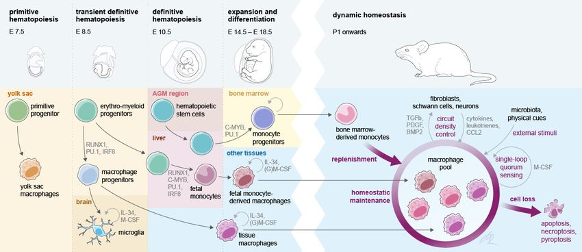
Figure 1. Interlinked pre- and postnatal development, adaptation, and maintenance of tissue macrophages. At birth, tissue MF originate from three
sources that prenatally seed developing body sites in sequential waves. Postnatally, self-maintenance and renewal by bone marrow-derived monocytes
are subject to microanatomical, soluble endogenous and exogenous cues. Establishment of a dynamic, tissue-specific MF homeostasis relies on tightly
regulated late prenatal and early postnatal events.
Henneke et al. eLife 2021;10:e67793. DOI: https://doi.org/10.7554/eLife.67793 3 of 21Review Article Immunology and Inflammation
tissues by late EMP-derived fetal monocytes, which develop in the fetal liver (Gomez Perdiguero
et al., 2015; Hoeffel et al., 2015). These cells colonize the organs pre- and perinatally and then
reside within most organs throughout the whole lifespan and maintain themselves via endogenous
proliferation rather than continuous exchange by circulating myeloid progenitors (Hashimoto et al.,
2013; Schulz et al., 2012). Thus, the fetal liver in the mouse serves as the main hematopoietic hub
during development, for example, as the source of fetal monocytes but also as a side for HSC-
derived progenitors until HSCs move to the bone marrow as the primary site of hematopoiesis from
the perinatal period onward (Figure 1). Interestingly, also other immune cells such as mast cells or
gd T-cells in the epidermis are derived from an HSC-independent embryonic source (Gentek et al.,
2018a; Gentek et al., 2018b). As noted above, EMP-derived MF (either from the early or late
wave) persist in selected and functionally distinct tissues into adulthood, including the brain (micro-
glia), the liver (Kupffer cells), or the epidermis (Langerhans cells). However, in several specialized tis-
sue niches, MF populations are found that are indeed replaced by circulating HSC-derived
progenitors from birth onwards, including the majority of dermal MF, lamina propria MF in the
intestine, or stromal choroid plexus MF in the brain (Goldmann et al., 2016; Kolter et al., 2019;
Bain et al., 2014; Van Hove et al., 2019).
Each tissue-resident MF population carries a transcriptional signature imprinted by hematopoietic
origin (Hagemeyer et al., 2016; Lavin et al., 2014; Mass et al., 2016). Prenatally, YS EMP-derived
MF and EMP-derived fetal monocytes compete for the same tissue niche (e.g., the alveolar space),
yet fetal monocytes are transcriptionally substantially closer to adult bone marrow monocytes than
to YS MF (van de Laar et al., 2016). As an example, EMP-derived fetal monocytes outcompete YS
MF in the development to alveolar MF in late fetal stages, which is associated with the repression
of the transcription factors c-Maf and MafB and a prerequisite for proper lung function and immune
defense (Li et al., 2020).
Upon entry into the anatomical niche, tissue MF are slowly adapting to the tissue environment
and a tissue-specific transcriptional program is imprinted, which is defined by many factors such as
neighboring cells (Bonnardel et al., 2019; Sakai et al., 2019) metabolites, and growth factors in the
niche (Okabe and Medzhitov, 2014), but also the presence or absence of microbial stimuli
(Capucha et al., 2018). During the perinatal establishment of definitive hematopoiesis, immune cell
progenitor cells (HSPCs) require a specific supportive environment inside the bone marrow, which
regulates cell growth and retention, such as the establishment of the stem cell niche for the reten-
tion of quiescent HSCs by osteoblasts (Coşkun et al., 2014). Furthermore, endothelial cells and mes-
enchymal stem cells, as key sources of cytokines and stem cell factors that maintain HSPCs, appear
to be essential in this context (Sugiyama et al., 2006; Hooper et al., 2009). Upon bone marrow
egress, immune cells carry a developmental program, which is determined by genetic, epigenetic,
and environmental cues from the stem cell niche to the target tissue. Differentiation of circulating
innate immune cells, such as monocytes or granulocytes, is a hierarchical and tightly regulated pro-
cess via several progenitor subsets that are instructed by defined transcriptional programs
(Mildner et al., 2017; Liu et al., 2019). Furthermore, their egress from the bone marrow is tightly
regulated by signals sensed, for example, via CCR2, CXCR2, CXCR4, or G-CSFR to allow a dynamic
adjustment of circulating immune cell numbers during homeostasis and disease (Semerad et al.,
2002; Serbina and Pamer, 2006; Eash et al., 2009). Monocytes are highly plastic cells with different
subsets circulating in the blood, but are even more plastic upon entry into inflamed tissues
(Guilliams et al., 2018; Giladi et al., 2020). In a recent study, even short-lived neutrophils have
been shown to be highly adaptable and plastic to different tissue environment and develop distinct
functional programs depending on the host tissue environment (Ballesteros et al., 2020). Proper
differentiation of tissue-resident immune cells along predefined pathways is a prerequisite for tissue
development and regeneration. Yet at the same time, cellular transformation processes are subject
to hijacking by ‘latent’ microorganisms like mycobacteria and herpes viruses for the creation of a
chronic niche (Lösslein et al., 2021).
The perinatal phase is crucial for immune cell maturation and acquisition of an immunocompetent
phenotype, including tissue MF differentiation and adaptation to the anatomical host niche. Within
their niche, MF undergo rapid adaptation to the changing environment and acquire often wide
changes in their transcriptional landscape, as, for example, seen for the stepwise differentiation of
microglia (Matcovitch-Natan et al., 2016; Thion et al., 2018). These differentiation processes are
Henneke et al. eLife 2021;10:e67793. DOI: https://doi.org/10.7554/eLife.67793 4 of 21Review Article Immunology and Inflammation
implicated both in keeping numeric MF homeostasis as well as a balanced response to infections
(Figure 1; Kolter et al., 2019; Feuerstein et al., 2020).
It has become clear, based on multiple independent observations, that early immune cell dissemi-
nation to hematopoietic and non-hematopoietic organs is associated with important functions
throughout life. However, it remains largely enigmatic, whether the effects later in life are secondary
to early tissue seeding, which imprints defined programs on the tissue environment (Cossı́o et al.,
2019). Especially around birth, immense adaptations and restructuring in the immune cell compart-
ment are required since a variety of environmental factors get in contact with the developing organ-
ism. The origin of cells that perinatally seed tissues needs to be rigorously deciphered to resolve
perinatal development of tissue immunity since early life immune seeding may have lasting conse-
quences, that is, represents a window of vulnerability as well as opportunity.
Perinatal adaptation of skin resident immune cells
Perinatal immune development may follow common principles, including genetic determination, in
utero modulation, and postnatal adaptation, yet it may further depend on spatial and temporal sig-
nals at distinct body sites. Moreover, cellular immune development runs in parallel with the perinatal
development of other cell types, for example, fibroblasts, endothelial and epithelial cells, or neurons.
These cells, on the one hand, harbor restricted immunological properties, like cytokine secretion,
and, on the other hand, build physical niches for immune cell interaction including residency. Thus,
they reciprocally interact with transient and tissue residing immune cells and form cellular niches as –
conceptionally – discrete working units for tissue immunity (Guilliams et al., 2020). In addition, MF
renewal patterns are related to the tissue substructure, for example, highly specialized sensory
nerve-associated MF are largely self-maintained (Kolter et al., 2019). In contrast, multicellular net-
works involving non-immune cells, physical cues, cytokines, stem cell factors, incoming cells, etc.,
tightly regulate renewal and density of the majority of tissue MF via only partially understood mech-
anisms, including quorum sensing [(Lohrmann et al., 2021) and Figure 1].
The skin is particularly interesting with respect to its highly structured microanatomy, its perinatal
development, and the dynamic integration of highly diverse cues into maintaining intercellular
immune cell control. In the skin, immune cell subsets of MF, dendritic cells, and gd T cell lineages
can be discriminated not only in view of their hematopoietic origin, but also with respect to the influ-
ence of environmental cues on cellular renewal patterns. Specifically, whereas nerve-associated MF
in the dermis, the immune-cell-rich layer bridging subcutaneous tissue and the epidermis, and Lan-
gerhans cells (LCs) in the latter, seem to maintain their populations size in a microbiota-independent
fashion (Capucha et al., 2018), the expansion and activation of other MF and Tgd17 cells in the skin
(Ridaura et al., 2018) is heavily influenced by the regular bacterial and fungal skin flora. Moreover,
the network of professional resident immune cells in the dermis depends on the interaction with
non-professional immune cells. Mechanistically, keratinocytes fortify skin barrier integrity by regulat-
ing Tgd17 cell activation, which itself is promoted by IL-23 signals and skin microbiota (Zhang et al.,
2019a). In addition, fibroblasts have the potential to produce tissue growth factors (like FGF2,
FGF7, tumor growth factor b1 [TGF-b1]) in response to type I IFN, which consecutively leads to plas-
macytoid dendritic cells being activated by neutrophils responding to translocating skin microbiota
(Di Domizio et al., 2020).
Immune cells that are taking up residency in the target tissue have to adapt to heterogeneous tis-
sue substructures. This process is steered by a predefined cellular program, for example, the surface
expression pattern of integrins and chemokine receptors. At the same time, a large variety of cues
need to be processed, such as those delivered by the extracellular matrix and non-professional
immune cells in the tissue. This localization to heterogeneous tissue structures providing functional
cues can result in modulation of immune responses, for example, antibacterial and anti-inflammatory
functions attributed to perivascular tissue MF (Abtin et al., 2014; Barreiro et al., 2016). Very
recently, paradigmatic evidence has linked the long-term dedication of MF subsets to microanatom-
ical tissue niches and distinct transcriptomic programs. This has been exemplified for the already
mentioned sensory nerve-associated MF of the skin (Goldmann et al., 2016; Kolter et al., 2019;
Bain et al., 2014; Van Hove et al., 2019) and to MF localizing to the myenteric plexus in the intesti-
nal tract (De Schepper et al., 2018). These MF, which function in nerve surveillance during steady
state and provide cues for axon outgrowth during activation, largely keep their population size by
self-renewal once they have adapted to the nerve niche. In contrast, MF in immediate vicinity and in
Henneke et al. eLife 2021;10:e67793. DOI: https://doi.org/10.7554/eLife.67793 5 of 21Review Article Immunology and Inflammation
the same tissue are replaced by HSC-derived progenitors, which exhibit a distinct transcriptional
program (definitive hematopoiesis). Yet, upon injury and nerve outgrowth and thus niche expansion,
MF of other origin can take up the nerve-niche specific program, independent of their origin
(Kolter et al., 2019).
Epidermal LC are – due to their high antigen-presenting capacities – functionally positioned
between MF and dendritic cells. Notably, LCs develop similar to the second immune cell lineage
residing in the mouse epidermis, gd T cells (dendritic epidermal T cells), from both early YS (YS) pro-
genitors and fetal liver monocytes before locally self-renewing in the adult (Gentek et al., 2018a;
Merad et al., 2002). The skin is colonized by EMP-derived MF during embryogenesis
(Hoeffel et al., 2012) similar to other tissues. Starting from E14.5 onwards, EMP-derived fetal mono-
cytes are entering the developing skin most likely via blood vessels and start to differentiate to LC
progenitors (Hoeffel et al., 2012). Shortly before birth, the first LC progenitors begin to colonize
the separating epidermis by transmigration from the underlying dermis into the epidermis where
they peri- and postnatally differentiate to mature LCs and expand by endogenous proliferation dur-
ing postnatal stages (Hoeffel et al., 2012; Chorro et al., 2009). Upon entry in the epidermis, LCs
subsequently loose monocyte markers and acquire LC maturation markers including Langerin,
CD11c, or major histocompatibility complex (MHC) class II. Maturation and maintenance of the adult
LC population are instructed by signals of the surrounding adjacent keratinocytes, including IL-34/
Csf1r signaling (Greter et al., 2012); however, the signals instructing differentiation and recruitment
during perinatal development have not been identified yet. Similar to nerve associated MF in the
dermis, the LC population is maintained by endogenous proliferation during steady state within the
niche, but upon injury such as UV-B irradiation the resident LCs are eliminated and efficiently
replaced by HSC-derived progenitors from the circulation (Merad et al., 2002; Seré et al., 2012).
This replacement is also mirrored in part in gd T cells in the dermis, where an IL-17-secreting subset
(Tgd17) develops before birth and is self-renewing, in contrast to IFN-g–producing CD27+gd T cells,
which are constantly replenished by newly generated gd-T cells from the thymus (Sandrock et al.,
2018).
Building up local immune defense in the developing lung
In the adult lung, local immune defense is mainly provided by tissue-resident MF. Two main sets of
tissue-resident MF can be distinguished, interstitial MF (IM) and alveolar MF (AM) (Evren et al.,
2020; Kopf et al., 2015). In the fetal and newborn mouse, the lung immune defense system is
established by tissue-resident MF via several waves of cell recruitment (Evren et al., 2020;
Kopf et al., 2015). The first wave consists of YS-derived MF, which can be detected in the lung
from around E10. These MF have a heterogeneous shape with multiple pseudopods and are charac-
terized by high expression of F4/80 and low expression of CD11b. The second wave of MF recruit-
ment to the lung consists of fetal liver-derived monocytes, which appear in the lung at E12. These
MF exhibit a round cell shape, low expression of F4/80, and high expression of CD11b
(Guilliams et al., 2013; Hashimoto et al., 2013; Tan and Krasnow, 2016). Both populations are dis-
tributed diffusely in the fetal lung interstitium between E14 and 17 (Tan and Krasnow, 2016). The
first wave of YS-derived resident MF does not substantially proliferate and remains as IM in the
interstitial space. Postnatally, these cells are continuously renewed by circulating bone marrow-
derived monocytes. The second wave of MF shows strong proliferative activity starting at E16 in the
lung interstitium, leading to the establishment of the uniquely localized alveolar MF, which – in the
mouse – start to develop around birth. In other words, fetal monocytes outcompete YS MF in the
development to alveolar MF late in fetal development, which has lasting consequences for proper
lung function and immune defense (Guilliams et al., 2013; Li et al., 2020; Tan and Krasnow, 2016).
The third wave of lung MF is recruited to the lung from bone marrow-derived circulating mono-
cytes starting immediately after birth to fill up any niche of interstitial MF throughout postnatal life.
Similar mechanisms concerning ontogeny of tissue-resident MF may also apply for the development
of interstitial and alveolar MF during human ontogeny, as addressed in a recently published study
(Evren et al., 2020).
Alveolar MF play a central role in the immediate postnatal protection of the lung from infections.
Importantly, alveolar MF exhibit a unique territorial behavior with on average three alveoli being sur-
veilled by one AM (Neupane et al., 2020). The cues for guiding alveolar MF function and distribu-
tion in an organ that undergoes a fourfold increase in weight in the first three weeks of life (in mice)
Henneke et al. eLife 2021;10:e67793. DOI: https://doi.org/10.7554/eLife.67793 6 of 21Review Article Immunology and Inflammation
are only partially understood. Yet, the positioning of prealveolar MF to the alveoli and the cell-intrin-
sic transition to AM appear to require the actin-bundling protein L-plastin (Todd et al., 2016). This
depends on GM-CSF produced by alveolar epithelial cells (Kopf et al., 2015). GM-CSF – through
binding to the GM-CSF receptor – in turn activates the transcription factor PPAR-g, which controls
the program for alveolar MF maturation and maintenance (Schneider et al., 2014). The unique
dependence of alveolar MF on GM-CSF signaling among tissue MF in the body is illustrated by the
disease alveolar proteinosis, where genetic or antibody-mediated subversion of this signaling
machinery results in abnormal accumulation of surfactant-derived lipoproteins (Trapnell et al.,
2019).
Alveolar MF keep their capacity for self-renewal throughout postnatal life (Epelman et al., 2014).
Yet, they can be replaced by circulating monocytes in cases of severe damage to the alveolar MF
population, for example, after infection or lung irradiation therapy (Machiels et al., 2017). The prop-
erties of the lung environment for alveolar MF function are highlighted by the ability of intratra-
cheally transferred ectopic MF to adopt an alveolar MF phenotype (Lavin et al., 2014; van de Laar
et al., 2016).
Both pulmonary development and establishment of an immune defense system in the lung are
still ongoing immediately after birth. Thus, any disturbance of lung homeostasis, not only during fetal
life but also in the early postnatal period, may inevitably affect lung development and local host
defense. This becomes evident in premature infants, where various noxious agents or procedures (e.
g., high oxygen levels, artificial ventilation, microbial products) can lead to bronchopulmonary dys-
plasia, a severe lung disease with long-term consequences (Blackwell et al., 2011). It is therefore
instrumental to elucidate the mechanisms regulating interstitial and alveolar MF function in the
developing lung including pathological conditions during this vulnerable postnatal time.
Hidden immune development: the brain
The embryonic colonization by microglial progenitors in the central nervous system (CNS) and their
subsequent maturation process during pre- and postnatal life is a particularly well-studied example
for the development of tissue-resident immune cells. Microglia arise from the early wave of YS
EMPs, and the progenitors colonize the developing brain rudiment early on during embryonic devel-
opment around E9.5 (Ginhoux et al., 2010; Kierdorf et al., 2013). Once emigrated into the CNS in
this early wave, the cells expand via endogenous proliferation without input of fetal monocytes or
HSC-derived progenitors during later developmental stages and adulthood (Schulz et al., 2012;
Hoeffel et al., 2015; Goldmann et al., 2013). With the establishment of the blood-brain barrier
around E15.5, the CNS parenchyma separates from the periphery (Haddad-Tóvolli et al., 2017). In
line with this separation, the microglial population is dependent on endogenous self-renewal via pro-
liferation. Therefore, microglia show a high proliferation rate during prenatal and postnatal stages to
expand and distribute in the developing CNS, but also exhibit a dramatic decline in proliferation in
adult stages, once the microglial network is set up (Matcovitch-Natan et al., 2016; Thion et al.,
2018). In the healthy adult CNS, the microglial entity is maintained via random proliferation with a
very low turnover rate in most brain regions (Tay et al., 2017; Füger et al., 2017). Several intrinsic
regulators of transcriptional programs but also exogenous cues were defined, which are involved in
the stepwise maturation process of these resident immune cells in the CNS parenchyma (Matco-
vitch-Natan et al., 2016; Thion et al., 2018; Hammond et al., 2019; Li et al., 2019). Microglial pro-
genitor differentiation from early EMPs in the YS relies on cell-intrinsic programs, in particular driven
by transcription factors, for example, Sfpi/Pu.1 and Irf8 (Matcovitch-Natan et al., 2016;
Thion et al., 2018; Kierdorf et al., 2013). Sfpi/Pu.1-deficient mice do not have any microglia in the
CNS parenchyma, whereas Irf8-deficient mice show a reduced number of progenitors and embryonic
microglia (Kierdorf et al., 2013). Adult Irf8-deficient mice have an altered number of microglia in
the parenchyma and show severe maturation defects (Hagemeyer et al., 2016). Next to Irf8, the
transcription factor Sall1 was identified as a core transcriptional regulator of microglial differentia-
tion. Loss of Sall1 abrogates microglial development and the developing cells are highly activated
and cause alterations in neurogenesis (Buttgereit et al., 2016). Moreover, microglial development is
highly dependent on different cues from cells in the physiological vicinity. One outstanding example
is the dependence of microglial development on colony stimulating factor 1 (CSF1) receptor signal-
ing. Deletion or inhibition of the CSF1 receptor during development results in loss of microglia (Mat-
covitch-Natan et al., 2016; Thion et al., 2018; Ginhoux et al., 2010; Erblich et al., 2011). The
Henneke et al. eLife 2021;10:e67793. DOI: https://doi.org/10.7554/eLife.67793 7 of 21Review Article Immunology and Inflammation
CSF1 receptor has two ligands, IL-34 and CSF1. Both are expressed in the CNS, but – interestingly –
with a spatially distinct expression pattern, which establishes postnatally (Easley-Neal et al., 2019;
Kana et al., 2019). As an example, gray matter microglia depend predominantly on IL-34 produced
by neurons, while white matter microglia depend on CSF1 secreted by astrocytes during postnatal
phases and adulthood (Kana et al., 2019). Of note, this distinct expression pattern of the two
ligands starts to emerge around birth and establishes during postnatal phases. Besides CSF1 and IL-
34, microglial expansion and homeostasis is highly dependent on TGF-b. Loss of TGF-b signaling in
adult microglia results in a hyperactivated phenotype and disturbance of homeostasis
(Butovsky et al., 2014; Zöller et al., 2018). Loss of TGF-b signaling during microglial development
results in impairment of microglial expansion throughout the developing CNS (Utz et al., 2020).
Even though microglia reside behind the closed blood-brain barrier (BBB) within the CNS paren-
chyma and are shielded early on during development, microglial homeostasis and development are
also highly influenced by external environmental cues with one interesting example being the depen-
dence of microglial maturation on the gut microbiota. Germ-free mice show a maturation defect of
adult microglia, resulting in an altered reaction to inflammatory cues such as lipopolysaccharides
(LPS) (Matcovitch-Natan et al., 2016; Thion et al., 2018; Erny et al., 2015). Therefore, microglial
sensing of the microbiota seems to provide essential signals during development. Microbiota-
derived signals highly affect microglia in postnatal development, whereas embryonic microglia until
birth is less affected by the absence of the microbiota (Thion et al., 2018).
Microglial development is shaped by different cues derived from the CNS but also the peripheral
environment. Especially during and shortly after birth manifold changes are seen in the transcrip-
tional differentiation program of microglia. Therefore, the perinatal phase is an important time for
CNS immune development and maturation. Furthermore, postnatal microglia are serving a plethora
of different functions and are very dynamic in terms of mobility and proliferation, making insults in
their developmental program detrimental for the CNS during this vulnerable phase.
Perinatal adaptation of the intestinal mucosa: a timed succession of
multidimensional phases to establish host-microbial homeostasis
The intestinal mucosa transits from sterility – and thus largely deprived from microbial innate
immune stimuli – before birth to being densely colonized by commensal bacteria, and threatened by
enteropathogens after birth. With the introduction of solid food, the intestinal mucosa is additionally
subjected to a large variety of dietary antigens. The parallel exposure to food and microbiota is ben-
eficial since microorganisms in neonatal gastrointestinal tract improve energy harvesting for growth
(Ganal-Vonarburg et al., 2020). On the other hand, the challenges for the neonatal intestinal
mucosa are tremendous since it has to (i) endure bacterial colonization and prevent inappropriate
inflammation, (ii) direct bacterial composition of a beneficial microbiota, (iii) generate and maintain
adaptive immune tolerance to dietary and microbial antigens, and (iv) establish innate and adaptive
antimicrobial host reactivity to overcome challenges by enteropathogens. A timed succession of
non-redundant phases facilitates postnatal immune development and the step-by-step establishment
of mucosal homeostasis (Hornef and Torow, 2020). Moreover, microbiota-immune interactions
starting immediately after birth have been implicated in a variety of both ‘non-communicable’ gas-
trointestinal diseases, like inflammatory bowel disease, as well as extraintestinal disorders ranging
from psoriasis to rheumatic arthritis, metabolic syndrome, neurodegeneration, and cancer. Notably,
a causal link has not been firmly established in all cases (Zheng et al., 2020).
The healthy fetal intestine is sterile, albeit low levels of microbial constituents derived from the
maternal microbiota reach the neonate and contribute to early immune priming (de Goffau et al.,
2019; Gomez de Agüero et al., 2016). With rupture of the amniotic membranes, the neonatal
organism gets into contact with the bacteria colonizing the maternal vaginal and gastrointestinal
tract (Ganal-Vonarburg et al., 2020). Whereas maternal fecal bacteria represent the dominant
source in vaginally delivered neonates, C-section is associated with a more environment- or skin-like
neonatal microbiota (Dominguez-Bello et al., 2010). Upon initial exposure, bacterial density quickly
reaches threshold levels (van Best et al., 2020a). In contrast, bacterial diversity and compositional
stability evolve slowly. During this time, dietary (breast milk versus formula feeding), genetic and
environmental factors (pets, siblings, geography), as well as medical interventions (antibiotics, probi-
otics) influence microbial composition (van Best et al., 2020b; Bokulich et al., 2016; van Best
et al., 2015). In general, the microbiota built-up follows programmed succession of highly abundant
Henneke et al. eLife 2021;10:e67793. DOI: https://doi.org/10.7554/eLife.67793 8 of 21Review Article Immunology and Inflammation
bacterial species (van Best et al., 2020a; Planer et al., 2016; Feng et al., 2020). In humans, it
requires 2–3 years after birth for bacterial diversity and composition to reach adult levels and stabil-
ity (Bäckhed et al., 2015; Yatsunenko et al., 2012). Whereas microbial antigens are already present
in the intestinal lumen at high concentrations early after birth, direct exposure to dietary non-self
antigens starts later, in particular with the introduction of solid food. Neonatal sepsis and meningitis
caused by bacterial pathogens, such as Streptococcus agalactiae, occur early and represent an
important cause of neonatal mortality worldwide (Kolter and Henneke, 2017). In contrast, only spe-
cific viruses, for example, cytomegalovirus contaminating breast milk and respiratory syncytial virus,
infect infants already shortly after birth, whereas the bulk of viral infections occur later with increas-
ing human contact and the cessation of maternal protection by transferred IgG and SIgA
(Ershad et al., 2019; Baasch et al., 2020).
An essential issue in this context relates to the specific mechanisms that allow the neonatal intesti-
nal mucosa to accommodate microorganisms and their potentially highly activating effector mole-
cules. Both the intestinal epithelium and resident immune cells express innate immune receptors
such as Toll-like receptors (TLRs), Nod-like receptors (NLRs), RIG-I-like helicases, and thus sense
microbial stimuli and secrete chemotactic and proinflammatory mediators. Age-dependent differen-
ces in the expression levels of innate immune receptors may regulate immune cell activation at this
critical time point (Pott et al., 2012; Gribar et al., 2009). In addition, adaptive mechanisms adjust
cellular reactivity in order to maintain tissue integrity. For example, shortly after birth the epithelium
undergoes reprogramming and thus acquires innate immune tolerance via miR-146a-mediated trans-
lational repression of the TLR signaling molecule IL-1 receptor-associated kinase 1 (Lotz et al.,
2006; Chassin et al., 2010). At the same time, immune cells undergo reprogramming via the endog-
enous alarmin S100A8/A9 interacting with TLR4 (Ulas et al., 2017). Other regulatory molecules,
for example, A20, single immunoglobulin IL-1 receptor-related molecule (SIGIRR), IL-1 receptor-asso-
ciated kinase-M, and Toll-interacting protein (TOLLIP), contribute to controlling the activation state
of the intestinal cells (Vereecke et al., 2010; Nanthakumar et al., 2011). Furthermore, molecules
such as IL-10 or arginase 2, secreted, for example, by neonatal B cells and CD71-positive erythroid
cells, as well as the immunosuppressive properties of amniotic fluid and breast milk constituents,
may provide synergistic cues in this context (Zhang et al., 2007; Elahi et al., 2013; Good et al.,
2012). Notably, the ability to control inappropriate innate immune stimulation depends on the
organism’s gestational age. Necrotizing enterocolitis (NEC), which is mainly observed in preterm
infants and is associated with severe colonic inflammation, is thought to result from a failure to con-
trol microbiota-induced immune stimulation (Neu, 2020; Gopalakrishna et al., 2019). Currently,
specific causal microorganisms have not been identified. However, frequent alterations in the micro-
biota of the preterm infants, and here in particular an elevated abundance of members of the phy-
lum proteobacteria, are likely to be significant. Accordingly, oral administration of probiotic
Bifidobacterium and Lactobacillus spp. modifies the microbial composition in these children and low-
ers the incidence of NEC and sepsis (van Best et al., 2020b; Neu, 2020; Panigrahi et al., 2017).
The fact that maternal fecal bacteria represent an important source for the infant’s microbiota
ensures the transmission of a usually ‘successful’ microbiota to the next generation. Delivery mode
impacts newborn gut colonization efficiency (preprint BioRxiv. doi: https://doi.org/10.1101/2020.01.
29.91999). In addition, the newborn infants actively shape a beneficial microbiota composition. For
example, and as outlined in detail below, human milk oligosaccharides (HMOs) are highly abundant
complex carbohydrates that are indigestible for humans. They foster the growth of Bifidobacterium,
the hallmark bacterium of the healthy infant microbiota (Zivkovic et al., 2011). In mice, hepatic
secretion of bile acids into the proximal small intestine promotes the expansion of lactobacilli and
accelerates microbiota maturation (van Best et al., 2020a). Finally, specific expression of the flagel-
lin receptor TLR5 by the neonatal gut epithelium favors colonization by non-flagellated bacteria in
comparative colonization and microbiota transfer experiments (Fulde et al., 2018). These and other
mechanisms might foster regular development of the neonatal microbiota-intestine interface to
counteract predictable disturbances, such as infections (Pop et al., 2014).
Despite early homing of lymphocytes and antigen-presenting cells to the mucosal lymphoid tis-
sue, the murine adaptive immune system appears to underlie active suppression during the first two
weeks of life (Torow et al., 2015). This may allow establishment of adaptive immunity, while – at the
same time – avoid autoimmunity. The cessation of breastfeeding causes significant alterations of the
microbiota composition and induces goblet cell-associated antigen passage (Kulkarni et al., 2020).
Henneke et al. eLife 2021;10:e67793. DOI: https://doi.org/10.7554/eLife.67793 9 of 21Review Article Immunology and Inflammation
It causes the so-called ‘weaning reaction’, which leads to a transient innate immune stimulation,
including accelerated replacement of fetal MF in the lamina propria by monocyte-derived MF, and
the initiation of adaptive mucosal immunity (Al Nabhani et al., 2019). This reaction propagates the
maturation of regulatory T cells and thus the maintenance of immune tolerance (Kulkarni et al.,
2020; Al Nabhani et al., 2019; Yang et al., 2015; Vatanen et al., 2016). Only then effector T lym-
phocytes are generated that provide antimicrobial protection. However, these early T lymphocytes
derive from separate progenitors and exhibit significant differences to their adult counterparts
(Mold et al., 2010). Their unique properties such as rapid effector responses, but reduced ability to
form memory, match the requirements of neonatal life, such as limited metabolic resources
(Rudd, 2020).
Infants provide efficient antimicrobial responses to control pathogen challenge despite the
described mucosal immune restrictions, First, exogenous antimicrobial factors such as lactoferrin,
antimicrobial peptides, and SIgA in breast milk support pathogen resistance. Also, the neonatal epi-
thelium appears to discriminate between microbial colonization and invasive infection via the forma-
tion of chemotactic and antimicrobial mediators (Zhang et al., 2014; Dupont et al., 2016). Finally,
newborn infants in the event of viral infection are able to mount a fast and effective cellular immune
response although the emerging T lymphocytes are more prone to rapid effector function than last-
ing memory function (Pott et al., 2012; Sarzotti et al., 1996). Much needs to be learnt to improve
understanding of the functional and structural particularities of the neonatal intestinal mucosa, which
may form the basis for improved preventive and therapeutic strategies at the beginning of life.
Breast milk and gut microbiota are intertwined regarding their impact
on perinatal immune development
Breast milk is a complex cocktail of biologically active components and nutritional substrates with
substantial compositional differences between individual mothers and changing over time of lacta-
tion (i.e., between colostrum and mature milk). Key components such as immunoglobulins, chemo-
kines/cytokines, bioactive micro- and macronutrients, oligosaccharides, microRNAs, hormones,
immune cells, and microorganisms have been associated with direct and indirect perinatal immune
programming.
HMOs comprise ~200 different bioactive glycans. They contain direct immune-mediated effects,
such as antiadhesive, antimicrobial, and immune programming activity (Walsh et al., 2020). HMOs
promote immune cell homeostasis and reinforce intestinal epithelial barrier integrity (Xiao et al.,
2019; Šuligoj et al., 2020; Zhang et al., 2019b). Consistently, the concentration of the HMO disia-
lyllacto-N-tetraose is inversely correlated with the infant’s risk for NEC (Masi et al., 2020). In addi-
tion, there is limited data suggesting that HMOs circulate in the maternal blood throughout
gestation and may cross the placenta (Hirschmugl et al., 2019; Wise et al., 2018). However, the
functional relevance of these findings for mother and fetus remains to be explored. In addition,
HMOs act as prebiotics promoting the growth of beneficial bacteria that in turn impact on the muco-
sal and systemic immune system. HMOs are poorly metabolized by the host, but are favored growth
substrates for certain beneficial Bifidobacterium species (Fulde et al., 2018). Immune-modulatory
effects may occur through recognition of cell surface-associated microbial components including bifi-
dobacterial exopolysaccharides and tad pili, and the production of immune-modulatory metabolites,
for example, short chain fatty acids (SCFAs) (Alessandri et al., 2019). Strikingly, an increased Bifido-
bacterium abundance in the human and mouse gut was described already during pregnancy. Thus
these effects may start prior to birth via placental crossing of microbial metabolites and in utero pro-
gramming (Nuriel-Ohayon et al., 2019). It remains unclear, however, how specific strains of Bifido-
bacterium and other early life microbiota constituents and their metabolites trigger
immunomodulatory responses that maintain immunological homeostasis during the perinatal period.
Breast milk and, in particular, colostrum provide other micro- and macronutrients with high
immune-modulatory activity, such as amino acids, lipids, vitamins, and trace elements (Dror and
Allen, 2018). Specific amino acids like glutamate and glutamine (the most abundant free amino
acids) are essential for immune and tissue development (Dror and Allen, 2018). The second largest
nutrient fraction comprises lipids, including the long-chain poly-unsaturated fatty acids arachidonic
acid and docosahexaenoic acid. These have been shown to influence lymphocyte proliferation and
cytokine production. In addition, iron (bound to lactoferrin) and zinc are central in perinatal immune
development and may act indirectly via growth modulation of specific gut bacteria. Importantly,
Henneke et al. eLife 2021;10:e67793. DOI: https://doi.org/10.7554/eLife.67793 10 of 21Review Article Immunology and Inflammation
obese or malnourished mothers may potentially lack an appropriate balance of immune-modulatory
components impacting perinatal immune programming in their infants (Samuel et al., 2020). Also,
the direct transfer of immune cells, such as T cells, NK cells, and MF, via breast milk may influence
perinatal immune development (Laouar, 2020). Whereas it remains unclear whether intact immune
cells can translocate through the infant’s mucosal barrier and populate neonatal tissues
(Torow et al., 2015), recent work on CD8+ memory T cells indicates that even cellular lysate may
still represent a mechanism for passive transfer of cellular immunity (Myles and Datta, 2021). More-
over, stem-cell-like cells in breast milk have high multilineage potential and can traffic to different
body sites including the brain. Yet, whereas their differentiation potential into neuronal and glial cell
types has been demonstrated, it remains unclear whether they can also differentiate into immune
cells (Ninkina et al., 2019). Breast milk also contains exosomes enriched in immune-modulating pro-
teins, peptides, and miRNAs (e.g., miR-30d-5p, let-7b-5p, and let-7a-5p), which seem to withstand
digestion and may promote intestinal epithelial cell growth and mucosal barrier development
(Hock et al., 2017). Other directly immunomodulatory components include immunoglobulins (Ig),
with secretory (S)IgA being dominant (>90%), followed by SIgM. IgG in humans is mainly found in
mature milk. SIgA carries major antimicrobial properties, but recent data suggest an additional role
of SIgA in enhancing bacterial colonization and homeostatic immunity (Cacho and Lawrence, 2017;
Pabst and Slack, 2020). T cell help is required to generate appropriate affinity maturation to bacte-
rial antigens including antigens from segmented filamentous bacteria (Kawamoto et al., 2012).
Growth factors and cytokines such as EGF, TGF-b, or the S100 calcium binding proteins are passed
from mother to infant via breast milk and modulate mucosal epithelial and immune responses (Kocis-
zewska-Najman et al., 2020). This transfer is influenced by birth mode and/or gestational age. For
example, preterm infants exhibit lower levels of fecal S100 proteins (i.e., fecal calprotectin), leading
to changes in the gut microbiota, including alterations in the abundance of members of the Bifido-
bacteriaceae family (Willers et al., 2020). Breast milk also contains microorganisms such as Staphylo-
coccus, Streptococcus, as well as low numbers of Bifidobacterium and Lactobacillus species
(Demmelmair et al., 2020; Cortes-Macias et al., 2020). An associated influence of immune devel-
opment may occur through bacterial metabolites, such as SCFAs. These modulate immune cell activ-
ity, for example, via the inhibition of histone deacetylases and the activation of G protein-coupled
receptors. Butyrate at concentrations found in breast milk modulated immune responses in an in
vivo food allergy model (Paparo et al., 2020).
The enteric microbiota impacts on the development and function of resident immune cells at
mucosal sites but also well beyond mucocutaneous surfaces, for example, in the CNS
(Oschwald et al., 2020; Elinav et al., 2011). Tissue-resident MF seeded in embryonic life are post-
natally replaced by bone marrow, monocyte-derived MF in a highly tissue-specific fashion
(Lohrmann et al., 2021). This process is influenced by the acquisition of the microbiota (Bain et al.,
2014) or can result from low-grade infections, for example, by herpes viruses in case of alveolar MF
(Machiels et al., 2017). Notably, the comparison of SPF mice with germ-free or antibiotic-treated
mice may underestimate the contribution of the endogenous microbiota since SPF mice are rela-
tively close to GF mice and distant to mice with a more diverse, ‘natural’ flora (Rosshart et al.,
2019). In contrast, the role of the microenvironment including the microbiota on perinatal develop-
ment of non-professional immune cells that contribute to immune niches, like fibroblasts and epithe-
lial cells in the skin, has not been sufficiently taken into consideration. In addition, the impact of the
maternal microbiota on the placenta as an important fetal immune organ has remained largely
elusive.
Blood circulation and immediate immune responses
During fetal ontogeny, various immune cells populate peripheral organs and install a broad defense
network to gradually prepare for the challenges after birth. The distribution of immune cells and
their precursors throughout the organism relies on effective transportation by the blood circulation.
In the mouse fetus, the onset of blood circulation is observed at around E8.5 and is fully established
at around E10.5 (Cacho and Lawrence, 2017). During this time, the first monocytes and neutrophils
derived from EMPs of the YS are released into the blood circulation (Stremmel et al., 2018;
McGrath et al., 2003). At later time points, they are replaced by neutrophils/monocytes derived
from HSCs originating from sites of intraembryonic definitive hematopoiesis, which takes place first
in the fetal liver and then gradually moves to the bone marrow. While early fetal circulating
Henneke et al. eLife 2021;10:e67793. DOI: https://doi.org/10.7554/eLife.67793 11 of 21Review Article Immunology and Inflammation
monocytes have been identified to seed peripheral organs and differentiate into tissue-resident MF,
as described above, the fate and function of early circulating neutrophils are less clear. Yet, several
lines of evidence support the hypothesis that circulating neutrophils already provide the first line of
immediate immune response, just as in postnatal life.
During fetal life, neutrophils exhibit an ontogenetically regulated gain of recruitment efficiency
into inflamed mouse YS vessels, as revealed by intravital imaging. The scrupulous analysis of neutro-
phils from preterm and term infants after birth showed that fetal as well as postnatal neutrophil
development follows a cell-intrinsic functional program, which intriguingly does not seem to be sig-
nificantly altered by cues from early postnatal life (Sperandio et al., 2013; Nussbaum et al., 2013).
This indicates that the ontogenetic neutrophil-specific program is tuned to perfectly adapt to the
specific needs and challenges of the growing organism within the uterus leaving room for develop-
ment and at the same time provides enough protection against exogenous threats. However, adap-
tion of neutrophils to premature postnatal life is rather limited. Paucity and altered activation of
neutrophils have been traditionally linked to infection susceptibility and high-related mortality in
newborns, especially preterm infants (Gessler et al., 1995; Henneke et al., 2003).
As compared to granulocytes, induced activation of circulating mononuclear phagocytes , in par-
ticular monocytes and dendritic cells, from newborn infants has been subject to rather detailed anal-
ysis. The results point to complex and dynamic alterations as compared to similar cells from older
children and adults. Overall, mononuclear phagocytes from cord blood, that is, late fetal cells at the
transition to independent postnatal life, show robust responses to whole bacteria and purified bacte-
rial effectors with respect to some cytokines (e.g., TNFa, IL-6, and IL-10), yet respond weakly with
type I interferons to cytomegalovirus infection (Kollmann et al., 2017). Unique properties of cord
blood mononuclear cells as compared to those from young infants in terms of cytokine formation
have been confirmed by high-dimensional analysis (Olin et al., 2018). Yet, the functional implications
of high activation at baseline and after stimulation around birth remain largely enigmatic.
Summary and outlook
Distinct immune cell niches exist in complex tissues and undergo major adaptations and changes
during the perinatal period. They can be discriminated by distinct cellular programs. Both systemic
regulators and microenvironment-specific signals steer homing and homeostatic maintenance of resi-
dent immune cells by transcriptionally redirecting progenitors from different sources.
The impact of exogenous signals,such as the establishing microbiota and microbial metabolites
as well as tissue-intrinsic factors, such as the perinatal change in oxygen tension on local cell compo-
sition, cell differentiation, and immune maturation and the interplay between local and systemic reg-
ulatory mechanisms are largely elusive. Understanding of these issues is pivotal, since perinatal
changes set the stage for tissue adaptation, immune homeostasis, and long-term health and may
therefore facilitate the development of interventional strategies to prevent disease disposition dur-
ing this critical period.
Acknowledgements
We are grateful for artwork by Michal Rössler (Freiburg).
Additional information
Funding
Funder Grant reference number Author
Bundesministerium für Bildung 01GL1746A Philipp Henneke
und Forschung
Deutsche Forschungsge- SFB-TRR 167 Philipp Henneke
meinschaft
Deutsche Forschungsge- SPP2225 Ho 2236/18-1 Mathias Hornef
meinschaft
Deutsche Forschungsge- SFB1382 ID403224013 Mathias Hornef
Henneke et al. eLife 2021;10:e67793. DOI: https://doi.org/10.7554/eLife.67793 12 of 21You can also read