Functional trade-offs and environmental variation shaped ancient trajectories in the evolution of dim-light vision
←
→
Page content transcription
If your browser does not render page correctly, please read the page content below
RESEARCH ARTICLE

                                     Functional trade-offs and environmental
                                     variation shaped ancient trajectories in
                                     the evolution of dim-light vision
                                     Gianni M Castiglione1,2†, Belinda SW Chang1,2,3*
                                     1
                                      Department of Cell and Systems Biology, University of Toronto, Toronto, Canada;
                                     2
                                      Department of Ecology and Evolutionary Biology, University of Toronto, Toronto,
                                     Canada; 3Centre for the Analysis of Genome Evolution and Function, University of
                                     Toronto, Toronto, Canada

                                     Abstract Trade-offs between protein stability and activity can restrict access to evolutionary
                                     trajectories, but widespread epistasis may facilitate indirect routes to adaptation. This may be
                                     enhanced by natural environmental variation, but in multicellular organisms this process is poorly
                                     understood. We investigated a paradoxical trajectory taken during the evolution of tetrapod dim-
                                     light vision, where in the rod visual pigment rhodopsin, E122 was fixed 350 million years ago, a
                                     residue associated with increased active-state (MII) stability but greatly diminished rod
                                     photosensitivity. Here, we demonstrate that high MII stability could have likely evolved without
                                     E122, but instead, selection appears to have entrenched E122 in tetrapods via epistatic interactions
                                     with nearby coevolving sites. In fishes by contrast, selection may have exploited these epistatic
                                     effects to explore alternative trajectories, but via indirect routes with low MII stability. Our results
*For correspondence:
belinda.chang@utoronto.ca
                                     suggest that within tetrapods, E122 and high MII stability cannot be sacrificed—not even for
                                     improvements to rod photosensitivity.
Present address: †Department         DOI: https://doi.org/10.7554/eLife.35957.001
of Ophthalmology, Johns
Hopkins University School of
Medicine, Baltimore, United
States
                                     Introduction
Competing interests: The             Nature-inspired strategies are increasingly recruited toward engineering objectives in protein design
authors declare that no
                                     (Khersonsky and Fleishman, 2016; Jacobs et al., 2016; Goldenzweig and Fleishman, 2018, a cen-
competing interests exist.
                                     tral challenge of which is to successfully manipulate backbone structure to modulate stability without
Funding: See page 24                 introducing undesirable pleiotropic effects on protein activity (Khersonsky and Fleishman, 2016;
Received: 14 February 2018           Goldenzweig and Fleishman, 2018; Starr and Thornton, 2017; Tokuriki and Tawfik, 2009). Engi-
Accepted: 09 September 2018          neering protein stability and activity requires an understanding of a protein’s sequence-function rela-
Published: 26 October 2018           tionship, or landscape (Pál and Papp, 2017; Wu et al., 2016; Starr et al., 2017), where billions of
                                     possible pair-wise and third-order interactions can exist between amino acids (Starr and Thornton,
Reviewing editor: Andrei N
                                     2017; Storz, 2016), and only a limited number of amino acid combinations will confer the function
Lupas, Max Planck Institute for
Developmental Biology,
                                     of interest (Wu et al., 2016; Starr et al., 2017; McMurrough et al., 2014; Mateu and Fersht,
Germany                              1999; Tarvin et al., 2017). To understand the context-dependence of amino acid functional effects
                                     (also known as intramolecular epistasis [Starr et al., 2017; Storz, 2016; Echave et al., 2016]),
   Copyright Castiglione and
                                     approaches such as deep mutational scanning (Wu et al., 2016; Starr et al., 2017; Sailer and
Chang. This article is distributed
                                     Harms, 2017) can explore a subset of sequence-function space formed in response to a limited set
under the terms of the Creative
Commons Attribution License,         of artificial selection pressures (Starr and Thornton, 2017). By contrast, natural protein sequence
which permits unrestricted use       variation reflects the range of protein function that evolved in response to changing ecological varia-
and redistribution provided that     bles (Starr and Thornton, 2017; Pál and Papp, 2017; Ogbunugafor et al., 2016), where conver-
the original author and source are   gent ‘solutions’ for protein function and stability can be derived through the evolution of alternative
credited.                            protein sequences (McMurrough et al., 2014; Mateu and Fersht, 1999; Tarvin et al., 2017). This

Castiglione and Chang. eLife 2018;7:e35957. DOI: https://doi.org/10.7554/eLife.35957                                                  1 of 30
Research article                                                           Biochemistry and Chemical Biology Evolutionary Biology

                                   eLife digest People can see in dim light because of cells at the back of the eye known as rods.
                                   These cells contain two key components: molecules called retinal, which are bound to proteins
                                   called rhodopsin. When light hits a rod cell, it kicks off a cascade of reactions beginning with the
                                   retinal molecule changing into an activated shape and ending with a nerve impulse travelling to the
                                   brain. The activated form of retinal is toxic, and as long as it remains bound to the rhodopsin protein
                                   it will not damage the rod or surrounding cells. The toxic retinal also cannot respond to light. It must
                                   be released from the protein and converted back to its original shape to restore dim light vision.
                                       As with all proteins, rhodopsin’s structure comprises a chain of building blocks called amino
                                   acids. Every land animal with a backbone has the same amino acid at position 122 in its rhodopsin.
                                   This amino acid, named E122, helps to stabilize the activated rhodopsin, slowing the release of the
                                   toxic retinal. Yet E122 also makes the rod cells less sensitive, resulting in poorer vision in dim light.
                                   In contrast, some fish do not have E122 but rather one of several different amino acids takes its
                                   place. What remains unclear is why all land animals have stuck with E122, and whether there were
                                   other options that evolution could have explored to overcome the trade-off between sensitivity and
                                   stability.
                                       By looking at the make-up of rhodopsins from many animals, Castiglione and Chang found other
                                   sites in the protein where the amino acid changed whenever position 122 changed. The amino acids
                                   at these so-called “coevolving sites” were then swapped into the version of rhodopsin that is found
                                   in cows, which had also been engineered to lack E122. These changes fully compensated for the
                                   destabilizing loss of E122 on activated rhodopsin but without sacrificing its sensitivity to light.
                                   Further experiments then confirmed that unless all amino acids were substituted at once, the
                                   activated rhodopsin was very unstable. Indeed, it was almost as unstable as mutated rhodopsins
                                   found in some human diseases. These findings suggest that, while there was in principle another
                                   solution available to land animals, the routes to it were closed off because they all came with an
                                   increased risk of eye disease.
                                       These findings highlight that rhodopsin likely plays a more important role in protecting humans
                                   and many other land animals against eye disease than previously assumed. More knowledge about
                                   this protective role may lead to new therapies for these conditions. Also, investigating similar
                                   evolutionary trade-offs could help to explain how and why different proteins work the way that they
                                   do today.
                                   DOI: https://doi.org/10.7554/eLife.35957.002

                                   suggests that closer examination of natural sequence variation may reveal new blueprints for protein
                                   design.
                                      The dim-light visual pigment rhodopsin (RH1/RHO) is an excellent model for understanding how
                                   both ecological variables and biophysical pleiotropy may interact to determine the availability of
                                   functional evolutionary solutions for environmental challenges (Kojima et al., 2017; Gozem et al.,
                                   2012; Dungan and Chang, 2017; Castiglione et al., 2018). Spectral tuning mutations that shift the
                                   RH1 wavelength of maximum absorbance (lMAX) can adapt dim-light vision to a remarkable range of
                                   spectral conditions across aquatic and terrestrial visual ecologies (Hunt et al., 2001; Hauser and
                                   Chang, 2017a; Dungan et al., 2016). Recently, lMAX was revealed to exist within a complex series
                                   of epistasis-mediated trade-offs with the non-spectral functional properties of RH1 long understood
                                   as adaptations for dim-light (Gozem et al., 2012; Dungan and Chang, 2017; Castiglione et al.,
                                   2017; Hauser et al., 2017b). These include an elevated barrier to spontaneous thermal-activation,
                                   which minimizes rod dark noise and is promoted by blue-shifts in lMAX (Kojima et al., 2017;
                                   Gozem et al., 2012; Kefalov et al., 2003; Yue et al., 2017); and a slow decay of its light-activated
                                   conformation, which we refer to here as metarhodopsin-II (MII) for simplicity (Imai et al., 1997;
                                   Lamb et al., 2016; Kojima et al., 2014; Sommer et al., 2012; Schafer et al., 2016; Van Eps et al.,
                                   2017). The RH1 MII active conformation is associated with rapid and efficient activation of G-protein
                                   transducin (Gt) (Kojima et al., 2014; Sugawara et al., 2010), yet the reasons for its long-decay after
                                   Gt signaling remain unclear (Kefalov et al., 2003; Imai et al., 1997; Imai et al., 2007).

Castiglione and Chang. eLife 2018;7:e35957. DOI: https://doi.org/10.7554/eLife.35957                                                  2 of 30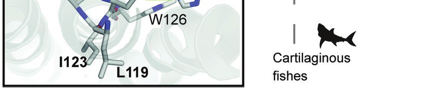
Research article                                                           Biochemistry and Chemical Biology Evolutionary Biology

                                       To sustain vision, all-trans retinal (atRAL) chromophore must be released from MII after Gt signal-
                                   ing (Palczewski, 2006)—a process that depends on the conformational stability of the MII-active-
                                   state structure (Schafer et al., 2016; Schafer and Farrens, 2015). Cone opsins have low MII stability
                                   and therefore rapidly release atRAL (Imai et al., 1997; Chen et al., 2012a), where it is quickly
                                   recycled back into 11-cis retinal (11CR) through the cone visual (retinoid) cycle, enabling rapid regen-
                                   eration of cone pigments for bright-light vision (Wang and Kefalov, 2011; Tsybovsky and Palczew-
                                   ski, 2015). Rods, in contrast, regenerate thousands of times slower than cones after bright-light
                                   exposure (Mata et al., 2002). Indeed, rod exposure to bright flashes of light leads to atRAL release
                                   that can outpace clearance by visual cycle enzymes (Sommer et al., 2014; Rózanowska and Sarna,
                                   2005), thus leading to accumulation (Saari et al., 1998; Lee et al., 2010) and light-induced retinop-
                                   athy through various modes of cellular toxicity involving oxidative stress (Maeda et al., 2009;
                                   Chen et al., 2012b). Interestingly, recent biochemical evidence suggests MII may play a role in reti-
                                   nal photoprotection by complexing with arrestin after Gt signaling to re-uptake and thus provide a
                                   sink for toxic atRAL after rod photobleaching (Sommer et al., 2014). This suggests the evolution of
                                   rhodopsin’s high conformational selectivity for toxic atRAL may be a functional specialization
                                   (Schafer et al., 2016; Schafer and Farrens, 2015), which could in turn reflect differences in retinoid
                                   metabolism between rods vs. cones (Wang and Kefalov, 2011; Tsybovsky and Palczewski, 2015;
                                   Imai et al., 2005).
                                       Consistent with the overlapping mechanisms of RH1 spectral and non-spectral functions via the
                                   highly constrained RH1 structure (Gozem et al., 2012; Yue et al., 2017), this biophysical pleiotropy
                                   likely necessitates costly trade-offs between the spectral and non-spectral functions of RH1 in natural
                                   systems (Dungan and Chang, 2017; Luk et al., 2016). By comparison, directed evolution and syn-
                                   thetic biology approaches have successfully engineered either spectral, or non-spectral aspects of
                                   rhodopsin function, but did not address trade-offs arising from shifts in function. It has thus been
                                   possible to shift the spectral absorbance of archaea and bacterial rhodopsins close to the limit of the
                                   visible spectrum (Herwig et al., 2017; McIsaac et al., 2014), and to engineer tetrapod rhodopsins
                                   with high thermal stability (Xie et al., 2003), constitutive activation (Deupi et al., 2012;
                                   Standfuss et al., 2011), and alternative chromophore-binding sites (Devine et al., 2013). However,
                                   it has not been investigated whether rod visual pigments with novel combinations of spectral and
                                   non-spectral functional properties can be engineered by manipulating the biophysical pleiotropy of
                                   RH1 otherwise exploited by natural selection.
                                       Site 122 (Bos taurus RH1 numbering) is a molecular determinant of both the spectral and non-
                                   spectral functional properties of rhodopsin and the cone opsins (Hunt et al., 2001; Yue et al., 2017;
                                   Imai et al., 1997; Imai et al., 2007; Yokoyama et al., 1999). Intriguingly, vertebrate visual pigment
                                   families show differences in which amino acid variants predominate at this site (Figure 1A), with I122
                                   strongly conserved in the most ancestrally diverging cone opsins such as the long-wave sensitive
                                   opsins (LWS) (Lamb et al., 2007), whereas in the most derived opsin group, the rhodopsins (RH1),
                                   E122 predominates (Figure 1B,C) (Imai et al., 1997; Lamb et al., 2007; Imai et al., 2007;
                                   Carleton et al., 2005). E122 is a key component of an important hydrogen-bonding network with
                                   H211 that is known to stabilize the MII active-conformation (Choe et al., 2011). This stability
                                   increase is so dramatic that E122 is considered a functional determinant distinguishing rhodopsin
                                   from cone opsins (Figure 1B) (Imai et al., 1997; Lamb et al., 2016; Kojima et al., 2014). Paradoxi-
                                   cally, by conferring this increase in MII stability, the evolution of E122 likely involved a costly fitness
                                   trade-off that diminished tetrapod rod photosensitivity (Yue et al., 2017), which can affect visual
                                   performance in animals (Kojima et al., 2017; Aho et al., 1988). Indeed, it is possible to improve tet-
                                   rapod rod photoreceptor sensitivity by decreasing rod dark noise in vivo by replacing E122 with a
                                   cone opsin amino acid variant (COV; Figure 1A) at site 122, such as Q122, which predominates in
                                   RH2 cone opsins (Yue et al., 2017; Lin et al., 2017). The strict conservation of E122 in all tetrapod
                                   rhodopsins (Figure 1C, Table 1, Supplementary file 1) therefore suggests that during the evolution
                                   of tetrapod dim-light vision, natural selection may have prioritized MII stability (Figure 1B,D) at the
                                   expense of rod sensitivity. This apparent evolutionary trade-off is perplexing given that the low
                                   spontaneous thermal activation of rhodopsin (and therefore rod dark noise) is a functional hallmark
                                   of rhodopsin divergence from the cone opsins (Kojima et al., 2017; Gozem et al., 2012;
                                   Kefalov et al., 2003; Lamb et al., 2016).
                                       Why has tetrapod RH1 been constrained to this paradoxical compromise at site 122 for the last
                                   350 million years? Interestingly, and in contrast to tetrapod rhodopsins, fish rhodopsins show

Castiglione and Chang. eLife 2018;7:e35957. DOI: https://doi.org/10.7554/eLife.35957                                                   3 of 30
Research article Biochemistry and Chemical Biology Evolutionary Biology Figure 1. Natural variation at site 122 determines rhodopsin function and stability. (A) Amino acid consensus residues at site 122 across vertebrate rod opsins (rhodopsin; RH1) and the cone opsins (long-wave (LWS), short-wave (SWS1 and SWS2) and middle-wave (RH2) sensitive). Modified from (Lamb et al., 2007). (B) Relative stability of the rod and cone opsin active-conformation (MII) in different vertebrates (Imai et al., 2005). (C) Schematic representation of naturally occurring cone opsin variants (COVs) and other amino acids across vertebrate RH1 (see Figure 1—figure supplements 1–2; Figure 1 continued on next page Castiglione and Chang. eLife 2018;7:e35957. DOI: https://doi.org/10.7554/eLife.35957 4 of 30
Research article                                                                  Biochemistry and Chemical Biology Evolutionary Biology

Figure 1 continued
Tables 1–2, Supplementary files 1–2). E122 is invariant in all Tetrapod RH1 genes sequenced to date. Natural deep-sea amino acid variants
(Hunt et al., 2001; Yokoyama et al., 1999) are identified with an asterisk (*; Table 2). (D) Introduction of the ancestral cone opsin (LWS) variant I122
blue shifts tetrapod RH1 spectral absorbance and accelerates decay of the MII light-activated conformation.
DOI: https://doi.org/10.7554/eLife.35957.003
The following figure supplements are available for figure 1:
Figure supplement 1. Schematic of RH1 site 122 variation across the vertebrate phylogeny.
DOI: https://doi.org/10.7554/eLife.35957.004
Figure supplement 2. Vertebrate phylogeny used in computational analyses.
DOI: https://doi.org/10.7554/eLife.35957.005

                                         variation at site 122, such as in the Coelacanth (Latimeria chalumnae), Lungfish (Neoceratodus for-
                                         steri), and deep-sea fish lineages, where COV (I, Q, M) and other residues at site 122 (V, D) are
                                         found (Figure 1C; Figure 1—figure supplement 1; Tables 1–2, Supplementary file 2) (Hunt et al.,
                                         2001; Yokoyama et al., 1999; Carleton et al., 2005). These substitutions have been shown to blue-
                                         shift lMAX by up to ~10 nm (Hunt et al., 2001; Yokoyama et al., 1999), and may improve dim-light
                                         sensitivity in poorly-lit aquatic environments (Yue et al., 2017). Strikingly, one of the largest freshwa-
                                         ter groups—the Characiphysi (which includes piranhas, electric eels, and catfishes [Chen et al.,
                                         2013]) —has the COV I122 residue completely fixed (Figure 1—figure supplement 1,
                                         Supplementary file 3). In tetrapods by contrast, the red-shifting E122 mutation is strictly main-
                                         tained, increasing MII stability (Imai et al., 1997) but greatly decreasing rod sensitivity (Yue et al.,
                                         2017). Why the strong constraints on high MII stability and E122 are relaxed only within certain
                                         aquatic visual ecologies, remains unknown.
                                             In light of these ecological patterns, we questioned whether it was possible to synthesize an evo-
                                         lutionary alternative: a tetrapod RH1 that never lost COV at site 122 but still developed high MII sta-
                                         bility. We reasoned that relative to tetrapods, the diversity and complexity of fish visual ecologies

Table 1. Variation at sites 119-122-123-124 in Tetrapods and Outgroup rh1.
Sites with variation relative to the Vertebrate consensus (LEIA) are in bold and highlighted grey. Subterranean species are denoted (*).
                     Species                              Accession             Common name                      119         122          123          124
Outgroups            Callorhinchus milii                  XP_007888679          Elephant shark                   L           E            I            G
                     Orectolobus ornatus                  AFS63882              Ornate wobbegong                 L           E            V            S
                     Latimeria chalumnae                  XP_005997879          Coelacanth                       L           Q            V            A
                     Neoceratodus forsteri                ABS89278              Australian lungfish              F           I            I            A
Mammals              Dasypus novemcinctus                 XP_004477303          9-banded armadillo*              I           E            I            A
                     Eptesicus fuscus                     XP_008150514          Big brown bat                    L           E            V            A
                     Chrysochloris asiatica               XP_006868732          Cape golden mole*                M           E            I            A
                     Sorex araneus                        XP_004613289          Common Shrew*                    L           E            V            A
                     Tupaia chinensis                     XP_006160726          Tree Shrew                       L           E            V            A
                     Ictidomys tridecemlineatus           XP_005333841          13-line ground squirrel          L           E            V            A
                     Rattus norvegicus                    NP_254276             brown rat                        L           E            I            G
                     Sarcophilus harrisii                 XP_003762497          Tasmanian devil                  T           E            V            A
Reptiles             Alligator mississippiensis           XM_006274155          American alligator               L           E            V            A
                     Alligator sinensis                   XP_006039462          Chinese alligator                L           E            V            A
                     Anolis carolinensis                  NP_001278316          Carolina anole                   L           E            M            G
                     Python bivittatus                    XP_007423324          Burmese python                   L           E            M            A
Amphibians           Ambystoma tigrinum                   U36574                Tiger salamander                 M           E            I            A
                     Cynops pyrrhogaster                  BAB55452              Jap. Fire belly newt             L           E            I            G
                     Xenopus tropicalis                   NP_001090803          Western clawed frog              L           E            M            A
                     Xenopus laevis                       NP_001080517          African clawed frog              L           E            V            A

DOI: https://doi.org/10.7554/eLife.35957.006

Castiglione and Chang. eLife 2018;7:e35957. DOI: https://doi.org/10.7554/eLife.35957                                                                5 of 30
Research article Biochemistry and Chemical Biology Evolutionary Biology
Table 2. Fish rh1 with variation at site 122 do not necessarily have variation at coevolving sites 119, 123, and 124.
Sites with variation relative to the Vertebrate consensus (LEIA) are in bold and highlighted grey.
Order Species Accession Common name 119 122 123 124 Ecology notes from FishBase
Lepisosteiformes Lepisosteus JN230969.1 spotted gar L M I S Freshwater; brackish; demersal. (Ref. 2060)
oculatus
Atractosteus JN230970.1 Tropical Gar L M L S Freshwater; demersal
tropicus
Osteoglossiformes Mormyrops JN230973.1 Cornish Jack T I I A Freshwater; demersal; potamodromous (Ref. 51243)
anguilloides
Osteoglossum KY026030.1 Silver arowana T I I A Freshwater; benthopelagic
bicirrhosum
Alepocephalifromes Alepocephalus JN230974.1 Bicolor slickhead L Q I A Marine; bathydemersal; depth range 439–1080 m (Ref.
bicolor 44023).
Bathytroctes JN544540.1 Smallscale L D I A Marine; bathypelagic; depth range 0–4900 m (Ref.
microlepis smooth-head 58018)
Conocara JN412577.1 Salmon smooth- L Q I A Marine; bathypelagic; depth range 2400–4500 m (Ref.
salmoneum head 40643)
Galaxiiformes Galaxias JN231000.1 Inanga L M I G Marine; freshwater; brackish; benthopelagic;
maculatus catadromous (Ref. 51243).
Stomiatiformes Argyropelecus JN412571.1 Lovely H Q I A Marine; bathypelagic; depth range 100–2056 m (Ref.
aculeatus Hatchetfish 27311)
Vinciguerria JN412570.1 Oceanic lightfish H Q V A Marine; bathypelagic; depth range 20–5000 m (Ref.
nimbaria 4470)
Ateleopodiformes Ateleopus KC442218.1 Pacific Jellynose L M I S Marine; bathydemersal; depth range 140–600 m (Ref.
japonicus Fish 44036).
Myctophiformes Benthosema JN412576.1 Smallfin H Q V G Marine; bathypelagic; oceanodromous; depth range
suborbitale lanternfish 50–2500 m (Ref. 26165)
Lampanyctus JN412575.1 Winged H Q V A Marine; bathypelagic; oceanodromous; depth range
alatus lanternfish 40–1500 m (Ref. 26165)
Neoscopelus KC442224.1 Shortfin L Q I A Marine; bathypelagic; depth range 250–700 m (Ref.
microchir neoscopelid 4481)
Gadiiformes Coryphaenoides JN412578.1 Gunther’s L V I A Marine; bathydemersal; depth range 831–2830 (Ref.
guentheri grenadier 1371)
Beryciformes Melamphaes JN231006.1 Shoulderspine L Q I A Marine; brackish; bathypelagic; depth range 500–1000
suborbitalis bigscale m (Ref. 31511).
Holocentriformes Holocentrus rufus KC442230.1 Longspine L M I S Marine; reef-associated; depth range 0–32 m (Ref.
squirrelfish 3724).
Myripristis KC442231.1 Pinecone L M I G Marine; reef-associated; depth range 1–50 m (Ref.
murdjan soldierfish 9710)
Scombriformes Aphanopus carbo EU637938.1 Black H Q I G Marine; bathypelagic; oceanodromous (Ref 108735);
scabbardfish 200–2300 m (Ref. 108733)
Cubiceps gracilis EU637952.1 Driftfish - Q I A Marine; pelagic-oceanic; oceanodromous (Ref. 51243);
DOI: https://doi.org/10.7554/eLife.35957.007
(Hunt et al., 2001; Hauser and Chang, 2017a) may have allowed selection the opportunity to
explore the pleiotropic potential of site 122 through the evolution of novel structural interactions
with nearby sites that could compensate for the destabilizing loss of the E122-H211 hydrogen bond.
To identify these interactions, our goal was to use analyses of evolutionary rates to predict sites
coevolving with site 122, and to investigate the functional consequences of coevolving sites with
experimental site-directed mutagenesis studies. Ultimately, we used our analyses of natural variation
as a guide to artificially engineer a tetrapod rhodopsin with increased MII stability, but within a non-
E122 sequence background. We demonstrated that this synthetic alternative is possible, even if evo-
lution did not proceed down this mechanistic trajectory toward a dim-light adapted visual pigment.
Castiglione and Chang. eLife 2018;7:e35957. DOI: https://doi.org/10.7554/eLife.35957 6 of 30Research article Biochemistry and Chemical Biology Evolutionary Biology Figure 2. Local coevolutionary forces govern the evolution of site 122 differentially between tetrapods and fish (teleost) RH1. (A) Extant and reconstructed codon variation at site 122 (Materials and methods). Despite a variety of residues at site 122 across the Coelacanth (Q122), Lungfish (I122; Ceratodontiformes), and Tetrapods (E122), GAA codons encoding for E122 are nevertheless predicted as the ancestral state with high posterior probabilities (shown in parentheses). E122 (GAA/GAG) is also likely to have been present in the last common ancestor of Cypriniformes and the Characiphysi, although with low posterior probabilities and therefore high uncertainty. I122 codon ATC is fixed in all Characiphysi rhodopsin to our knowledge (Supplementary file 3). Approximate divergence times are from (Hedges et al., 2015). (B) Mutual information (MI) analyses (MISTIC [Simonetti et al., 2013]) reveal all sites coevolving with site 122 are within 6 Å. Significance thresholds were determined by reference to the highest MI z-score from all sites across analyses of randomized datasets (n = 150; z-score cut-off = 21.6), as previously described (Ashenberg and Laub, 2013). (C) Sites within this radius displayed decreased amino acid variation in tetrapod and characiphysi RH1, where E122 and I122 are fixed, respectively (asterisks). (D) In tetrapods and characiphysi RH1, reduction in amino acid variation (relative to teleosts) at positions within the 6 Å radius were driven by increases in purifying selection on non-synonymous codons. Statistically significant gene-wide increases in purifying selection (*) between lineages were detected by likelihood ratio tests of alternative (Clade model C [Bielawski and Yang, 2004]) and null (M2a_REL [Weadick and Chang, 2012]) model analyses of codon substitution rates (dN/dS) ((p
Research article                                                                    Biochemistry and Chemical Biology Evolutionary Biology

Table 3. Results of Clade Model C (CmC) analyses of vertebrate rh1 under various partitions.
                                                                            Parameters
Model and
Foreground†                            DAIC‡           lnL                  !0                  !1             !2/!d                 Null              P [df]
     M2a_rel                           225.5             47185.37           0.02 (69%)          1 (3%)         0.20 (28%)            N/A               -
CmC_Tetrapod Branch                    97.44             47119.33           0.20                1              0.02 (69%)            M2a_rel           0.000 [1]
                                                                            (28%)               (3%)           Tetra Br: 0.00
CmC_Tetrapod                           4.92              47073.06           0.02 (67%)          1 (3%)         0.24 (30%)            M2a_rel           0.000 [1]
                                                                                                               Tetra: 0.13
CmC_Teleost                            1.88              47071.54           0.02 (67%)          1 (3%)         0.14 (30%)            M2a_rel           0.000 [1]
                                                                                                               Teleost: 0.24
CmC_Teleost vs Tetrapod                0*                47069.60           0.02 (67%)          1 (3%)         0.17 (30%)            M2a_rel           0.000 [2]
                                                                                                               Tetra: 0.13
                                                                                                               Teleost: 0.24
†
    The foreground partition is listed after the underscore for the clade models and consists of either: the clade of Teleost fishes (Teleost); the clade Tetra-
pods (Tetrapod;Tetra) or branch leading to tetrapods (Tetrapod branch; Tetra Br); or the clades of both the teleost fishes and tetrapods as two separate
foregrounds (Teleost vs Tetrapods). In any partitioning scheme, the entire clade was tested, and all non-foreground data are present in the background
partition.
‡
    All DAIC values are calculated from the lowest AIC model. The best fit is shown with an asterisk (*).
!d is the divergent site class, which has a separate value for the foreground and background partitions.
¶
    Significant p-values (a 0.05) are bolded. Degrees of freedom are given in square brackets after the p-values.
Abbreviations—lnL, ln Likelihood; p, p-value; AIC, Akaike information criterion.
DOI: https://doi.org/10.7554/eLife.35957.010

                                         Results
                                         Phylogenetic identification of an intramolecular coevolutionary network
                                         To better understand the selection pressures that may be constraining E122 to fixation during tetra-
                                         pod evolution, we constructed a large vertebrate rhodopsin phylogenetic dataset (Figure 1—figure
                                         supplements 1 and 2, Supplementary files 1–2) and investigated the evolutionary history of site
                                         122 using ancestral reconstruction (Materials and methods). We found that E122 (codon GAA;
                                         Figure 2A) has been fixed in tetrapod RH1 since the most recent common ancestor ~350 million
                                         years ago (MYA) (Hedges et al., 2015), where it appears along the ancestral branch leading to tetra-
                                         pods (Figure 2A; Table 3) following the diversification from lungfishes (I122, codon ATA, Figure 2A;
                                         Supplementary file 1) and the coelacanth (Q122, codon CAA, Figure 2A; Supplementary file 1).
                                         This transition period in vertebrate evolution is characterized by extensive morphological modifica-
                                         tions for vision within terrestrial environments, and likely included large increases in environmental
                                         light irradiance (MacIver et al., 2017; Warrant and Johnsen, 2013). Apart from the lungfishes and
                                         coelacanth, the high conservation of E122 in tetrapods is also reflected in other vertebrate rhodop-
                                         sins (Figure 1, Figure 1—figure supplement 1; Tables 1–2; Supplementary files 1–2), but there
                                         are important exceptions within certain lineages of teleost fishes, such as the Characiphysi. Within
                                         this group, the COV residue I122 was introduced likely through E122I (codon ATC; Figure 2A),
                                         where I122 is now completely fixed across the extant Characiphysi (Supplementary file 3).
                                             Since fishes (Teleosts), unlike tetrapods, display amino acid variation at site 122 (Figure 1C), we
                                         hypothesized that compensatory mutations may be coevolving with site 122 across fish RH1. To test
                                         this hypothesis, we investigated across the entire transmembrane domain of rhodopsin (residues 53–
                                         302) for evidence of sites coevolving with site 122 within an alignment of Teleost RH1 (Materials and
                                         methods; Supplementary file 2). Using phylogenetically corrected mutual information (MI) analyses
                                         (MISTIC; [(Simonetti et al., 2013]) with z-score cut-off determined by analyses of randomized data-
                                         sets (Ashenberg and Laub, 2013), we found significant evidence of coevolution with site 122 at sev-
                                         eral RH1 positions, all of which clustered within 6 Å of E122 (Figure 2B) in the MII crystal structure
                                         (Choe et al., 2011. This is within the range at which intramolecular forces such as Van der Waals and
                                         hydrophobic interactions between amino acids are thought to occur (Ivankov et al., 2014). It is
                                         known, however, that there is a tendency of covariation analyses such as MI to identify coevolving
                                         sites proximal to each other, which may in turn overlook more distal coevolving sites potentially

Castiglione and Chang. eLife 2018;7:e35957. DOI: https://doi.org/10.7554/eLife.35957                                                                       8 of 30
Research article Biochemistry and Chemical Biology Evolutionary Biology
Table 4. Analyses used to elucidate sites coevolving with site 122 in Vertebrate rhodopsins (rh1).
In bold are the results of interest described in the main text, including: elevated dN/dS, long-term shifts in selection between teleosts
and tetrapods, amino acid statistical covariation with site 122 in the teleost dataset, and phylogenetically correlated amino acid varia-
tion with site 122.
Posterior probability of long-term
Distance to site Tetrapod M8 Teleost M8 Characiphysi shift in selection Z-score Significant correlated
Site 122 (Å)* dN/dS† dN/dS† M8 dN/dS† (tetrapod/characiphysi)‡ covariation§ evolution?
118 5 0.05 0.05 0.05 0.00/0.00 1.54 No
119 3.5 0.14 0.168 0.05 0.57/0.19 30.2 Yes
120 3.1 0.05 0.05 0.05 0.00/0.00 1.91 No
121 N/A 0.05 0.05 0.05 0.00/0.00 0.94 No
122 N/A 0.05 0.322 0.05 1.00/1.00 N/A N/A
123 N/A 0.19 0.094 0.05 0.00/0.00 20.4 No
124 3.2 0.05 0.411 0.05 1.00/1.00 5.53 No
125 3.4 0.05 0.05 0.05 0.00/0.00 1.19 No
126 3.7 0.05 0.05 0.05 0.00/0.00 1.60 No
127 4.9 0.05 0.065 0.05 0.00/0.00 27.1 Yes
160 5.1 0.05 0.05 0.05 0.00/0.00 3.90 No
164 4.2 0.05 0.05 0.05 0.00/0.00 7.88 No
167 3.8 0.05 0.05 0.05 0.00/0.00 1.38 No
168 5.9 0.05 0.308 0.192 1.00/1.00 34.2 No
207 4.9 0.05 0.05 0.05 0.00/0.00 1.21 No
211 2.7 0.05 0.05 0.05 0.00/0.00 1.38 No
265 5.1 0.05 0.05 0.05 0.00/0.00 1.44 No
*
From structural analysis of distances between amino acids and site 122 within the MII crystal structure 3PQR (Choe et al., 2011).
†
Post mean dN/dS from M8 analyses described in Tables 8–10.
‡
Bayes empirical Bayes posterior probability of long-term shift in selection calculated in Clade model C (CmC) (Yang, 2007) analyses (CmC_Teleost vs Tet-
rapod/CmC_Characi clade) described in Tables 3 and 5, respectively.
§
Phylogenetically corrected MI z-scores (MISTIC; [Simonetti et al., 2013]) of covariation with site 122 from analyses on Teleost RH1 dataset. Values were
considered significant if greater than the top absolute z-score (21.6) from all site-wise comparisons from all analyses of 150 randomized datasets, as
described (Ashenberg and Laub, 2013).
¶
Tests of correlated evolution in amino acid variation (Pagel, 1994) between a given site and site 122. p-values were calculated by performing Monte Carlo
tests using data from simulations (n > 1000) in MESQUITE (Maddison and Maddison, 2017). p-Values were subjected to a Bonferroni-correction to deter-
mine significance (pResearch article Biochemistry and Chemical Biology Evolutionary Biology
Table 5. Results of Clade Model C (CmC) analyses of teleost rh1 under various partitions.
Parameters
Model and
Foreground† DAIC‡ lnL !0 !1 !2/!d Null P [df]
M2a_rel 17.1 30987.99 0.01 (60%) 1 (5%) 0.19 (35%) N/A -
CmC_Characi branch 19.05 30986.96 0.01 (60%) 1 (5%) 0.19 (35%) M2a_rel 0.794 [1]
Char Br: 0.20
CmC_Characi clade 0* 30977.43 0.00 (60%) 1 (5%) 0.20 (20%) M2a_rel 0.000 [1]
Char Cl: 0.10
The foreground partition is listed after the underscore for the clade models and consists of either: the ancestral branch leading to the Characiphysi (Char-
aci branch; Char Br) or the entire Characiphysi clade (Characi clade; Char Cl). In any partitioning scheme, the entire clade was tested, and all non-fore-
ground data are present in the background partition.
‡
All DAIC values are calculated from the lowest AIC model. The best fit is bolded with an asterisk (*).
§
!d is the divergent site class, which has a separate value for the foreground and background partitions.
Significant p-values (a 0.05) are bolded. Degrees of freedom are given in square brackets after the p-values.
Abbreviations—lnL, ln Likelihood; p, p-value; AIC, Akaike information criterion.
DOI: https://doi.org/10.7554/eLife.35957.012
amino acid variation observed in these lineages would be driven by an increase in purifying selection
on non-synonymous codons, ultimately reflecting the entrenchment of compensatory amino acid res-
idues by natural selection.
We therefore employed codon-based phylogenetic likelihood methods to test for a relative
increase of purifying selection at RH1 sites within 6 Å of site 122, within Tetrapod vs Teleosts, as
well as in Characiphysi vs other Teleosts (Yang, 2007) (Materials and methods). Using likelihood
ratio tests of alternative (Clade model C [(Bielawski and Yang, 2004]) and null (M2a_REL
([Weadick and Chang, 2012)]) model analyses of codon substitution rates (dN/dS) across the RH1
coding-sequence, we identified statistically significant evidence of gene-wide increases in purifying
selection within Tetrapods (Table 3) and Characiphysi (Table 5) relative to teleosts ((pResearch article Biochemistry and Chemical Biology Evolutionary Biology
Figure 3. Coevolving sites form the LxxEIA and FxxINS motifs. (A) Overview of tetrapod RH1 MII rhodopsin crystal structure (Choe et al., 2011 with
coevolving sites. The green highlight and dashed line indicate the stabilizing hydrogen bond between E122-H211. (B) Reconstruction of residues at site
122 (Figure 2—figure supplement 1) and coevolving positions for ancestral characiphysi, tetrapod and outgroup rhodopsins indicates the
entrenchment of two structural motifs centering around site 122 (Materials and methods). The LxxEIA (or LEIA) motif was also predicted as present
within the ancestral Osteichthyes. Approximate divergence times are from (Hedges et al., 2015.
DOI: https://doi.org/10.7554/eLife.35957.013
increase in purifying selection on non-synonymous codons relative to other Teleosts, site 168 never-
theless displayed amino acid variation in the Characiphysi (T/V168; Figure 2C, D), suggesting it may
not necessarily play a functionally compensatory role for the ancient E122I mutation, especially since
T vs. V168 may be reasonably expected to have biochemically and/or structurally dissimilar effects
on this region of the rhodopsin TM3-TM5 microdomain (Choe et al., 2011). Conversely, although
C127 has been fixed in Characiphysi RH1 relative to other Teleosts (asterisks, Figure 2C) and may
therefore be functionally important, there was no increase in purifying selection on non-synonymous
codons at site 127 relative to Teleosts (Figure 2D), suggesting that the fixation of C127 in Characi-
physi RH1 may be a historical contingency that does not necessarily reflect intramolecular entrench-
ment by the ancient E122I mutation (Goldstein and Pollock, 2017. Although this same logic
ostensibly applies to site 123, unlike C127—a residue shared with some tetrapods (Figure 2C;
Supplementary files 1–3)—we observed a striking fixation of a rare amino acid residue in Characi-
physi RH1 (N123, asterisks, Figure 2C) which is not, to our knowledge, observed within any verte-
brate rhodopsin other than the Characiphysi where it is completely fixed (Tables 1–
2, Supplementary files 1–3), and located between coevolving sites 119, 122 and 124 which are also
Castiglione and Chang. eLife 2018;7:e35957. DOI: https://doi.org/10.7554/eLife.35957 11 of 30Research article Biochemistry and Chemical Biology Evolutionary Biology Figure 4. Coevolving sites modulate the pleiotropic functional effects of site 122. The LEIA and FINS motifs are convergent solutions for high tetrapod RH1 active state (MII) stability but with different spectral absorbances. (A) The introduction of the ancestral cone opsin variant into tetrapod RH1 (E122I) blue-shifts rhodopsin absorbance lMAX and dramatically destabilizes the MII active-conformation (Figure 4—figure supplement 1; Table 6). Bar graphs show retinal release half-life values. (B) Substituting FINS motif residues into coevolving sites have varied effects on rhodopsin spectral tuning Figure 4 continued on next page Castiglione and Chang. eLife 2018;7:e35957. DOI: https://doi.org/10.7554/eLife.35957 12 of 30
Research article                                                                   Biochemistry and Chemical Biology Evolutionary Biology

Figure 4 continued
and the stability of the active-conformation. (C) Within the E122I background, FINS motif substitutions at coevolving sites have marked effects on
spectral tuning, but no rescue effect on MII active-conformation stability. (D) Partial incorporation of the FINS motif within tetrapod rhodopsin produces
further blue-shifting effects and has a significant but small stabilizing effect within the E122I background. (E) Full incorporation of the FINS motif into
tetrapod RH1 maintains the absorbance blue-shift while fully rescuing the destabilizing effects of E122I on tetrapod RH1. Statistically significant
differences in MII stability were calculated using two-tailed t-tests with unequal variance, with standard error reported in bar graphs (*p
Research article                                                               Biochemistry and Chemical Biology Evolutionary Biology

                                   Figure 5. LEIA and FINS motifs are alternative solutions for high tetrapod RH1 MII stability within a limited
                                   sequence-function landscape. Spectral absorbance (lMAX) and stability of the active-conformation (MII) of wild
                                   type and mutant tetrapod RH1 with E122 (green) and I122 (blue), respectively. The only natural intermediate
                                   between the wild-type tetrapod consensus motif (LEIA) and the wild-type Characiphysi motif (FINS) is ‘FIIA’ from
                                   Lungfish RH1. The mutation I123N has opposite effects on MII stability depending on background sequence (sign-
                                   epistasis), which may have closed the LEIA to FINS motif evolutionary trajectory (dashed line) for tetrapod RH1.
                                   Although reflecting a limited experimental dataset, these epistatic effects may have created indirect routes to the
                                   high MII stability of the FINS motif via intermediates with low MII stability.
                                   DOI: https://doi.org/10.7554/eLife.35957.017
                                   The following figure supplement is available for figure 5:
                                   Figure supplement 1. Compensatory effects at coevolving sites are mediated by a diversity of possible structural
                                   mechanisms.
                                   DOI: https://doi.org/10.7554/eLife.35957.018

                                   Experimental characterization of natural variation at coevolving sites
                                   We therefore tested the ability of coevolving sites 119, 123 and 124 to affect tetrapod rhodopsin
                                   function and the potential for natural variation at these sites to compensate for the destabilizing loss
                                   of the E122-H211 hydrogen bond. We conducted site-directed mutagenesis and in vitro expression
                                   of mutant rhodopsins using detergent micelles (Materials and methods). This was followed by in vitro
                                   functional characterization using spectroscopic absorbance- and fluorescence-based measurements
                                   of both lMAX and the stability of the active-state conformation (Figure 4; Figure 4—figure supple-
                                   ment 1; Table 6; Materials and methods), both of which can provide information on relative differen-
                                   ces that exist within natural systems (Schafer et al., 2016; Van Eps et al., 2017; Schott et al.,
                                   2016a. Tetrapod RH1 with E122I (Figure 4A) and other FINS motif single substitutions to the
                                   coevolving sites (L119F, I123N, A124S; Figure 4B) displayed large shifts in rhodopsin lMAX and MII
                                   stability, with two single mutations (L119F, A124S) significantly increasing the stability of the active-
                                   conformation but producing opposite spectral tuning effects (Figure 4B; Table 6). Meanwhile,
                                   I123N destabilized the active-conformation almost as dramatically as E122I but produced no spectral
                                   tuning effect (Figure 4B; Table 6). This suggested that FINS substitutions at coevolving sites could
                                   functionally compensate for some of the pleiotropic effects of E122I on tetrapod rhodopsin.

Castiglione and Chang. eLife 2018;7:e35957. DOI: https://doi.org/10.7554/eLife.35957                                                          14 of 30
Research article Biochemistry and Chemical Biology Evolutionary Biology
We created double and triple mutants representing partial replacements of the LEIA with the
FINS motif, which tended to blue-shift lMAX (Figure 4C–D; Table 6). Yet, none of these intermedi-
ates were sufficient to restore WT-levels of MII stability within the COV I122 background
(Figure 4C–D; Table 6). We therefore reasoned that the complete recapitulation of the FINS motif
within tetrapod rhodopsin may be required for a full restoration of WT active-conformation stability.
We found, incredibly, that the L119F/I123N/A124S triple mutation fully restored the MII stability of
E122I tetrapod rhodopsin to WT levels, while even further blue-shifting lMAX relative to E122I
(Figure 4E; Table 6). The LEIA and FINS motifs are therefore two configurations conferring conver-
gent MII stabilities but different spectral sensitivities, with the blue-shifting I122-containing FINS
motif likely also decreasing rod dark noise in vivo (Gozem et al., 2012; Yue et al., 2017).
Our experiments demonstrate that N123, which is not, to our knowledge, observed within any
vertebrate rhodopsin other than the Characiphysi (Tables 12, Supplementary files 1–3) is neverthe-
less required for a complete rescue of MII stability within the LWS COV I122 background, where it
has opposite functional effects depending on E vs. I122 backgrounds (also known as sign-epistasis
[Storz, 2016; Weinreich et al., 2006]) (Figure 4D–E; Figure 5). Structural analysis of a homology
model of the MII active-state structure (Materials and methods; Figure 5—figure supplement 1)
suggests the conformation of the FINS motif mediates a series of context-dependent structural rear-
rangements promoting novel interactions (F119 with W161; N123 with N78/T160; Figure 5—figure
supplement 1) that can interact with existing GPCR hydrogen bond networks known to stabilize the
MII active conformation (S124 with D83-S298- N302; Figure 5—figure supplement 1; [Choe et al.,
2011]). These epistatic structural interactions produce correspondingly variable pleiotropic effects
on RH1 spectral absorbance and MII stability (Figure 4), which were consistent with patterns of natu-
ral sequence variation at these positions across vertebrate rhodopsins. Using these patterns of natu-
rally occurring sequence variation, we could successfully navigate a complex sequence-function
landscape (Figure 5) to engineer the spectral and non-spectral functions of rhodopsin
simultaneously.
Discussion
We questioned why E122, a residue that diminishes rod photosensitivity, was retained in the evolu-
tion of all tetrapod rod pigments. We investigated if it was possible to engineer a tetrapod RH1 with
high active-state conformational stability without E122. We uncovered a natural solution—over 150
million years ago the ‘FINS’ motif originated within the rhodopsin of an ancestral population of the
Characiphysi freshwater fish lineage. Although we explore here only a limited subsection of the total
sequence-function space of tetrapod vs. Characiphysi RH1 (notably excluding sites 127 and 168 in
our experimental analyses), this natural variation, nevertheless, inspired us to engineer a synthetic
alternative that nature never produced: a tetrapod RH1 with high MII stability without E122, result-
ing in a blue-shifted pigment predicted to increase rod photosensitivity in vivo. These results, along
with recent advances in molecular evolutionary theory, and studies of rhodopsin biochemistry, sug-
gest a plausible model of why the FINS motif might have been the road less traveled in evolutionary
history.
Physiological relevance of MII stability—a proposed role in
photoprotection in the eye
Did a physiological advantage related to high MII stability drive the fixation of the LEIA and FINS
motifs? Consistent with predictions of intramolecular coevolutionary theory (Talavera et al., 2015;
Pollock et al., 2012; Shah et al., 2015), evolutionary trajectories from the LEIA to FINS motifs must
pass through sub-optimal sequence-function intermediates which include variants associated with
active-state instability and human rhodopsin disease phenotypes (e.g. A164V) (Stojanovic et al.,
2003) (Figure 5). Similar to E122I, disease variants such as A164V are likely pathogenic through dis-
ruption of the E122-H211 hydrogen bond, which has been shown to stabilize the active-state confor-
mation but can be affected indirectly through mutations at nearby sites 119 and 123 (Imai et al.,
1997; Stojanovic et al., 2003; Morrow and Chang, 2015. Although correlations between dark-
state stability and active-state (MII) stability have been recently postulated (Kojima et al., 2017),
there exists substantial conformational differences in the TM3-TM5 region thought to stabilize both
structures, including the reconfiguration of E122-H211 and E122-W126 hydrogen bonds upon light
Castiglione and Chang. eLife 2018;7:e35957. DOI: https://doi.org/10.7554/eLife.35957 15 of 30Research article Biochemistry and Chemical Biology Evolutionary Biology
activation (Choe et al., 2011; Ahuja et al., 2009; Okada et al., 2004; Lin and Sakmar, 1996). While
it is unclear if such structural differences exist within the cone opsins, the lack of E122 (e.g. Q122 in
Rh2 (except the lamprey [(Lin et al., 2017; Davies et al., 2007)), I122 in LWS (Lamb et al., 2007;
Carleton et al., 2005)) strongly suggests that natural selection has prioritized dark-state stability
over MII stability within the cone opsins, which may be related to mitigating the high noise of cone
photoreceptors, especially in red-shifted LWS (Gozem et al., 2012; Kefalov et al., 2003;
Imai et al., 1997; Chen et al., 2012a; Kefalov et al., 2005). By contrast, in tetrapod rhodopsins,
E122 predominates, increasing MII stability while red shifting spectral absorbance, and therefore
also decreasing in vivo rod photosensitivity (Gozem et al., 2012; Yue et al., 2017). This suggests
that selection has maintained E122, and therefore the stability of the MII active-conformation, for
reasons distinct from those maintaining the stability of the dark-state conformation, which modulates
rod photosensitivity.
Why has the increased rod photosensitivity conferred by COV at site 122 been sacrificed in all tet-
rapods? In addition to setting the limit on rod photosensitivity (Baylor et al., 1980), rhodopsin is
also associated with light-induced photodamage (Grimm et al., 2000; Williams and Howell, 1983),
where retinal susceptibility strongly correlates with rhodopsin expression levels, and can be altered
through ambient lighting conditions in some animals (Rózanowska and Sarna, 2005;
Organisciak and Vaughan, 2010). Below we describe the mounting indirect evidence that the high
stability and long decay of the rhodopsin MII active conformation may be a photoprotective mecha-
nism against light-induced retinal damage (Sommer et al., 2014; Imai et al., 2005), and we postu-
late that this likely accompanied the evolution of dim-light vision.
First, one of the most promising strategies to increase retinal resistance to photodamage is to
slow the rate of rhodopsin regeneration, which can be achieved via mutations or molecules inhibiting
the normal functioning of visual cycle proteins responsible for synthesizing 11-cis retinal (11CR)
(Wenzel et al., 2001; Saari et al., 2001; Mandal et al., 2011). This can also be done through block-
ing rhodopsin regeneration and binding of 11CR (Radu et al., 2003; Sieving et al., 2001), reducing
the light-dependent accumulation of atRAL condensation products such as diretinoid-pyridinium-
ethanolamine (A2E), which contributes to lipofuscin deposits in the retinal pigment epithelium asso-
ciated with human retinal diseases (Maeda et al., 2009; Chen et al., 2012b; Radu et al., 2003;
Sparrow, 2003). Importantly, whether rhodopsin will bind available 11CR, or atRAL is dictated by
the conformational selectivity of the rhodopsin dark- and active-state (MII) conformations, respec-
tively (Schafer et al., 2016; Schafer and Farrens, 2015; Chen et al., 2012a)—a new finding consis-
tent with previous observations that rhodopsins with high MII stability tend to also have slowed
11CR regeneration rates, which is a key distinguishing feature from the cone opsins (Chen et al.,
2012a; Imai et al., 2005). These observations suggest that rhodopsin conformational selectivity may
be an overlooked functional specialization of dim-light vision—one that may be associated with rates
of regeneration and therefore photoprotection in the eye.
Although multiple molecular mechanisms within the visual cycle appear to have evolved to pre-
vent the accumulation of toxic atRAL (Rózanowska and Sarna, 2005; Chen et al., 2012c) as well as
excess 11CR (Quazi and Molday, 2014, a possible role for rhodopsin’s intrinsic conformational
selectivity in sequestering these retinal ligands has been mostly overlooked. Recent advances in rho-
dopsin biochemistry suggest that such a photoprotective mechanism may indeed exist. In contrast
to cones, which have an expanded retinoid recycling capacity (Wang and Kefalov, 2011;
Tsybovsky and Palczewski, 2015), in rods atRAL clearance is limited by the activity of retinal dehy-
drogenases (RDH) (Saari et al., 1998; Chen et al., 2012b; Chen et al., 2012c), and can transiently
accumulate to toxic levels (Sommer et al., 2014 causing light induced-retinopathy through a variety
of mechanisms involving oxidative stress (Maeda et al., 2009; Chen et al., 2012b. Unlike cone
opsins, which have low active-conformation stability, the rhodopsin active-conformation is highly sta-
ble due in large part to the evolution of E122 (Imai et al., 1997), and when phosphorylated and
bound to rod arrestin, contains a binding affinity for atRAL sufficient for sequestration and reduction
below toxic levels (Sommer et al., 2014; Rózanowska and Sarna, 2005). Indeed, it is now known
that elevated atRAL concentrations will increase atRAL re-uptake by active-conformation rhodopsin
in vitro (Schafer et al., 2016), and in bright light levels when the risk of photodamage and atRAL lev-
els are highest (Rózanowska and Sarna, 2005; Organisciak and Vaughan, 2010, this process is fur-
ther promoted by the constitutive binding of arrestin to the active rhodopsin conformation, which
importantly does not block RDH access to atRAL (Gurevich et al., 2011; Sommer et al., 2012). This
Castiglione and Chang. eLife 2018;7:e35957. DOI: https://doi.org/10.7554/eLife.35957 16 of 30Research article Biochemistry and Chemical Biology Evolutionary Biology
proposed survival mechanism not only precludes Gt signaling but also promotes re-uptake of atRAL
by rhodopsin, therefore delaying MII decay and regeneration of the dark state, and enhancing atRAL
sequestration (Sommer et al., 2012; Sommer and Farrens, 2006). Although the other rhodopsin in
the homodimer bound by arrestin is likely free to decay to the inactive conformation, permitting
regeneration with 11CR (Sommer et al., 2012; Schafer and Farrens, 2015; Beyrière et al., 2015), a
higher intrinsic stability of the active conformation would not only likely delay regeneration with
11CR, but also likely push the equilibrium toward atRAL re-uptake —a process that could be even
further promoted if atRAL levels are high (Sommer et al., 2012; Schafer et al., 2016), such as within
dark-adapted animals exposed to bright flashes (Saari et al., 1998; Lee et al., 2010, and within dis-
ease models where atRAL clearance is delayed (Maeda et al., 2009; Chen et al., 2012c. The evolu-
tion of high conformational selectivity of active-state rhodopsin for atRAL—a distinguishing feature
from the cone opsinsmay therefore play a key role within these putatively photoprotective ternary
complexes, which have been previously proposed to provide an atRAL sink for rods in bright light
(Sommer et al., 2014).
While only detailed experimental investigations can determine the relationships between rhodop-
sin regeneration rates, atRAL-associated photodamage, and the recently expanded ensemble of
spectrally identical MII conformational substates (Van Eps et al., 2017, a putative photoprotective
role for the intrinsic stability of the rhodopsin MII active conformation would imply the presence of
strong rhodopsin functional constraints in addition to those canonical constraints associated with rod
photosensitivity. This model is consistent with the fact that high rod photosensitivity and susceptibil-
ity to photodamage appear to be a trade-off that accompanied the evolution of rhodopsin-mediated
dim-light vision (Grimm et al., 2000; Williams and Howell, 198390,91. Indeed, a trade-off model of
rhodopsin evolution may clarify why the experimental relevance of long MII decay still remains
unclear, as the focus has been mostly on mutational effects to photosensitivity, rather than photo-
damage ( Kojima et al., 2014; Imai et al., 2007). Below, we outline a trade-off model of rhodopsin
evolution, and describe in detail how it may help to unravel the paradoxical distribution of natural
sequence variation at rhodopsin site 122.
Trade-offs between rhodopsin-mediated photosensitivity and
photoprotection may explain the E122 paradox
As discussed above, tetrapod susceptibility to photodamage is a necessary side-effect of rhodopsin-
mediated dim-light vision (Grimm et al., 2000; ), yet it has been an often-overlooked possibility that
the functional constraints governing rhodopsin evolution could have also been shaped by those
associated with rhodopsin-mediated photodamage, which induces oxidative stress leading to retinal
degenerative diseases via toxic atRAL (Tsybovsky and Palczewski, 2015; Sommer et al., 2014;
Rózanowska and Sarna, 2005; Wenzel et al., 2005). By delaying both atRAL release and 11CR
binding, high rhodopsin MII stability could provide an additional protective mechanism against rho-
dopsin-mediated photodamage outside the visual cycle—one which could be modulated parsimoni-
ously in response to light conditions through mutations altering MII stability (Dungan and Chang,
2017; Gutierrez et al., 2018; Hauser et al., 2017b. Our model would therefore predict that the
evolution of rhodopsin after divergence from the cone opsins involved unique functional specializa-
tions for both photosensitivity and photoprotection. These photodamage-related constraints associ-
ated with the evolution of dim-light vision may have been especially relevant within dim-light
adapted animals with high levels of rhodopsin and an increased susceptibility to photodamage
(Rózanowska and Sarna, 2005; Organisciak and Vaughan, 2010 where exposures to bright light
flashes can dramatically increase toxic ATR accumulation levels, leading to photoreceptor degenera-
tion (Saari et al., 1998; Chen et al., 2012b).
Interestingly, due to environmental differences, variation in the selective constraints associated
with rhodopsin’s role in photosensitivity vs. photodamage may explain the paradoxical ecological
patterns associated with natural variation at site 122. Specifically, our results demonstrate that the
only visual ecologies where the selective constraints on E122 are repeatedly relaxed across the phy-
logeny is within the constant darkness of deep-dwelling fish environments (Hunt et al., 2001;
Yokoyama et al., 1999)—the natural system where one might expect the fitness effects of the rho-
dopsin-mediated trade-off between photosensitivity and photoprotection to drastically shift, as
there is likely little photodamage risk for fishes living below 1000 m in near-permanent darkness
(Denton, 1990). All whales by contrast—some of which routinely dive into complete darkness at
Castiglione and Chang. eLife 2018;7:e35957. DOI: https://doi.org/10.7554/eLife.35957 17 of 30Research article Biochemistry and Chemical Biology Evolutionary Biology
depths near 2000 m (e.g. the sperm whale) (Denton, 1990; Watkins et al., 1993)—strictly maintain
E122, thereby forgoing the photosensitivity increase conferred by COV at site 122 that would other-
wise likely prove advantageous within these dark marine environments (Hunt et al., 2001;
Yue et al., 2017; Yokoyama et al., 1999). Yet, unlike deep dwelling fishes, all whales must resur-
face, suggesting that photoprotection-associated constraints may be maintaining E122 despite the
cost of decreased photosensitivity: a prediction consistent with our model and with increases to MII
stability as a key feature of whale evolution (Dungan and Chang, 2017. In contrast, in Characiphysi
fishes, the FINS motif evolved—a novel molecular mechanism likely increasing rod photosensitivity
without the consequent trade-off on MII stability. Although this may be related to mitigating the
increased dark noise that can arise as consequence of spectral tuning to some red-shifted freshwater
environments (Gozem et al., 2012; Van Nynatten et al., 2015), this remains unclear, as the ances-
tral condition is uncertain, and the distribution of characiphysian fishes occur in a wide range of envi-
ronments (Castiglione et al., 2017; Chen et al., 2013). Whether other sequence motifs within this
network represent different ‘tuning solutions’ for the visual system across different environments is
unknown, yet the possible permutations appear to have been dramatically limited by a combination
of natural selection, historical contingency and epistasis (Tables 1–2) (Storz, 2016). This would be
an interesting avenue of future investigation.
In tetrapods, none of these coevolutionary motifs include COV at site 122 (Tables 1,
Supplementary files 1–2). This suggests that tetrapods have been confined to a local optimum
(E122), which makes it tempting to speculate that this evolutionary constraint could only be main-
tained by the existence of a strongly detrimental pleiotropic effect, which we propose to be that of
rhodopsin-mediated photodamage. Potential caveats to this theory include the existence of subter-
ranean tetrapods maintaining E122 (Table 1)—a system where one might expect the putative photo-
damage-associated constraints on rhodopsin to relax, as may have occurred within a variety of
deep-sea fishes (although it remains unclear if increases to photosensitivity would even be prioritized
within these animals if the putative constraints on photoprotection were indeed relaxed
(Partha et al., 2017)). Although speculative, our trade-off model of rhodopsin evolution, combined
with fitness landscape theory (Hartl, 2014) could potentially explain why the evolutionary trajectories
between the LEIA and FINS motifs have been traversed by some freshwater fishes, but never by a
tetrapod lineage.
Exploring inferred fitness landscapes using natural variation
Evolutionary pathways often include compensatory mutations (Talavera et al., 2015; Pollock et al.,
2012; Shah et al., 2015; Tokuriki et al., 2008), where adaptive mutations are permitted by non-
adaptive neutral mutations (Pál and Papp, 2017; Starr et al., 2017; Tarvin et al., 2017) (also known
as ‘pre-adaptations’ [Pál and Papp, 2017], or ‘pre-adjustments’ [(Goldstein and Pollock, 2017)) and
this contingency opens new evolutionary paths by accommodating the subsequent mutational per-
turbations to protein activity and/or stability (Tokuriki and Tawfik, 2009; Ivankov et al., 2014;
DePristo et al., 2005). Similarly, we find that a triple mutation (L119F/I123N/A124S) would be
required to functionally compensate for the detrimental effects of E122I on tetrapod rhodopsin
active-conformation stability. Yet, one of these ’pre-adaptations’ (I123N) is as destabilizing as E122I
itself, and displays strong sign-epistasis (Figure 4), which may be sufficient to close the evolutionary
trajectory leading from the LEIA to FINS motifs (Storz, 2016; Weinreich et al., 2005;
Poelwijk et al., 2007). Accordingly, a wide variety of indirect evolutionary paths may cut through
these valleys to access fitness peaks (Wu et al., 2016; Starr et al., 2017; Weinreich et al., 2006)—a
scenario which has not been extensively characterized in protein systems evolving within natural
environments (Pál and Papp, 2017; Hartl, 2014).
Detrimental intermediates and historical contingency (Pál and Papp, 2017; Wu et al., 2016;
Starr et al., 2017; Weinreich et al., 2006; Palmer et al., 2015) may ultimately explain why the road
to the FINS motif was less travelled by in evolutionary history; although tetrapods could have in the-
ory evolved both higher rod photosensitivity and high MII stability via the FINS motif (Gozem et al.,
2012; Yue et al., 2017; Imai et al., 1997), the sign-epistasis of site 123 (Figure 4) may have con-
strained tetrapod RH1 to the local fitness optima of E122 and the LEIA motif (Storz, 2016;
Weinreich et al., 2005; Poelwijk et al., 2007). E122 as a historical contingency may have promoted
MII-mediated photoprotection within terrestrial environments and was therefore entrenched by puri-
fying selection pressures and epistatic interactions with nearby sites (Pollock et al., 2012;
Castiglione and Chang. eLife 2018;7:e35957. DOI: https://doi.org/10.7554/eLife.35957 18 of 30You can also read