Ph.D. DEGREE IN NEUROSCIENCE
←
→
Page content transcription
If your browser does not render page correctly, please read the page content below
Ph.D. DEGREE IN
                           NEUROSCIENCE
                              Cycle XXXV

                    TITLE OF THE Ph.D. THESIS
      THE DIAGNOSIS OF DYSTONIA, AN ISSUE YET TO BE SOLVED
                     Scientific Disciplinary Sector
                               MED/26

Ph.D. Student:                 Dott. Tommaso Ercoli

Supervisor                     Prof. Giovanni Defazio

                 Final exam. Academic Year 2021/2022
                 Thesis defence: January 2023 Session
Fervet opus

Virgilio, Georgiche, IV, 169

                          2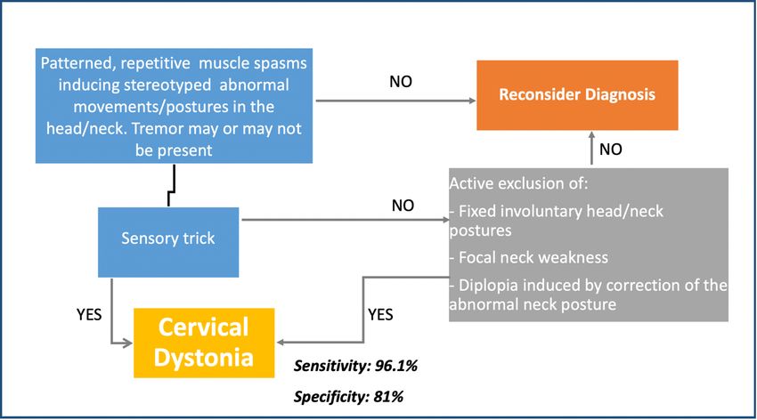
SUMMARY

According to the most recent consensus update, dystonia is defined as a condition characterized by

“sustained or intermittent muscle contractions causing abnormal, often repetitive, movements,

postures, or both. Dystonic movements are typically patterned, twisting, and may be tremulous.

Dystonia is often initiated or worsened by voluntary action and associated with overflow muscle

activation”. Prevalence of dystonia in general population is probably underestimated, mainly due to

the lack of validated diagnostic criteria for most type of dystonia, and the presence of a significant

proportion of individuals with mild symptoms who are usually not referred to neurology clinics.

Due to the lack of validated diagnostic biomarkers, the diagnosis of dystonia is based on clinical

examination and therefore may be challenging and open to bias. The factors contributing to

misdiagnosis of dystonia can be summarized in two main points: i) the huge variability in the clinical

phenomenology of dystonia; ii) the existence of a bunch of medical conditions (i.e., pseudodystonia)

mimicking the abnormal postures/movements induced by dystonia. Within this context, the most

common neurological and non-neurological imitators of dystonia are: functional dystonia; tics; head

tilt; camptocormia/scoliosis; atlanto-axial and shoulder subluxation; Arnold-Chiari malformation;

soft tissue neck mass; trigger digits; neuromuscular causes (such as myasthenia gravis etc.); spasms;

orthopedic and rheumatological causes.

       Functional dystonia is a clinical manifestation of functional motor disorder which is a

common presentation of functional neurological disorder. Functional neurological disorder is a very

common condition in clinical practice, and it is considered the second most frequent reason for a new

outpatient neurological consultation. Indeed, functional motor disorder accounts for 2-10% of

patients seen in movement disorder clinics. The diagnosis of functional motor disorder should not be

considered a diagnosis of exclusion, and it should rely on positive clinical features for which

laboratory findings may help. The two most important features that guide the clinical diagnosis of all

FMDs are: i) inconsistency (i.e., clinical features may vary over time with susceptibility to

                                                                                                    3
distraction); ii) incongruence (i.e., signs are incompatible with known determined patterns). The
diagnosis of FMD, especially for some motor symptoms (such as functional distonia), may be very
challenging.
This work is organized in two different part (Study 1 and Study 2) and the overall aim of the
work is to help clinicians to better diagnosis idiopathic dystonia and functional dystonia.
The objective of Study 1 (Sudden onset, fixed dystonia and acute peripheral trauma as
diagnostic clues for functional dystonia) is to identify clinical features suggestive of functional
dystonia to guide physicians to distinguish functional dystonia from idiopathic dystonia. For this
purpose, patient data were extracted from the Italian Registry of Functional Motor Disorders and the
Italian Registry of Adult Dystonia. Patients with functional and idiopathic dystonia were followed up
at the same clinical sites, and they were similar in age and sex. We identified 113 patients with
functional dystonia and 125 with idiopathic dystonia. Sudden onset of dystonia, evidence of fixed
dystonia, and acute peripheral trauma before dystonia onset were more frequent in the functional
dystonia group. No study variable alone achieved satisfactory sensitivity and specificity, whereas a
combination of variables yielded 85% sensitivity and 98% specificity. A diagnostic algorithm was
developed to reduce the risk of misclassifying functional dystonia. The findings of Study 1 extend
the current diagnostic approach to functional dystonia by showing that clinical information about
symptom onset, fixed dystonia, and history of peripheral trauma may provide key clues in the
diagnosis of functional dystonia.
Study 2 (Validation of a guideline to reduce variability in diagnosing cervical dystonia) was
designed to provide practical guidance for clinicians in confirming or refuting suspected cervical
dystonia, which is the most frequent type of dystonia. For this reason, participants of Study 2 were
video-recorded according to a standardized protocol to assess 6 main clinical features possibly
contributing to cervical dystonia diagnosis: presence of repetitive, patterned head/neck
movements/postures inducing head/neck deviation from neutral position (item 1); sensory trick (item
2); and red flags related to conditions mimicking dystonia that should be absent in dystonia (items 3
4to 6). Inter/intra-rater agreement among three independent raters was assessed by k statistics. To
estimate sensitivity and specificity, the gold standard was cervical dystonia diagnosis reviewed at
each site by independent senior neurologists. The validation sample included 43 idiopathic cervical
dystonia patients and 21 control subjects (6 normal subjects, 2 patients with isolated head tremor, 2
with dyskinesia/chorea, 3 with tics, 2 with head ptosis, 4 with orthopedic/rheumatologic neck
diseases, and 2 with ocular torticollis). The best combination of sensitivity and specificity was
observed considering all the items except for an item related to capability to voluntarily suppress
spasms (sensitivity: 96.1%; specificity: 81%). The findings of Study 2 show that an accurate diagnosis
of cervical dystonia can be achieved if, in addition to the core motor features, we also consider some
clinical features related to dystonia mimics that should be absent in dystonia.
In conclusion, this work sheds more light on the complex topic of the diagnosis of dystonia.
Indeed, the algorithms proposed in Study 1 and Study 2 provide a helpful tool for clinicians in their
practice. The findings of Study 1 may help in the differential diagnosis between functional dystonia
and idiopathic dystonia; and the guideline proposed in Study 2 will reduce variability in diagnosing
cervical dystonia, which was a crucial need in the field.
5TABLE OF CONTENTS
AUTHOR’S PUBBLICATIONS
ABBREVIATIONS
INTRODUCTION
DYSTONIA
Definition of dystonia
Epidemiology
Clinical features
Classification
Diagnosis of dystonia
FUNCTIONAL MOTOR DISORDERS
Definition of functional motor disorders
Epidemiology
Clinical features
Diagnosis of functional motor disorders
STUDY AIMS
Study 1
Study 2
CONCLUSIONS AND PERSPECTIVE
REFERENCES
6AUTHOR’S PUBBLICATIONS
1: Fadda L, Floris G, Polizzi L, Meleddu L, Ercoli T, Garofalo P, Saba L, Muroni A, Defazio G.
Pulvinar sign in a case of anti-CV2 encephalitis. J Neurol Sci. 2018 Oct 15;393:69-71. doi:
10.1016/j.jns.2018.08.010. Epub 2018 Aug 7. PMID: 30121006.
2: Ercoli T, Dagostino S, Pierri V, Cannas A, Solla P, Uselli S, Scapin E, Saba L, Defazio G. Internal
carotid artery dissection causing ischemic stroke during pole sport practice. J Sports Med Phys
Fitness. 2019 May;59(5):892-893. doi: 10.23736/S0022-4707.18.08904-1. Epub 2019 Feb 12.
PMID: 30758162.
3: Dagostino S, Ercoli T, Gigante AF, Pellicciari R, Fadda L, Defazio G. Sensory trick in upper limb
dystonia. Parkinsonism Relat Disord. 2019 Jun;63:221-223. doi: 10.1016/j.parkreldis.2019.01.006.
Epub 2019 Jan 6. PMID: 30655163.
4: Fadda L, Corona F, Floris G, Mascia MM, Cossa B, Ercoli T, Pau M, Defazio G. Upper limb
movements in dementia with Lewy body: a quantitative analysis. Exp Brain Res. 2019
Aug;237(8):2105-2110. doi: 10.1007/s00221-019-05575-2. Epub 2019 Jun 8. PMID: 31177296.
5: Ercoli T, Defazio G, Muroni A. Status epilepticus in Hashimoto's encephalopathy. Seizure. 2019
Aug;70:1-5. doi: 10.1016/j.seizure.2019.06.020. Epub 2019 Jun 13. PMID: 31228700.
6: Ercoli T, Defazio G, Muroni A. Cerebellar Syndrome Associated with Thyroid Disorders.
Cerebellum. 2019 Oct;18(5):932-940. doi: 10.1007/s12311-019-01059-9. PMID: 31388971.
7: Solla P, Masala C, Liscia A, Piras R, Ercoli T, Fadda L, Hummel T, Haenher A, Defazio G. Sex-
related differences in olfactory function and evaluation of possible confounding factors among
patients with Parkinson's disease. J Neurol. 2020 Jan;267(1):57-63. doi: 10.1007/s00415-019-09551-
2. Epub 2019 Sep 25. PMID: 31555978.
8: Defazio G, Fabbrini G, Erro R, Albanese A, Barone P, Zibetti M, Esposito M, Pellicciari R,
Avanzino L, Bono F, Eleopra R, Bertolasi L, Altavista MC, Cotelli MS, Ceravolo R, Scaglione C,
Bentivoglio AR, Cossu G, Coletti Moja M, Girlanda P, Misceo S, Pisani A, Mascia MM, Ercoli T,
7Tinazzi M, Maderna L, Minafra B, Magistrelli L, Romano M, Aguggia M, Tambasco N, Castagna
A, Cassano D, Berardelli A; Italian Dystonia Registry Participants. Does acute peripheral trauma
contribute to idiopathic adult-onset dystonia? Parkinsonism Relat Disord. 2020 Feb;71:40-43. doi:
10.1016/j.parkreldis.2020.01.002. Epub 2020 Jan 13. PMID: 32007783.
9: Ercoli T, Stone J. False Positive Hoover's Sign in Apraxia. Mov Disord Clin Pract. 2020 May
21;7(5):567-568. doi: 10.1002/mdc3.12970. PMID: 32626806; PMCID: PMC7328420.
10: Tinazzi M, Erro R, Mascia MM, Esposito M, Ercoli T, Ferrazzano G, Di Biasio F, Pellicciari R,
Eleopra R, Bono F, Bertolasi L, Barone P, Scaglione CLM, Pisani A, Altavista MC, Cotelli MS,
Ceravolo R, Cossu G, Zibetti M, Moja MC, Girlanda P, Maderna L, Albanese A, Petracca M,
Magistrelli L, Misceo S, Minafra B, Romano M, Squintani GM, Modugno N, Aguggia M, Cassano
D, Castagna A, Morgante F, Berardelli A, Defazio G. Demographic and clinical determinants of neck
pain in idiopathic cervical dystonia. J Neural Transm (Vienna). 2020 Oct;127(10):1435-1439. doi:
10.1007/s00702-020-02245-4. Epub 2020 Aug 26. Erratum in: J Neural Transm (Vienna). 2020 Oct
20;: PMID: 32851476.
11: Defazio G, Ercoli T, Erro R, Pellicciari R, Mascia MM, Fabbrini G, Albanese A, Lalli S, Eleopra
R, Barone P, Marchese R, Ceravolo R, Scaglione C, Liguori R, Esposito M, Bentivoglio AR,
Bertolasi L, Altavista MC, Bono F, Pisani A, Girlanda P, Berardelli A; Italian Dystonia Registry
Participants. Idiopathic Non-task-Specific Upper Limb Dystonia, a Neglected Form of Dystonia.
Mov Disord. 2020 Nov;35(11):2038-2045. doi: 10.1002/mds.28199. Epub 2020 Jul 14. PMID:
32662572.
12: Muroni A, Murru MR, Sechi M, Ercoli T, Marrosu F, Bentivoglio AR, Petracca M, Maria
Scaglione CL, Soliveri P, Cocco E, Pedron M, Murgia M, Deriu M, Cuccu S, Ulgheri L, Zuccato C,
Defazio G. Prevalence of Huntington's disease in Southern Sardinia, Italy. Parkinsonism Relat
Disord. 2020 Nov;80:54-57. doi: 10.1016/j.parkreldis.2020.09.011. Epub 2020 Sep 14. PMID:
32956974.
813: Ferrazzano G, Muroni A, Conte A, Ercoli T, Tamburini G, Fabbrini G, Berardelli A, Defazio G.
Development of a Clinical Rating Scale for the Severity of Apraxia of Eyelid Opening, Either
Isolated or Associated with Blepharospasm. Mov Disord Clin Pract. 2020 Sep 22;7(8):950-954. doi:
10.1002/mdc3.13083. PMID: 33163566; PMCID: PMC7604665.
14: Belvisi D, Pellicciari R, Fabbrini A, Costanzo M, Pietracupa S, De Lucia M, Modugno N,
Magrinelli F, Dallocchio C, Ercoli T, Terravecchia C, Nicoletti A, Solla P, Fabbrini G, Tinazzi M,
Berardelli A, Defazio G. Risk factors of Parkinson disease: Simultaneous assessment, interactions,
and etiologic subtypes. Neurology. 2020 Nov 3;95(18):e2500-e2508. doi:
10.1212/WNL.0000000000010813. Epub 2020 Sep 17. PMID: 32943485; PMCID: PMC7682833.
15: Di Lorenzo F, Ercoli T, Cuffaro L, Barbato F, Iodice F, Tedeschi G, Bombaci A; SIgN. COVID-
19 impact on neurology training program in Italy. Neurol Sci. 2021 Mar;42(3):817-823. doi:
10.1007/s10072-020-04991-5. Epub 2021 Jan 14. PMID: 33443668; PMCID: PMC7807224.
16: Tinazzi M, Geroin C, Erro R, Marcuzzo E, Cuoco S, Ceravolo R, Mazzucchi S, Pilotto A,
Padovani A, Romito LM, Eleopra R, Zappia M, Nicoletti A, Dallocchio C, Arbasino C, Bono F,
Pascarella A, Demartini B, Gambini O, Modugno N, Olivola E, Bonanni L, Antelmi E, Zanolin E,
Albanese A, Ferrazzano G, de Micco R, Lopiano L, Calandra-Buonaura G, Petracca M, Esposito M,
Pisani A, Manganotti P, Stocchi F, Coletti Moja M, Antonini A, Ercoli T, Morgante F. Functional
motor disorders associated with other neurological diseases: Beyond the boundaries of "organic"
neurology. Eur J Neurol. 2021 May;28(5):1752-1758. doi: 10.1111/ene.14674. Epub 2021 Jan 2.
PMID: 33300269.
17: Ercoli T, Erro R, Fabbrini G, Pellicciari R, Girlanda P, Terranova C, Avanzino L, Di Biasio F,
Barone P, Esposito M, De Joanna G, Eleopra R, Bono F, Manzo L, Bentivoglio AR, Petracca M,
Mascia MM, Albanese A, Castagna A, Ceravolo R, Altavista MC, Scaglione C, Magistrelli L, Zibetti
M, Bertolasi L, Coletti Moja M, Cotelli MS, Cossu G, Minafra B, Pisani A, Misceo S, Modugno N,
Romano M, Cassano D, Berardelli A, Defazio G; Italian Dystonia Registry Participants. Spread of
9segmental/multifocal idiopathic adult-onset dystonia to a third body site. Parkinsonism Relat Disord.
2021 Jun;87:70-74. doi: 10.1016/j.parkreldis.2021.04.022. Epub 2021 May 12. PMID: 33991781.
18: Solla P, Masala C, Pinna I, Ercoli T, Loy F, Orofino G, Fadda L, Defazio G. Frequency and
Determinants of Olfactory Hallucinations in Parkinson's Disease Patients. Brain Sci. 2021 Jun
24;11(7):841. doi: 10.3390/brainsci11070841. PMID: 34202903; PMCID: PMC8301996.
19: Fabbrini G, Conte A, Ferrazzano G, Esposito M, Albanese A, Pellicciari R, Di Biasio F, Bono F,
Eleopra R, Ercoli T, Altavista MC, Berardelli A, Defazio G; Italian Dystonia Registry participants.
Neuroimaging in idiopathic adult-onset focal dystonia. Neurol Sci. 2021 Jul;42(7):2947-2950. doi:
10.1007/s10072-020-05025-w. Epub 2021 Jan 3. PMID: 33389253.
20: Ercoli T, Lutzoni L, Orofino G, Muroni A, Defazio G. Functional neurological disorder after
COVID-19 vaccination. Neurol Sci. 2021 Oct;42(10):3989-3990. doi: 10.1007/s10072-021-05504-
8. Epub 2021 Jul 29. PMID: 34324120; PMCID: PMC8319586.
21: Tinazzi M, Pilotto A, Morgante F, Marcuzzo E, Cuoco S, Ceravolo R, Mazzucchi S, Padovani
A, Romito LM, Eleopra R, Nicoletti A, Dallocchio C, Arbasino C, Bono F, Magro G, Demartini B,
Gambini O, Modugno N, Olivola E, Bonanni L, Zanolin E, Albanese A, Ferrazzano G, Tessitore A,
Lopiano L, Calandra-Buonaura G, Petracca M, Esposito M, Pisani A, Manganotti P, Tesolin L,
Teatini F, Defazio G, Ercoli T, Stocchi F, Erro R, Zappia M, Geroin C. Functional gait disorders:
Demographic and clinical correlations. Parkinsonism Relat Disord. 2021 Oct;91:32-36. doi:
10.1016/j.parkreldis.2021.08.012. Epub 2021 Aug 25. PMID: 34479056.
22: Defazio G, Jinnah HA, Berardelli A, Perlmutter JS, Berkmen GK, Berman BD, Jankovic J,
Bäumer T, Comella C, Cotton AC, Ercoli T, Ferrazzano G, Fox S, Kim HJ, Moukheiber ES,
Richardson SP, Weissbach A, Wrigth LJ, Hallett M. Diagnostic criteria for blepharospasm: A
multicenter international study. Parkinsonism Relat Disord. 2021 Oct;91:109-114. doi:
10.1016/j.parkreldis.2021.09.004. Epub 2021 Sep 8. PMID: 34583301; PMCID: PMC9048224.
23: Ercoli T, Defazio G, Geroin C, Marcuzzo E, Fabbrini G, Bono F, Mechelli A, Ceravolo R,
Romito LM, Albanese A, Pisani A, Zibetti M, Altavista MC, Maderna L, Petracca M, Girlanda P,
10Mascia MM, Berardelli A, Tinazzi M; Italian Registry of Functional Motor Disorders Study Group;
Italian Registry of Adult Dystonia Study Group. Sudden Onset, Fixed Dystonia and Acute Peripheral
Trauma as Diagnostic Clues for Functional Dystonia. Mov Disord Clin Pract. 2021 Sep
10;8(7):1107-1111. doi: 10.1002/mdc3.13322. PMID: 34631946; PMCID: PMC8485608.
24: Muroni A, Murru MR, Ulgheri L, Sechi M, Ercoli T, Marrosu F, Scaglione CL, Bentivoglio AR,
Petracca M, Soliveri P, Cocco E, Cuccu S, Deriu M, Zuccato C, Defazio G. Geographic differences
in the incidence of Huntington's disease in Sardinia, Italy. Neurol Sci. 2021 Dec;42(12):5177-5181.
doi: 10.1007/s10072-021-05217-y. Epub 2021 Apr 1. PMID: 33792825.
25: Tinazzi M, Geroin C, Marcuzzo E, Cuoco S, Ceravolo R, Mazzucchi S, Pilotto A, Padovani A,
Romito LM, Eleopra R, Zappia M, Nicoletti A, Dallocchio C, Arbasino C, Bono F, Magro G,
Demartini B, Gambini O, Modugno N, Olivola E, Bonanni L, Zanolin E, Albanese A, Ferrazzano G,
De Micco R, Lopiano L, Calandra- Buonaura G, Petracca M, Esposito M, Pisani A, Manganotti P,
Tesolin L, Teatini F, Ercoli T, Morgante F, Erro R. Functional motor phenotypes: to lump or to split?
J Neurol. 2021 Dec;268(12):4737-4743. doi: 10.1007/s00415-021-10583-w. Epub 2021 May 7.
PMID: 33961091; PMCID: PMC8563631.
26: Ercoli T, Masala C, Pinna I, Orofino G, Solla P, Rocchi L, Defazio G. Qualitative smell/taste
disorders as sequelae of acute COVID-19. Neurol Sci. 2021 Dec;42(12):4921-4926. doi:
10.1007/s10072-021-05611-6. Epub 2021 Sep 23. PMID: 34557966; PMCID: PMC8459812.
27: Pierri V, Dagostino S, Vasta R, Ercoli T, Piga G, Melas V, Bruder F, Conti C, Cappai PF,
Manieli C, Melis M, Floris G, Melis M, Muroni A, Maleci A, Defazio G. Incidence and spatial
distribution of adult-onset primary malignant and other central nervous system tumors in Southern
Sardinia, Italy. Neurol Sci. 2022 Jan;43(1):419-425. doi: 10.1007/s10072-021-05747-5. Epub 2021
Nov 17. PMID: 34791565.
28: Solla P, Masala C, Ercoli T, Orofino G, Loy F, Pinna I, Fadda L, Defazio G. Olfactory
Impairment in Parkinson's Disease Patients with Tremor Dominant Subtype Compared to Those with
11Akinetic Rigid Dominant Subtype: A Pilot Study. Brain Sci. 2022 Jan 31;12(2):196. doi:
10.3390/brainsci12020196. PMID: 35203959; PMCID: PMC8869930.
29: Solla P, Ercoli T, Masala C, Orofino G, Fadda L, Corda DG, Zarbo IR, Meloni M, Sechi E,
Bagella CF, Defazio G. Rasagiline Withdrawal Syndrome in Parkinson's Disease. Brain Sci. 2022
Feb 5;12(2):219. doi: 10.3390/brainsci12020219. PMID: 35203982; PMCID: PMC8870166.
30: Borghero G, Pierri V, Vasta R, Ercoli T, Primicerio G, Pili F, Gigante AF, Rocchi L, Chiò A,
Defazio G. Incidence of amyotrophic lateral sclerosis in Sardinia, Italy: age-sex interaction and
spatial-temporal variability. Amyotroph Lateral Scler Frontotemporal Degener. 2022 Feb 21:1-7.
doi: 10.1080/21678421.2022.2041670. Epub ahead of print. PMID: 35188026.
31: Ercoli T, Masala C, Cadeddu G, Mascia MM, Orofino G, Gigante AF, Solla P, Defazio G,
Rocchi L. Does Olfactory Dysfunction Correlate with Disease Progression in Parkinson's Disease?
A Systematic Review of the Current Literature. Brain Sci. 2022 Apr 19;12(5):513. doi:
10.3390/brainsci12050513. PMID: 35624900; PMCID: PMC9139278.
32: Belvisi D, Pellicciari R, Fabbrini A, Costanzo M, Ressa G, Pietracupa S, De Lucia M, Modugno
N, Magrinelli F, Dallocchio C, Ercoli T, Nicoletti A, Zappia M, Solla P, Bologna M, Fabbrini G,
Tinazzi M, Conte A, Berardelli A, Defazio G. Relationship between risk and protective factors and
clinical features of Parkinson's disease. Parkinsonism Relat Disord. 2022 May;98:80-85. doi:
10.1016/j.parkreldis.2022.04.017. Epub 2022 Apr 29. PMID: 35526494.
33: Lidstone SC, Costa-Parke M, Robinson EJ, Ercoli T, Stone J; FMD GAP Study Group.
Functional movement disorder gender, age and phenotype study: a systematic review and individual
patient meta-analysis of 4905 cases. J Neurol Neurosurg Psychiatry. 2022 Jun;93(6):609-616. doi:
10.1136/jnnp-2021-328462. Epub 2022 Feb 25. PMID: 35217516.
34: Solla P, Masala C, Pinna I, Frau C, Ercoli T, Defazio G. Olfactory hallucinations in Parkinson's
disease patients and the role of their evaluation in clinical practice. Parkinsonism Relat Disord. 2022
Sep;102:141. doi: 10.1016/j.parkreldis.2022.07.020. Epub 2022 Aug 5. PMID: 35948466.
1235: Ercoli T, Tinazzi M, Geroin C, Marcuzzo E, Erro R, Cuoco S, Ceravolo R, Mazzucchi S, Pilotto
A, Padovani A, Romito LM, Eleopra R, Zappia M, Nicoletti A, Dallocchio C, Arbasino C, Bono F,
Spano G, Demartini B, Gambini O, Modugno N, Olivola E, Bonanni L, Albanese A, Ferrazzano G,
Tessitore A, Lopiano L, Calandra-Buonaura G, Petracca M, Morgante F, Esposito M, Pisani A,
Manganotti P, Tesolin L, Teatini F, Stocchi F, Defazio G. Do demographic and clinical features and
comorbidities affect the risk of spread to an additional body site in functional motor disorders? J
Neural Transm (Vienna). 2022 Oct;129(10):1271-1276. doi: 10.1007/s00702-022-02537-x. Epub
2022 Aug 16. PMID: 35972697; PMCID: PMC9468120.
36: Romano M, Bagnato S, Altavista MC, Avanzino L, Belvisi D, Bologna M, Bono F, Carecchio
M, Castagna A, Ceravolo R, Conte A, Cosentino G, Eleopra R, Ercoli T, Esposito M, Fabbrini G,
Ferrazzano G, Lalli S, Mascia MM, Osio M, Pellicciari R, Petrucci S, Valente EM, Valentino F,
Zappia M, Zibetti M, Girlanda P, Tinazzi M, Defazio G, Berardelli A. Diagnostic and therapeutic
recommendations in adult dystonia: a joint document by the Italian Society of Neurology, the Italian
Academy for the Study of Parkinson's Disease and Movement Disorders, and the Italian Network on
Botulinum Toxin. Neurol Sci. 2022 Oct 3. doi: 10.1007/s10072-022-06424-x. Epub ahead of print.
PMID: 36190683.
37: Frau C, Masala C, Solla P, Ercoli T, Defazio G. Association between olfactory dysfunction and
motor subtypes in Parkinson's disease: are non-tremor- dominant subtypes really uncorrelated to
olfactory impairment? Neurol Sci. 2022 Oct 11. doi: 10.1007/s10072-022-06445-6. Epub ahead of
print. PMID: 36221040.
38: Ercoli T, Barbato F, Cuffaro L, Iodice F, Romoli M, Tedeschi G, Berardelli A, Di Lorenzo F,
Bombaci A; SIgN. The future of neurology after the COVID-19 pandemic according to neurology
residents. Neurol Sci. 2022 Oct 13:1–4. doi: 10.1007/s10072-022-06450-9. Epub ahead of print.
PMID: 36227386; PMCID: PMC9559160.
1339: C. Ballini, F. Destro, P. Garofalo, J.S. Suri, T. Ercoli, A. Muroni, G. Caddeo, Y. Qi, G. Defazio,
L. Saba, Magnetic resonance imaging of Baló’s concentric sclerosis: Literature review and
presentation of two focused cases, Clin. Exp. Neuroimmunol. (2020) 1–9.
40: T. Ercoli, F.M. Genitori, G. Defazio, A. Muroni, Serum anti-thyroid antibodies in Hashimoto’s
encephalopathy: A never-ending story, Clin. Exp. Neuroimmunol. 11 (2020) 198–199.
41: Defazio, G.; Hallett, M.; Berardelli, A.; Perlmutter, J. S.; Berman, B. D.; Jankovic, J.; Bäumer,
T.; Comella, C.; Ercoli, T.; Ferrazzano, G.; Fox, S. H.; Kim, H.; Moukheiber, E. S.; Pirio Richardson,
S.; Weissbach, A.; Gigante, A. F.; Jinnah, H. A. Measurement Properties of Clinical Scales Rating
the Severity of Blepharospasm: A Multicenter Observational Study. Mov. Disord. Clin. Pract. 2022,
mdc3.13530
42: Scorr, L. M.; Cho, H. J.; Kilic-Berkmen, G.; McKay, J. L.; Hallett, M.; Klein, C.; Baumer, T.;
Berman, B. D.; Feuerstein, J. S.; Perlmutter, J. S.; Berardelli, A.; Ferrazzano, G.; Wagle-Shukla, A.;
Malaty, I. A.; Jankovic, J.; Bellows, S. T.; Barbano, R. L.; Vidailhet, M.; Roze, E.; Bonnet, C.;
Mahajan, A.; LeDoux, M. S.; Fung, V. S. C.; Chang, F. C. F.; Defazio, G.; Ercoli, T.; Factor, S.;
Wojno, T.; Jinnah, H. A. Clinical Features and Evolution of Blepharospasm: A Multicenter
International Cohort and Systematic Literature Review. Dystonia 2022, 1, 10359.
43: Mascia MM, Orofino G, Cimino P, Cadeddu G, Ercoli T, Defazio G. Writing tremor in
Parkinson's disease: frequency and associated clinical features. J Neural Transm (Vienna). 2022
44: Mostile G, Geroin C, Erro R, Luca A, Marcuzzo E, Barone P, Ceravolo R, Mazzucchi S, Pilotto
A, Padovani A, Romito LM, Eleopra R, Dallocchio C, Arbasino C, Bono F, Bruno PA, Demartini B,
Gambini O, Modugno N, Olivola E, Bonanni L, Albanese A, Ferrazzano G, De Micco R, Zibetti M,
Calandra-Buonaura G, Petracca M, Morgante F, Esposito M, Pisani A, Manganotti P, Stocchi F,
Coletti Moja M, Di Vico IA, Tesolin L, De Bertoldi F, Ercoli T, Defazio G, Zappia M, Nicoletti A
and Tinazzi M (2022) Data-driven clustering of combined Functional Motor Disorders based on the
Italian registry. Front. Neurol. 13:987593. doi: 10.3389/fneur.2022.987593
1445: Vincenzo Pierri, Giuseppe Borghero, Francesca Pili, Tommaso Ercoli, Angelo Fabio Gigante,
Luigi Isaia Lecca, Rosario Vasta, Marcello Campagna, Adriano Chiò, Giovanni Defazio (2022):
Impact of occupational categories on the incidence of amyotrophic lateral sclerosis in Sardinia Island,
Italy, Amyotrophic Lateral Sclerosis and Frontotemporal Degeneration,
DOI:10.1080/21678421.2022.2153606
46: Defazio G, Gigante AF, Hallett M, Berardelli A, Perlmutter JS, Berman BD, Jankovic J, Bäumer
T, Comella C, Ercoli T, Ferrazzano G, Fox SH, Kim HJ, Moukheiber ES, Richardson SP, Weissbach
A, Jinnah HA. Motor and psychiatric features in idiopathic blepharospasm: A data-driven cluster
analysis. Parkinsonism Relat Disord. 2022 Nov;104:94-98. doi: 10.1016/j.parkreldis.2022.10.008.
Epub 2022 Oct 13. PMID: 36306537.
15ABBREVIATIONS
BoNT: Botulinum neurotoxin
BSP: Blepharospasm
CD: Cervical dystonia
FDYT: Functional dystonia
FMD: Functional motor disorder
FND: Functional neurological disorder
IDYT: Idiopathic dystonia
IRAD: Italian registry of adult dystonia
IRFMD: Italian registry of functional motor disorders
LD: Laryngeal dystonia
ULD: Upper limb dystonia
OMD: Oromandibular dystonia
WC: Writer’s cramp
16INTRODUCTION
DYSTONIA
Definition of dystonia
The term dystonia was first introduced by Oppenheim in 1911 in the form “dystonia musculorum
deformans”.1 Previous medical description of dystonia was made by Gowers, Destarac, and Schwalbe
who referred to the condition as “tetanoid chorea”, “torticolis spasmodique”, and “tonic cramps”
respectively.2–4 Interestingly, one the most famous artistic description of torcicollis was made by
Dante Aligheri in “La Divina Commedia (Inferno, Canto XX)”:
“Come ’l viso mi scese in lor più basso,
mirabilmente apparve esser travolto
ciascun tra ’l mento e ’l principio del casso;
ché da le reni era tornato ’l volto,
e in dietro venir li convenia,
perché ’l veder dinanzi era lor tolto.”
After Meige the term “dystonia” has been extensively used in the neurological studies in the
form “torsion dystonia”. Until the early 1980s, dystonia was considered a condition between organic
disorder and psychiatric condition. David Marsden was the first neurologist to postulate that dystonia
was a disorder related to the basal ganglia functioning. Marsden described dystonia as a syndrome
characterized by sustained involuntary muscle contraction, that frequently causes twisting or
17repetitive movements or abnormal postures.5,6 He also highlighted that dystonia would be considered
an involuntary movement. Marsden’s theory is a keystone for the understanding of dystonia, and the
recent definitions of dystonia were based on his concept.
Indeed, according to the recent and current definition “dystonia is a movement disorder
characterized by sustained or intermittent muscle contractions causing abnormal, often repetitive,
movements, postures, or both. Dystonic movements are typically patterned, twisting, and may be
tremulous. Dystonia is often initiated or worsened by voluntary action and associated with overflow
muscle activation.” 7
Epidemiology
Prevalence of dystonia in general population is probably underestimated, and it is not still clear if
dystonia should be considered a rare condition. Findings from different epidemiological studies had
a huge variability probably due to differences in methods and analysis.8
Overall, 27 studies assessed the prevalence of adult-onset dystonia: 2 population-based studies
showed a high prevalence; 5 reported a prevalence ranging from 40 to 70 cases per million
individuals; and the remain 20 estimated a prevalence ranging from 3 to 37 cases per million.9 Within
this context, a recent viewpoint has urged the necessity to really clarify the epidemiology of dystonia
in order to better assess risk and protective factors, neurological changes over time, and disease-
modifying treatment.10
Another important reason related to the great variability amongst epidemiological studies, is
the lack of validated diagnostic criteria for most type of dystonia, and the presence of a significant
proportion of individuals with mild symptoms who are usually not referred to neurology clinics.
With particular reference to adult-onset dystonia, focal presentation is more frequent than
generalized dystonia, as well as woman are more affected than men. Among focal dystonia, cervical
dystonia (CD) is the most frequent type, followed by blepharospasm (BSP). For adult-onset focal
18forms the genetic causes are rare, however, a positive family history has been described in about a
fifth of the patients.8
Clinical features
The main motor features of dystonia include dystonic postures and dystonic movements, which are
two physical signs that can be recognized during neurological examination. Dystonic postures
indicate that muscle contractions may be continuous (usually associated with a co-contraction of
agonist and antagonist muscles), forcing the involved body part into sustained postures (i.e., body
site is flexed or twisted along its longitudinal axis).7,11,12 Dystonic movements may be present at rest
and they are generally repetitive and patterned or twisting. Fatigue and stress may often worsen the
phenomenology of the movements, as well as the simultaneous presence of a voluntary
movement.13,14 A remarkable feature of dystonia, especially upper limb dystonia (ULD), is the task
specificity, meaning that the disorder appears only during the execution of specific motor activities
(e.g., writer’s cramp [WC]).15–17 Neurologists should recognize other clinical signs that are usually
reported in patients with dystonia:
i) Tremor: may appear as isolated tremor and is a feature of dystonic movements. This rhythmic
movement is often worsened by attempts to keep the primary posture.8,9
ii) Motor overflow: diffusion of dystonic movements or posture to an anatomically distinct body
region contiguous to those primarily affected.8,9
iii) Geste antagoniste or sensory trick: specific voluntary movements that relieve or temporarily
abolish dystonia. These manoeuvres involve the affected body site and are usually graceful and
natural (e.g., touching specific parts of the face, cheek, chin in CD; or shifting pen holding, and
writing with a closed fist in WC). Some individuals may also manifest multiple sensory tricks and
the phenomenology of the geste may vary from person-to-person.18,19
19iv) Mirroring: involuntary unilateral posture or movements (with the same or similar features of
patient’s dystonia) involving the contralateral upper or lower limbs that can be elicited during the
activation of the body part affected by dystonia.8,9
Dystonia is “isolated” when it is the only motor symptom, whereas it is called “combined”
when additional movement disorders occur (e.g., myoclonus dystonia or dystonia parkinsonism).7
Since the recognition of dystonia is mainly based on the clinical observation of the aforementioned
physical signs, the diagnosis can be difficult when the dystonic movements occur in isolation or in a
non-complete pattern. This is the reason why the diagnosis of dystonia can be missed or delayed very
frequently.
Classification
A recent classification of dystonia have been proposed in order guide clinician in patients’
assessment.7 The panel of expert identify two distinct axes (Figure 1):
i) Axis I shows clinical features (e.g. age at onset, body distribution, temporal pattern,
cooccurrence of other movement disorders or of other neurological manifestations) and
provides information at the time od examination.
ii) Axis II is focused on etiology and examines identifiable anatomical changes and pattern
of inheritance.
20Figure 1. Dystonia classification based on the hierarchical organization of the two axes (from
Albanese et al. 2018).
21At this point, some aspects of the classification should be highlighted.7 Indeed, according to
Axis I, five different age groups are identified for age at dystonia onset: infancy (birth to 2 years),
childhood (3-12 years), adolescence (13-20 years), early adulthood (21-40 years) and late adulthood
(>40 years). Following the definition provided by the experts, the body distribution of dystonia may
be focal (only one body region is affected), segmental (two or more contiguous body regions are
affected), multifocal (two noncontiguous or more body regions are involved), generalized (the trunk
and at least two other sites are involved) or hemidystonia (more body regions restricted to one body
side are involved). Interestingly body distribution can change over time and dystonia my spread from
the original body site to another one.15,20–22 As far as the etiology is concerned, Axis II provides
helpful indications. Anatomical change and pattern of inheritance should be used together for
etiological classification. Exploring the nervous system pathology may lead to the observation of
degeneration (namely a progressive structural abnormality); static lesions; or the absence of
degeneration/structural lesion. Moreover, dystonia may be i) inherited (dystonia forms of proven
genetic origin), ii) acquired (dystonia due to a known specific cause); iii) idiopathic (unknown cause).
Noteworthy several cases of focal or segmental isolated dystonia with onset in adulthood fall in the
latter group.7
Diagnosis of dystonia
Due to the lack of validated diagnostic biomarkers, the diagnosis of dystonia is based on clinical
examination and therefore may be challenging and open to bias.23 This is the reason why many
patients experienced numerous medical visits, delaying access to treatment.24 It has been showed that
experienced neurologists in movement disorders can diagnose dystonia with greater accuracy than
general neurologists.25
22The factors contributing to misdiagnosis of dystonia can be summarized in two main points:
i) the huge variability in the clinical phenomenology of dystonia; ii) the existence of a bunch of
medical conditions (i.e., pseudodystonia) mimicking the abnormal postures/movements induced by
dystonia. Within this context, the most common neurological and non-neurological imitators of
dystonia are: functional dystonia (FDYT); tics; head tilt; camptocormia/scoliosis; atlanto-axial and
shoulder subluxation; Arnold-Chiari malformation; soft tissue neck mass; trigger digits;
neuromuscular causes (such as myasthenia gravis etc.); spasms; orthopedic and rheumatological
causes.7
To date diagnostic guidelines have been proposed just for BSP26,27 and laryngeal dystonia
(LD)28, whereas the lack of diagnostic guidelines for the other types is unmet need in the field.
Recently a group of Italian Movement Disorder experts provided clinical diagnostic
recommendations for CD, oromandibular (OMD), and limb dystonia.23 These recommendation serve
as basis for future validated diagnostic guidelines.
23FUNCTIONAL MOTOR DISORDERS
Definition of functional motor disorders
Functional motor disorder (FMD) is a common presentation of functional neurological disorder
(FND), presenting with diverse phenotypes such as tremor, dystonia, tics, weakness and gait
disorder.29,30 According to a recent review published in Lancet Neurology, the authors “define the
term FND to denote clinical syndromes consisting of symptoms and signs of genuinely experienced
alterations in motor, sensory, or cognitive performance, which are distressing or impairing, and
manifest one or more patterns of deficits that are consistent predominantly with dysfunction of the
nervous system, and show variability in performance within the same task and between different
tasks.”29 FND is a very common condition in clinical practice and it is considered the second most
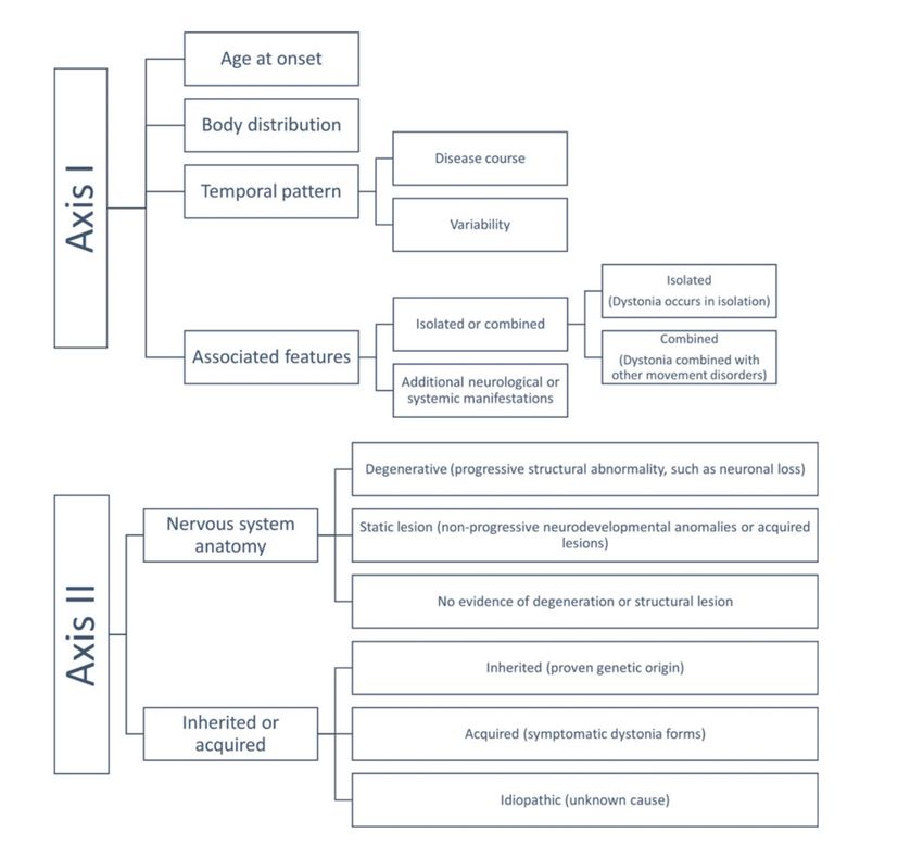
frequent reason for a new outpatient neurological consultation.31 The etiology of FND is
multifactorial and it has postulated that several factors may play an important role in the development
of the disorder (Figure 2).
24Figure 2. Multifactorial model explaining the complexity of FND (from Hallet et al. 2022)
25Epidemiology
Although the lack of focused studies on the topic, FND seems to have similar prevalence across
geographical areas.29 FMD accounts for 2-10% of patients seen in movement disorder clinics.32–34
Basic epidemiological features of FMD have been elucidated only recently by a large international
study, called GAP study (Gender, Age and Phenotype study), confirming female preponderance
(72.6%) and mean age at FMD onset of 39.6 + 16.1 years.30 Interestingly, men had a significantly
later age of onset than women (41.0 years vs 39.1 years).30
Clinical features
FMDs manifest with involuntary various symptoms of altered movement (such as tremor, dystonia,
weakness, gait disorders). The GAP study also described a clearer picture of FMD phenotype
frequency: mixed FMD (23%), tremor (22%), weakness (18%), dystonia (12%), gait disorder (8%),
myoclonus/jerks (5%), and parkinsonism (2%).30 Compared the mean age of FMD onset, patients
with gait disorders had a significantly later age at onset than tremor, while the mean ages at onset of
weakness and dystonia were significantly younger.30 Common features of FMDs included sudden
onset of symptoms (71%), anxiety (52%), fatigue (45%), and pain (42%).35 About 22% of FMD
patients experienced neurological and non-neurological diseases, highlighting the important overlap
among various conditions.36 Moreover, the disorder may start in a single body site in about half of
patients and then spread to additional body sites in 15–20% of cases.37
26Diagnosis of functional motor disorders
Diagnosis by the text revision of DSM-5 (Diagnostic and Statistical Manual of Mental Disorders) no
longer requires the presence of precipitating stressors, and FND is referred to as functional
neurological symptom disorder (conversion disorder).29 The diagnosis of FMD should not be
considered a diagnosis of exclusion, and it should rely on positive clinical features for which
laboratory findings may help.38 According to Gupta-Lang criteria,39 the two most important features
that guide the clinical diagnosis of all FMDs are: i) inconsistency (i.e., clinical features may vary over
time with susceptibility to distraction); ii) incongruence (i.e., signs are incompatible with known
determined patterns). The diagnosis is often supported by positive clinical signs, such as the Hoover’s
sign for functional leg weakness, and the entrainment test for functional tremor.38 Within this context,
the examination can establish a “rule-in” (positive) rather than a “rule-out” (exclusionary) diagnosis
for FMDs. The diagnosis of FMD, especially for some motor symptoms (such as FDYT), may be
very challenging. However, clinicians should bear in mind that missing the diagnosis of FMD could
be more frequent than making an erroneous diagnosis of FMD.40
27STUDY AIMS
This work is organized in two different part (Study 1 and Study 2) and the overall aim of the work is
to help clinicians to better diagnosis idiopathic dystonia (IDYT) and FDYT.
As the title suggests “Sudden onset, fixed dystonia and acute peripheral trauma as diagnostic
clues for functional dystonia”, Study 1 is focused on the complex differential diagnosis between
IDYT and FDYT. Indeed, the objective of the study is to identify clinical features suggestive of FYDT
to guide physicians to distinguish FDYT from IDYT. The study has been already published in an
international scientific journal specialized in movement disorders (Movement Disorders Clinical
Practice, Volume 8, Issue 7, October 2021, Pages 1107-1111).41 The study received the “BRONZE
award” for the Best Research Article in Movement Disorders Clinical Practice, 2021-2022.
Study 2 “Validation of a guideline to reduce variability in diagnosing cervical dystonia” was
designed to provide practical guidance for clinicians in confirming or refuting suspected CD, which
is the most frequent type of dystonia. To date CD diagnosis is based on clinical examination and is
therefore subjective, so a diagnostic test is still an unmet need. The final version of the study was
submitted to an international scientific journal.
28STUDY 1
Sudden onset, fixed dystonia and acute peripheral trauma as diagnostic clues for
functional dystonia41
Inconsistency (i.e., changing patterns over time with susceptibility to distraction) and/or
incongruence (i.e., a clinical picture incompatible with known determined patterns) are clinical
features of neurological examination that support clinically definite diagnosis of FDYT according
with the most recent set of diagnostic criteria proposed by Gupta and Lang.39
Demonstrating inconsistency/incongruence may be clinically challenging, however,42,43 and
straightforward laboratory-supported criteria for most forms of dystonia are lacking.44 The only
reliable neurophysiological discriminator between FDYT and IDYT proposed to date has been for
blepharospasm.45
Medical history and clinical features that may reveal some clues to the diagnosis of FDYT
include sudden symptom onset, evidence of fixed movement disorder, history of physical trauma,
psychiatric diseases, and comorbid functional somatic disorders. Their validity in supporting a
diagnosis of FDYT remains to be fully established. For this study, we compared the frequency of
sudden symptom onset, evidence of fixed dystonia, and prior acute peripheral trauma in patients with
adult-onset FDYT and IDYT. We also assessed their sensitivity and specificity either alone or
combined. We did not assess psychiatric diseases and comorbid functional somatic disorders because
of their high frequency in both FDYT and IDYT.46
29Methods
The study relied on information from the Italian Registry of Functional Motor Disorders (IRFMD)
and the Italian Registry of Adult Dystonia (IRAD), two multicenter initiatives coordinated by the
Italian Academy for the Study of Parkinson’s Disease and Other Movement Disorders (Accademia
LIMPE-DISMOV RADAC project) and Fondazione LIMPE. Patients in the IRFMD were referred
from 25 Italian centers for movement disorders with a diagnosis of clinically definite FMDs based
on Gupta and Lang’s diagnostic criteria.39 The IRAD includes patients with adult-onset dystonia from
37 secondary/tertiary referral centers for movement disorders throughout Italy. Diagnosis was made
according to published criteria.7,26 Core assessment characterizing IRFMD and IRAD has been
described in detail in other studies35,47; it comprises demographic, historical, and clinical information
on the movement disorder and possible predisposing/precipitating factors.36,48,49 Information was
collected about dystonia at different body sites (upper and lower limbs, trunk, cervical, cranial among
others), year of dystonia onset, and prior peripheral injury (at extracranial body sites). Onset of
dystonia can be defined as acute (abrupt with deterioration within a few days or weeks) or slowly
progressing.35 Peripheral injury had to be severe enough to require medical attention, hospitalization,
or surgery. Information about trauma included year and site of the injury. Dystonia was defined as
fixed when immobile dystonic postures did not return to the neutral position at rest.50 Patients with
FDYT from the IRFMD were frequency matched by age and sex with patients with IDYT from the
IRAD who were followed up at the same clinics. To include only patients who were idiopathic, tests
for Wilson’s disease, dopa-responsive dystonia, and common genetic variants (e.g., TOR1A, THAP1)
were performed as appropriate. Patients screening positive on genetic testing were not included in the
study sample.15
Statistical analysis was performed using the Stata 11 package (StataCorp, College Station,
TX) and descriptive and inferential statistics as appropriate (t test, chi-square test, Fisher’s exact test).
Data are expressed as mean + standard deviation unless otherwise indicated. Logistic regression
30models for unequal case-control ratios were computed to assess the association between history of
trauma and case-control status after adjusting for potentially confounding variables. Statistical
significance was set at 0.05. To assess sensitivity and specificity, the gold standard was the diagnosis
of FDYT or IDYT made at each site. Sensitivity was defined as the proportion of patients who
screened positive among those given a diagnosis of FDYT (true positives/true positives + false
negatives). Specificity was the proportion of patients who screened negative among those given a
diagnosis of IDYT (true negatives/false positives + true negatives).
Results
In December 2020, the data on 113 patients with FDYT were extracted from the IRFMD, which
included data from 410 patients with functional movement disorders,36,51 and 125 patients with IDYT
selected among the 1634 patients from the IRAD.22 The two groups were similar for sex, age, and
educational level but differed for disease duration, dystonia distribution, and frequency of focal
dystonia, which was more frequent in the patients with IDYT (Table 1).
31Table 1. Clinical and demographic features of patients with functional and idiopathic dystonia.
Patients with Patients with P-value
functional idiopathic
dystonia (n=113) dystonia
(n=125)
Women, no. (%) 87 (77) 94 (75) 0.4
Mean age, yrs. 47.2 + 14.4 49.4 + 10.0 0.2
Mean years of schooling 12.5 + 4.1 12.3 + 3.5 0.7
Mean age (years) at onset + SD 41.3 + 14.1 40.2 + 10.6 0.3
Mean years of disease duration 5.9 + 6.2 9.5 + 8.0 0.0002
Focal dystonia, no. (%) 67 (59.3) 104 (83.2)Limb/trunk dystonia was more frequent in the FDYT group, and cranial-cervical dystonia was
more frequent in the IDYT group (Table 1). Sudden onset of dystonia, evidence of fixed dystonia,
and acute peripheral trauma before dystonia onset were more frequent in the FDYT group (Table 1).
This finding was confirmed after limiting the analysis to trauma to the dystonic body part (Table 1)
and adjusting for disease duration, age, and sex on logistic regression analysis (adjusted odds ratio,
5.8; 95% confidence interval, 1.6–20.9; P = 0.007).
No study variable alone reached a satisfactory combination of sensitivity/specificity (sudden
onset of dystonia: sensitivity, 65% [73/113] and specificity, 100% [125/125]; fixed dystonia:
sensitivity, 50% [56/113] and specificity, 100% [125/125]; prior trauma: sensitivity, 15% [17/113]
and specificity, 98% [122/125]). However, screening positive to at least 1 of the 3 variables yielded
85% sensitivity (96/113) and 98% specificity (122/125) (Figure 3).
33Figure 3. Diagnosing functional dystonia.
34Discussion
Our findings indicate that sudden dystonia onset and fixed dystonia are more likely to occur in FDYT
and that acute peripheral trauma may be significantly associated with FDYT.43,50 Novel findings
indicate that each variable differentiated FDYT from IDYT with 15% to 65% sensitivity, but none of
these clinical features alone was crucial for diagnosing FDYT. Nonetheless, each of the 3 variables
carried a negligible risk of misclassifying FDYT cases and reached 98% to 100% specificity.
Although no study variable alone achieved satisfactory sensitivity and specificity, screening positive
to at least 1 of the 3 clinical features can correctly diagnose FDYT in more than 8/10 patients who
have the condition (85% sensitivity) and can correctly identify as not having FDYT about 10/10
subjects not affected by the condition (98% specificity). The sensitivity and specificity levels were
shared by those reported for a recently published decision tree that classified FDYT using a complex
case-finding procedure based on the serial application of about seven historical/clinical features and
diagnostic confirmation subsequently informed by recognition of incongruence/inconsistence on
neurological examination.52 At variance with such an approach, however, our algorithm is
considerably simpler and based on the combination of only three historical/clinical features.
Our study has several strengths. First, the populations of FDYT and IDYT were from
multicenter settings and probably representative of the general population of cases with similar
demographic/clinical features.35,47 The older age at onset of patients with FDYT compared with the
other reported cohorts probably reflects the inclusion of only adult-onset patients in the source
registries. The frequency of sudden onset and fixed dystonia in our sample was consistent with
previous series, whereas the trauma frequency was lower.50 Unlike other studies, however, we limited
recall bias by not including patients with mild trauma. Physical injury was more commonly recorded
preceding weakness than dystonia.53 Second, the standard for comparison was dystonia status based
on clinical examination by neurologists applying stringent diagnostic criteria. In addition, both groups
of patients with FDYT and IDYT were followed up by the same neurologists at the same center,
35which provided accuracy in data collection. The low frequency of peripheral trauma to a specific
body part in the IDYT group was consistent with the observations of several large controlled studies
and demonstrated a negligible effect of peripheral trauma on topographically related IDYT.54,55
The present study also has some limitations. Patients participating in the study were diagnosed
with clinically definite FMDs according to the Gupta and Lang criteria.39 hese criteria are largely
based on incongruence/inconsistency that may incorporate some of the issues we studied such as
sudden onset and fixed dystonia. This may lead to the “circular argument” of diagnosing FMDs with
new sets of criteria based on existing criteria. However, our aim was to measure the accuracy of these
3 simple aids alone in recognizing FDYT diagnosed according with the current gold standard, that is,
clinical diagnosis established by expert neurologists who relied on several additional aspects of
incongruence/inconsistency and positive clinical signs.19,38,56 Moreover, because the patients and the
physicians involved in the study were from the same country, data from other populations of patients
and movement disorder specialists are needed to confirm the present results. Also, because our study
focused on dystonia, not all findings may be extensible to other functional movement disorders.
Disease duration was significantly lower in the FDYT group, even though the estimated association
between FDYT and trauma did not change after adjusting for disease duration. Body distribution of
dystonia differed in the FDYT and the IDYT groups, which probably reflects the frequency
distribution of dystonia in the general population of adult-onset cases with functional and idiopathic
dystonia. Although we did not match patients with FDYT and IDYT by distribution of dystonia, we
were confident that our study variables, in particular sudden onset of dystonia and evidence of fixed
dystonia, were probably independent of the body localization of dystonia. Nevertheless, we
acknowledge that the higher frequency of limb dystonia in the FDYT group may limit the
generalizability of findings to all forms of FDYT.
In conclusion, our findings extend the current diagnostic approach to FDYT by showing that
clinical information about symptom onset, fixed dystonia, and previous peripheral trauma may
provide key clues for diagnosing FDYT in addition to incongruence/inconsistence. In this context,
36this large cohort corroborates the existing knowledge and presents sensitivity and specificity figures
for a few historical/clinical features that can aid clinicians to establish a positive diagnosis for FDYT.
37STUDY 2
Validation of a guideline to reduce variability in diagnosing cervical dystonia
Cervical dystonia, the most frequent form of focal dystonia, is characterized by a variable pattern of
neck muscle involvement, leading to clinically heterogeneous directional presentations, such as
torticollis, laterocollis, retrocollis, or anterocollis.57 Patients may also have additional signs and
symptoms, including shoulder elevation, neck/shoulder pain, or head tremor, and may benefit from
the use of sensory tricks.18,49,58
Due to the lack of a diagnostic test, CD diagnosis is based on clinical examination and is
therefore subjective.59 As an example, a study on CD incidence in northern California found that up
to 65% of patients may be incorrectly diagnosed prior to receiving a correct diagnosis.60 Diagnostic
errors may largely be due to the clinical variability of CD but also to the existence of several related
conditions, for example, pseudodystonia mimicking the abnormal movements or postures of CD.61,62
In the case of CD, dystonia mimics may include head tremor;63 neck chorea producing nonrepetitive
head movements;64 neck tics associated with ability to mentally suppress the spasms;65 orthopedic
neck diseases (like atlanto-axial and shoulder subluxation, or fracture of the cervical vertebrae),
rheumatologic neck diseases, and posterior fossa tumors, all leading to tonic postures or movement
of the head;66 lower motor neuron disease/myopathy/myasthenia gravis inducing weakness of the
neck muscles opposite to the abnormal posture;67,68 and ocular torticollis characterized by diplopia
caused by the voluntary correction of the abnormal neck posture.69
38Methods
Participants were identified from among outpatients attending the movement disorder clinic of the
University of Cagliari and Sapienza University of Rome. Inclusion criteria for both case and control
subjects were age 18 or older, any sex, and the willingness and mental/physical ability to sign
informed consent and participate in the protocol. Case patients were enrolled if they had a diagnosis
of focal idiopathic CD made by an experienced movement disorder neurologist.23 Exclusion criteria
were secondary CD and co-existing medical conditions/surgical interventions that could confound
assessment of CD. Botulinum neurotoxin (BoNT) treatment was performed at least 12 weeks before
the examination. The control group included normal subjects and a group of patients with head/neck
impairment that could be confused with CD,62 i.e., isolated head tremor; nonrepetitive head
movements due to chorea; head tics associated with the ability to mentally suppress spasms; fixed
involuntary neck postures due to orthopedic neck diseases (like atlanto-axial and shoulder subluxation
or cervical vertebrae fracture), rheumatologic neck diseases, or posterior fossa tumors; focal
weakness of the neck muscles opposite the side of abnormal posture due to lower motor neuron
disease/myopathy/myasthenia gravis; and diplopia caused by the voluntary correction of abnormal
neck posture due to ocular torticollis. To assess diagnostic accuracy we focused on the following
clinical items: (i) presence of repetitive, patterned head/neck movements/postures inducing head/neck
deviation from neutral position (item 1, derived from the 2013 revised definition of dystonia);7 (ii)
sensory trick (item 2); and (iii) red flags related to conditions mimicking dystonia that would be
expected to be absent in dystonia (items 3 to 6). In the latter group, we took into account fixed
head/neck deviation from neutral position (item 3, a feature distinguishing dystonia from orthopedic
or rheumatologic diseases inducing fixed postures); focal weakness of neck muscles antagonizing the
abnormal head/neck posture (item 4, a feature that may prove useful to differentiate lower motor
neuron diseases/myopathy from dystonia); diplopia induced by voluntary correction of the abnormal
head/neck posture (item 5, a feature that may distinguish CD from ocular torticollis); and ability to
39voluntarily suppress spasms defined as an inner volitional effort rather than voluntary compensatory
frontalis muscle overactivity (item 6, a feature that is potentially useful to distinguish dystonia and
tics). Attention was paid to distinguish suppressibility by willpower alone from compensatory
movements that often counteract dystonic movements or postures and are also the result of voluntary
action. There was no duration requirement for voluntary suppression. Participants were video
recorded according to a standardized protocol in order to assess all the major/distinctive clinical
features possibly contributing to CD diagnosis. The video protocol included standard maneuvers
triggering involuntary head movements, sensory trick if present, and the strength of neck muscles
under voluntary contraction. Subjects were also asked by the examiner about: (i) occurrence of
diplopia induced by voluntary correction of the abnormal head/neck posture, and (ii) capability to
voluntarily suppress involuntary neck movements. Inter/intra-rater agreement was assessed among
three independent raters who did not belong to the centers participating in the project. The number of
videos included in the reliability study (64 video recordings of 43 CD patients, 6 normal controls, and
15 disease controls) exceeded that based on recommended subject-to-item ratios (which usually
consider the assessment of 5-10 subjects for each item of a new scale) and on the number of items (n.
4) to be assessed by the three observers. Item 5 (diplopia induced by voluntary correction of the
abnormal head/neck posture) and item 6 (inability to voluntarily suppress spasms) were not included
in the reliability analysis because questions about these items were asked by the site examiner but not
captured in the video. Agreement among raters was assessed by k index, which measures the level of
agreement beyond chance and ranges from -1 (perfect disagreement) to +1 (perfect agreement). A k
index >0.4 (indicating moderate to substantial/almost perfect agreement) was considered to be
satisfactory. To estimate sensitivity and specificity, the gold standard was the diagnosis made at each
site by the senior neurologists. Sensitivity was defined as the proportion of subjects who screened
positive from among those who had a diagnosis of CD on clinical examination (true positives/true
positives + false negatives). Specificity was the proportion of subjects who screened negative from
40You can also read