Platelet-Rich Plasma-Derived Exosomal USP15 Promotes Cutaneous Wound Healing via Deubiquitinating EIF4A1 - Hindawi.com
←
→
Page content transcription
If your browser does not render page correctly, please read the page content below
Hindawi
Oxidative Medicine and Cellular Longevity
Volume 2021, Article ID 9674809, 14 pages
https://doi.org/10.1155/2021/9674809

Research Article
Platelet-Rich Plasma-Derived Exosomal USP15 Promotes
Cutaneous Wound Healing via Deubiquitinating EIF4A1

 Yan Xu ,1 Ze Lin ,1 Lei He ,1 Yanzhen Qu ,1 Liu Ouyang,1 Yu Han ,2 Chao Xu ,3
 and Deyu Duan 1
 1
 Department of Orthopaedics, Union Hospital, Tongji Medical College, Huazhong University of Science and Technology,
 Wuhan 430022, China
 2
 Department of Orthopaedic Surgery, Shanghai Key Laboratory of Orthopaedic Implants, Shanghai Ninth People’s Hospital,
 Shanghai Jiaotong University School of Medicine, Shanghai 200011, China
 3
 College of Life Science and Technology, Huazhong University of Science and Technology, China

 Correspondence should be addressed to Chao Xu; xuchao@hust.edu.cn and Deyu Duan; duandeyu@21cn.com

 Received 8 June 2021; Revised 4 July 2021; Accepted 23 July 2021; Published 10 August 2021

 Academic Editor: Alin Ciobica

 Copyright © 2021 Yan Xu et al. This is an open access article distributed under the Creative Commons Attribution License, which
 permits unrestricted use, distribution, and reproduction in any medium, provided the original work is properly cited.

 Epithelial regeneration is an essential wound healing process, and recent work suggests that different types of exosomes (Exos) can
 improve wound repair outcomes by promoting such epithelial regeneration. Platelet-rich plasma (PRP) is known to facilitate
 enhanced wound healing, yet the mechanisms underlying its activity are poorly understood. To explore these mechanisms, we
 first isolated PRP-derived Exos (PRP-Exos). Using immortalized keratinocytes (HaCaT cells) treated with PBS, PRP, or PRP-
 Exos, we conducted a series of in vitro Cell Counting Kit-8 (CCK-8), EdU, scratch wound, and transwell assays. We then
 established a wound defect model in vivo in mice and assessed differences in the mRNA expression within these wounds to
 better understand the basis for PRP-mediated wound healing. The functions of PRP-Exos and USP15 in the context of wound
 healing were then confirmed through additional in vitro and in vivo experiments. We found that PRP-Exos effectively promoted
 the in vitro proliferation, migration, and wound healing activity of HaCaT cells. USP15 was further identified as a key mediator
 through which these PRP-Exos were able to promote tissue repair both in vitro and in vivo. At a mechanistic level, USP15
 enhanced the functional properties of HaCaT cells by promoting EIF4A1 deubiquitination. Thus, PRP-Exos and USP15
 represent promising tools that can promote wound healing via enhancing epithelial regeneration.

1. Introduction be essential mediators of normal wound healing processes
 [5, 6]. Targeted efforts to accelerate reepithelialization in
Chronic wounds are defined as wounds that do not heal the context of wound repair thus represent a key area of
appropriately and that persist for three months or longer ongoing scholarly research.
[1, 2]. Such wounds most commonly arise in the context of Platelet-rich plasma (PRP) contains a diverse array of
diabetes, arterial ischemia, poor venous return, infections, physiologically important growth factors at high concentra-
pressure ulcers, or malignant tumors, resulting in pain for tions, including transforming growth factor-β (TGF-β), epi-
the affected patient while also compromising the integrity dermal growth factor (EGF), platelet-derived growth factor
of barriers that are essential for the prevention of bacterial (PDGF), and vascular endothelial growth factor (VEGF) [7,
entry into the human body [3]. As such, chronic wounds 8]. Some of these growth factors are closely related to the for-
can reduce patient quality of life while also imposing a signif- mation of blood vessels, which is an integral process in the
icant economic burden on their families and on society as a context of tissue regeneration. Indeed, VEGF-producing
whole [4]. There is still a lack of any standard treatments Schwann cells have been reported to accelerate peripheral
for chronic wounds, although keratinocytes are known to nerve repair [9]. As such, PRP has shown great promise as
2 Oxidative Medicine and Cellular Longevity

 100 nm
 Acc. voltage = 100.0 kV 100 nm
 Magnification = x60.0 k

 (a)

 PRP-Exos

 Control
 80 Calnexin 100 kDa

 60
 70 kDa
 TSG101
 Exents

 55 kDa
 40

 20
 CD9 25 kDa
 0
 30 40 60 80 100 120 150
 Size (nm)
 (b) (c)

Figure 1: PRP-Exo characterization: (a) TEM images of PRP-Exos; (b) PRP-Exo size distributions, as measured via DLS; (c) Western blotting
analyses of proteins within PRP-Exos.

an applied treatment capable of mediating various forms of 2. Materials and Methods
tissue regeneration. The therapeutic application of PRP has
been linked to accelerated angiogenesis and reepithelializa- 2.1. Cell Culture. HaCaT cells were purchased from the
tion, thereby promoting expedited wound healing [10]. The China Center for Type Culture Collection, Wuhan, China,
mechanisms whereby PRP can promote such regeneration, and were cultured in DMEM containing 10%FBS in a 5%
however, are unclear. In addition to the abovementioned CO2 incubator at 37°C. Lipofectamine was used to trans-
growth factors, platelets can produce a variety of different fect cells with siRNA constructs (50 μmol/L; GenePharma,
extracellular vesicles, including exosomes [11], which are Shanghai, China).
small vesicles that are approximately 100 nm in diameter
(range: 40–160 nm). These vesicles have been a focus of 2.2. PRP Preparation. Mice (n = 12) were anesthetized via
intensive research interest in recent years owing to their intraperitoneal injection with 1% sodium pentobarbital, after
essential role as mediators of cell-cell communication, shut- which blood was collected from the abdominal vena cava into
tling nucleic acids, proteins, metabolites, and other macro- a 1 mL syringe containing 0.1 mL of anticoagulant. The col-
molecules between cells such that they hold promise as lected blood was transferred into a fresh 1.5 mL tube and
tools for treating a range of diseases [12–15]. Owing to the stored at 4°C until the collection was completed, at which
unique biological properties of PRP and of exosomes, further time samples were spun for 10 min at 100 ×g at 4°C. The
research is warranted to establish the ability of PRP-derived supernatant and the layer containing the white blood cells
exosomes (PRP-Exos) to shape wound healing processes were then collected from each tube and transferred to a
in vitro and in vivo. new tube, leaving the red blood cell pellet undisturbed. In this
 The present study was therefore designed to evaluate the case, normal plasma is obtained. These samples were then
impact of PRP-Exos on epithelial cell function to explore the spun again for 10 min at 600 ×g at 4°C, after which 3/4 of
mechanistic basis for such activity in vitro and in vivo in the the supernatant was discarded with the remaining sample
context of wound healing. being gently mixed to yield PRP.
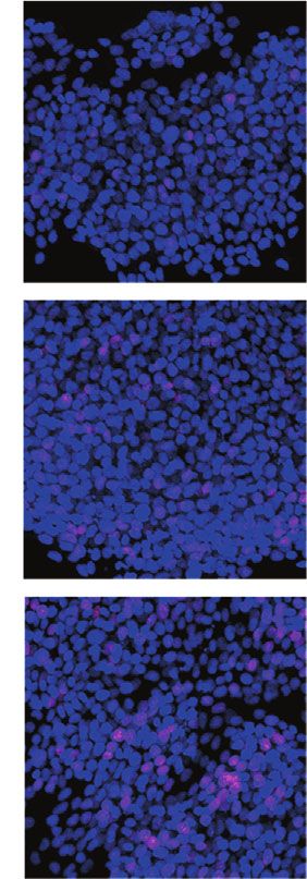
Oxidative Medicine and Cellular Longevity 3

 DAPI F-action

 20 m 20 m

 20 m 20 m

 PAK26 Merge
 (a)
 EDU Hoechst Merge

 2.0
 Control
 ⁎⁎⁎
 ⁎⁎⁎
 200 m
 OD value (450 nm)

 1.5

 1.0

 0.5 PRP

 0.0
 0 24 48 72
 h
 Control PRP-Exos
 PRP
 PRP-Exos

 (b) (c)

 Figure 2: Continued.

4 Oxidative Medicine and Cellular Longevity

 Diploid: 100.00 %
 Diploid: 100.00 %
 Dip G1: 39.49 % at 42.11 120 ⁎⁎⁎
 Dip G1: 53.91 % at 43.71 500
 800 Dip G2: 6.62 % at 85.23 Dip G2: 10.94 % at 82.12
 Dip S: 39.47 % G2/G1: 1.95 Dip S: 49.57 % G2/G1: 1.95 ⁎⁎⁎ ⁎⁎⁎
 400 %CV: 7.03

 % of cell in each phase
 600 %CV: 6.49
 Number

 Number
 300 80
 400
 200
 200
 100
 40
 0 0
 0 30 60 90 120 150 0 30 60 90 120 150
 Channels (PE-A) Channels (PE-A)
 Control PRP 0

 Control

 PRP

 PRP-Exos
 Diploid: 100.00 %
 500 Dip G1: 38.53 % at 43.37
 Dip G2: 5.85 % at 84.57
 400 Dip S: 55.63 % G2/G1: 1.95 S
 %CV: 5.97
 Number

 G2
 300 G1
 200

 100

 0
 0 30 60 90 120 150
 Channels (PE-A)
 PRP-Exos
 Debris Dip G2
 Aggregates Dip S
 Dip G1
 (d)
 PRP-Exos
 Control

 PRP

 Cyclin D1 35 kDa

 Cyclin D3 31 kDa

 DAPDH 37 kDa 500 m

 Control PRP PRP-Exos
 (e) (f)

 Figure 2: Continued.
Oxidative Medicine and Cellular Longevity 5

 ⁎⁎⁎⁎ Control PRP PRP-Exos
 150
 ⁎⁎⁎
 0h
 Migrated HaCTas
 100 ⁎⁎⁎ 500 m

 50 12 h

 0
 Control

 PRP

 PRP-Exos

 24 h

 (g) (h)

Figure 2: PRP-Exos enhance HaCaT cell proliferation and migration. (a) HaCaT cells were able to take up PKH26-labelled PRP-Exos. (b, c)
CCK-8 and EdU uptake assays were used to assess the impact of PRP-Exo treatment on HaCaT cells. (d) Flow cytometry was used to assess
how PRP-Exos affect cell cycle progression. (e) Western blotting was used to assess levels of cell-cycle associated proteins (cyclin D1 and
cyclin D3) following PRP-Exo treatment. (f) Wound healing and (g, h) Transwell assays were used to gauge the impact of PRP-Exos on
HaCaT cell migration. ∗ P < 0:05, ∗∗ P < 0:01, ∗∗∗ P < 0:001, and ∗∗∗∗ P < 0:0001.

2.3. PRP-Exo Isolation and Characterization. Samples of PRP scratched with sterile 10 μL pipette tips to generate linear
(1.5 mL) were centrifuged for 30 min at 2,000 ×g, after which scratch wounds. Cells were then incubated in the serum-
supernatants were collected and centrifuged for 45 min at free media for 12 or 24 h, after which they were imaged via
12,000 ×g at 4°C, and supernatants were then passed through inverted microscope.
a 0.45 μm filter membrane, after which they were centrifuged
for an additional 80 min at 13,000 ×g. Supernatants were then 2.7. Transwell Migration Assay. Cells were added to the upper
discarded, while pellets were resuspended in PBS and centri- chamber of a Transwell filter with an 8 μm pore size (3 ×
fuged for 70 min at 110,000 ×g, and the remaining particles 104 /well) in 200 μL of serum-free media, after which the
were suspended in chilled PBS for subsequent analysis. For lower chamber was filled with 600 μL of media containing
transmission electron microscopy analyses, these exosomes Exos or other appropriate reagents. Following incubation
were combined for 30 min with osmium tetroxide (4%) at for 24 h, the number of migratory cells was assessed via light
4°C in a 50 μL volume, after which they were transferred onto microscopy.
a copper grid. Next, 1% phosphotungstic acid was utilized to
 2.8. Cell Cycle Analysis. Cell cycle progression was assessed
stain these particles, and a transmission electron microscope
 via flow cytometry using a cell cycle and apoptosis analysis
(Hitachi, HT-7700) was used for their characterization. A
 kit (G1700; use G1700-50T, Servicebio) based on the pro-
NanoFCM™ instrument (N30E) was used for dynamic light
 vided directions.
scattering (DLS) analyses. Exosome marker protein expres-
sion was assessed via Western blotting. Exosomes collected 2.9. Western Blotting. After total protein extraction, 40 μg of
from each subject (n = 12) were examined individually. Exo- protein per sample was separated via 10% SDS-PAGE and
somes were pooled together for use in subsequent cell and transferred to PVDF membranes that were stained over-
animal experiments. night at 4°C with primary antibodies, after which they were
 probed for 1 h with HRP-conjugated secondary antibodies
2.4. CCK-8 Assay. HaCaT cells were cultured in 96-well plates at 37°C. The following antibodies were used: anti-CD9
(5 × 10³ ) for 24, 48, or 72 h, after which they were treated with (1 : 1000, Abcam, ab223052), anti-TSG101 (1 : 1000, Abcam,
the CCK-8 reagent for 2 h (G4103, Servicebio). Absorbance ab125011), anti-Calnexin (1 : 1000, Abcam, ab22595), anti-
was then measured at 450 nm to assess cell proliferation. USP15 (1 : 500, Abcam, ab71713), and anti-EIF4A1 (1 : 1000,
 Abcam, ab31217).
2.5. EdU Assay. HaCaT cells were plated in 24-well plates
(1 × 10⁵ /well) and treated as appropriate for 24 h, after which 2.10. Quantitative Real-Time PCR (qPCR). TRIzol (Invitro-
EdU staining was performed based on provided directions gen) was used to isolate total RNA from prepared samples,
(G1601, Servicebio). after which 1 μg of this RNA was used to prepare cDNA. A
 StepOne™ Real-Time PCR system (Life Technologies, CA,
2.6. Wound Healing Assay. Following appropriate treat- USA) was used to conduct all qPCR reactions, with the 2−ΔΔCt
ments, HaCaT cell monolayers cultured in 6-well plates were method being used to assess relative gene expression and
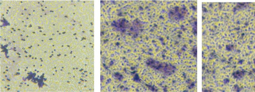
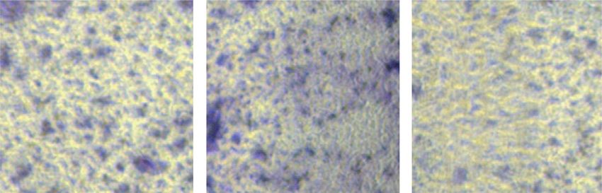
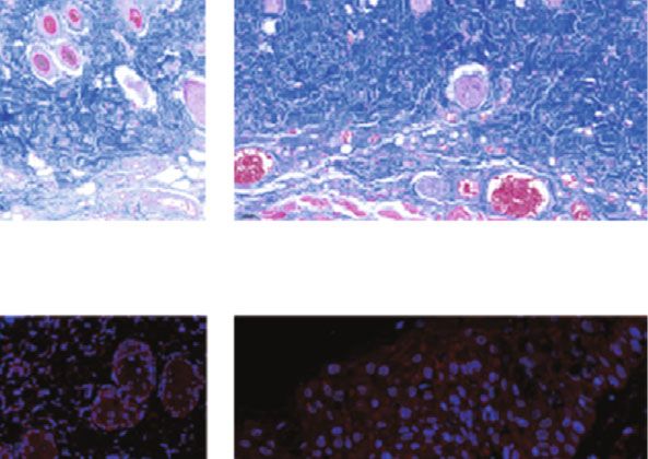
6 Oxidative Medicine and Cellular Longevity

 120
 Day 0 Day 3 Day 5 Day 7 Day 10 Day 14

 Wound closure (%)
 Control 80 ⁎⁎⁎
 ⁎⁎⁎
 40
 PRP
 0
 0 3 5 7 10 14
 PRP-Exos (Days)

 Control
 PRP
 PRP-Exos
 (a)
 Control PRP PRP-Exos 4000
 1000 m ⁎⁎⁎⁎

 Scar widths ( m)
 3000
 ⁎⁎⁎
 2000 ⁎⁎⁎
 HE 1000

 0

 Control

 PRP

 PRP-Exos
 (c)

 Collagen volume fraction (%)
 100
 1000 m
 80 ⁎⁎⁎⁎

 60 ⁎⁎⁎
 ⁎⁎⁎
 40
 Masson
 20
 0
 Control
 PRP
 PRP-Exos
 (d) ⁎⁎⁎⁎
 200
 ⁎⁎⁎
 100 m
 ⁎⁎⁎
 USP15 (%)

 100
 USP15

 0
 Control
 PRP
 PRP-Exos

 (e)

 Control
 PRP
 PRP-Exos

Figure 3: PRP-Exos promote wound healing in C57BL/6 mice. (a) Images of wounds in representative mice. (b) Wound healing rates in the
control, PRP, and PRP-Exo treatment groups. (c, d) H&E staining and Masson’s staining results from the three treatment groups with
corresponding quantification results. (e) USP15 immunohistochemistry results for wound sections in the three treatment groups. ∗ P <
0:05, ∗∗ P < 0:01, ∗∗∗ P < 0:001, and ∗∗∗∗ P < 0:0001. n = 6.

GAPDH serving as a normalization control. Primer sequences Experimental Animals, Tongji Medical College, Huazhong
were as follows: USP15: forward: 5 ′ -AAAACCTCGCTCCG University of Science and Technology, were individually
GAAAGG-3′ , reverse: 5 ′ -CCACCTTTCGTGCTATTGG-3′ ; housed in an 18°C facility with a 12 h light/dark cycle. Ani-
EIF4A1: forward: 5 ′ -TGTCTGCGAGCCAGGATTCCC-3′ , mals were anesthetized using intraperitoneal pentobarbital
 sodium with no signs of peritonitis, pain, or discomfort, after
reverse: 5 ′ -AGATGCCACGGAGAAGGGACTC-3 ′ .
 which a 10 mm diameter full-thickness excisional skin wound
 was generated on the dorsum of each animal. Mice were then
2.11. Murine Cutaneous Wound Model Establishment. Male randomly assigned to five treatment groups that were treated
C57BL/6 male mice (6–8 weeks old) from the Center of with PBS (100 μL), PRP-Exos (100 μg PRP-Exos in 100 μL
Oxidative Medicine and Cellular Longevity 7

 ⁎⁎⁎

 1.5

 Relative USP15 expression
 1.0

 PRP PRP-Exo Control PRP PRP-Exos

 USP15 120 kDa 0.5 USP15 120 kDa

 GAPDH 37 kDa GAPDH 37 kDa
 0.0
 PRP PRP-Exos
 (a) (b) (c)

Figure 4: PRP-Exos contain high levels of USP15, which promotes enhanced HaCaT cell functionality. (a) Western blotting was used to
assess USP15 levels in PRP and PRP-Exos. (b) USP15 mRNA levels were assessed via qPCR. (c) USP15 levels in murine wound tissues
were assessed via Western blotting. Data are from three independent experiments. ∗ P < 0:05, ∗∗ P < 0:01, and ∗∗∗ P < 0:001.

PBS), siRNA-NC (in PBS), siRNA-USP15 (in PBS), or siRNA- Western blotting were initially used to characterize isolated
USP15+PRP-Exos (in PBS). For siRNA-NC and siRNA- PRP-Exo samples. The obtained PRP-Exos ranged from 40
USP15 treatments, animals were administered 100 μL of a to 100 nm in size (Figures 1(a) and 1(b)), with the majority
20 μmol/L preparation of the corresponding siRNA, while of these particles exhibiting cup-shaped or spheroid morpho-
for the siRNA-USP15+PRP-Exos treatment group, mice were logical characteristics consistent with those of Exos. Western
administered 100 μL of a 10 μmol/L siRNA preparation and blotting analyses indicated the presence of the exosomal
10 μmol/L of PRP-Exos in PBS. All prepared solutions were markers CD9 and TSG101 in these samples (Figure 1(c)),
injected subcutaneously in appropriate mice at four sites adja- thus confirming the successful enrichment of exosomes from
cent to the wounded area (25 μL/site). Images of the wounds PRP samples.
were captured on days 0, 3, 7, 10, and 14 postwounding. Ani-
mals were euthanized on day 14, at which time skin samples 3.2. PRP-Exo Treatment Enhances In Vitro Keratinocyte
were collected for downstream analyses. Wound area was Responses. The impact of PRP-Exo treatment on immortal-
measured with the ImageJ software, and wound healing was ized human keratinocytes (HaCaT cells) was next assessed.
calculated as follows: Wound healing = ðWound area on Day To establish the uptake of these exosomes by HaCaT cells,
n/Wound area on Day 0Þ ðWound area on Day n/Wound area these particles were initially labeled using the lipophilic
on Day 0Þ × 100: PKH26 dye, followed by incubation in cell culture media
 The Animal Care and Use Committee of the Tongji Med- for 8 h at which time the uptake of these fluorescent particles
ical College, Huazhong University of Science and Technol- was clearly evident (Figure 2(a)). HaCaT cells were then
ogy, approved all animal studies detailed herein. treated with PBS, PRP, or PRP-Exos, and their proliferation
 was assessed through a series of CCK-8 and EdU uptake
2.12. Hematoxylin and Eosin Staining, Masson’s Trichrome assays, confirming that PRP-Exo exposure was associated
Staining, and Immunohistochemical Staining. Paraffin- with the enhanced proliferation of these keratinocytes
embedded tissue sections (7 μm thick) were subjected to (Figures 2(b) and 2(c)). PRP-Exo treatment was associated
hematoxylin and eosin (H&E) and Masson’s trichrome stain- with an increase in the frequency of HaCaT cells entering
ing. An immunofluorescent approach was used to detect the S stage of the cell cycle (Figure 2(d)). Following PRP-
USP15 in prepared tissue sections. Briefly, prepared sections Exo treatment, higher levels of cell cycle-associated proteins
were blocked for 30 min with 1% BSA, probed overnight with were found to be expressed in these cells (Figure 2(e)). In
anti-USP15 (1 : 500, Abcam, ab71713), and stained for 1 h with Transwell and wound healing assays, PRP-Exo treatment
an appropriate secondary antibody, and then, USP15-positive was also associated with significant improvements in HaCaT
cells area in three random fields of view were analyzed. All cell migration (Figures 2(f)–2(h)). Together, these data thus
stained tissue sections were independently assessed by three indicate that PRP-Exo treatment can significantly augment
observers blinded to experimental treatment protocols. HaCaT cell proliferation and migration.

3. Results 3.3. PRP-Exo Treatment Enhances In Vivo Wound Healing in
 Mice. An in vivo cutaneous wound model was next estab-
3.1. PRP-Exo Characterization. Transmission electron lished using C57BL/6 mice, with equal volumes of PBS,
microscopy (TEM), dynamic light scattering (DLS), and PRP, or PRP-Exo preparations being injected around the
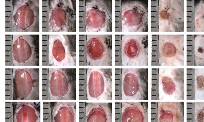
8 Oxidative Medicine and Cellular Longevity

 PRP-Exos+siUSP15
 PRP-Exos
 Control

 siRNA-Nc

 PRP-Exos
 PRP

 siUSP15
 Control
 USP15 120 kDa
 USP15 120 kDa

 GAPDH 37 kDa GAPDH 37 kDa

 (a) (b)
 siUSP15 PRP-Exos PRP-Exos+siUSP5

 0h
 500 m

 2.0

 OD value (450 nm)
 12 h 1.5 ⁎⁎⁎
 ⁎⁎⁎
 1.0

 24 h 0.5

 0.0
 0 24 48 72
 h
 PRP-Exos-siRNA-USP15
 siRNA-USP15
 PRP-Exo
 (c) (d)
 ⁎⁎⁎⁎
 150 ⁎⁎⁎⁎
 siUSP15 PRP-Exos PRP-Exos+siUSP5
 100 ⁎⁎⁎⁎

 50

 500 m
 0
 siRNA-USP15

 PRP-Exos

 siRNA-USP15
 +PRP-Exos

 siRNA-USP15
 PRP-Exos
 siRNA-USP15+PRP-Exos
 (e)

Figure 5: USP15 promotes in vitro wound healing. (a) Western blotting data demonstrating USP15 levels in control, PRP, and PRP-Exos
groups. (c, e) Wound healing and Transwell assays were used to assess the migratory activity of HaCaT cells. (d) The proliferation
of HaCaT cells in the indicated treatment groups was assessed via CCK-8 assay. Data are from three independent experiments. ∗ P < 0:05,
∗∗
 P < 0:01, ∗∗∗ P < 0:001.
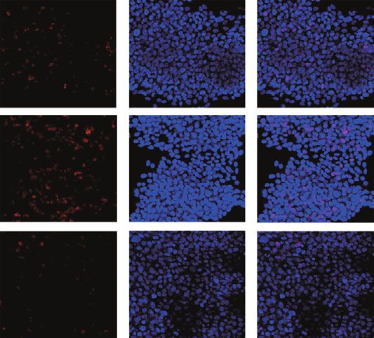
Oxidative Medicine and Cellular Longevity 9

 siRNA-USP15

 siRNA-USP15
 siRNA-NC

 siRNA-NC
 Control

 Control
 USP15 120 kDa EIF4A1 46 kDa

 GAPDH 37 kDa GAPDH 37 kDa

 (a) (b)
 1.5 ns 2.0
 Relative EIF4A1 expression

 ns

 OD value (450 nm)
 1.5
 1.0 ⁎⁎⁎ ⁎⁎⁎
 1.0
 0.5
 0.5

 0.0 0.0
 Control

 siRNA-NC

 siRNA-USP15

 0 24 48 72
 h

 Control
 siRNA-EIF4A1
 siRNA-NC
 (c) (d)
 EdU Hoechst Merge

 siRNA-EIF4A1
 Control
 siRNA-NC

 200 m
 Control

 siRNA-NC
 EIF4A1 46 kDa

 siRNA-EIF4A1
 GAPDH 37 kDa

 (e) (f)

 Figure 6: Continued.
10 Oxidative Medicine and Cellular Longevity

 Control siRNA-NC siRNA-EIF4A1 Control siRNA-NC siRNA-EIF4A1

 0h

 500 m

 500 m

 12 h
 80
 Migrated HaCaTs

 ⁎⁎⁎⁎
 60
 ⁎⁎⁎
 40
 20
 24 h
 0
 Control
 siRNA-NC
 siRNA-EIF4A1

 Control
 siRNA-NC
 siRNA-EIF4A1
 (g) (h)

Figure 6: USP15 promotes EIF4A1 deubiquitination to enhance HaCaT cell functionality. (a, b) Western blotting results demonstrating
USP15 and EIF4A1 levels in HaCaT cells following siRNA-USP15 treatment. (c) EIF4A1 levels in HaCaT cells following siRNA-USP15
treatment, as assessed via qPCR. (d, e) HaCaT cell proliferation was assessed via CCK-8 and EdU assays following siRNA-EIF4A1
treatment. (f) Western blotting analyses were used to assess EIF4A1 expression HaCaT cells following siRNA-EIF4A1 treatment. (g, h)
Wound healing and Transwell assays were used to assess the migratory activity of HaCaT cells following siRNA-EIF4A1 treatment. Data
are from three independent experiments. ∗ P < 0:05, ∗∗ P < 0:01, and ∗∗∗ P < 0:001.

wound site in each animal to assess the impact of such treat- levels were evident in epidermal keratinocytes in the PRP-
ment on wound healing. Relative to control animals, those Exo group in vivo (Figures 4(c) and Figure 3(e)), suggesting
treated with PRP and PRP-Exos exhibited faster wound that USP15 may be linked to the ability of PRP-Exos to pro-
repair, with such healing being even more rapid for PRP- mote wound healing.
Exo-treated mice relative to mice treated with PRP alone
(Figures 3(a) and 3(b)). The scars of mice in the PRP-Exo 3.5. PRP-Exos Promotes HaCaT Cell Migration and
group were smaller than those of mice in any other group Proliferation in a USP15-Dependent Manner. To understand
(Figure 3(c)), and these mice exhibited the highest levels of the functional effects of USP15 on HaCaT cells, they were
collagen formation (Figure 3(d)). Reepithelialization plays next treated with PBS, PRP, and PRP-Exos. Western blotting
an essential role in the wound healing process [16]. Deubi- indicated that USP15 levels were highest for HaCaT cells
quitinases (DUBs) can alter protein stability by removing a treated with PRP-Exos (Figure 5(a)). As shown in Figure 1,
ubiquitin chain from a given protein, thereby stabilizing it HaCaTs treated with PRP-Exos exhibited significantly aug-
[17], thus potentially accelerating the process of reepitheliali- mented cellular proliferation and migration. In contrast,
zation in the context of wound healing. The ubiquitin- siUSP15 treatment had the opposite effect on HaCaT cell
specific protease (USP) family is the best-studied group of proliferation and migration in these same assay systems
DUB proteins, and USP15 is an important member of this (Figures 5(b)–5(e)). As such, decreasing USP15 expression
family [18]. As such, we next evaluated the expression of in HaCaT cells can reduce the beneficial impact of PRP-Exo
USP15 in keratinocytes in wounded skin tissues, revealing treatment. Together, these findings suggest that USP15 is
it to be expressed at higher levels in the PRP-Exo treatment the primary mediator whereby PRP-Exos promote HaCaT
group (Figure 3(e)). cell migration and proliferation.

3.4. USP15 Is Enriched in PRP-Exo Preparations. To explore 3.6. USP15 Enhances HaCaT Cell Functionality by Promoting
the functional importance of USP15 as a driver of PRP- EIF4A1 Deubiquitination. We next explored the mechanisms
Exo-mediated wound healing, we assessed the levels of this whereby USP15 promotes HaCaT cell functionality. EIF4A1
protein in PRP and PRP-Exo samples via Western blotting, is a key eukaryotic initiation factor complex component that
revealing it to be present at significantly higher levels in has been linked to the proliferation of certain cell lines [19,
PRP-Exos (Figure 4(a)). Subsequent qPCR analyses addition- 20], although it has not been studied in detail in HaCaT
ally exhibited increased USP15 expression in PRP-Exos at the cells. As such, we hypothesized that USP15 may be able to
mRNA level (Figure 4(b)). Similarly, higher USP15 protein promote HaCaT cell proliferation in part via altering the
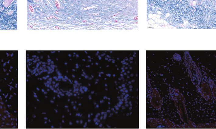
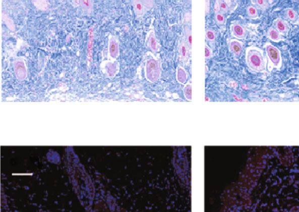
Oxidative Medicine and Cellular Longevity 11

 Day 0 Day 3 Day 5 Day 7 Day 10 Day 14 120

 Wound closure (%)
 Control
 80 ⁎⁎⁎ ⁎⁎⁎

 siRNA-NC 40

 siRNA-USP15
 0
 0 3 5 7 10 14
 siRNA-USP15 (Days)
 +PRP-Exos
 Control siRNA-USP15
 siRNA-NC siRNA-USP15+PRP-Exos
 (a) (b)

 Control siRNA-NC siRNA-USP15 siRNA-USP15+PRP-Exos
 1000 m

 HE

 (c)
 100 m

 Masson

 (d)
 100 m

 USP15

 (e)
 ⁎⁎⁎⁎
 Collagen volume fraction (%)

 ⁎⁎⁎ 200
 8000 100
 ⁎⁎⁎
 Scar widths ( m)

 6000 80
 ⁎⁎⁎ ⁎⁎⁎
 USP15 (%)

 ⁎⁎⁎
 60 ⁎⁎⁎⁎
 4000 ⁎⁎⁎ 100
 40
 2000 ⁎⁎⁎
 20 ⁎⁎⁎

 0 0 0
 Control

 siRNA-USP15
 siRNA-USP15
 +PRP-Exos

 Control

 siRNA-USP15
 siRNA-USP15
 +PRP-Exos

 Control

 siRNA-USP15
 siRNA-USP15
 +PRP-Exos
 siRNA-NC

 siRNA-NC

 siRNA-NC

 Control siRNA-USP15
 siRNA-NC siRNA-USP15+PRP-Exos
 (f)

Figure 7: USP15 promotes in vivo wound healing. (a) Images of mice in the three treatment groups. (b) Wound closure rates for mice in the
three treatment groups. (c) H&E staining results from the three treatment groups. (d) Masson’s trichrome staining results for wounds in the
three treatment groups. (e) USP15 immunohistochemical staining results for the three groups. (f) Quantification results for data in the three
treatment groups. ∗ P < 0:05, ∗∗ P < 0:01, ∗∗∗ P < 0:001, and ∗∗∗∗ P < 0:0001. n = 6.
12 Oxidative Medicine and Cellular Longevity

 HaCaTs USP15
 miRNA
 mRNA
 Proteins
 USP15
 DNA
 Deubiquitination

 PRP-Exos EIF4A1

 PRP

 Reepithelialization

Figure 8: USP15 is one of the main mRNA in PRP-Exos, which could be taken into HaCaTs, subsequently deubiquitinating EIF4A1, then
accelerating reepithelialization and promoting wound healing.

EIF4A1 expression. To examine the effects of USP15 on 4. Discussion
EIF4A1 stability in HaCaT cells, we conducted Western
blotting assays which revealed a significant decrease in both A number of clinical strategies have been employed in recent
USP15 and EIF4A1 expressions following siRNA-USP15 years in an effort to accelerate wound healing [21]. PRP-
treatment (Figure 6(a) and 6(b)), whereas qPCR analyses based therapies have emerged as promising means of pro-
indicated that the EIF4A1 mRNA levels in these cells were moting wound healing and have been studied in the fields
unchanged (Figure 6(c)). of orthopedics, dermatology, dentistry, and diabetic wound
 We then assessed the functional importance of EIF4A1 in management. However, there have been few studies to date
HaCaT cells by knocking down this gene with a specific of PRP-Exos. Herein, we determined that PRP-Exo treatment
siRNA construct. Subsequent CCK-8 and EdU assays sug- was sufficient to promote wound healing in a manner more
gested that EIF4A1 knockdown was sufficient to suppress efficient than direct PRP treatment. The process of epidermal
HaCaT cell proliferation (Figures 6(d) and 6(e)), with regeneration is a multistep process that is regulated by a
Western blotting being used to confirm that EIF4A1 pro- range of cytokine and cell types, ultimately leading to the
tein levels were reduced following siRNA transfection reconstruction of the damaged skin barrier [22]. Exosomes
(Figure 6(f)). In Transwell and wound healing assays, derived from mesenchymal stem cells have been repeatedly
siRNA EIF4A1 treatment significantly reduced the migra- shown to accelerate reepithelialization and to thereby
tory activity of these keratinocytes (Figures 6(g) and 6(h)). enhance wound healing [23, 24]. Our present results indi-
Together, these data suggested that USP15 can enhance cated that PRP-Exo treatment similarly promoted enhanced
the migration and proliferation of HaCaT cells by promot- epithelialization, highlighting the promise of these particles
ing EIF4A1 deubiquitination. for use in the treatment of chronic wounds.
 Exosomes are small, lipid bilayer-enclosed extracellular
3.7. USP15 Promotes Wound Healing In Vivo. To examine vesicles that are produced by most known cell types and that
the impact of USP15 on wound healing processes, equivalent can transmit macromolecules and other compounds between
amounts of PBS, siRNA-NC, siRNA-USP15, siRNA-USP15, cells [25]. When taken up by recipient cells, these exosomes
or PRP-Exos were injected surrounding wound sites in can thus alter cellular functionality via the delivery of specific
C57/BL6 mice, as above. USP15 knockdown was found to proteins, nucleic acids, and signaling molecules in a manner
significantly slow the wound healing process in these animals that makes them ideal for use as drug carriers in a range of
(Figures 7(a) and 7(b)), and H&E staining confirmed that disease types [24]. Herein, we found that HaCaT cells were
wounds in the siRNA-USP15 group were larger than those able to efficiently internalize PRP-Exos. It has been reported
in other groups (Figure 7(c)). Masson’s trichrome staining that a range of new types of hydrogels with specifically tai-
also revealed that collagen levels were lowest in the siRNA- lored biochemical functions can accelerate wound healing,
USP15 group (Figure 7(d)), and the number of USP15- thus exhibiting good prospects for therapeutic application
positive cells in murine wound sites was lower in siRNA- [26, 27]. However, such artificial hydrogels play a less direct
USP15-treated mice relative to other groups (Figures 7(e) role in normal physiological processes, and a combination
and 7(f)). Together, these results thus demonstrated that of exosomes and hydrogels may thus offer greater advantages
USP15 knockdown was sufficient to largely ablate the benefi- further expediting the wound healing process.
cial effects of PRP-Exo treatment in the context of in vivo Protein ubiquitination has been shown to play diverse
cutaneous wound healing. regulatory roles in the context of wound healing [28, 29].
Oxidative Medicine and Cellular Longevity 13

USP family proteins, including USP15, are key mediators of References
protein deubiquitination. Others have reported that PRP
can promote wound healing by driving accelerated epithelial- [1] M. H. Kathawala, W. L. Ng, D. Liu et al., “Healing of chronic
ization [30], and we herein found that PRP-Exo treatment wounds: an update of recent developments and future possibil-
was superior to PRP treatment as a means of enhancing ities,” Tissue Engineering Part B: Reviews, vol. 25, no. 5,
 pp. 429–444, 2019.
wound healing in a manner associated with increased
USP15 protein levels in wound tissues relative to those [2] F. Werdin, M. Tenenhaus, and H. O. Rennekampff, “Chronic
 wound care,” Lancet, vol. 372, no. 9653, pp. 1860–1862, 2008.
detected upon PBS or PRP administration. Through a series
of in vitro and in vivo experiments, we further confirmed that [3] L. M. Morton and T. J. Phillips, “Wound healing and treating
USP15 was able to promote wound healing, suggesting that wounds: differential diagnosis and evaluation of chronic
 wounds,” Journal of the American Academy of Dermatology,
PRP-Exo-derived USP15 is a key mechanism driving this
 vol. 74, no. 4, pp. 589–605, 2016.
regenerative process. Tao et al. have reported that PRP-
 [4] C. K. Sen, “Human wounds and its burden: an updated com-
Exos can suppress apoptosis in a rat model of femoral head
 pendium of estimates,” Adv Wound Care (New Rochelle),
osteonecrosis via the Akt/Bad/Bcl-2 signaling pathway [31], vol. 8, no. 2, pp. 39–48, 2019.
while Iyer et al. revealed the ability of PRP-Exo treatment
 [5] S. C. Pan, C. Y. Li, C. Y. Kuo et al., “The p53-S100A2 positive
to promote functional recovery following muscle injury
 feedback loop negatively regulates epithelialization in cutane-
[32]. Owing to their unique properties, exosomes have been ous wound healing,” Scientific Reports, vol. 8, no. 1, p. 5458,
shown to be of value for the treatment of many diseases 2018.
and thus warrant further clinical study. [6] K. Liu, C. Chen, H. Zhang, Y. Chen, and S. Zhou, “Adipose
 In prior studies, USP15 was shown to interact with stem cell-derived exosomes in combination with hyaluronic
EIF4A1 and to thereby accelerate wound healing [33]. Con- acid accelerate wound healing through enhancing re-
sistent with such a model, we found that USP15 knock- epithelialization and vascularization,” The British Journal of
down was sufficient to reduce EIF4A1 expression, while Dermatology, vol. 181, no. 4, pp. 854–856, 2019.
EIF4A1 knockdown directly impaired HaCaT cell migra- [7] T. Yuan, S. C. Guo, P. Han, C. Q. Zhang, and B. F. Zeng,
tion and proliferation. Together, these results suggest that “Applications of leukocyte- and platelet-rich plasma (L-PRP)
the USP15-EIF4A1 axis is a key mediator of the reepithelia- in trauma surgery,” Current Pharmaceutical Biotechnology,
lization process. vol. 13, no. 7, pp. 1173–1184, 2012.
 [8] P. Everts, K. Onishi, P. Jayaram, J. F. Lana, and K. Mautner,
 “Platelet-rich plasma: new performance understandings and
5. Conclusion therapeutic considerations in 2020,” International Journal of
 Molecular Sciences, vol. 21, no. 20, p. 7794, 2020.
In conclusion, the results of this study indicate that PRP- [9] P. Wu, Z. Tong, L. Luo et al., “Comprehensive strategy of con-
Exo-derived USP15 is a key mediator of HaCaT cell sur- duit guidance combined with VEGF producing Schwann cells
vival and migratory activity, with EIF4A1 playing an accelerates peripheral nerve repair,” Bioact Mater, vol. 6,
important role in the process of USP15-induced epithelial- no. 10, pp. 3515–3527, 2021.
ization (Figure 8). Together, these findings provide a robust [10] I. Iacopetti, M. Patruno, L. Melotti et al., “Autologous
foundation for future studies of the therapeutic potential of platelet-rich plasma enhances the healing of large cutaneous
PRP-Exo treatment as a means of promoting improved wounds in dogs,” Frontiers in Veterinary Science, vol. 7,
wound healing. p. 575449, 2020.
 [11] E. Torreggiani, F. Perut, L. Roncuzzi, N. Zini, S. R. Baglìo, and
 N. Baldini, “Exosomes: novel effectors of human platelet lysate
Data Availability activity,” European Cells and Materials, vol. 28, pp. 137–151,
 2014.
Data available on request. [12] R. Kalluri and V. S. LeBleu, “The biology, function, and bio-
 medical applications of exosomes,” Science, vol. 367,
 no. 6478, p. eaau6977, 2020.
Conflicts of Interest [13] N. Kosaka, Y. Yoshioka, K. Hagiwara, N. Tominaga, and
 T. Ochiya, “Functional analysis of exosomal microRNA in
All authors report no conflicts of interest in this work.
 cell-cell communication research,” Methods in Molecular Biol-
 ogy, vol. 1024, pp. 1–10, 2013.
Authors’ Contributions [14] B. Mi, L. Chen, Y. Xiong et al., “Saliva exosomes-derived
 UBE2O mRNA promotes angiogenesis in cutaneous wounds
Yan Xu and Ze Lin contributed equally to this work and by targeting SMAD6,” Journal of Nanobiotechnology, vol. 18,
should be regarded as co-first authors. no. 1, p. 68, 2020.
 [15] Y. Xiong, L. Chen, T. Yu et al., “Inhibition of circulating exo-
 somal microRNA-15a-3p accelerates diabetic wound repair,”
Acknowledgments Aging (Albany NY), vol. 12, no. 10, pp. 8968–8986, 2020.
 [16] M. Rodrigues, N. Kosaric, C. A. Bonham, and G. C. Gurtner,
This study was supported by the National Key Research and “Wound healing: a cellular perspective,” Physiological Reviews,
Development Program of China (2017YFC1103804). vol. 99, no. 1, pp. 665–706, 2019.
14 Oxidative Medicine and Cellular Longevity

[17] D. Popovic, D. Vucic, and I. Dikic, “Ubiquitination in disease prevent apoptosis induced by glucocorticoid-associated endo-
 pathogenesis and treatment,” Nature Medicine, vol. 20, plasmic reticulum stress in rat osteonecrosis of the femoral
 no. 11, pp. 1242–1253, 2014. head via the Akt/Bad/Bcl-2 signal pathway,” Theranostics,
[18] Y. Zhao, Z. Wang, C. Ho, G. Zhang, and Q. Li, “Ubiquitin-spe- vol. 7, no. 3, pp. 733–750, 2017.
 cific protease 15 maintains transforming growth factor-β path- [32] S. R. Iyer, A. L. Scheiber, P. Yarowsky, R. F. Henn 3rd,
 way activity by deubiquitinating transforming growth factor-β S. Otsuru, and R. M. Lovering, “Exosomes isolated from
 receptor I during wound healing,” The American Journal of platelet-rich plasma and mesenchymal stem cells promote
 Pathology, vol. 189, no. 7, pp. 1351–1362, 2019. recovery of function after muscle injury,” The American Jour-
[19] C. Li, Y. Tian, Y. Liang, and Q. Li, “Circ_0008035 contributes nal of Sports Medicine, vol. 48, no. 9, pp. 2277–2286, 2020.
 to cell proliferation and inhibits apoptosis and ferroptosis in [33] Y. Zhao, X. Huang, Z. Zhang et al., “USP15 enhances re-
 gastric cancer via miR-599/EIF4A1 axis,” Cancer Cell Interna- epithelialization through deubiquitinating EIF4A1 during
 tional, vol. 20, no. 1, p. 84, 2020. cutaneous wound repair,” Frontiers in Cell and Development
[20] X. Ma, B. Li, J. Liu, Y. Fu, and Y. Luo, “Phosphoglycerate dehy- Biology, vol. 8, p. 529, 2020.
 drogenase promotes pancreatic cancer development by inter-
 acting with eIF4A1 and eIF4E,” Journal of Experimental &
 Clinical Cancer Research, vol. 38, no. 1, p. 66, 2019.
[21] P. Martin and R. Nunan, “Cellular and molecular mechanisms
 of repair in acute and chronic wound healing,” The British
 Journal of Dermatology, vol. 173, no. 2, pp. 370–378, 2015.
[22] N. Deshayes, F. Bloas, F. Boissout, J. Lecardonnel, and
 M. Paris, “3D in vitro model of the re-epithelialization phase
 in the wound-healing process,” Experimental Dermatology,
 vol. 27, no. 5, pp. 460–462, 2018.
[23] B. Zhang, M. Wang, A. Gong et al., “HucMSC-exosome
 mediated-Wnt4 signaling is required for cutaneous wound
 healing,” Stem Cells, vol. 33, no. 7, pp. 2158–2168, 2015.
[24] S. C. Tao, S. C. Guo, M. Li, Q. F. Ke, Y. P. Guo, and C. Q.
 Zhang, “Chitosan wound dressings incorporating exosomes
 derived from microRNA-126-overexpressing synovium mes-
 enchymal stem cells provide sustained release of exosomes
 and heal full-thickness skin defects in a diabetic rat model,”
 Stem Cells Translational Medicine, vol. 6, no. 3, pp. 736–747,
 2017.
[25] D. Perocheau, L. Touramanidou, S. Gurung, P. Gissen, and
 J. Baruteau, “Clinical applications for exosomes: are we there
 yet?,” British Journal of Pharmacology, vol. 178, no. 12,
 pp. 2375–2392, 2021.
[26] J. Cao, P. Wu, Q. Cheng, C. He, Y. Chen, and J. Zhou, “Ultra-
 fast fabrication of self-healing and injectable carboxymethyl
 chitosan hydrogel dressing for wound healing,” ACS Applied
 Materials & Interfaces, vol. 13, no. 20, pp. 24095–24105, 2021.
[27] M. Li, Y. Liang, J. He, H. Zhang, and B. Guo, “Two-pronged
 strategy of biomechanically active and biochemically multi-
 functional hydrogel wound dressing to accelerate wound clo-
 sure and wound healing,” Chemistry of Materials, vol. 32,
 no. 23, pp. 9937–9953, 2020.
[28] S. Cheng, Z. Xi, G. Chen, K. Liu, R. Ma, and C. Zhou, “Extra-
 cellular vesicle-carried microRNA-27b derived from mesen-
 chymal stem cells accelerates cutaneous wound healing via
 E3 ubiquitin ligase ITCH,” Journal of Cellular and Molecular
 Medicine, vol. 24, no. 19, pp. 11254–11271, 2020.
[29] H. Qu, T. Miao, Y. Wang et al., “Dedicator of cytokinesis 5 reg-
 ulates keratinocyte function and promotes diabetic wound
 healing,” Diabetes, vol. 70, no. 5, pp. 1170–1184, 2021.
[30] S. C. Guo, S. C. Tao, W. J. Yin, X. Qi, T. Yuan, and C. Q. Zhang,
 “Exosomes derived from platelet-rich plasma promote the re-
 epithelization of chronic cutaneous wounds via activation of
 YAP in a diabetic rat model,” Theranostics, vol. 7, no. 1,
 pp. 81–96, 2017.
[31] S. C. Tao, T. Yuan, B. Y. Rui, Z. Z. Zhu, S. C. Guo, and C. Q.
 Zhang, “Exosomes derived from human platelet-rich plasma
You can also read