Polymerization of misfolded Z alpha-1 antitrypsin protein lowers CX3CR1 expression in human PBMCs
←
→
Page content transcription
If your browser does not render page correctly, please read the page content below
SHORT REPORT

                                   Polymerization of misfolded Z alpha-1
                                   antitrypsin protein lowers CX3CR1
                                   expression in human PBMCs
                                   Srinu Tumpara1, Matthias Ballmaier2, Sabine Wrenger1, Mandy König3,
                                   Matthias Lehmann3, Ralf Lichtinghagen4, Beatriz Martinez-Delgado5,
                                   Elena Korenbaum6, David DeLuca1, Nils Jedicke7, Tobias Welte1, Malin Fromme8,
                                   Pavel Strnad8, Jan Stolk9, Sabina Janciauskiene1,9*
                                   1
                                    Department of Respiratory Medicine, Hannover Medical School, Biomedical
                                   Research in Endstage and Obstructive Lung Disease Hannover (BREATH), Member
                                   of the German Center for Lung Research (DZL), Hannover, Germany; 2Cell Sorting
                                   Core Facility Hannover Medical School, Hannover, Germany; 38sens.biognostic
                                   GmbH, Berlin, Germany; 4Institute of Clinical Chemistry, Hannover Medical School,
                                   Hannover, Germany; 5Department of Molecular Genetics, Institute of Health Carlos
                                   III, Center for Biomedical Research in the Network of Rare Diseases (CIBERER),
                                   Majadahonda, Spain; 6Institute for Biophysical Chemistry, Hannover Medical School,
                                   Hannover, Germany; 7Department of Gastroenterology, Hepatology and
                                   Endocrinology, Hannover Medical School, Hannover, Germany; 8Medical Clinic III,
                                   Gastroenterology, Metabolic Diseases and Intensive Care, University Hospital
                                   RWTH Aachen, Aachen, Germany; 9Department of Pulmonology, Leiden University
                                   Medical Center, Member of European Reference Network LUNG, section Alpha-1-
                                   antitrypsin Deficiency, Leiden, Netherlands

                                   Abstract Expression levels of CX3CR1 (C-X3-C motif chemokine receptor 1) on immune cells
                                   have significant importance in maintaining tissue homeostasis under physiological and pathological
                                   conditions. The factors implicated in the regulation of CX3CR1 and its specific ligand CX3CL1
*For correspondence:
janciauskiene.sabina@mh-           (fractalkine) expression remain largely unknown. Recent studies provide evidence that host’s
hannover.de                        misfolded proteins occurring in the forms of polymers or amyloid fibrils can regulate CX3CR1
                                   expression. Herein, a novel example demonstrates that polymers of human ZZ alpha-1 antitrypsin
Competing interest: See
                                   (Z-AAT) protein, resulting from its conformational misfolding due to the Z (Glu342Lys) mutation in
page 12
                                   SERPINA1 gene, strongly lower CX3CR1 mRNA expression in human peripheral blood mononuclear
Funding: See page 13               cells (PBMCs). This parallels with increase of intracellular levels of CX3CR1 and Z-AAT proteins.
Received: 13 November 2020         Presented data indicate the involvement of the CX3CR1 pathway in the Z-AAT-related disorders
Accepted: 16 May 2021              and further support the role of misfolded proteins in CX3CR1 regulation.
Published: 18 May 2021

Reviewing editor: Koyeli Mapa,
Shiv Nadar University, India
                                   Introduction
   Copyright Tumpara et al. This
                                   Interactions between the chemokine receptors and chemokines, but also other proteins, peptides,
article is distributed under the
                                   lipids, and microbial products, play a critical role in the recruitment of inflammatory cells into
terms of the Creative Commons
Attribution License, which         injured/diseased tissues (Bachelerie et al., 2014). Many human diseases involve altered surface
permits unrestricted use and       expression of chemokine receptors, which can lead to a defective cell migration and inappropriate
redistribution provided that the   immune response. Most of the human peripheral blood mononuclear cells (PBMCs) express CX3CR1
original author and source are     (Bazan et al., 1997), also known as the G-protein-coupled receptor 13 (GPR13) or fractalkine recep-
credited.                          tor, a mediator of leukocyte migration and adhesion. In the central nervous system, CX3CR1 is

Tumpara et al. eLife 2021;10:e64881. DOI: https://doi.org/10.7554/eLife.64881                                                  1 of 16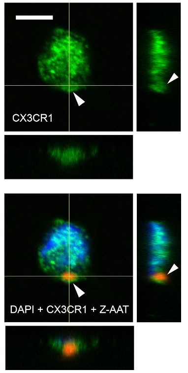
Short report                                                                                                Cell Biology Medicine

                                   eLife digest Proteins can lose their structure and form polymers because of mutations or
                                   changes in their immediate environment which can lead to cell damage and disease. Interestingly,
                                   polymers formed by a variety of proteins can reduce the levels of CX3C chemokine receptor 1
                                   (CX3CR1 for short) that controls the behaviour of immune cells and is implicated in a range of
                                   illnesses.
                                       Inherited ZZ alpha-1 antitrypsin deficiency is a rare genetic condition that highly increases the risk
                                   of liver and lung diseases. This disorder is characterised by mutant alpha-1 antitrypsin proteins (AAT
                                   for short) reacting together to form polymers; yet it remains unclear how the polymers affect
                                   different cells or organs, and lead to diseases. To investigate this question, Tumpara et al. examined
                                   whether polymers of mutant AAT influence the level of the CX3CR1 protein in specific classes of
                                   immune cells.
                                       Experiments revealed that in people with AAT deficiency, certain blood immune cells express
                                   lower levels of CX3CR1. Regardless of age, clinical diagnosis, or treatment regimen, all individuals
                                   with ZZ alpha-1 antitrypsin deficiency had AAT polymers circulating in their blood: the higher the
                                   levels of polymers measured, the lower the expression of CX3CR1 recorded in the specific immune
                                   cells. When Tumpara et al. added polymers of mutant AAT to the immune cells of healthy donors,
                                   the expression of CX3CR1 dropped in a manner dependent on the polymer concentration.
                                   According to microscopy data, AAT polymers occurred inside cells alongside the CX3CR1 protein,
                                   suggesting that the two molecular actors interact. In the future, new drugs that remove these
                                   polymers, either from inside cells or as they circulate in the body, could help patients suffering from
                                   conditions associated with this abnormal protein aggregation.

                                   largely expressed by microglial cells (brain macrophages) (Ransohoff, 2009), which are involved in
                                   neurodegenerative diseases like Alzheimer’s disease. The major role of CX3CR1-expressing cells is
                                   to recognize and enter tissue following CX3CL1 (fractalkine or also called neurotactin) gradient, and
                                   to crawl or ‘patrol’ in the lumen of blood vessels (Auffray et al., 2007). Since CX3CR1/CX3CL1 axis
                                   is also involved in the synthesis of anti-inflammatory cytokines and has a significant role in cytoskele-
                                   tal rearrangement, migration, apoptosis, and proliferation, its dysregulation is associated with the
                                   development of cardiovascular diseases, kidney ischemia–reperfusion injury, cancer, chronic obstruc-
                                   tive pulmonary disease (COPD), neurodegenerative disorders, and others (Harrison et al., 1998;
                                   Ning et al., 2004; Rius et al., 2013). Some studies indicate that CX3CR1 deficiency contributes to
                                   the severity of infectious diseases (Bonduelle et al., 2012) and promotes lung pathology in respira-
                                   tory syncytial virus-infected mice (Das et al., 2017). Animals with deletion of CX3CR1 show impaired
                                   phagocytosis (Thome et al., 2015), which is vital to prevent unwanted inflammation. It is clear that
                                   CX3CR1-expressing cells have tissue-specific roles in different pathophysiological conditions. Never-
                                   theless, a comprehensive knowledge on the regulation of CX3CR1 expression is still missing.
                                       Current findings suggest that divergent proteins with a common propensity to form extracellular
                                   oligomers interact with chemokine receptors and affect their expression levels. For example, Alz-
                                   heimer’s peptide, Ab, interacts with CX3CR1 and significantly reduces its expression in cultured
                                   microglial cells and in Alzheimer’s brain (Cho et al., 2011). Similarly, highly aggregated extracellular
                                   Tau protein binds to CX3CR1, promotes its internalization, and reduces expression in microglial cells
                                   (Bolós et al., 2017). In concordance, polymers of human Z alpha-1 antitrypsin (Z-AAT), resulting
                                   from protein misfolding due to the Z (Glu342Lys) mutation in SERPINA1 gene, lower CX3CR1 mRNA
                                   expression in human PBMCs, which parallels with increased intracellular CX3CR1 and Z-AAT protein
                                   levels.

                                   Results and discussion
                                   Inherited alpha-1 antitrypsin deficiency (AATD) is a rare genetic condition caused by SERPINA1 gene
                                   mutations. Homozygous Z AATD mutation is the most clinically relevant among Caucasians (preva-
                                   lence is about 1:2000-1:5000) that is characterized by low plasma levels of AAT protein (10–15%
                                   compared to the wild type, MM AAT, 1.3–2 g/l) and the presence of intracellular and circulating
                                   Z-AAT polymers (Tan et al., 2014). The liver is the major producer of AAT, therefore the

Tumpara et al. eLife 2021;10:e64881. DOI: https://doi.org/10.7554/eLife.64881                                                         2 of 16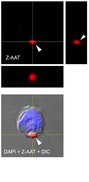
Short report                                                                                                        Cell Biology Medicine

                                    accumulation of Z-AAT polymers in hepatocytes is a marker for diagnosing AATD
                                    (Janciauskiene et al., 2011). The intracellular Z-AAT polymers have also been identified in other
                                    AAT-expressing cells like monocytes and macrophages (Belchamber et al., 2020). The accumulation
                                    of polymers is harmful for AAT-producing cells, whereas the circulating Z-AAT polymers are not able
                                    to execute the tasks of AAT protein, a major inhibitor of serine proteases having a strong immuno-
                                    modulatory potential. Based on the facts that: (i) circulating Z-AAT polymers contribute to the risk of
                                    developing pathologies (Parmar et al., 2002; Strnad et al., 2020), (ii) pathogenic oligomeric pro-
                                    teins affect CX3CR1 expression (Bolós et al., 2017), and (iii) CX3CR1/CX3CL1 axis plays a significant
                                    role in immunity (Imai and Yasuda, 2016), we aimed to investigate CX3CR1 expression in PBMCs of
                                    ZZ AATD individuals. For this, in collaboration with German Alpha1 Patient Association and Aachen
                                    University, was prepared RNA from freshly isolated PBMCs of 41 clinically stable ZZ AATD volun-
                                    teers independently of their clinical diagnosis or treatment with intravenous AAT, a specific augmen-
                                    tation therapy (Janciauskiene and Welte, 2016). For comparison, PBMCs isolated from healthy
                                    volunteers having normal plasma AAT levels were used. Additionally, a limited amount of RNA sam-
                                    ple was available from PBMCs isolated from a cohort of 12 ZZ AATD emphysema patients at Leiden
                                    University Medical Center, The Netherlands (Figure 1—figure supplement 1).
                                       Independent of individual’s age, clinical diagnosis (healthy, lung or liver disease), or augmentation
                                    therapy, the CX3CR1 mRNA expression turned to be much lower in ZZ AATD PBMCs than in PBMCs
                                    from non-AATD controls (median [range]: 4.1 [2.7–5.5] vs. 18.5 [13–26.6], p
Short report Cell Biology Medicine
CX3CR1 expression in human CD56+ NK cells (Barlic et al., 2003; Sechler et al., 2004). However,
plasma levels of IL-15 were lower in ZZ AATD than in non-AATD (pg/ml, median [range]: 6.6 [5.9–
6.9], n = 23 vs. 7.63 [6.63–8.1], n = 21, p=0.001), excluding a possible link between IL-15 and
CX3CR1 mRNA levels.
Because ZZ AATD individuals, differently from non-AATD, have about 90% lower blood concen-
tration of Z-AAT protein, which may influence cellular microenvironment (Ramos et al., 2010), a rela-
tionship between CX3CR1 and Z-AAT plasma levels cannot be excluded. However, no correlation
was found between CX3CR1 mRNA in PBMCs and plasma levels of Z-AAT measured by nephelome-
try (data not shown). Next, plasma Z-AAT polymers were measured, as the biomarkers of all carriers
of the Z allele (Tan et al., 2014; Janciauskiene et al., 2002). As anticipated, only minor amounts of
polymers were detected in plasma of non-AATD individuals while plasma of ZZ AATD contained
high amounts of polymers (mg/ml, mean [SD]: 4.1 [6], n = 18 vs. 1399.8 [750], n = 20, respectively).
Since most of the ZZ AATD individuals received intravenous augmentation therapy with plasma puri-
fied AAT protein, ZZ AATD individuals were segregated into subgroups who receive or not receive
therapy. There were no significant differences in Z-AAT polymer levels between the subgroups: (mg/
ml, median [range]: non-augmented 1506.6 [854–1781], n = 17 vs. augmented 1348.5 [779.5–1529],
n = 23, respectively). A previous study used a sandwich ELISA based on 2C1 antibody and found
that circulating Z-AAT polymers range between 8.2 and 230.2 mg/ml in ZZ AATD (Tan et al., 2014)
whereas much higher circulating levels of Z-AAT polymers were detected by using the single mono-
clonal antibody (LG96)-based ELISA. These discrepancies can be due to the differences between
antibody specificities. For example, 2C1 showed high affinity for polymers formed by heating M- or
Z-AAT at 60˚C (Miranda et al., 2010) while LG96 antibody recognizes naturally occurring/native
Z-AAT polymers without requiring sample heating. To answer, why some individuals have higher
plasma levels of Z-AAT polymers than monomers (measured by nephelometry) is of great impor-
tance for the further studies.
Most interestingly, in ZZ AATD individuals, was found a trend toward an inverse relationship
between CX3CR1 mRNA in PBMCS and plasma Z-AAT polymers (r2 = 0.31, n = 38, p=0.055)
(Figure 1C). This latter finding prompted more extensive investigation whether Z-AAT polymers
affect CX3CR1 expression when added to healthy donor PBMCs for 18 hr, ex vivo. Lipopolysaccha-
ride (LPS, from Escherichia coli, 1 mg/ml) was included as a known reducer of CX3CR1 expression
(Pachot et al., 2008; Sica et al., 1997). Indeed, polymeric Z-AAT in a concentration-dependent
manner lowered CX3CR1 mRNA (Figure 2—figure supplement 2) whereas repeated experiments
using Z-AAT at a constant concentration of 0.5 mg/ml reduced CX3CR1 mRNA more than twice as
compared to non-treated controls (Figure 2A). Accordingly, LPS and polymer containing Z-AAT
preparation significantly decreased surface expression of CX3CR1, specifically in CD14+ monocytes
and NK cells (Figure 3).
By contrast, cellular levels of CX3CR1 protein increased in PBMCs treated with Z-AAT polymers
or LPS (used as a positive control) as compared to non-treated controls (Figure 2B). The CX3CR1
protein was present in detergent resistant lipid raft fraction of PBMCs treated with Z-AAT (Fig-
ure 2—figure supplement 3A). Total cell lysates and lipid raft fractions from Z-AAT-treated PBMCs,
in contrast to those prepared from M-AAT-treated or non-treated PBMCs, contained high amounts
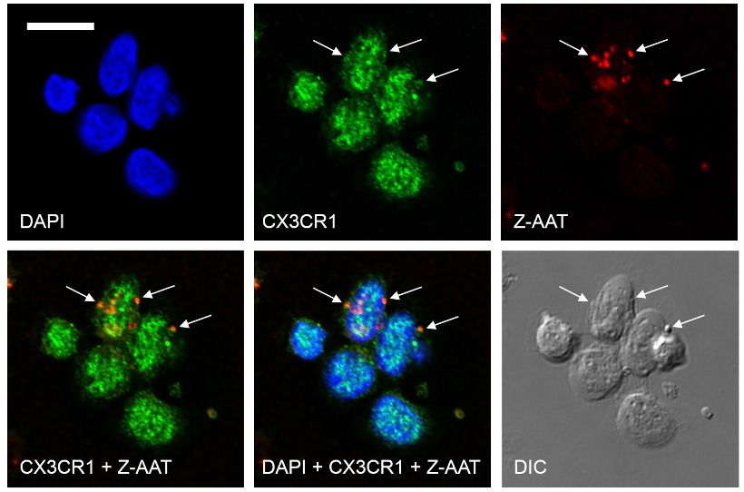
of AAT polymers (Figure 2C, Figure 2—figure supplement 3B). The laser scanning confocal micros-
copy of double-labeled specimens showed a co-localization of Z-AAT polymers with CX3CR1 protein
(Figure 2D). Furthermore, 3D reconstruction of cross sections visualized larger Z-AAT aggregates
surrounded by cellular extensions in a cap-like formation, suggesting that cells may react differently
depending on the size of Z-AAT polymers (Figure 2E). It cannot be excluded that Z-AAT polymers,
similar like polymers of Tau protein, interact with CX3CR1 and get internalized
(Chidambaram et al., 2020). This may determine the fate of CX3CR1 mRNA expression, that is,
sequestered intracellularly and not returning to the cell surface, CX3CR1 protein may induce signal-
ing pathways lowering CX3CR1 expression. To achieve a definitive answer how Z-AAT or other types
of protein polymers regulate CX3CR1 levels, detailed mechanistic studies are required. In general,
along with transcriptional regulation, chemokine receptors trafficking is of great importance to
understand (Kershaw et al., 2009).
Under the same experimental conditions, monomeric M-AAT had no effect on CX3CR1 mRNA
expression relative to housekeeping gene HPRT1 (mean [SD]: 24.9 [2.9] controls, n = 5 vs. 23.7 [1.3],
n = 5, NS), and protein levels (Figure 2B). Likewise, monomeric Z-AAT protein does not affect
Tumpara et al. eLife 2021;10:e64881. DOI: https://doi.org/10.7554/eLife.64881 4 of 16Short report Cell Biology Medicine
A B Control LPS M-AAT Z-AAT C Control LPS M-AAT Z-AAT
25
CX3CR1 expression relative to
55 kDa 130 kDa
housekeeping gene HPRT1
20
100 kDa
15 P < 0.001 70 kDa
CX3CR1
55 kDa
10
P < 0.001 β-Actin
5
Relative intensity (fold change)
Control LPS M-AAT Z-AAT AAT
0
Control LPS Z-AAT 1.00 7.147 0.990 4.531
1 µg/ml 0.5 mg/ml 1.00 9.693 1.528 5.888
1.00 4.211 0.399 3.687 β-Actin
D E
Figure 2. Effects of Z alpha-1 antitrypsin (Z-AAT) and M-AAT on CX3CR1 mRNA and protein expression. (A) CX3CR1 gene expression relative to
HPRT1 housekeeping gene was determined by real-time PCR using Taqman gene expression assays. Peripheral blood mononuclear cells (PBMCs) were
incubated for 18 hr with plasma-derived Z-AAT, lipopolysaccharide (LPS), or M-AAT in the concentrations as indicated, or with RPMI medium alone
(control). The data from n = 6 independent experiments are presented as median (IQR) in box and whisker plot format; lines represent medians in each
box. Measurements were carried out in duplicates. p-Value was calculated by nonparametric Kruskal-Wallis test. (B) Representative uncut Western blot
(n = 3 independent experiments) of CX3CR1 in RIPA lysates prepared from PBMCs incubated for 18 hr alone or with LPS (1 mg/ml), M-AAT (1 mg/ml), or
Z-AAT (0.5 mg/ml). For analysis of CX3CR1, equal amounts of protein were separated by SDS-PAGE under reducing conditions. Relative intensities
were calculated for each band using the ratio relative to b-actin, as a loading control, and then normalized by the experimental control. (C) For analysis
of cellular AAT, the same lysates were separated under non-reducing conditions. Western blots were probed with polyclonal rabbit anti-human AAT
recognizing monomeric, polymeric, or truncated forms of AAT. One representative blot from n = 3 independent experiments is shown. b-Actin was
used for a loading control. (D) and (E) Co-distribution of Z-AAT polymers with CX3CR1 in human total PBMCs incubated with 0.5 mg/ml Z-AAT
polymers for 18 hr. (D) Immunofluorescence microscopy revealed co-localization of Z-AAT polymers (red) with CX3CR1-positive structures (green).
Arrows point areas of co-localization. Scale bar, 10 mm. (E) Confocal microscopy 3D stack with orthogonal reconstruction shows an aggregate of Z-AAT
polymers (red) surrounded by CX3CR1-positive (green) cellular extensions forming a cap-like structure (arrowhead). Scale bar, 5 mm. The images with
indicated channels merged and the corresponding differential interference contrast (DIC) image are presented. 4’, 6- Diamidino 2-phenylindole (DAPI)
was used for nuclei staining (blue).
The online version of this article includes the following source data and figure supplement(s) for figure 2:
Source data 1. Source file, containing original data for Figure 2A, to document reduced CX3CR1 expression in peripheral blood mononuclear
cells (PBMCs) treated with Z alpha-1 antitrypsin (Z-AAT) or lipopolysaccharide (LPS) (A).
Figure supplement 1. Quality control of isolated M- and Z-AAT (alpha-1 antitrypsin) proteins by SDS-PAGE.
Figure supplement 2. Z alpha-1 antitrypsin (Z-AAT) in a concentration-dependent manner reduces CX3CR1 mRNA expression in peripheral blood
mononuclear cells (PBMCs) isolated from healthy donors.
Figure supplement 3. Z alpha-1 antitrypsin (Z-AAT) induces association of CX3CR1 with lipid rafts.
Figure supplement 4. CX3CR1 transcript and protein expressions in the presence of Z alpha-1 antitrypsin (Z-AAT) monomer, Z-AAT polymer, native
M-AAT, and M-AAT polymer.
Figure supplement 5. Effect of CX3CL1 alone or in combination with Z alpha-1 antitrypsin (Z-AAT) on CX3CR1 transcript and protein expression.
Figure 2 continued on next page
Tumpara et al. eLife 2021;10:e64881. DOI: https://doi.org/10.7554/eLife.64881 5 of 16Short report Cell Biology Medicine
Figure 2 continued
Figure supplement 5—source data 1. Source file, containing original data for Figure 2—figure supplement 5A, to document CX3CR1 gene expres-
sion in peripheral blood mononuclear cell (PBMC).
CX3CR1 mRNA and protein levels, and heat-induced polymers of M-AAT showed no effect on
CX3CR1 expression as well (Figure 2—figure supplement 4). Probably, specific conformational
properties and/or molecular sizes of Z-AAT polymers are required for their interaction with CX3CR1.
For example, cell surfaces express CX3CL1 as a constitutive oligomer (three to seven molecules),
which is essential for efficient interaction with CX3CR1 (Hermand et al., 2008; Ostuni et al., 2020).
Numerous chemokines tend to self-associate that determines their activity (Proudfoot et al., 2003),
and therefore certain Z-AAT polymers may resemble chemokine structures competing for the same
receptor(s). In some experimental models, Z-AAT polymers expressed strong chemotactic properties
(Parmar et al., 2002; Lomas and Mahadeva, 2002). When chemokine receptors are engaged in
chemotaxis, they can be removed from the cell surface by the ligand–receptor internalization
(Springer, 1994), which might explain a decrease of CX3CR1 in ZZ PBMCs. Interestingly, the soluble
form of CX3CL1, even when used at a high concentration of 500 ng/ml, does not antagonize Z-AAT
polymer effects on CX3CR1 mRNA and protein levels, and by itself showed no effect on CX3CR1
A Monocytes 2500
CX3CR1 mean fluorescence
2000
Control
intensity
1500
1000
LPS (1 µg/ml)
500 p < 0.001
p < 0.001
Z-AAT (0.5 mg/ml) 0
l S T
ro LP AA
nt Z-
CX3CR1-PE Co
NK cells (CD56lo CD16+)
B 3000
CX3CR1 mean fluorescence
2500
Control 2000
intensity
1500 p < 0.001
LPS (1 µg/ml) p < 0.001
1000
500
Z-AAT (0.5 mg/ml) 0
l S T
ro LP
nt AA
CX3CR1-PE Co Z-
Figure 3. Flow cytometric analysis of CX3CR1 surface expression in peripheral blood mononuclear cells (PBMCs)
after incubation with RPMI alone (control), Z alpha-1 antitrypsin (Z-AAT), or lipopolysaccharide (LPS) in the
concentrations as indicated for 18 hr. CX3CR1-expressing cells were found in the monocyte gate (A) and in the NK
cell gate (CD56lo CD16+) (B). Histograms show representative results and bars represent mean (SD) of n = 4
independent biological repeats each measured one time. After incubation with Z-AAT or LPS, monocytes and NK
cells show significantly reduced CX3CR1 surface expression in comparison to untreated control cells. p-Values
were calculated by one-way ANOVA.
The online version of this article includes the following source data and figure supplement(s) for figure 3:
Source data 1. Source files, containing original data for Figure 3A,B, to document reduced CX3CR1 surface
expression in monocytes (A) and NK cells (B) after treatment with Z alpha-1 antitrypsin (Z-AAT) or lipopolysaccha-
ride (LPS) (A).
Figure supplement 1. Gating strategy: sequential gating to identify monocytes and NK cells from total peripheral
blood mononuclear cells (PBMCs).
Tumpara et al. eLife 2021;10:e64881. DOI: https://doi.org/10.7554/eLife.64881 6 of 16Short report Cell Biology Medicine
mRNA or protein levels (Figure 2—figure supplement 5), although some studies reported that
CX3CL1 reduces CX3CR1 expression (Pachot et al., 2008; White et al., 2014). In solution CX3CL1
remains monomeric, even at high concentrations (Hermand et al., 2008; Mizoue et al., 1999)
whereas, as mentioned above, membrane-bound CX3CL1 occurs as oligomer. These two forms of
the CX3CL1 perform differential roles (Winter et al., 2020), and therefore it cannot be excluded
that oligomeric, but not a soluble, form of CX3CL1 would compete with Z-AAT for CX3CR1 interac-
tion in vivo.
The CX3CR1 helps to define the major subsets of human monocytes because classical monocytes
express much lower levels of CX3CR1 than non-classical monocytes (Ziegler-Heitbrock et al.,
2010). After in vitro challenge with LPS for longer periods (like for 18 hr), human monocytes are
known to increase in the mRNA and membrane expression of CD14, a receptor for LPS
(Landmann et al., 1996). The enhancement of CD14 expression after treatment of PBMCs with
Z-AAT strikingly resembled LPS (Figure 4 and Figure 4—figure supplement 1). This raised a suspi-
cion that Z-AAT preparations might contain endotoxin. According to the limulus amebocyte lysate
test, endotoxin levels of Z-AAT preparations were below detection limit (0.01 EU/ml). Moreover, LPS
significantly induced expression of TNFa, IL-6, and IL-1b while polymer containing Z-AAT prepara-
tions had no effect (Figure 4—figure supplement 2). Besides, LPS but not Z-AAT significantly
increased release of cytokines (IL-1b, pg/ml, median [range]: LPS 1342.9 [1008–1834] vs. Z-AAT 3.2
[2.5–5.9] vs. controls 2.5 [2.1–3.7], n = 4 independent experiments; TNFa, ng/ml, mean [SD]: LPS
19.5 [2.5] vs. Z-AAT vs. controls, undetectable, n = 4 and IL-6, ng/ml, median [range]: LPS 15903.5
[14,626–17,262] vs. Z-AAT 5.4 [2.9–6.1] vs. control [1.7 (1.0–2.3), n = 4]). Therefore, the effect of
Z-AAT preparations on CD14 is valid and unrelated to a potential LPS contamination. Although both
Z-AAT polymers and LPS induce CD14 expression and similarly affect CX3CR1 expression and pro-
tein levels, data imply that Z-AAT polymers and LPS do not share the same signaling mechanisms.
As a side note, it has been reported that CD14++ monocytes have the lowest expression of
CX3CR1 (Appleby et al., 2013). Low and high surface CX3CR1 levels are suggested to delineate
A 40
B 25000
p < 0.001 p < 0.001
CD14 expression relative to
housekeeping gene HPRT1
fluorescence intensity
20000
30
Control
CD14 mean
15000
20 p < 0.001
LPS (1 µg/ml) 10000
10
5000
Z-AAT (0.5 mg/ml)
0 0
l S T
ro LP AA
Control Z-AAT CD14-FITC nt Z-
0.5 mg/ml Co
Figure 4. Z alpha-1 antitrypsin (Z-AAT) and lipopolysaccharide (LPS) induce CD14 expression. (A) Z-AAT increases CD14 gene expression. Peripheral
blood mononuclear cells (PBMCs) were incubated for 18 hr with 0.5 mg/ml Z-AAT or RPMI medium alone (control). CD14 mRNA expression relative to
HPRT1 was determined by real-time PCR using Taqman gene expression assays. Measurements were carried out in duplicates. Data are represented as
median (IQR) in boxplots, lines represent medians of n = 14 independent biological repeats. Outliers are defined as data points located outside the
whiskers. p-Values were calculated using nonparametric Kruskal-Wallis test. (B) Z-AAT increases monocyte CD14 surface expression. PBMCs were
cultured with RPMI (control), Z-AAT, or LPS for 18 hr. CD14 mean fluorescence intensities of monocytic cells were determined by flow cytometry.
Histograms show representative results and bars represent mean (SD) of n = 4 independent biological repeats each measured one time. p-Values were
calculated from ANOVA.
The online version of this article includes the following source data and figure supplement(s) for figure 4:
Source data 1. Source files, containing original data for Figure 4A, B to document CD14 gene expression in peripheral blood mononuclear
cells (PBMCs) (A) and CD14 surface expression in monocytes (B).
Figure supplement 1. Inverse changes in CD14 and CX3CR1 mRNA expression in peripheral blood mononuclear cells (PBMCs) treated with different
concentrations of Z alpha-1 antitrypsin (Z-AAT).
Figure supplement 2. Z alpha-1 antitrypsin (Z-AAT) does not induce cytokine expression.
Figure supplement 2—source data 1. Source files, containing original data for Figure 4—figure supplement 2 to document IL1B (a), IL6 (b), and
TNFA (c) gene expression in peripheral blood mononuclear cells (PBMCs).
Tumpara et al. eLife 2021;10:e64881. DOI: https://doi.org/10.7554/eLife.64881 7 of 16Short report Cell Biology Medicine
two functional subsets of murine blood monocytes: ‘inflammatory’ and ‘resident monocytes’
(Geissmann et al., 2003). This dichotomy appears conserved in humans as CD14+ CD16 , and
CD14low CD16+ monocytes resemble ‘inflammatory’ and ‘resident’ monocytes. Previous study dem-
onstrated that peripheral blood monocytes of clinically healthy young adults (30 years of age) with
ZZ AATD have significantly higher mRNA and surface expression of CD14 as compared to age
matched MM subjects (Sandström et al., 2008). Authors thought that the higher CD14 expression
reflects early pathological processes whereas according to the current findings this phenomenon
seems to relate with the circulating Z-AAT polymers.
During steady state, non-classical monocytes expressing CX3CR1 patrol healthy tissues through
crawling on the resting endothelium but these monocytes are required for a rapid tissue invasion at
the site of infection or inflammation (Auffray et al., 2009; Cros et al., 2010). Previous work evi-
denced that the non-classical subset of monocytes, characterized by high expression of CX3CR1, is
almost absent in ZZ AATD emphysema patients (Stolk et al., 2019). Moreover, plasma levels of AAT
polymers were found to correlate with the levels of endothelium-related markers like sE-selectin and
sICAM-1 (Aldonyte et al., 2004). Beyond, in a small cohort of ZZ AATD emphysema patients, was
found a strong inverse association between lung function, based on percentage (%) predicted trans-
fer factor for carbon monoxide (TLCO%pred) and forced expiratory volume in 1 s (FEV1%pred), and
plasma levels of Z-AAT polymers: (TLCO%pred [r2 = 0.75, n = 9, p=0.02] and FEV1%pred
[r2 = 0.82, n = 9, p=0.006]). Thus, higher levels of Z-AAT polymers and lower numbers of CX3CR1-
positive cells may favor the development of lung injury and disease. A decrease in the expression of
CX3CR1 on human monocytes has been shown in patients with atopic dermatitis (Echigo et al.,
2004) and septic shock (Pachot et al., 2008).
To date, many functional aspects of the CX3CR1–CX3CL1 axis have been suggested, including
the adhesion of immune cells to vascular endothelial cells, chemotaxis, the crawling of the mono-
cytes that patrol on vascular endothelial cells, the retention of monocytes of the inflamed endothe-
lium to recruit inflammatory cells, and the survival of the macrophage. Considering the above, these
different aspects of interactions between PBMCs and Z-AAT or other polymers occurring due to
genetic or post-translational protein modifications require further investigations in dedicated clinical
and experimental studies.
Materials and methods
Key resources table
Reagent type
(species) or
resource Designation Source or reference Identifiers Additional information
Biological PBMCs Blood samples Collected from 41
sample (Homo ZZ AATD and 21
sapiens) non-AATD healthy
controls
Sequenced- CX3CR1 Thermo Fisher Scientific Taqman assay 4331182 Hs00365842_m1
based reagent
(human)
Sequenced- TNFA Thermo Fisher Scientific Taqman assay 4331182 Hs01113624_g1
based reagent
(human)
Sequenced- IL6 Thermo Fisher Scientific Taqman assay 4331182 Hs00985639_m1
based reagent
(human)
Sequenced- IL1B Thermo Fisher Scientific Taqman assay 4331182 Hs01555410_m1
based reagent
(human)
Sequenced- HPRT1 Thermo Fisher Scientific Taqman assay 4331182 Hs02800695_m1
based reagent
(human)
Continued on next page
Tumpara et al. eLife 2021;10:e64881. DOI: https://doi.org/10.7554/eLife.64881 8 of 16Short report Cell Biology Medicine
Continued
Reagent type
(species) or
resource Designation Source or reference Identifiers Additional information
Sequenced- CD14 Thermo Fisher Scientific Taqman assay 4331182 Hs00169122_g1
based reagent
(human)
Other Non-adherent 12-well plates Greiner Bio-One 665970
Other Alpha-1 Antitrypsin Select matrix GE Healthcare Life Sciences, Cytiva 17547201
Other Plasma purified human AAT CSL Behring Respreeza
Other LPS Sigma-Aldrich L2880 Escherichia coli
O55:B5
Commercial Pierce Chromogenic Endotoxin Thermo Fisher Scientific A39553
assay or kit Quant Kit
Commercial UltraRIPA kit BioDynamics Laboratory F015
assay or kit
Antibody Anti-AAT polymer, Deposited at LG96 ELISA: Coating: 1 mg/ml;
LG96 (mouse German Collection LG96-HRP conjugate: (1:2000)
monoclonal) of Microorganisms Immunofluorescence: (1:5000)
and Cell Cultures:
DSM ACC3092
Antibody Anti-human AAT Agilent Dako A001202-2 (1:800)
(rabbit polyclonal)
Antibody Anti-AAT Hycult Biotech Clone 2C1; HM2289 (1:500)
polymer
(mouse monoclonal)
Antibody Anti-CX3CR1 Abcam ab8021 (1:500)
(rabbit polyclonal)
Antibody Anti-b-actin Sigma-Aldrich Clone AC-15; HRP-conjugated; (1:20,000)
(mouse monoclonal) A3854
Antibody Anti-CX3CR1 (rabbit polyclonal) Abcam ab8021 (1:500)
Antibody Anti-CX3CR1 (mouse monoclonal) Invitrogen, Thermo Fisher Scientific Clone 2A9-1; 12-6099-42 PE-conjugated; 5 ml per test
Antibody Anti-CD14 (mouse monoclonal) Invitrogen, Thermo Fisher Scientific Clone TuK4; MHCD1401 FITC-conjugated; 5 ml per test
Antibody Anti-CD16 (mouse monoclonal) Immunotools Clone 3G8; 21278166 APC-conjugated; 5 ml per test
Antibody Anti-CD56 (mouse monoclonal) BD Biosciences Clone NCAM16.2; 566124 BV-480 conjugated; 5 ml per test
Peptide, Human CX3CL1/Fractalkine Bio-Techne 365-FR-025
recombinant
protein
Software, FlowJo Becton, Dickinson and Company v10
algorithm
Software, SigmaPlot 14 Systat Software v14.0
algorithm
Study approval
The study cohort consists of 41 clinically stable ZZ AATD volunteers collected in collaboration with
German Alpha1 Patient Association and Aachen University independently on their clinical diagnosis
or treatment with intravenous AAT and 21 non-AATD healthy controls. The institutional review board
of Aachen University (EK 173/15) provided ethical approval for individuals recruited in Germany. For
Z-AAT polymer determination, we added 12 ZZ AATD emphysema patients recruited at Leiden Uni-
versity Medical Center. In addition, ZZ AATD emphysema patients (four males and five females)
were enrolled with mean (SD): age 51 (6.6) years, forced expiratory volume in 1 s percent predicted
(FEV1%pred, 66.3; Pachot et al., 2008) and transfer factor of the lung for carbon monoxide percent
predicted (TLCO%pred, 64; Sica et al., 1997). The plasma levels of Z-AAT polymers in these cases
were median (range) 714.2 ([412.9–2270.4] mg/ml). Leiden University Medical Center provided ethical
approval (project P00.083 and P01.101) for the additional study groups. For all individuals, detailed
medical records data were anonymized. All participants issued a written informed consent according
Tumpara et al. eLife 2021;10:e64881. DOI: https://doi.org/10.7554/eLife.64881 9 of 16Short report Cell Biology Medicine
to the ethical guidelines of the Helsinki Declaration (Hong Kong Amendment) as well as Good Clini-
cal Practice (European guidelines).
Isolation of PBMCs
Total PBMCs were isolated from freshly obtained peripheral blood (within 6 hr) using Lymphosep (C-
C-Pro, Oberdorla, Germany) discontinuous gradient centrifugation according to the manufacturer’s
instructions as described previously (Frenzel et al., 2014). Thereafter, cells were lysed with RLT
buffer for RNA analysis or suspended in RPMI-1640 medium (Gibco, Thermo Fisher Scientific, Wal-
tham, MA) and plated into non-adherent 12-well plates (Greiner Bio-One, Kremsmünster, Austria)
for the further analyses.
Real-time PCR analysis
Isolation of total RNA, synthesis of cDNA and mRNA analysis using Taqman gene expression assays
(Thermo Fisher Scientific, Waltham, MA, Table 1) were performed as described previously
(Frenzel et al., 2014). Real-time PCR was carried out in duplicates. RNA quality was checked on
agarose gels.
AAT polymer ELISA
The AAT polymer ELISA using the monoclonal antibody LG96 (deposited under access number DSM
ACC3092 at German Collection of Microorganisms and Cell Cultures) was developed by Candor Bio-
sciences. Normal M-AAT was used for a negative control. Recovery ratio, signal-to-noise ratio, cali-
bration curve, sample stability under different storage conditions were tested and all tests passed. A
cross-reactivity with M-AAT was not reported in any of the tests. Nunc MaxiSorp flat-bottom 96-well
plates (Thermo Fisher, Waltham, MA) were coated overnight at 2–8˚C with monoclonal antibody
LG96, at 1 mg/ml in coating buffer pH 7.4 (Candor Biosciences, Wangen, Germany). After a 2 hr
blocking step, the plasma samples were applied in the previously determined dilutions made in Low-
Cross-Buffer (Candor Biosciences), which also served as a blank. Incubation was performed for 2 hr
at room temperature (RT). For detection, the captured antigen was incubated with antibody (LG96)-
horseradish peroxidase (HRP) conjugate (1:2000) for 2 hr. The conjugate was prepared in advance
with the HRP Conjugation Kit Lightning-Link (Abcam, Cambridge, UK) according to the manufac-
turer’s instructions. For signal development SeramunBlau fast2 microwell peroxidase substrate (Sera-
mun, Heidesee, Germany) was used. The incubation was performed at RT for 12 min in the dark and
the reaction was stopped with 2 M H2SO4. Plates were analyzed at 450 nm by microplate reader
(Dynex, Chantilly, VA) equipped with Dynex Revelation 4.21 software. Measurements were carried
out in triplicates.
Preparation of AAT proteins
Plasma M- and Z-AAT was isolated by affinity chromatography using the AAT-specific Alpha-1 Anti-
trypsin Select matrix (GE Healthcare Life Sciences, Cytiva, Sheffield, UK) according to the manufac-
turer’s recommendations. For Z-AAT preparation plasma from volunteers not receiving AAT
augmentation therapy was pooled. To change the buffer in the M- and Z-AAT protein pools to
Hank’s balanced salt solution (HBSS, Merck, Darmstadt, Germany), Vivaspin centrifugal concentra-
tors with 10,000 MWCO (Vivaproducts, Littleton, MA) were used. Plasma purified human AAT (99%
purity, Respreeza, Zemaira, CSL Behring, Marburg, Germany) was changed to HBSS by the same
Table 1. Primers for gene expression analysis.
Primer Assay ID
CX3CR1 Hs00365842_m1
TNFA Hs01113624_m1
IL6 Hs00985639_m1
IL1B Hs01555410_s1
HPRT1 Hs02800695_m1
CD14 Hs00169122_g1
Tumpara et al. eLife 2021;10:e64881. DOI: https://doi.org/10.7554/eLife.64881 10 of 16Short report Cell Biology Medicine
method. Protein concentrations were determined using Pierce BCA Protein Assay Kit
(Thermo Fisher, Waltham, MA). The quality of the M- and Z-AAT preparations was confirmed on
Coomassie gels (10% SDS-PAGE, Figure 2—figure supplement 1A) and by analyzing endotoxin lev-
els with Pierce Chromogenic Endotoxin Quant Kit according to the manufacturer’s guidelines
(Thermo Fisher, Waltham, MA) using TECAN Infinite M200 PRO (Männedorf, Switzerland). In both,
M- and Z-AAT preparations, endotoxin levels were below the detection limit (assay sensitivity: 0.01–
0.1 EU/ml).
Preparation of Z-AAT monomers
Z-AAT was isolated by affinity chromatography using AAT-specific Alpha-1 Antitrypsin Select matrix
as described above. After the isolation Z-AAT, protein was diluted with sterile 0.9% NaCl (Fresenius
Kabi, Bad Homburg, Germany), and Vivaspin-20, 100 kDa centrifugal column units (Sartorius, Göttin-
gen, Germany) were used to separate Z-AAT monomers from polymers. Protein concentrations were
determined using the Pierce BCA Protein Assay Kit (Thermo Fisher Scientific, Carlsbad, CA) accord-
ing to manufacturer’s instructions. The Z-AAT protein monomers were confirmed by using 7.5%
SDS-PAGE without sample heating and without b-mercaptoethanol (Figure 2—figure supplement
1B).
In vitro experiments with PBMCs from healthy donors
PBMCs (5 106 cells/ml) were incubated for 18 hr at 37˚C, 5% CO2 either alone, or with Z- or
M-AAT proteins, or LPS (1 mg/ml, E. coli O55:B5, Sigma-Aldrich, Merck, St. Louis, MO). In some
experiments, a recombinant CX3CL1 protein (R&D Systems, Bio-Techne, Minneapolis, MN) was
used. Protein was reconstituted at a concentration of 25 mg/ml in sterile PBS containing 0.1% BSA
(Sigma-Aldrich) and added to PBMCs at various concentrations up to 500 ng/ml either alone or
together with Z-AAT (0.5 mg/ml) for 18 hr. Afterward, cells were used for RNA isolation, flow cytom-
etry or Western blot analysis. For Western blot, PBMCs were lysed in RIPA buffer (Sigma-Aldrich),
supplemented with protease inhibitor cocktail (Sigma-Aldrich). For some Western blot experiments,
we extracted detergent resistant lipid raft associated proteins from insoluble cell fractions using
UltraRIPA kit according to the supplier’s instructions (BioDynamics Laboratory, Tokyo, Japan).
Western blot
Equal amounts of lysed proteins were separated by 7.5% or 10% SDS-polyacrylamide gels (under
reducing conditions for CX3CR1 and non-reducing for total AAT or AAT polymer analysis) prior to
transfer onto polyvinylidene difluoride membranes (Merck-Millipore, Burlington, MA). Membranes
were blocked for 1 hr with 5% low fat milk (Carl Roth, Karlsruhe, Germany) followed by overnight
incubation at 4˚C with specific primary antibodies: polyclonal rabbit anti-human AAT (1:800) (DAKO
A/S, Glostrup, Denmark), mouse monoclonal anti-AAT polymer antibody (clone 2C1, 1:500, Hycult
Biotech, Uden, The Netherlands), rabbit polyclonal anti-CX3CR1 (1:500, Abcam, Cambridge, UK), or
HRP-conjugated monoclonal anti-b-actin antibody (1:20,000, Sigma-Aldrich, Merck, St. Louis, MO)
for a loading control. The immune complexes were visualized with anti-rabbit or anti-mouse HPR-
conjugated secondary antibodies (DAKO A/S) and enhanced by Clarity Western ECL Substrate (Bio-
Rad, Hercules, CA). Images were acquired by using Chemidoc Touch imaging system (BioRad) under
optimal exposure conditions and processed using Image Lab v5.2.1 software (BioRad). For quantifi-
cation, the signal intensity of the CX3CR1 protein band in each lane was divided by the correspond-
ing b-actin band intensity (normalization factor or loading control). Afterward, the normalized signal
of each lane was divided by the normalized target signal observed in the control sample to get the
abundance of the CX3CR1 protein as a fold change relative to the control.
ELISA
Plasma samples from 22 ZZ AATD and 21 non-AATD controls were analyzed for CX3CL1/Fractalkine
using Duoset kit (R&D Systems, Minneapolis, MN, assay sensitivity 0.072 ng/ml, detection range 0.2–
10 ng/ml). Cell-free culture supernatants were analyzed directly or stored at 80˚C. ELISA Duoset
kits for TNF-a (assay detection range 15.6–1000 pg/ml), IL-1b/IL-1F2 (assay detection range 3.91–
250 pg/ml), and IL-6 (assay detection range 9.38–600 pg/ml) were purchased from R&D Systems
(Minneapolis, MN) and were used according to the manufacturer’s instructions. Plates were
Tumpara et al. eLife 2021;10:e64881. DOI: https://doi.org/10.7554/eLife.64881 11 of 16Short report Cell Biology Medicine
measured on Infinite M200 microplate reader (Tecan, Männedorf, Switzerland). Measurements were
carried out in duplicates.
Flow cytometry analysis
PBMCs (2 106 cells per condition) were incubated with LPS (1 mg/ml), M-AAT (1 mg/ml), or Z-AAT
(0.5 mg/ml) for 18 hr. Staining was performed with phycoerythrin (PE)-conjugated mouse monoclonal
anti-CX3CR1 antibody (clone 2A9-1 Invitrogen, Thermo Fisher Scientific, Carlsbad, CA), fluorescein
(FITC)-conjugated mouse monoclonal anti-CD14 antibody (clone TuK4, Life Technologies,
Thermo Fisher Scientific, Carlsbad, CA), allophycocyanin (APC)-conjugated mouse monoclonal anti-
CD16 antibody (clone 3G8, Immunotools, Friesoythe, Germany), or BV-480-conjugated anti-CD56
mouse monoclonal antibody (Clone NCAM16.2, BD Biosciences, San Jose, CA) alone or in combina-
tions. Dead cells were excluded by a staining with 7-amino-actinomycin D. Samples were measured
on a BD FACSAria Fusion machine and analyzed with FlowJo v10 (Becton, Dickinson and Company,
Franklin Lakes, NJ). The gating strategy is shown in Figure 3—figure supplement 1.
Immunofluorescence confocal laser microscopy
Human total PBMCs (2 106) were plated onto glass coverslips and incubated alone or with Z-AAT
polymers (0.5 mg/ml) in RPMI medium for 18 hr at 37˚C and 5% CO2. Cells were then washed with
PBS, fixed with 3% paraformaldehyde in PBS for 20 min, and continued with or without permeabiliza-
tion with 0.5% Triton X-100 in PBS for 5 min at RT. For immunolabeling, cells were co-incubated with
primary antibodies against human CX3CR1 (rabbit polyclonal IgG [1:500], Abcam, Cambridge, UK)
and anti-AAT polymer antibody, LG96 (1:5000, mouse monoclonal) for 1 hr, at RT. After washing,
the cells were incubated with corresponding secondary antibodies (1:1000) conjugated to Alexa-
Fluor-488 (goat anti-rabbit) or AlexaFlour-594 (goat anti-mouse) both from Thermo Fisher Scientific,
Rockford, IL. After final wash, the cells were mounted on microscope slides using ProLong Gold
Antifade Mountant with DAPI (Thermo Fisher Scientific, Carlsbad, CA). Images were acquired using
confocal laser microscope FluoView 1000 (Olympus, Shinjuku, Japan) equipped with a 60 oil
immersion objective and differential interference contrast in sequential mode. Confocal z-stacks
were collected with a 0.25 mm increment.
Statistics
Data were analyzed and visualized by using Sigma Plot 14.0. One-tailed Student’s t-test was applied
to compare two sample means on one variable. When more than two groups were involved in the
comparison, one-way ANOVA was used. Data were presented as mean (SD). If normality test failed,
the nonparametric Kruskal-Wallis one-way analysis or Mann-Whitney rank sum test was performed,
and data were presented as median (range). For correlation analysis, the Pearson’s linear correlation
method was used to measure the correlation for a given pair. A p-value of less than 0.05 was consid-
ered significant.
Acknowledgements
We thank the German society Alpha1 Deutschand eV and society members for support.
Additional information
Competing interests
Tobias Welte: reports grants from German Ministry of Research and Education, during the conduct
of the study; personal fees from Grifols, CSL Behring, outside the submitted work. Pavel Strnad:
reports grants and personal fees from CSL Behring, grants and personal fees from Grifols Inc, per-
sonal fees from Dicerna Inc, grants from Vertex, grants from Arrowhead, outside the submitted
work. Jan Stolk: reports grants from CSL Behring, grants from Kamada LtD, during the conduct of
the study. The other authors declare that no competing interests exist.
Tumpara et al. eLife 2021;10:e64881. DOI: https://doi.org/10.7554/eLife.64881 12 of 16Short report Cell Biology Medicine
Funding
Funder Grant reference number Author
Deutsche Forschungsge- STR 1095/6-1 Pavel Strnad
meinschaft
Deutsche Zentrum für Lungen- 82DZL002A Sabina Janciauskiene
forschung
Deutsche Forschungsge- SFB/TRR57 Pavel Strnad
meinschaft
The funders had no role in study design, data collection and interpretation, or the
decision to submit the work for publication.
Author contributions
Srinu Tumpara, Data curation, Validation, Investigation, Methodology, Writing - original draft, Writ-
ing - review and editing; Matthias Ballmaier, Elena Korenbaum, Data curation, Investigation, Visuali-
zation, Methodology, Writing - review and editing; Sabine Wrenger, Data curation, Investigation,
Visualization, Methodology, Writing - original draft, Writing - review and editing; Mandy König, Mat-
thias Lehmann, Ralf Lichtinghagen, Beatriz Martinez-Delgado, David DeLuca, Data curation, Investi-
gation, Methodology, Writing - review and editing; Nils Jedicke, Malin Fromme, Data curation,
Investigation, Writing - review and editing; Tobias Welte, Conceptualization, Resources, Investiga-
tion, Writing - review and editing; Pavel Strnad, Conceptualization, Resources, Data curation, Fund-
ing acquisition, Investigation, Writing - review and editing; Jan Stolk, Conceptualization, Resources,
Data curation, Supervision, Validation, Investigation, Writing - original draft, Project administration,
Writing - review and editing; Sabina Janciauskiene, Conceptualization, Resources, Data curation,
Supervision, Funding acquisition, Validation, Investigation, Visualization, Writing - original draft, Proj-
ect administration, Writing - review and editing
Author ORCIDs
Srinu Tumpara https://orcid.org/0000-0003-0363-3914
Matthias Ballmaier http://orcid.org/0000-0002-1352-5995
Sabine Wrenger https://orcid.org/0000-0002-9733-6162
Beatriz Martinez-Delgado http://orcid.org/0000-0001-6834-350X
David DeLuca http://orcid.org/0000-0002-0141-9116
Sabina Janciauskiene https://orcid.org/0000-0001-7687-5258
Ethics
Human subjects: The institutional review board of Aachen University (EK 173/15) provided ethical
approval for individuals recruited in Germany. Leiden University Medical Center provided ethical
approval (project P00.083 and P01.101) for the second study group. For all individuals detailed med-
ical records data were anonymized. All participants issued a written informed consent according to
the ethical guidelines of the Helsinki Declaration (Hong Kong Amendment) as well as Good Clinical
Practice (European guidelines).
Decision letter and Author response
Decision letter https://doi.org/10.7554/eLife.64881.sa1
Author response https://doi.org/10.7554/eLife.64881.sa2
Additional files
Supplementary files
. Transparent reporting form
Tumpara et al. eLife 2021;10:e64881. DOI: https://doi.org/10.7554/eLife.64881 13 of 16Short report Cell Biology Medicine
Data availability
All data generated or analysed during this study are included in the manuscript and supporting files.
Source data files have been provided for Figures.
References
Aldonyte R, Eriksson S, Piitulainen E, Wallmark A, Janciauskiene S. 2004. Analysis of systemic biomarkers in
COPD patients. COPD: Journal of Chronic Obstructive Pulmonary Disease 1:155–164. DOI: https://doi.org/10.
1081/COPD-120030828, PMID: 17136983
Appleby LJ, Nausch N, Midzi N, Mduluza T, Allen JE, Mutapi F. 2013. Sources of heterogeneity in human
monocyte subsets. Immunology Letters 152:32–41. DOI: https://doi.org/10.1016/j.imlet.2013.03.004,
PMID: 23557598
Auffray C, Fogg D, Garfa M, Elain G, Join-Lambert O, Kayal S, Sarnacki S, Cumano A, Lauvau G, Geissmann F.
2007. Monitoring of blood vessels and tissues by a population of monocytes with patrolling behavior. Science
317:666–670. DOI: https://doi.org/10.1126/science.1142883, PMID: 17673663
Auffray C, Sieweke MH, Geissmann F. 2009. Blood monocytes: development, heterogeneity, and relationship
with dendritic cells. Annual Review of Immunology 27:669–692. DOI: https://doi.org/10.1146/annurev.immunol.
021908.132557, PMID: 19132917
Bachelerie F, Ben-Baruch A, Burkhardt AM, Combadiere C, Farber JM, Graham GJ, Horuk R, Sparre-Ulrich AH,
Locati M, Luster AD, Mantovani A, Matsushima K, Murphy PM, Nibbs R, Nomiyama H, Power CA, Proudfoot
AE, Rosenkilde MM, Rot A, Sozzani S, et al. 2014. International union of basic and clinical pharmacology.
[corrected]. LXXXIX. Update on the extended family of chemokine receptors and introducing a new
nomenclature for atypical chemokine receptors. Pharmacological Reviews 66:1–79. DOI: https://doi.org/10.
1124/pr.113.007724, PMID: 24218476
Barlic J, Sechler JM, Murphy PM. 2003. IL-15 and IL-2 oppositely regulate expression of the chemokine receptor
CX3CR1. Blood 102:3494–3503. DOI: https://doi.org/10.1182/blood-2003-03-0946, PMID: 12881312
Bazan JF, Bacon KB, Hardiman G, Wang W, Soo K, Rossi D, Greaves DR, Zlotnik A, Schall TJ. 1997. A new class
of membrane-bound chemokine with a CX3C motif. Nature 385:640–644. DOI: https://doi.org/10.1038/
385640a0, PMID: 9024663
Belchamber KBR, Walker EM, Stockley RA, Sapey E. 2020. Monocytes and Macrophages in Alpha-1 Antitrypsin
Deficiency. International journal of chronic obstructive pulmonary disease 15:3183–3192. DOI: https://doi.org/
10.2147/COPD.S276792, PMID: 33311976
Bolós M, Llorens-Martı́n M, Perea JR, Jurado-Arjona J, Rábano A, Hernández F, Avila J. 2017. Absence of
CX3CR1 impairs the internalization of Tau by microglia. Molecular Neurodegeneration 12:59. DOI: https://doi.
org/10.1186/s13024-017-0200-1, PMID: 28810892
Bonduelle O, Duffy D, Verrier B, Combadière C, Combadière B. 2012. Cutting edge: protective effect of
CX3CR1+ dendritic cells in a vaccinia virus pulmonary infection model. The Journal of Immunology 188:952–
956. DOI: https://doi.org/10.4049/jimmunol.1004164, PMID: 22219332
Cardona AE, Sasse ME, Liu L, Cardona SM, Mizutani M, Savarin C, Hu T, Ransohoff RM. 2008. Scavenging roles
of chemokine receptors: chemokine receptor deficiency is associated with increased levels of ligand in
circulation and tissues. Blood 112:256–263. DOI: https://doi.org/10.1182/blood-2007-10-118497, PMID: 1
8347198
Chidambaram H, Das R, Chinnathambi S. 2020. Interaction of tau with the chemokine receptor, CX3CR1 and its
effect on microglial activation, migration and proliferation. Cell & Bioscience 10:109. DOI: https://doi.org/10.
1186/s13578-020-00474-4, PMID: 32944223
Cho SH, Sun B, Zhou Y, Kauppinen TM, Halabisky B, Wes P, Ransohoff RM, Gan L. 2011. CX3CR1 protein
signaling modulates microglial activation and protects against plaque-independent cognitive deficits in a
mouse model of Alzheimer disease. Journal of Biological Chemistry 286:32713–32722. DOI: https://doi.org/10.
1074/jbc.M111.254268, PMID: 21771791
Cros J, Cagnard N, Woollard K, Patey N, Zhang SY, Senechal B, Puel A, Biswas SK, Moshous D, Picard C, Jais JP,
D’Cruz D, Casanova JL, Trouillet C, Geissmann F. 2010. Human CD14dim monocytes patrol and sense nucleic
acids and viruses via TLR7 and TLR8 receptors. Immunity 33:375–386. DOI: https://doi.org/10.1016/j.immuni.
2010.08.012, PMID: 20832340
Das S, Raundhal M, Chen J, Oriss TB, Huff R, Williams JV, Ray A, Ray P. 2017. Respiratory syncytial virus infection
of newborn CX3CR1-deficient mice induces a pathogenic pulmonary innate immune response. JCI Insight 2:
94605. DOI: https://doi.org/10.1172/jci.insight.94605, PMID: 28878128
Echigo T, Hasegawa M, Shimada Y, Takehara K, Sato S. 2004. Expression of fractalkine and its receptor, CX3CR1,
in atopic dermatitis: possible contribution to skin inflammation. Journal of Allergy and Clinical Immunology
113:940–948. DOI: https://doi.org/10.1016/j.jaci.2004.02.030
Frenzel E, Wrenger S, Immenschuh S, Koczulla R, Mahadeva R, Deeg HJ, Dinarello CA, Welte T, Marcondes AM,
Janciauskiene S. 2014. Acute-phase protein a1-antitrypsin–a novel regulator of angiopoietin-like protein 4
transcription and secretion. The Journal of Immunology 192:5354–5362. DOI: https://doi.org/10.4049/
jimmunol.1400378, PMID: 24760148
Geissmann F, Jung S, Littman DR. 2003. Blood monocytes consist of two principal subsets with distinct migratory
properties. Immunity 19:71–82. DOI: https://doi.org/10.1016/S1074-7613(03)00174-2, PMID: 12871640
Tumpara et al. eLife 2021;10:e64881. DOI: https://doi.org/10.7554/eLife.64881 14 of 16Short report Cell Biology Medicine
Harrison JK, Jiang Y, Chen S, Xia Y, Maciejewski D, McNamara RK, Streit WJ, Salafranca MN, Adhikari S,
Thompson DA, Botti P, Bacon KB, Feng L. 1998. Role for neuronally derived fractalkine in mediating
interactions between neurons and CX3CR1-expressing microglia. PNAS 95:10896–10901. DOI: https://doi.org/
10.1073/pnas.95.18.10896, PMID: 9724801
Hermand P, Pincet F, Carvalho S, Ansanay H, Trinquet E, Daoudi M, Combadière C, Deterre P. 2008. Functional
adhesiveness of the CX3CL1 chemokine requires its aggregation. Role of the transmembrane domain. The
Journal of Biological Chemistry 283:30225–30234. DOI: https://doi.org/10.1074/jbc.M802638200, PMID: 1
8725411
Imai T, Yasuda N. 2016. Therapeutic intervention of inflammatory/immune diseases by inhibition of the
fractalkine (CX3CL1)-CX3CR1 pathway. Inflammation and Regeneration 36:9. DOI: https://doi.org/10.1186/
s41232-016-0017-2, PMID: 29259682
Janciauskiene S, Dominaitiene R, Sternby NH, Piitulainen E, Eriksson S. 2002. Detection of circulating and
endothelial cell polymers of Z and wild type a1-Antitrypsin by a monoclonal antibody. Journal of Biological
Chemistry 277:26540–26546. DOI: https://doi.org/10.1074/jbc.M203832200
Janciauskiene SM, Bals R, Koczulla R, Vogelmeier C, Köhnlein T, Welte T. 2011. The discovery of a1-antitrypsin
and its role in health and disease. Respiratory Medicine 105:1129–1139. DOI: https://doi.org/10.1016/j.rmed.
2011.02.002, PMID: 21367592
Janciauskiene S, Welte T. 2016. Well-Known and less Well-Known functions of Alpha-1 antitrypsin. Its role in
chronic obstructive pulmonary disease and other disease developments. Annals of the American Thoracic
Society 13 Suppl 4:S280–S288. DOI: https://doi.org/10.1513/AnnalsATS.201507-468KV, PMID: 27564662
Kershaw T, Wavre-Shapton ST, Signoret N, Marsh M. 2009. Analysis of chemokine receptor endocytosis and
intracellular trafficking. Methods in Enzymology 460:357–377. DOI: https://doi.org/10.1016/S0076-6879(09)
05218-5, PMID: 19446735
Landmann R, Knopf HP, Link S, Sansano S, Schumann R, Zimmerli W. 1996. Human monocyte CD14 is
upregulated by lipopolysaccharide. Infection and Immunity 64:1762–1769. DOI: https://doi.org/10.1128/IAI.64.
5.1762-1769.1996, PMID: 8613389
Landsman L, Bar-On L, Zernecke A, Kim KW, Krauthgamer R, Shagdarsuren E, Lira SA, Weissman IL, Weber C,
Jung S. 2009. CX3CR1 is required for monocyte homeostasis and atherogenesis by promoting cell survival.
Blood 113:963–972. DOI: https://doi.org/10.1182/blood-2008-07-170787, PMID: 18971423
Lomas DA, Mahadeva R. 2002. Alpha1-antitrypsin polymerization and the serpinopathies: pathobiology and
prospects for therapy. Journal of Clinical Investigation 110:1585–1590. DOI: https://doi.org/10.1172/
JCI0216782, PMID: 12464660
Miranda E, Pérez J, Ekeowa UI, Hadzic N, Kalsheker N, Gooptu B, Portmann B, Belorgey D, Hill M, Chambers S,
Teckman J, Alexander GJ, Marciniak SJ, Lomas DA. 2010. A novel monoclonal antibody to characterize
pathogenic polymers in liver disease associated with alpha1-antitrypsin deficiency. Hepatology 52:1078–1088.
DOI: https://doi.org/10.1002/hep.23760, PMID: 20583215
Mizoue LS, Bazan JF, Johnson EC, Handel TM. 1999. Solution structure and dynamics of the CX3C chemokine
domain of fractalkine and its interaction with an N-terminal fragment of CX3CR1. Biochemistry 38:1402–1414.
DOI: https://doi.org/10.1021/bi9820614, PMID: 9931005
Ning W, Li CJ, Kaminski N, Feghali-Bostwick CA, Alber SM, Di YP, Otterbein SL, Song R, Hayashi S, Zhou Z,
Pinsky DJ, Watkins SC, Pilewski JM, Sciurba FC, Peters DG, Hogg JC, Choi AM. 2004. Comprehensive gene
expression profiles reveal pathways related to the pathogenesis of chronic obstructive pulmonary disease.
PNAS 101:14895–14900. DOI: https://doi.org/10.1073/pnas.0401168101, PMID: 15469929
Ostuni MA, Hermand P, Saindoy E, Guillou N, Guellec J, Coens A, Hattab C, Desuzinges-Mandon E, Jawhari A,
Iatmanen-Harbi S, Lequin O, Fuchs P, Lacapere JJ, Combadière C, Pincet F, Deterre P. 2020. CX3CL1 homo-
oligomerization drives cell-to-cell adherence. Scientific Reports 10:9069. DOI: https://doi.org/10.1038/s41598-
020-65988-w, PMID: 32494000
Pachot A, Cazalis MA, Venet F, Turrel F, Faudot C, Voirin N, Diasparra J, Bourgoin N, Poitevin F, Mougin B,
Lepape A, Monneret G. 2008. Decreased expression of the fractalkine receptor CX3CR1 on circulating
monocytes as new feature of sepsis-induced immunosuppression. The Journal of Immunology 180:6421–6429.
DOI: https://doi.org/10.4049/jimmunol.180.9.6421, PMID: 18424766
Parmar JS, Mahadeva R, Reed BJ, Farahi N, Cadwallader KA, Keogan MT, Bilton D, Chilvers ER, Lomas DA.
2002. Polymers of alpha(1)-antitrypsin are chemotactic for human neutrophils: a new paradigm for the
pathogenesis of emphysema. American journal of respiratory cell and molecular biology 26:723–730.
DOI: https://doi.org/10.1165/ajrcmb.26.6.4739, PMID: 12034572
Proudfoot AE, Handel TM, Johnson Z, Lau EK, LiWang P, Clark-Lewis I, Borlat F, Wells TN, Kosco-Vilbois MH.
2003. Glycosaminoglycan binding and oligomerization are essential for the in vivo activity of certain
chemokines. PNAS 100:1885–1890. DOI: https://doi.org/10.1073/pnas.0334864100, PMID: 12571364
Ramos MV, Fernández GC, Brando RJ, Panek CA, Bentancor LV, Landoni VI, Isturiz MA, Palermo MS. 2010.
Interleukin-10 and interferon-gamma modulate surface expression of fractalkine-receptor (CX(3)CR1) via PI3K in
monocytes. Immunology 129:600–609. DOI: https://doi.org/10.1111/j.1365-2567.2009.03181.x,
PMID: 20102414
Ransohoff RM. 2009. Chemokines and chemokine receptors: standing at the crossroads of immunobiology and
neurobiology. Immunity 31:711–721. DOI: https://doi.org/10.1016/j.immuni.2009.09.010, PMID: 19836265
Rius C, Company C, Piqueras L, Cerdá-Nicolás JM, González C, Servera E, Ludwig A, Morcillo EJ, Sanz MJ. 2013.
Critical role of fractalkine (CX3CL1) in cigarette smoke-induced mononuclear cell adhesion to the arterial
endothelium. Thorax 68:177–186. DOI: https://doi.org/10.1136/thoraxjnl-2012-202212, PMID: 23143793
Tumpara et al. eLife 2021;10:e64881. DOI: https://doi.org/10.7554/eLife.64881 15 of 16You can also read