SARS-COV-2 PEPTIDE SCAFFOLD ANTIDOTES AND VACCINES - LIGANDAL
←
→
Page content transcription
If your browser does not render page correctly, please read the page content below
SARS-CoV-2 Peptide Scaffold Antidotes and Vaccines
                                   ANDRE WATSON
                                  CEO, LIGANDAL INC.
                                    APRIL 16, 2020

Ligandal has designed a prospective antidote and vaccine for SARS-CoV-2 (the novel
coronavirus) to be formulated and developed in response to the worldwide pandemic.
This approach utilizes peptide scaffolds (think of them as nanorobots) that are designed
with supercomputing and AI to mimic the critical immune binding and responsive
elements of the virus, in addition to being designed to serve as rapid-response antidotes
to an ongoing infection. In essence, this approach is expected to aid in eliminating the
virus from an already-infected host, while bolstering the immune response.

In other words, the approach creates vaccines and antidotes against SARS-CoV-2,
whereby the damage of SARS-CoV-2 / COVID-19 is offset, and whereby the viral immune
cloaking mechanisms of the virus are eliminated, while the peptides simultaneously
display immuno-epitopes in order to instill an ultra-specific immune response, in
comparison to existing vaccine and therapeutic options which may not adequately form
an immune response, and frequently treat symptomatic conditions without addressing
the root cause of viral propagation and infection.

In the present instance, we mimic the viral binding elements of SARS-CoV-2 (“COVID”) to
competitively displace ACE2 from the spike protein receptor binding domain (RBD), in
order for the virus to no longer display what we hypothesize to be an ACE2 “cloak” from
the immune system, while also offsetting the need for alternative therapeutic solutions,
because the peptide scaffolds will expose the virus for the host’s own immune system to
learn and deal with while also presenting key B cell and T cell epitopes for bolstering
immure response.

This approach is fully synthetic, which lends itself to rapid prototyping, personalized
approaches for various viruses, and scale-up on the order of days or weeks instead of
years. We have successfully completed peptide synthesis and are now engaging in a series
of studies to determine binding affinity for ACE2 and CD147, in addition to immune
recognition of the “peptide scaffolds” by antibodies from recovered COVID-19 patient
blood.

This is a potential breakthrough medical moment. A new approach to curing the cause,
versus treating the symptoms, has been made possible by the tragic coronavirus
pandemic. COVID has made possible for a new way of thinking about medicine and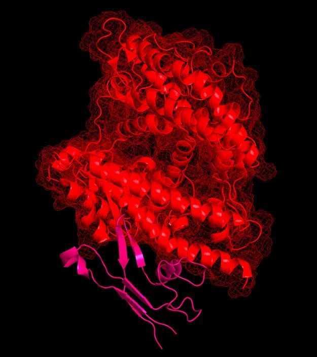
healthcare, through creating atomically-precise, genetically tailored, peptide and
drug/gene delivery nanosystems for broad therapeutic, vaccine, and preventative
medical purposes.

This, coupled with the virus’ other mechanisms for burying antibody immuno-epitope
sites, poses a significant problem in eliminating the virus from the body. These peptide
scaffolds are designed to be ultra-high-affinity, fully synthetic constructs that can serve
as competitive displacers to the viral entry mechanisms, as well as preventing ACE2
from forming its cloak around the virus. Furthermore, the scaffolds are designed to act
as immuno-boosters, presenting precise antibody immuno-epitopes as well as T cell
receptor (TCR) immuno-epitopes against the key viral immunogenic sites.
To begin, please see Ligandal’s prior work on Rapid Simulation and Design of Synthetic
  Peptide Mini-Scaffold Vaccines for Novel Coronavirus (SARS-CoV-2) and Pandemic
  Preparedness for rationale, and a video explaining our strategy for aligning a SWISS-
 MODEL structure of SARS-CoV-2 (the novel coronavirus)i to its binding site with Gibbs
 free energy plots of key interacting residues,ii as well as comparing to the Veesler Lab
  cryo-EM structuresiii of the viral spike protein in open and closed conformations. We
  worked with Dr. Jinbo Xu to simulate our proprietary sequences in their final, folded
                                       conformations.iv
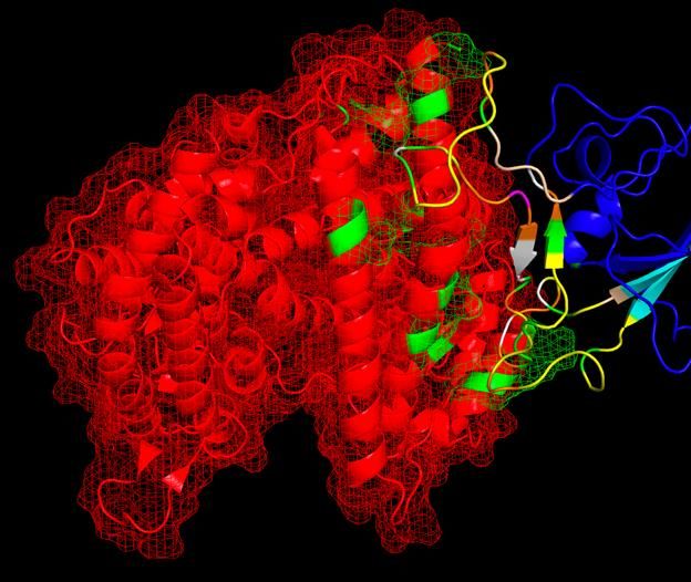
On February 11, we correctly predicted the binding domain of SARS-CoV-2 to the ACE2
   receptor. Shown is an overlap of our structural modeling of ACE2 bind to a SWISS-
MODEL simulated spike protein (red and blue/multicolored, respectively), in comparison
 to PDBID 6M0J showing a crystal structure via X-ray crystallography of the SARS-CoV-2
              receptor binding domain (RBD) bound to ACE2 (both yellow).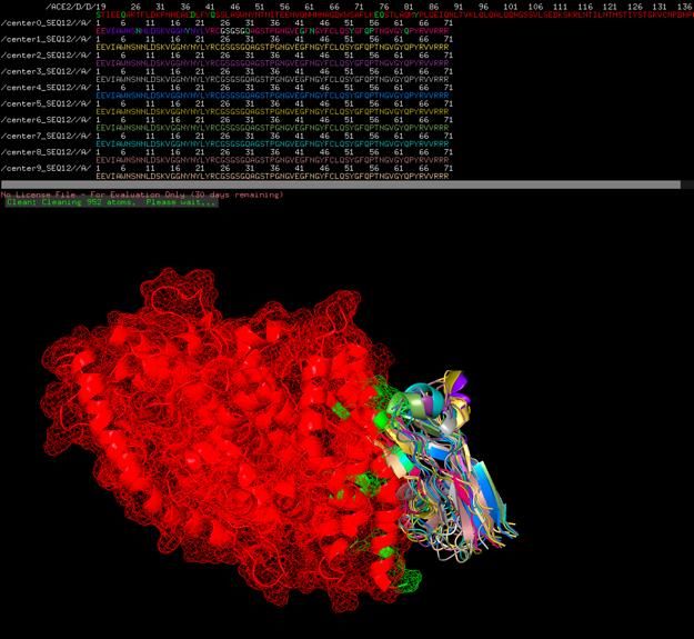
Shown are SEQ12 peptide scaffolds built upon a C60 buckminsterfullerene. This
particular sequence is designed to mimic the binding cleft of the virus to ACE2, whereby
it is anticipated to competitively displace the virus from ACE2 receptors and soluble
ACE2. The fullerene substrate acts to multivalently display a tetrad or greater amount of
each peptide.
The vaccine/antidote “scaffolds” are designed to prevent the viral spike protein (green) from
                                   binding to ACE2 (red).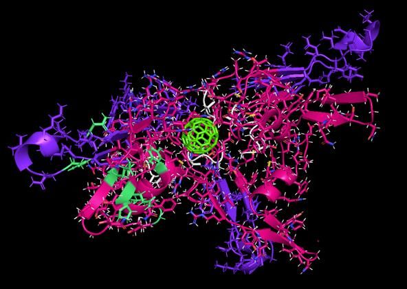
Shown are SEQ12-C60 scaffolds (magenta, pink), whereby one of the scaffolds is inhibiting the
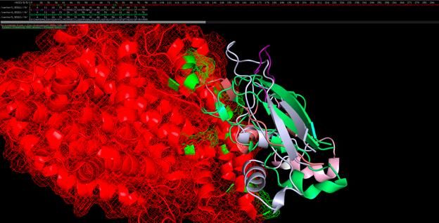
PEPTIDE SCAFFOLDS ARE DESIGNED TO INHIBIT THE VIRUS FROM BINDING TO ACE2, WHILE
    EXPOSING ITS IMMUNE RESPONSIVE SITES FOR ANTIBODY AND TCR GENERATION.
Buckminsterfullerene (C60) with peptide scaffolds bound to soluble ACE2 receptor.
Peptide scaffold SEQ13 (purple) bound to viral spike protein at its ACE2-binding site, preventing
binding to ACE2, while allowing for presentation of antibody immuno-epitopes on the viral
protein surface. Shown also is the SARS-CoV-2 spike protein (green) with T cell immuno-epitopes
(pink).
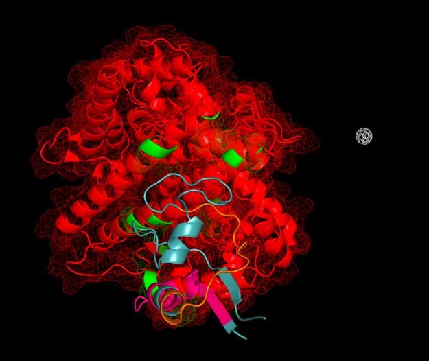
Ligandal peptide scaffolds assembled upon buckminsterfullerene, with ACE2 shown in red. The
peptide scaffolds can act as rapid antidotes to the SARS-CoV-2 coronavirus, preventing the virus
from binding to and entering cells, as well as exposing the sophisticated immune cloaking
mechanisms, whereby the virus gets coated in soluble ACE2, in order to displace soluble ACE2
from the virus and allow for natural immune recognition of the viral surface.
An individual peptide scaffold bound to ACE2 can serve as a competitive inhibitor to viral
                                           entry.

Predicted thermodynamics of binding and dissociation constant of SEQ12_center0 with ACE2:v

                                                  ΔG (kcal mol-   Kd (M) at 37.0
                  Protein-protein complex
                                                       1)               ℃

                     ACE2 with
                                                     -16.1          4.7E-12
                   SEQ12_center0
Wildtype SARS-CoV-2 (coronavirus) SWISS-MODEL truncated structure, bound to ACE2 receptor
based on alignment to SARS-CoV-1; binding residues are colored green, repulsory residues are
colored orange, neutral residues are colored yellow.vi,viiAdditionally, we report a finding that antibodies found against fragments of the virus, such as those against the WHOLE-SPIKE or spike fragment
(reference PDB ID 6W41 for a human antibody against a spike RBD https://www.rcsb.org/structure/6W41) are actually not neutralizing
antibodies against the fully-formed virus… See the whole spike overlapping with the antibody; therefore, the wrong immune responses may
actually be detrimental to vaccine development.Veesler Lab Cryo-EM atomic resolution structure of SARS-CoV-2.
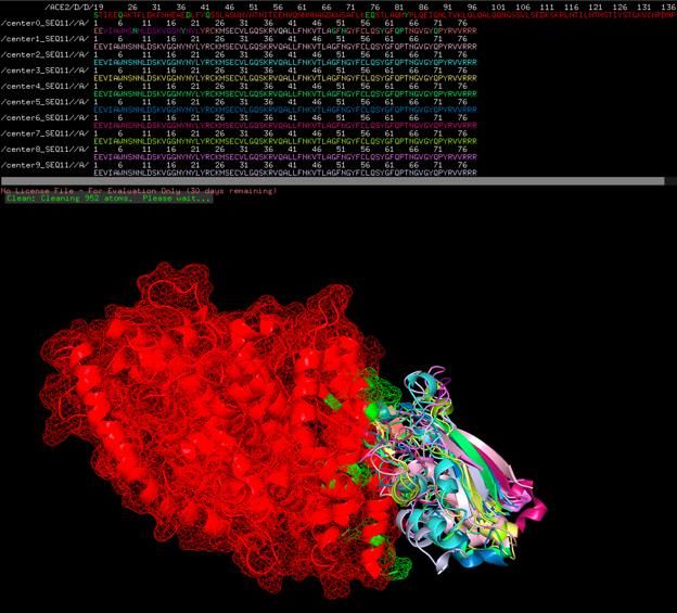
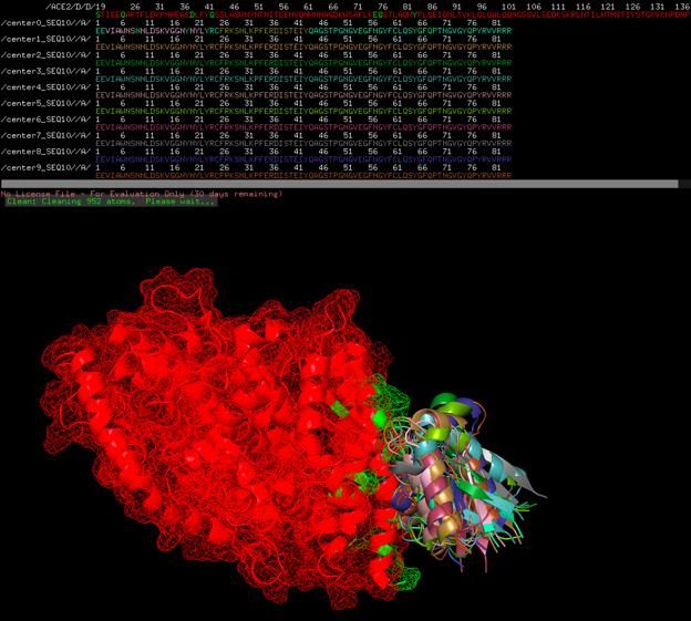
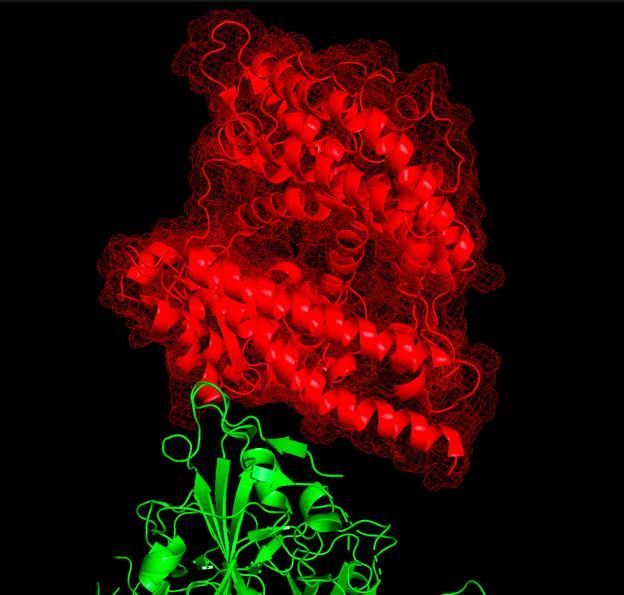
One folded state of SEQ12 with ACE2.
Two folded states (center0 and center9) of SEQ12 with ACE2

Chaotic assortment of 9 folded states (center0 – center9) with ACE2, probably showing
reasonable average folding and locations of all possible folded states given Heisenberg
                                 Uncertainty Principle
SWISS-MODEL spike protein aligned to SARS-CoV-1 binding site, with binding residues colored
green.ACE2 bound to synthetic peptide antidote.
ACKNOWLEDGEMENTS:

We thank Dr. Jinbo Xu for granting access to RaptorX,4 and simulation of scaffolds based on
Ligandal’s sequences.

Eric Greenberg, as a board member of Ligandal, provided commentary and feedback during the
compilation of this document.

i
   SWISS-MODEL:
      a) Waterhouse, A., Bertoni, M., Bienert, S., Studer, G., Tauriello, G., Gumienny, R., Heer, F.T., de
          Beer, T.A.P., Rempfer, C., Bordoli, L., Lepore, R., Schwede, T. SWISS-MODEL: homology
          modelling of protein structures and complexes. Nucleic Acids Res. 46(W1), W296-W303 (2018).
      b) Bienert, S., Waterhouse, A., de Beer, T.A.P., Tauriello, G., Studer, G., Bordoli, L., Schwede, T.
          The SWISS-MODEL Repository - new features and functionality. Nucleic Acids Res. 45, D313-
          D319 (2017).
      c) Guex, N., Peitsch, M.C., Schwede, T. Automated comparative protein structure modeling with
          SWISS-MODEL and Swiss-PdbViewer: A historical perspective. Electrophoresis 30, S162-S173
          (2009).
      d) Studer, G., Rempfer, C., Waterhouse, A.M., Gumienny, G., Haas, J., Schwede, T. QMEANDisCo
          - distance constraints applied on model quality estimation. Bioinformatics 36, 1765-1771 (2020).
      e) Bertoni, M., Kiefer, F., Biasini, M., Bordoli, L., Schwede, T. Modeling protein quaternary structure
          of homo- and hetero-oligomers beyond binary interactions by homology. Scientific Reports 7
          (2017).
ii
    PDBePISA:
      a) E. Krissinel and K. Henrick (2007). 'Inference of macromolecular assemblies from crystalline
          state.'. J. Mol. Biol. 372, 774--797.
      b) E. Krissinel (2009). Crystal contacts as nature's docking solutions. J Comput Chem. 2010 Jan
          15;31(1):133-43.; DOI 10.1002/jcc.21303
      c) E. Krissinel and K. Henrick (2007). 'Inference of macromolecular assemblies from crystalline
          state.'. J. Mol. Biol. 372, 774--797.
      d) E. Krissinel (2009). Crystal contacts as nature's docking solutions. J Comput Chem. 2010 Jan
          15;31(1):133-43.; DOI 10.1002/jcc.21303
iii
    Walls, A. C., Park, Y. J., Tortorici, M. A., Wall, A., McGuire, A. T., & Veesler, D. (2020). Structure,
function, and antigenicity of the SARS-CoV-2 spike glycoprotein. Cell.
iv
    RAPTORX:
      a) Jianwei Zhu, Sheng Wang, Dongbo Bu and Jinbo Xu. Protein threading using residue co-variation
          and deep learning. Bioinformatics, 34(13): i263-i273, 2018.
      b) Jinbo Xu. Distance-based protein folding powered by deep learning. BioRxiv, 2018.
      c) Morten Kallberg, Haipeng Wang, Sheng Wang, Jian Peng, Zhiyong Wang, Hui Lu and Jinbo Xu.
          Template-based protein structure modeling using the RaptorX web server. Nature Protocols, 7(8):
          1511-1522, 2012.
      d) Jianzhu Ma, Jian Peng, Sheng Wang and Jinbo Xu. A Conditional Nueral Fields model for protein
          threading. Bioinformatics, Vol. 28, Issue 12, i59-i66, 2012.
      e) Jianzhu Ma, Sheng Wang, Feng Zhao and Jinbo Xu. Protein threading using context-specific
          alignment potential. Bioinformatics, Vol. 29, Issue 13, pp. i257-i265.
v PRODIGY:

      a) Vangone A. and Bonvin A.M.J.J. "Contact-based prediction of binding affinity in protein-protein
          complexes", eLife, 4, e07454 (2015).
      b) Xue L., Rodrigues J., Kastritis P., Bonvin A.M.J.J.*, Vangone A.*, "PRODIGY: a web-server for
          predicting the binding affinity in protein-protein complexes", Bioinformatics,
          doi:10.1093/bioinformatics/btw514 (2016).
You can also read