Schisandrin Protects against Norepinephrine-Induced Myocardial Hypertrophic Injury by Inhibiting the JAK2/STAT3 Signaling Pathway
←
→
Page content transcription
If your browser does not render page correctly, please read the page content below
Hindawi
Evidence-Based Complementary and Alternative Medicine
Volume 2021, Article ID 8129512, 8 pages
https://doi.org/10.1155/2021/8129512

Research Article
Schisandrin Protects against Norepinephrine-Induced
Myocardial Hypertrophic Injury by Inhibiting the JAK2/STAT3
Signaling Pathway

          Min Yang ,1,2 Xing-Can Jiang,2 Lei Wang,1,2 Dong-An Cui,2 Jing-Yan Zhang,1,2
          Xu-Rong Wang,2 Hai-Peng Feng,1,2 Kang Zhang,1,2 Kai Zhang ,1,2 Jian-Xi Li ,1,2
          and Xue-Zhi Wang 1,2
          1
              Engineering & Technology Research Center of Traditional Chinese Veterinary Medicine, Gansu, Lanzhou, China
          2
              Lanzhou Institute of Husbandry and Pharmaceutical Sciences, Chinese Academy of Agricultural Sciences (CAAS),
              Lanzhou, China

          Correspondence should be addressed to Kai Zhang; 18690165@qq.com, Jian-Xi Li; 1196674841@qq.com, and Xue-Zhi Wang;
          469359626@qq.com

          Received 27 May 2020; Revised 8 March 2021; Accepted 17 March 2021; Published 16 June 2021

          Academic Editor: José L. Rios

          Copyright © 2021 Min Yang et al. This is an open access article distributed under the Creative Commons Attribution License,
          which permits unrestricted use, distribution, and reproduction in any medium, provided the original work is properly cited.
          Aims. Heart failure is closely associated with norepinephrine-(NE-) induced cardiomyocyte hypertrophy. Schisandrin is derived
          from the traditional Chinese medicine Schisandra; it has a variety of pharmacological activities, and the mechanism of schi-
          sandrin-mediated protection of the cardiovascular system is not clear. Main Methods. NE was used to establish a cardiomyocyte
          hypertrophy model to explore the mechanism of action of schisandrin. An MTT assay was used for cell viability; Hoechst
          fluorescence staining was used to observe the cell morphology and calculate the apoptosis rate. The cell surface area was measured
          and the protein to DNA ratio was calculated, changes in mitochondrial membrane potential were detected, and the degree of
          hypertrophic cell damage was evaluated. WB, QRT-PCR, and immunofluorescence were used to qualitatively, quantitatively, and
          quantitatively detect apoptotic proteins in the JAK2/STAT3 signaling pathway. Key Findings. In the NE-induced model,
          schisandrin treatment reduced the apoptosis rate of cardiomyocytes, increased the ratio of the cell surface area to cardiomyocyte
          protein/DNA, and also, increased the membrane potential of the mitochondria. The expression of both JAK2 and STAT3 was
          downregulated, and the BAX/Bcl-2 ratio was significantly reduced. In conclusion, schisandrin may protect against NE-induced
          cardiomyocyte hypertrophy by inhibiting the JAK2/STAT3 signaling pathway and reducing cardiomyocyte apoptosis.

1. Introduction                                                        schisandrin A. Schisandrin may play a role in the treatment
                                                                       of cardiovascular disease.
Schisandrin is one of the dibenzocyclooctadiene lignans in                 Heart failure is associated with chronic increases in
Schisandra and has antioxidant, anti-inflammatory, anti-                plasma NE and mast cell numbers, and increased NE is a key
apoptotic effects [1–5]. Studies have found that Schisandra             marker of pathological heart failure [7–11]. Thus, blocking
and lignans have potentially beneficial effects on the treat-            NE is conducive to cell survival and also inhibits cardiac
ment of cardiovascular diseases. Currently, research reports           hypertrophy [10]. Modern studies have shown that JAK2/
have found that Schisandra chinensis extracts schisandrin B,           STAT3 signaling pathway activation is associated with
gomisin N, schisandrin A, and schisandrin C exert various              cardiac hypertrophy [12–14]; it is one of the important
pharmacological activities for the treatment of cardiovas-             pathways of cardiac apoptosis [15] and is of great signifi-
cular disease and have studied the molecular mechanism of              cance in cardiomyocyte signal transduction pathways. JAK2
action [6]. The structure of schisandrin is similar to that of         is considered to be a new treatment target for inhibiting the
2                                                                 Evidence-Based Complementary and Alternative Medicine

progression of hypertrophy to heart failure. Studies have        Beijing, China) with a 20× objective (scale bar: 25 μm).
found that STAT3 activation occurs in cardiac hypertrophy        ImageJ software was used to process the images, and the
and is involved in cell growth. Key regulators may be im-        surface area of more than 100 cardiomyocytes in each field of
portant regulators of cardiovascular system growth [13]. In      view was measured and counted.
this study, based on the pharmacological activity of schi-            Measurement of the protein/DNA ratio: the adherent
sandrin and the important significance of JAK2/STAT3 in           cells were digested with 0.25% trypsin to prepare a cell
the regulation of cardiomyocyte hypertrophy, we pretreated       suspension, and the cells were processed using an automated
H9C2 cardiomyocytes with schisandrin and induced car-            cell counter (Nexcelom Bioscience) for cell counting. Then,
diomyocyte hypertrophy with NE. We found that the JAK2/          the cells were lysed with M-PER cell lysis buffer (Thermo
STAT3 signaling pathway was inhibited, the BAX/Bcl-2 ratio       Fisher Scientific, USA); the lysate was centrifuged, and the
was decreased, the mitochondrial membrane potential was          supernatant was collected. Total protein detection was
stabilized, the cardiomyocytes were protected, and apoptosis     performed according to the instructions of the Micro BCA
was inhibited.                                                   Protein Assay Kit (Thermo Fisher Scientific, USA). The DNA
                                                                 content was determined using a Green II dsDNA Quanti-
2. Materials and Methods                                         tation Kit Plus (Yu heng, Su zhou, China), and the DNA
                                                                 concentration in the sample was further determined based
2.1. Passage and Culture of Rat H9C2 Cardiomyocytes.             on the generated standard curve. Finally, the ratio of protein/
H9C2 cells were from the Cell Bank of the Chinese Academy        DNA was calculated.
of Sciences [16], maintained in Dulbecco’s modified Eagle’s
medium (DMEM; USA) containing 10% fetal bovine serum
(Gibico; USA) at 37°C with 5% CO2. The cells were rinsed in      2.3. Hoechst 33342 Staining. After the cells were treated with
phosphate-buffered saline (PBS) and treated with 0.25%            drugs, the cell culture solution was sucked away, and the
trypsinat 37°C (Gibico, USA) for passaging cells.                cells were washed with PBS and fixed in a 4% cell fixation
                                                                 solution (Solarbio, Beijing, China) at room temperature for
                                                                 20 min. Hoechst 33342 fluorescent dye (5 μg/mL; Biyun-
2.2. Methyl Thiazolyl Tetrazolium (MTT) Assay. Cells with a      tian, Shanghai, China) was added to each well, and the plate
density of 1 × 10⁵ cells/mL per well were inoculated into 96-    was incubated at 37°C for 20 minutes. Subsequently, the
well plates. After 24 h, in the positive drug group [17], a      cells were washed with PBS and photographed with a
DMEM complete culture medium containing 5 μg/mL was              universal fluorescence inverted microscope (OLYMPUS
added to each well with captopril [15, 18], and a schisandrin-   IX71, Japan) (scale bar: 25 μm). Fifty visual fields were
DMEM complete cell culture medium containing 5 μg/mL,            randomly selected in each group. The number of apoptotic
10 μg/mL, and 20 μg/mL was added to each well in the low-,       cells and the total number of cells were recorded. The H9C2
middle-, and high-dose groups, respectively. A certain           apoptotic rate was calculated by the following formula:
concentration of norepinephrine can induce cardiac hy-
                                                                                            Apoptotic number of H9C2
pertrophy [19, 20]. After 48 hours culturing, the complete       H9C2 apoptotic rate(%) �                            × 100%.
                                                                                              Total number of H9C2
culture medium of DMEM containing 10−6 mol/L NE was
added to each well of the model group, positive drug group,                                                                 (2)
low-, middle-, and high-dose groups of schisandrin for 48
hours, and pictures were taken under the microscope
(Olympus, Tokyo, Japan) to observe the morphological             2.4. Mitochondrial Membrane Potential Staining. The cells
changes of the cells (scale bar: 500 μm). Cell viability was     were stained with a JC-1 mitochondrial membrane potential
evaluated by the 3-(4, 5-dimethylthiazol-2-yl)-2, 5-diphenyl     detection kit (Biyuntian, Beijing, China) according to the
tetrazolium bromide (MTT) assay based on the reduction of        manufacturer’s instructions. The CCCP group was used as
soluble yellow tetrazolium compound into insoluble purple        positive control to induce mitochondrial membrane po-
formazan crystals produced in living cells. After the treat-     tential decline. JC-1 staining solution was added to cell
ment procedure, MTT was added to each well and incubated         culture medium and mixed well. The cells were incubated
for 4 h at 37°C in darkness. The supernatant was removed,        with the stain at 37°C for 20 min. Then, the supernatant was
and then, 150uL dimethyl sulfoxide was added to dissolve         discarded, and the cells were washed twice with JC-1 staining
the formazan compound. The absorbance was measured at            buffer (1×) and photographed under a laser scanning con-
490 nm using a microplate reader. The H9C2 survival rate         focal microscope (ZEISS, Shanghai, China) (scale bar:
was calculated by the following formula:                         25 μm). A fluorescence microplate reader was used to detect
                                                                 the fluorescence intensity; the excitation wavelength was set
                          OD value of treated group
H9C2 survival rate(%) �                             × 100%.      at 490 nm, and the emission wavelength was set at 530 nm to
                          OD value of control group              detect the JC-1 monomer. The excitation wavelength was set
                                                        (1)      at 525 nm, and the emission wavelength was 590 nm to
                                                                 detect the JC-1 polymer.
    Measurement of the cell surface area: the cardiomyocytes
were cultured in a 24-well plate, and 6 wells of the three
groups of cells were cultured in parallel. Photographs were      2.5. Immunofluorescence. The cells were fixed in 4% para-
taken using a phase contrast microscope (Leica DMi1,             formaldehyde (Solarbio, Beijing, China) for 20 min, washed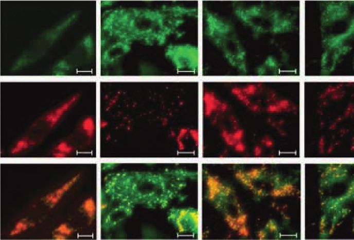
Evidence-Based Complementary and Alternative Medicine 3
with PBS, and permeabilized in 0.5% Triton X-100 for 2.8. Statistics. The experimental data were analyzed using
15 min. Subsequently, the cells were treated with 5% BSA at SPSS 19.0 (SPSS, Inc., USA). The data are expressed as the
room temperature for 1 h. The cells were then incubated with mean ± standard deviation (SD). A t-test was used for
the corresponding dilution ratio of the primary antibody comparisons between groups. The different symbols in the
overnight at 4°C. Then, the cells were washed with PBST and table represent P < 0.05, and the difference was considered
incubated with diluted fluorescent secondary goat anti- statistically significant.
rabbit IgG H&L (FITC; 1 : 500 diluted; Abcam, USA) and
conjugated goat anti-rabbit IgG H&L (1 : 500 diluted; Pro- 3. Results and Discussion
teintech, USA) antibodies at 37°C for 1 hour. Subsequently,
the cells were stained with 5 μg/mL DAPI solution for 15 min 3.1. Schisandrin Increases the Viability of Cardiomyocytes.
at 37°C. A fluorescence microscope (ZEISS, Shanghai, Figure 1 shows the changes in cardiomyocyte morphology
China) was used to observe the cells and collect pictures and cell viability in different groups, the H9C2 car-
(scale bar: 25 μm). diomyocytes of the control group showed a long spindle
shape, and the cardiomyocytes of the model group increased
in volume, mostly triangular and elliptical (Figure 1(a)).
2.6. Western Blot (WB) Assay. The cells in each group were Compared with the control group, the survival rate of
lysed in M-PER cell lysis buffer (Thermo Fisher Scientific, cardiomyocytes in the model group was significantly re-
USA). The total protein of each group was collected and duced (P < 0.05). Compared with that of the model group,
quantified measuring with a BCA total protein kit (Thermo the cell viability of the captopril group and schisandrin
Fisher Scientific, USA). Proteins of the same volume were medium- and high-dose groups was significantly increased
separated by SDS-PAGE. Then, the proteins were transferred (P < 0.05), but there was no significant difference in the low-
to the semidry membrane. Subsequently, membranes were dose schisandrin group (P > 0.05), indicating that captopril
blocked in 5% BSA for 1 hour at room temperature and and schisandrin can protect cardiomyocytes and increase the
incubated with β-actin (1 : 2500, Proteintech), rabbit anti- viability of H9C2 cells (Figure 1(b)).
BAX (1 : 250, Proteintech), rabbit anti-Bcl-2 (1 : 1000, Abcam),
rabbit anti-JAK2 (1 : 1000, Cell Signaling Technology), and
rabbit anti-STAT3 (1 : 1000, Cell Signaling Technology) an- 3.2. Hoechst Staining Assay. Hoechst fluorescence staining
tibodies at 4°C overnight. Subsequently, the membranes were was used to detect the apoptotic morphology and apoptosis
incubated with secondary antibodies labeled with horseradish rate of H9C2 cardiomyocytes, as shown in Figure 2. The
peroxidase (HRP) for 2 hour. The membrane was exposed to nuclei of the control group were clearly visible, the mor-
the developing solution of electrogenerated chem- phology was normal, and the blue fluorescence was normal.
iluminescence (ECL) (Advansta, CA, USA). The optical Bright blue fluorescence appeared in the model group, the
density of protein bands was analyzed by ImageJ image nuclear morphology was incomplete, and there were ir-
analysis software. regular shape changes, such as crescent shapes. Compared
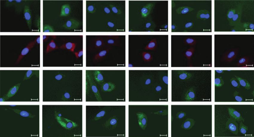
with that of the control group, the apoptosis rate of the other
six groups was decreased (P < 0.05). Compared with that of
2.7. Quantitative Real-Time PCR. Quantitative real-time the model group, the apoptosis rate of the medium- and
PCR (QRT-PCR) was performed for the quantitative de- high-dose schisandrin groups and the captopril group was
tection of mRNA. Total RNA was extracted using a High- significantly reduced (P < 0.05), but there was no significant
purity Total RNA Rapid Extraction Kit (BIOTEKE COR- change in the low-dose schisandrin group (P > 0.05).
PORATION, Beijing, China). RNA is transcribed into Compared with that of the captopril group, the apoptosis
cDNA with a PrimeScriptTMRT reagent Kit with a gDNA rate of the schisandrin medium-dose group did not increase
Eraser (Takara, Dalian, China). QT-PCR was performed significantly (P < 0.05). These results indicate that schisan-
using a QuantStudio 5 real-time PCR instrument (Thermo drin and captopril both protect cardiomyocytes.
Fisher Scientific, USA) and TB GreenTM Premix Ex TaqTM
II (Takara, Dalian, China). The primer sequences for qRT-
PCR were as follows: β-actin mRNA, 5′-GGAGATTACTGC- 3.3. Schisandrin Improves Cardiomyocyte Hypertrophy In-
CCTGGCTCCTAGC-3′ (forward), 5′-GGCCGGACT- duced by NE. By measuring changes in the myocardial cell
CATCGTACTCCTGCTT-3′ (reverse); JAK2 mRNA, 5′- surface area and protein/DNA ratio, the degree of myo-
TCATAAACCTGGAGACCCT-3′ (forward), 5′-ATGTTTC- cardial hypertrophy was further determined (Figure 2);
CCTCTTGACCAC-3′ (reverse); STAT3 mRNA, 5′-TAA- compared with that of the model group, the myocardial cell
CATTCTGGGCACGAACA-3′ (forward), 5′-GGCATCA- surface area of the captopril group and the medium-dose
CAATTGGCACGG-3′ (reverse); Bax mRNA, 5′-GGCTGG- schisandrin group was significantly decreased (P < 0.05).
ACACTGGACTTCCT-3′ (forward), 5′-GGTGAGGACTC- Compared with that of the captopril positive-control group,
CAGCCACAA-3′ (reverse); and Bcl-2 mRNA, 5′-ACGGT- that of the medium-dose schisandrin group was not sig-
GGTGGAGGAACTCTT-3’ (forward), 5′-GCAGATGC- nificantly increased (P > 0.05). The medium dose of schi-
CGGTTCAGGTA-3′ (reverse). The Ct values obtained for sandrin and 5 μg/mL captopril had similar effects on the
each sample were calculated. The results were analyzed surface area of cardiomyocytes treated with NE. Further-
according to the 2-ΔΔCt method. β-Actin was used as an more, 10 μg/mL schisandrin had the greatest effect on the
internal reference gene. promotion of myocardial cell surface area recovery. We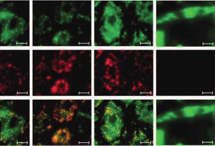
4 Evidence-Based Complementary and Alternative Medicine
H9C2 cell survival rate (%)
Control Model Captopril 100
ab
acd abc abc
a
50
Ld-SCH Md-SCH Hd-SCH 0
Control
Model
Captopril
Ld-SCH
Md-SCH
Hd-SCH
(a) (b)
Figure 1: (a) (Scale bar: 500 μm) morphological changes of myocardial cells in each group were studied; control: H9C2 cells growth without
any treatment; model: norepinephrine-induced myocardial hypertrophic injury and without any treatment. Coptopril: H9C2 cells were
pretreated with captopril. Different dosages of SCH were administrated in the cell model with myocardial hypertrophic injury (Ld-SCH,
Md-SCH, and Hd-SCH). (b) The applied MTT assay for the determination of the effect of schisandrin pretreatment on NE-treated
cardiomyocyte survival. SCH, schisandrin; Ld, low dose; Md, medium dose; Hd, high dose; a, P < 0.05 vs. the control group; b, P < 0.05 vs.
the model group; c, P < 0.05 vs. the captopril group; and d, P < 0.05 vs. the medium-dose schisandrin group.
Control Model Captopril a
40 acd
H9C2 apoptosis rate (%)
35 abcd
30 abc
25 ab
20
15
Ld-SCH Md-SCH Hd-SCH 10
5
0
Control
Model
Captopril
Ld-SCH
Md-SCH
(a) (b) Hd-SCH
1800 250
H9C2 surface area (μm2)
a acd
1600 abc acd
1400 200 a
ab
Protein/DNA
acd
1200 abc abc
1000 150 ab
800
100
600
400 50
200
0 0
Captopril
Control
Model
Ld-SCH
Md-SCH
Hd-SCH
Captopril
Control
Model
Ld-SCH
Md-SCH
Hd-SCH
(c) (d)
Figure 2: Detection of the apoptosis rate by Hoechst fluorescence staining (scale bar: 25 μm). (a) Morphological changes of nuclei assessed
by Hoechst fluorescence staining. (b) Statistical chart of the apoptosis rate of myocardial cells in each group. (c) Statistical graph of changes
in the surface area of myocardial cells in each group. (d) Statistical graph of changes in the protein/DNA ratio of cardiomyocytes in each
group. Ld, low dose; Md, medium dose; Hd, high dose; a, P < 0.05 vs. the control group; b, P < 0.05 vs. the model group; c, P < 0.05 vs. the
captopril group; and d, P < 0.05 vs. the medium-dose schisandrin group.Evidence-Based Complementary and Alternative Medicine 5
observed the protein/DNA ratio and found that the ratio of immunofluorescence analysis of Bcl-2 protein, the fluores-
protein/DNA was significantly increased under 10−6 mol/L cence intensity in the model group was significantly lower
NE induction conditions (P < 0.05 compared with the than that in the control group. We found that, after pre-
control group). After pretreatment with schisandrin and treatment with schisandrin, Bcl-2 fluorescence in the me-
captopril, the ratio of protein/DNA decreased, but the dium- and high-dose schisandrin groups was significantly
change in the captopril and schisandrin groups was the most increased compared with that in the model group. The
significant (P < 0.05). This finding indicates that schisandrin expression level of the captopril positive control group was
has a protective effect against NE-induced cardiomyocyte similar to that of the medium- and high-dose schisandrin
hypertrophy. groups. Among the different experimental groups, the
fluorescence intensity of BAX protein in the model group
and low-dose and high-dose schisandrin groups was
3.4. Mitochondrial Membrane Potential Assay. The changes stronger than that in the control group. Compared with that
in mitochondrial membrane potential are shown in Figure 3. of the model group, the fluorescence intensity of the low-
The mitochondrial membrane potential of the experimental dose and high-dose schisandrin groups was similar, and the
group was significantly increased compared with that of the fluorescence intensity of the medium-dose schisandrin
CCCP group (P < 0.05). In the comparison of the model group was reduced. Therefore, schisandrin has an inhibitory
groups, the membrane potential of the captopril group and effect on the JAK2/STAT3 signaling pathway but increases
the medium-dose schisandrin group decreased (P < 0.05). In the fluorescence intensity of Bcl-2 and decreases the fluo-
the low-dose schisandrin group, there was no significant rescence intensity of BAX.
change in mitochondrial membrane potential (P > 0.05).
Compared with that of the captopril group, there was no
obvious difference in the medium-dose schisandrin group 4. Discussion
(P > 0.05), and the mitochondrial membrane potential was We first studied the ability of schisandrin pretreatment to
similar to that of the captopril group. reduce the degree of noradrenaline-induced cardiomyocyte
hypertrophy in cardiomyocytes. The results of this experi-
3.5. Western Blot and Quantitative Real-Time PCR Assays. ment indicated that schisandrin may inhibit the JAK2/
The results of the WB and RT-PCR are shown in Figure 3. STAT3 signaling pathway, downregulate BAX expression,
Compared with that of the control group, the relative ex- upregulate Bcl-2 expression, and play a protective role in
pression of JAK2 and STAT3 protein and mRNA in the cardiomyocytes.
model group was increased (P < 0.05). Compared with the Cardiac hypertrophy is an independent risk factor, and it
model group, the schisandrin and captopril groups all easily progresses to heart failure, which is one of the main
displayed decreased relative protein and mRNA expression causes of harm to human life [21]. The hypertrophic changes
of JAK2 and STAT3; the medium-dose schisandrin group in cardiomyocytes are mainly characterized by an increase in
displayed the most significant decrease (P < 0.05). The ex- cell volume and an increase in the amount of protein
pression in the captopril control group was similar to that of synthesis. Many experimental studies have shown that the
the schisandrin group. Compared with that in the control JAK/STAT signaling pathway is an important signal
group, the relative expression of BAX and Bcl-2 protein and transduction pathway often involved in the process of
mRNA in each experimental group was downregulated cardiac hypertrophy, cardiac apoptosis, and cardiac pa-
(P < 0.05). Compared with the model group, the medium- thology [22–24]. JAK2 is of great significance in the signal
dose schisandrin group displayed significantly upregulated transduction associated with cardiac growth and dysfunc-
BAX protein and mRNA expression (P < 0.05) and down- tion. It can help reduce hypertrophy by inhibiting the ac-
regulated Bcl-2 protein and mRNA expression (P < 0.05). tivity of JAK2 [25]. Similarly, the activation of STAT3 causes
There was no significant difference in the BAX and Bcl-2 cardiomyocyte hypertrophy and plays a role in the regula-
protein and mRNA expression in the captopril group and tion of hypertrophic growth. The phosphorylation of STAT3
the medium-dose schisandrin group. We found that the is dependent on JAK2 [26, 27]. These findings are consistent
gene expression trend detected by RT-PCR was consistent with those of our study. In NE-induced cardiac hypertrophy,
with the protein expression in the WB, indicating that the protein expression of JAK2 and STAT3 is significantly
schisandrin inhibits the JAK2/STAT3 pathway, down- upregulated, further promoting the development of car-
regulates BAX expression, and upregulates Bcl-2 expression. diomyocyte hypertrophy. When cardiomyocytes were pre-
treated with schisandrin and then treated with NE, the
expression of JAK2 and STAT3 protein was downregulated
3.6. Immunofluorescence Assay. The expression of JAK2, and the surface area, protein/DNA ratio, cell viability, and
STAT3, BAX, and Bcl-2 was detected by immunofluores- apoptosis rate of cardiomyocytes were detected. The use of
cence (Figure 4), and JAK2, STAT3, BAX, and Bcl-2 were NE alone induced a decrease in cardiomyocyte viability.
positively expressed in the cytoplasm. The fluorescence Therefore, we speculate that schisandrin protects car-
intensity of JAK2 and STAT3 was the highest in the model diomyocytes and inhibits apoptosis by inhibiting the JAK2/
group and the lowest in the control group. After pretreat- STAT3 signaling pathway.
ment with low, medium, and high doses of schisandrin, the Compared with those in the model group, the myo-
fluorescence intensity decreased accordingly. In the cardial cells treated with schisandrin had an increased cell6 Evidence-Based Complementary and Alternative Medicine
Control Model Captopril Ld-SCH Md-SCH Hd-SCH CCCP e
monomers 1.0
JC-1
fluorescence ratio
JC-1 aggregates/
abe
monomers
ae acde abce acde
0.5
aggregates
JC-1
a
0.0
Merge
Control
Model
Captopril
Ld-SCH
Md-SCH
Hd-SCH
CCCP
(a) (b)
BAX
Relative protein level
Bcl-2 a
4
STAT3 c
ab c
ab
c a a
JAK2 ab
ab
c
c
ab c
c ab
2 ab
ab c
c ab ab
β-actin
ab
ab
c
c c
ab ab ab ab
a
Control
Model
Captopril
Ld-SCH
Md-SCH
Hd-SCH
0
JAK2 STAT3 BAX Bcl-2
Control Model
Captopril Ld-SCH
Md-SCH Hd-SCH
(c) (d)
1.6 a
1.4 c
c
a c
ab
c
ab
c c
Relative mRNA level
ab a
c ab ab c c ab
1.2 ab
ab ab ab
ab
ab
1.2
1.0
ab c
c ab c
0.8 ab ab
a
0.6
0.4
0.2
0.0
JAK2 STAT3 BAX Bcl-2
Control Model
Captopril Ld-SCH
Md-SCH Hd-SCH
(e)
Figure 3: (a) Fluorescence picture of mitochondrial membrane potential changes (scale bar: 25 μm). (b) Red fluorescence intensity/green
fluorescence intensity ratio statistics. (c) Band diagram of β-actin, JAK2, STAT3, BAX, and Bcl-2 protein expression detected by WB. (d)
Statistical comparison of the gray value of JAK2/β-actin, STAT3/β-actin, BAX/β-actin, and Bcl-2/β-actin proteins. (e) The statistical value of
the ratio of JAK2/β-actin, STAT3/β-actin, BAX/β-actin, and Bcl-2/β-actin mRNA. Ld, low dose; Md, medium dose; Hd, high dose; a,
P < 0.05 vs. the control group; b, P < 0.05 vs. the model group; c, P < 0.05 vs. the captopril group; d, P < 0.05 vs. the medium-dose schisandrin
group; and e, P < 0.05 vs. the CCCP group.
survival rate and a decreased apoptosis rate. We found that release, stabilize the mitochondrial membrane potential, and
the effect of schisandrin was the greatest at 10 μg/mL. In downregulate caspase-3 protein expression, reducing the
addition, the ratio of BAX/Bcl-2 after pretreatment with apoptosis rate [30]. We speculate that schisandrin may have
schisandrin was reduced, and the mitochondrial membrane a similar effect on the mitochondria of cardiomyocytes. The
potential was increased. Heart failure and cardiac hyper- results of this experiment showed that, in NE-induced hy-
trophy are often associated with mitochondrial dysfunction pertrophic cells, schisandrin pretreatment induced the
and insufficient energy, and the mitochondria play an im- upregulation of Bcl-2 expression, downregulated Bax ex-
portant role in the pathogenesis of heart failure [28]. When pression, decreased mitochondrial membrane potential, and
the mitochondrial membrane is destroyed, the permeability decreased apoptosis. Both Bcl-2 and BAX are members of
changes, the membrane potential decreases, and the pro- the Bcl-2 family of proteins; Bcl-2 is a protein that inhibits
duction of reactive oxygen species (ROS) increases. Con- apoptosis, and Bax is a protein that promotes apoptosis, but
sequently, a large amount of cytochrome c flows into the they are all closely associated with the mitochondrial-me-
cytoplasm and causes apoptosis-inducing damage [29]. diated apoptosis pathway. Bcl-2 protein protects the mito-
Studies have reported that, in rat cortical cells, schisandrin chondrial membrane and inhibits apoptosis by inhibiting
can inhibit ROS production and excessive cytochrome c the release of cytochrome C into the cytosol [31]. The BaxEvidence-Based Complementary and Alternative Medicine 7
Control Model Captopril Ld-SCH Md-SCH Hd-SCH
JAK2
STAT3
Bcl-2
BAX
Figure 4: Immunofluorescence assay showing the expression of JAK2, STAT3, Bax, and Bcl-2 in cells of each experimental group (scale bar:
25 μm).
protein is located in the cytoplasm. After cell damage, the the Central Public-Interest Scientific Institution Basal
BAX protein binds to Bcl-2 to form a heterodimer, which Research Fund (1610322019005).
reduces the activity of Bcl-2, damages the mitochondrial
membrane, and causes apoptosis [32]. Therefore, we spec- References
ulate that, in this experiment, schisandrin upregulates the
BAX/Bcl-2 ratio, which is related to the protection of the [1] M.-Y. Lee, C.-S. Seo, N.-H. Lee et al., “Anti-asthmatic effect of
mitochondria. Schisandrin protects the normal function of schizandrin on OVA-induced airway inflammation in a
the mitochondria, provides energy for cell functioning, murine asthma model,” International Immunopharmacology,
inhibits cardiomyocyte apoptosis, and protects against heart vol. 10, no. 11, pp. 1374–1379, 2010.
failure due to hypertrophy. [2] P. D. Moon, H. J. Jeong, and H. M. Kim, “Effects of schi-
In conclusion, schisandrin may reduce the ratio of BAX/ zandrin on the expression of thymic stromal lymphopoietin in
human mast cell line HMC-1,” Life Sciences, vol. 91, no. 11-12,
Bcl-2 and inhibit the apoptosis of cardiomyocytes by
pp. 384–388, 2012.
inhibiting the JAK2/STAT3 signaling pathway. This research [3] S. I. Jeong, S. J. Kim, T. H. Kwon, K. Y. Yu, and S. Y. Kim,
can further inform studies of the changes in other charac- “Schizandrin prevents damage of murine mesangial cells via
teristics modulated by schisandrin in NE-induced car- blocking NADPH oxidase-induced ROS signaling in high
diomyocyte hypertrophy and provide a reference for the glucose,” Food and Chemical Toxicology, vol. 50, no. 3-4,
development of new drugs for the prevention and treatment pp. 1045–1053, 2012.
of cardiovascular diseases. However, our research is limited [4] C. K. Wan, A. K. Tse, Z. L. Yu, G. Y. Zhu, H. Wang, and
to the cellular level, and subsequent in vivo animal exper- D. W. Fong, “Inhibition of cytochrome P450 3A4 activity by
iments are needed for validation. schisandrol A and gomisin A isolated from Fructus Schi-
sandrae chinensis,” Phytomedicine International Journal of
Phytotherapy & Phytopharmacology, vol. 17, no. 8-9,
Data Availability pp. 702–705, 2010.
[5] E. J. Jeong, H. K. Lee, K. Y. Lee et al., “The effects of lignan-
The data used to support the findings of this study are riched extract of Shisandra chinensis on amyloid-β-induced
available from the corresponding author upon request. cognitive impairment and neurotoxicity in the cortex and
hippocampus of mouse,” Journal of Ethnopharmacology,
Conflicts of Interest vol. 146, no. 1, pp. 347–354, 2013.
[6] J. N. Chun, M. Cho, I. So, and J.-H. Jeon, “The protective
There are no conflicts of interest to declare. effects of Schisandra chinensis fruit extract and its lignans
against cardiovascular disease: a review of the molecular
mechanisms,” Fitoterapia, vol. 97, pp. 224–233, 2014.
Acknowledgments [7] A. Jain, N. Atale, S. Kohli, M. Sharma, and V. Rani, “An
assessment of norepinephrine mediated hypertrophy to ap-
This study was supported by the Chinese Traditional optosis transition in cardiac cells: a signal for cell death,”
Veterinary Medicine and Clinical Science and Technology Chemico-Biological Interactions, vol. 225, pp. 54–62, 2015.
Innovation Projects (CAAS-ASTIP-2015-LIHPS), National [8] M. B. Patel, A. V. Loud, B. D. King, P. Anversa, D. Sack, and
Natural Science Foundation of China (no. 31702288), and T. H. Hintze, “Global myocardial hypertrophy in conscious8 Evidence-Based Complementary and Alternative Medicine
dogs with chronic elevation of plasma norepinephrine levels,” [23] J. Lammerding, R. D. Kamm, and R. T. Lee, “Mechano-
Journal of Molecular and Cellular Cardiology, vol. 21, transduction in cardiac myocytes,” Annals of the New York
pp. 49–61, 1989. Academy of Sciences, vol. 1015, no. 1, pp. 53–70, 2004.
[9] P. Simpson, “Norepinephrine-stimulated hypertrophy of [24] I. S. Harris, S. Zhang, I. Treskov, A. Kovacs, C. Weinheimer,
cultured rat myocardial cells is an alpha 1 adrenergic re- and A. J. Muslin, “Raf-1 kinase is required for cardiac hy-
sponse,” Journal of Clinical Investigation, vol. 72, no. 2, pertrophy and cardiomyocyte survival in response to pressure
pp. 732–738, 1983. overload,” Circulation, vol. 110, no. 6, pp. 718–723, 2004.
[10] T. Tsutamoto, K. Nishiyama, H. Sakai et al., “Transcardiac [25] D. L. Beckles, E. Mascareno, and M. A. Q. Siddiqui, “Inhi-
increase in norepinephrine and prognosis in patients with bition of Jak2 phosphorylation attenuates pressure overload
chronic heart failure,” European Journal of Heart Failure, cardiac hypertrophy,” Vascular Pharmacology, vol. 45, no. 6,
vol. 10, no. 12, p. S157, 2008. pp. 350–357, 2006.
[11] M. Tavares, E. Rezlan, I. Vostroknoutova, H. Khouadja, and [26] L. Wang, Z. Li, Y. Tan et al., “PARP1 interacts with STAT3
A. Mebazaa, “New pharmacologic therapies for acute heart and retains active phosphorylated-STAT3 in nucleus during
failure,” Critical Care Medicine, vol. 36, no. 1, pp. S112–S120, pathological myocardial hypertrophy,” Molecular and Cel-
2008. lular Endocrinology, vol. 474, pp. 137–150, 2018.
[12] J. Lammerding, R. D. Kamm, and R. T. Lee, “Mechano- [27] M. Kurdi and G. M. Booz, “JAK redux: a second look at the
transduction in cardiac myocytes,” New York Academy of regulation and role of JAKs in the heart,” American Journal of
Sciences, vol. 1015, no. 1, pp. 57–70, 2004. Physiology-Heart and Circulatory Physiology, vol. 297, no. 5,
[13] E. Mascareno and M. A. Q. Siddiqui, “The role of Jak/STAT pp. H1545–H1556, 2009.
signaling in heart tissue renin-angiotensin system,” Control of [28] S. Hügel, M. Horn, H. Remkes, C. Dienesch, and S. Neubauer,
Gene Expression by Catecholamines and the Renin-Angio- “Preservation of cardiac function and energy reserve by the
tensin System, vol. 212, no. 1, pp. 171–175, 2000. angiotensin-converting enzyme inhibitor quinapril during
[14] J. J. Jacoby, A. Kalinowski, M.-G. Liu et al., “Cardiomyocyte- postmyocardial infarction remodeling in the rat,” Journal of
restricted knockout of STAT3 results in higher sensitivity to Cardiovascular Magnetic Resonance (Print), vol. 3, no. 3,
inflammation, cardiac fibrosis, and heart failure with ad- p. 215, 2001.
vanced age,” Proceedings of the National Academy of Sciences, [29] J. D. Schilling, “The mitochondria in diabetic heart failure:
vol. 100, no. 22, pp. 12929–12934, 2003. from pathogenesis to therapeutic promise,” Antioxidants &
[15] Y. Zhang, L. Zhang, X. Fan et al., “Captopril attenuates TAC- Redox Signaling, vol. 22, no. 17, pp. 1515–1526, 2015.
induced heart failure via inhibiting Wnt3a/β-catenin and [30] H.-Y. Cheng, M.-T. Hsieh, C.-R. Wu et al., “Schizandrin
Jak2/Stat3 pathways,” Biomedicine and Pharmacotherapy, protects primary cultures of rat cortical cells from glutamate-
vol. 113, Article ID 108780, 2019. induced excitotoxicity,” Journal of Pharmacological Sciences,
[16] A. Aminzadeh and S. Mehrzadi, “Cardioprotective effect of vol. 107, no. 1, p. 21, 2008.
levosimendan against homocysteine-induced mitochondrial [31] M. Allessie, J. Ausma, and U. Schotten, “Electrical, contractile
stress and apoptotic cell death in H9C2,” Biochemical and and structural remodeling during atrial fibrillation,” Car-
Biophysical Research Communications, vol. 507, no. 1–4, diovascular Research, vol. 54, no. 2, pp. 230–246, 2002.
pp. 395–399, 2018. [32] Z.-Q. Zhao, D. A. Velez, N.-P. Wang et al., “Progressively
[17] K. Zhang, J. Zhang, X. Wang et al., “Cardioprotection of developed myocardial apoptotic cell death during late phase
Sheng Mai Yin a classic formula on adriamycin induced of reperfusion,” Apoptosis, vol. 6, no. 4, pp. 279–290, 2001.
myocardial injury in Wistar rats,” Phytomedicine, vol. 38,
pp. 1–11, 2018.
[18] S. K. Laycock, K. A. Kane, J. McMurray, and J. R. Parratt,
“Captopril and norepinephrine-induced hypertrophy and
haemodynamics in rats,” Journal of Cardiovascular Phar-
macology, vol. 27, no. 5, p. 667, 1996.
[19] C. Zhang, X. L. Shan, Y. L. Liao et al., “Effects of stachydrine
on norepinephrine-induced neonatal rat cardiac myocytes
hypertrophy and intracellular calcium transients,” BMC
Complementary and Alternative Medicine, vol. 14, no. 1,
p. 474, 2014.
[20] S. Ahuja, S. Kohli, S. Krishnan, D. Dogra, D. Sharma, and
V. Rani, “Curcumin: a potential therapeutic polyphenol,
prevents noradrenaline-induced hypertrophy in rat cardiac
myocytes,” Journal of Pharmacy and Pharmacology, vol. 63,
no. 12, pp. 1604–1612, 2011.
[21] C. J. Watson, S. Horgan, R. Neary et al., “Epigenetic therapy
for the treatment of hypertension-induced cardiac hyper-
trophy and fibrosis,” Journal of Cardiovascular Pharmacology
and Therapeutics, vol. 21, no. 1, pp. 127–137, 2016.
[22] N. M. Al-Rasheed, M. M. Al-Oteibi, R. Z. Al-Manee et al.,
“Simvastatin prevents isoproterenol-induced cardiac hyper-
trophy through modulation of the JAK/STAT pathway,” Drug
Design Development and Therapy, vol. 9, pp. 3217–3229, 2015.You can also read