Circular RNA ATAD1 is upregulated in acute myeloid leukemia and promotes cancer cell proliferation by downregulating miR 34b via promoter methylation
←
→
Page content transcription
If your browser does not render page correctly, please read the page content below

ONCOLOGY LETTERS 22: 799, 2021

            Circular RNA ATAD1 is upregulated in acute myeloid
             leukemia and promotes cancer cell proliferation by
             downregulating miR‑34b via promoter methylation
                        YARONG WU1, BINGJUN GAO2, XIAOLEI QI3, LIYUN BAI3, BIXIN LI3,
                           HONGJING BAO4, XI WU5, XIAOYUN WU6 and YUXIA ZHAO3

       Departments of 1Hematology and 2Osteology, The People's Hospital of Danyang, Affiliated Danyang Hospital of
        Nantong University, Danyang, Jiangsu 212300; Departments of 3Hematology, 4Ultrasound and 5Neurosurgery,
              The People's Hospital of Xing'an League, Ulanhot, Inner Mongolia Autonomous Region 137499;
              6
               Department of Technology, Research Center for Hua‑Da Precision Medicine of Inner Mongolia
                  Autonomous Region, Hohhot, Inner Mongolia Autonomous Region 010000, P.R. China

                                      Received November 21, 2020; Accepted July 16, 2021

                                                   DOI: 10.3892/ol.2021.13060

Abstract. A previous study has reported the oncogenic role         methylation. Moreover, AML cell proliferation was increased
of circular RNA (circ)‑ATAD1 in gastric cancer. The aim of         by circ‑ATAD1 overexpression, but decreased by miR‑34b
the present study was to investigate the role of circ‑ATAD1        overexpression, and the effect of circ‑ATAD1 overexpression
in acute myeloid leukemia (AML). Bone marrow mono‑                 on AML cell proliferation was reduced by miR‑34b overex‑
nuclear cells were collected from 60 patients with AML and         pression. Together, these results indicate circ‑ATAD1 as a
60 healthy controls, followed by RNA isolation and reverse         nucleus‑specific circRNA in AML, which promotes AML cell
transcription‑quantitative PCR to assess the expression of         proliferation by downregulating miR‑34b via methylation.
circ‑ATAD1 and microRNA (miR)‑34b. A subcellular frac‑
tionation assay was used to determine the subcellular location     Introduction
of circ‑ATAD1 in AML cells. Furthermore, circ‑ATAD1 and
miR‑34b were overexpressed in AML cells to study crosstalk         Acute myelogenous leukemia (AML) is the most common type
between the two molecules. The effect of circ‑ATAD1 over‑          of acute leukemia, accounting for ~80% of all cases world‑
expression on miR‑34b gene methylation was also analyzed           wide (1). AML develops from the bone marrow and blood, and
by methylation‑specific PCR, and the roles of circ‑ATAD1           is characterized by rapid progression and a highly aggressive
and miR‑34b in the regulation of AML cell proliferation were       nature (2). Although AML is a severe malignancy, it is treat‑
analyzed by BrdU assay. circ‑ATAD1 expression was found to         able and often curable with chemotherapy in combination with
be elevated, and inversely correlated with that of miR‑34b, in     targeted drugs (3,4). However, chemoresistance frequently
patients with AML. Subcellular fractionation assays showed         occurs after long‑term therapy, resulting in chemotherapeutic
that circ‑ATAD1 was specifically expressed in the nucleus.         failure and poor patient survival times (5,6). Therefore, the
In addition, circ‑ATAD1 overexpression in AML cells                development of novel therapeutic approaches to further
decreased miR‑34b expression and increased miR‑34b gene            improve patient survival is urgently required.
                                                                       Molecular‑targeted therapies are emerging novel
                                                                   approaches for AML treatment, which regulate the expression
                                                                   of related genes (7‑9). With an increasing understanding of the
Correspondence to: Dr Yuxia Zhao, Department of Hematology,        molecular mechanisms of AML, certain molecular pathways,
The People's Hospital of Xing'an League, 66 Hanshan West Street,   such as PI3K/Akt/mTOR signaling and glutathione metabo‑
Ulanhot, Inner Mongolia Autonomous Region 137499, P.R. China       lism pathways, have been indicated as potential targets for
E‑mail: YuxiaZhaoInner@163.com                                     anti‑AML therapy (10,11). However, molecular‑targeted therapy
                                                                   is still in the research stages. Assessment of therapeutic safety,
Abbreviations: AML, acute myelogenous leukemia; BMMNCs,
                                                                   and the identification of more effective drug targets, are being
bone marrow mononuclear cells; FISH, fluorescence in situ
hybridization; IF, immunofluorescence; MSP, methylation‑specific   widely researched. As covalently closed, single‑strand RNA
PCR; USP, unmethylation‑specific PCR; LICs, leukemia initiating    transcripts, circular RNAs (circRNAs) are involved in various
cells                                                              cancer types by regulating the expression of cancer‑related
                                                                   genes (12). Noncoding RNAs have been regarded as novel regu‑
Key words: acute myeloid leukemia, circular RNA ATAD1,             lators of cancer progression (13). microRNAs (miRNAs/miRs)
microRNA‑34b, proliferation, methylation                           and circRNAs are two important types of noncoding RNA.
                                                                   MiR‑34b is one of the hallmark miRNAs that are associ‑
                                                                   ated with AML chemotherapy resistance (14). Furthermore,
2 WU et al: circ‑ATAD1 UPREGULATION IN AML PROMOTES CANCER CELL PROLIFERATION
miR‑34b regulates ubiquitin‑specific protease 2a expression Kasumi‑6 cells with a circ‑ATAD1 expression vector (1 µg;
to increase intracellular glutathione content and indirectly pcDNA3.1 backbone vector; Invitrogen; Thermo Fisher
interfere with the oxidative cascade, triggered by chemothera‑ Scientific, Inc.) or miR‑34b mimics (10 nM; Sigma‑Aldrich;
peutic agents (15). Circ‑ATAD1 is a newly identified circRNA Merck KGaA) using Lipofectamine ® 2000 (Invitrogen;
contributing to gastric cancer cell progression (16). However, Thermo Fisher Scientific, Inc.) (1:2). Untransfected and
to the best of our knowledge, its role in other cancer types is empty vector‑ or NC miRNA‑(Sigma‑Aldrich; Merck KGaA)
yet to be reported. Therefore, the present study was conducted transfected cells were used as the normal control and negative
to investigate the role of circ‑ATAD1 and miR‑34b in AML. control (NC) cells, respectively. The sequences of the miR‑34b
mimics and miR‑NC are displayed in the Table I. After incu‑
Materials and methods bation with the transfection mixture for 6 h at 37˚C, cells were
immediately washed with fresh medium, followed by culture
Research subjects. A total of 60 patients with AML (38 men in fresh medium for another 48 h prior to use.
and 22 women; age, 62.2+/‑5.7 years) and 60 healthy controls
(38 men and 22 women; age, 62.1+/‑5.8 years) who were RNA isolation and reverse transcription‑quantitative (RT‑q)
admitted to the Xing'an League People's Hospital (Ulanhot, PCR. Total RNA was isolated from BMMNCs and in vitro
China) between May 2017 and May 2020, were enrolled in cultured cells using RNAzol reagent (Sigma‑Aldrich; Merck
the present study. All healthy controls showed normal physi‑ KGaA), and treated with DNA eraser (Takara Bio, Inc.)
ological parameters in systemic physiological examination, at 37˚C until the OD260 nm/280 nm ratio had reached ~2.0
including bone marrow blasts 20% (with a mean of 21.5%) and no previous history of AML Script RT reagent kit (Takara Bio, Inc.) per the manufacturer's
or other malignancies. Patients with other clinical disorders instructions. Circ‑ATAD1 expression was determined using
(such as metabolic disorders, chronic diseases and severe infec‑ the SYBR® Green Quantitative RT‑qPCR Kit (Sigma‑Aldrich;
tion), and who had initiated therapy for such disorders within Merck KGaA) with GAPDH as the internal control. Mature
3 months prior to admission, were excluded. The present study miR‑34b expression was analyzed using the All‑in‑One™
was approved by the Ethics Committee of Xing'an League miRNA qRT‑PCR Detection Kit (GeneCopoeia, Inc.) with
People's Hospital (approval no. 323SE), and all patients and U6 as the internal control. All operations were completed
control subjects provided written informed consent. following the manufacturers' instructions. The qPCR thermo‑
cycling conditions for all genes, cirRNA and miRNAs were
Bone marrow mononuclear cells (BMMNCs) and AML as follows: 95˚C for 1 min, followed by 40 cycles of 95˚C for
cell lines. Bone marrow was collected from all patients and 10 sec and 60˚C for 45 sec. PCR reactions were performed
healthy controls by biopsy, and used to isolate BMMNCs using using the CFX96 Touch Real‑Time PCR Detection System
lymphocyte separation medium (TBD Science; Tian Jin Hao (Bio‑Rad Laboratories, Inc.). Ct values of the targeted genes
Yang Biological Manufacture Co., Ltd.). The isolation proce‑ were normalized to the corresponding internal controls
dure was conducted following the manufacturer's instructions. based on the 2‑ΔΔCq method, which was used to quantify gene
Briefly, 2 ml bone marrow mixed with medium was centrifuged expression (18). The qPCR primers are listed in Table II.
at 400 x g for 15 min. The second layer of the supernatant
(containing the lymphocytes) was then used for cell culture. Subcellular fractionation assay. Both the nuclear and cyto‑
The Beckman MoFlo Astrios high‑performance, live‑cell plasmic fractions of Kasumi‑3 and Kasumi‑6 cells were
sorting system (Beckman Coulter, Inc.) was used to isolate prepared using the Nuclei Isolation Kit: Nuclei EZ Prep
BMMNC subpopulations, as outlined in a previous study (17). (Sigma‑Aldrich; Merck KGaA) according to the manufac‑
PE‑conjugated mouse anti‑human CXCR4 (cat. no. 60089PE.1; turer's instructions, and used for RNA isolation and RT‑qPCR
Stemcell Technologies, Inc.) and APC‑conjugated rat to detect circ‑ATAD1, with GAPDH as the internal control.
anti‑human CD45 (cat. no. 28145‑1; Signalway Antibody LLC)
were used to isolate the BMMNCs, per the sorting identification Flu o res ce n ce i n s it u hy b r i d iz a t i o n (FI S H) a n d
strategy of a previous study (17). The cells were resuspended immunofluorescence (IF). IF staining with anti‑histone
in minimal essential medium with Earle's salts (Gibco; Thermo H3 (1:300; cat. no. ab6002; Abcam) was in reference to a
Fisher Scientific, Inc.) containing 10% fetal calf serum and previous study (19). The Blocking buffer was PBS with 1%
1% antibiotics (penicillin/streptomycin; Gibco; Thermo Fisher BSA (Thermo Fisher Scientific, Inc.). The Alexa fluor 555
Scientific, Inc.); 1x10⁷ resuspended bone marrow cells were anti‑rabbit antibody (cat. no. ab150078; Abcam) was used as
seeded into 100‑mm cell culture dishes and incubated at 37˚C in the secondary antibody (1:500). FISH was performed using
a humidified incubator with 5% CO2. Kasumi‑3 and Kasumi‑6 Kasumi‑3 and Kasumi‑6 cells as described previously (19).
AML cell lines were purchased from the ATCC, and cultured The probe of cir‑ATAD1 (TAC CAC AGC C TG GAG G CC
in RPMI‑1640 medium (10% FBS) at 37˚C in an incubator with CATAG) was synthesized and labeled with digoxigenin
5% CO2 and 95% humidity. Cells used in the subsequent assays (DIG‑dUTP) by Sangon Biotech (Shanghai) Co., Ltd.
were collected at ~85% confluence. Specifically, the slices covered by cells were fixed in 4%
paraformaldehyde (MilliporeSigma) for 10 min at room
Transient transfection. Overexpression of circ‑ATAD1 and temperature. Before pre‑hybridization, cells were permeabi‑
miR‑34b was achieved by transfecting 1x10 6 Kasumi‑3 or lized with cold 0.1% Triton X‑100 and pre‑hybridized with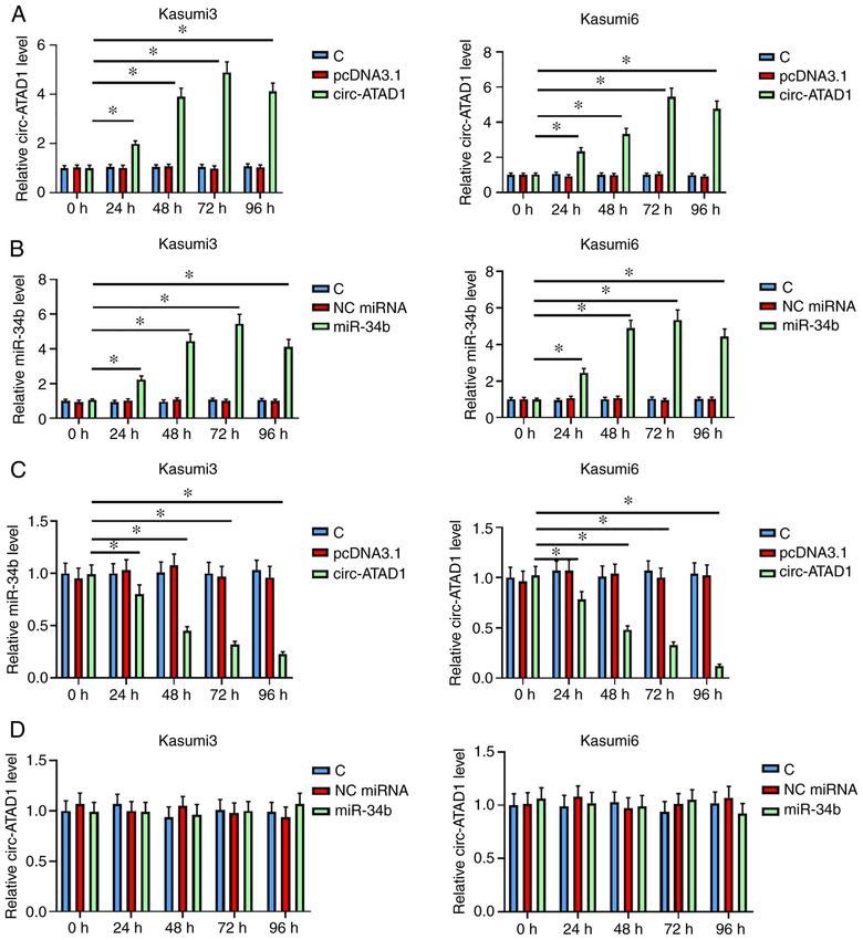
ONCOLOGY LETTERS 22: 799, 2021                                                 3

Table I. Sequences of oligonucleotides.

Oligonucleotide                                                                                     Sequence (5'‑3')

Mimics NC sense                                                                         UUCUCCGAACGUGUCACGUU
Mimics NC antisense                                                                     AACGUGACACGUUCGGAGAA
miR‑34b mimics sense                                                                    UAGGCAGUGUCAUUAGCUGAUUG
miR‑34b mimics antisense                                                                CAAUCAGCUAAUGACACUGCCUA

NC, negative control; miR, microRNA.

Table II. Sequences of quantitative PCR primers.

Gene                                      Forward primer (5'‑3')                                   Reverse primer (5'‑3')

Circ‑ATAD1                      GTTTCCTTCCTGTGTGAGGC                                      GGTCCGAGACGGTCCTTAAA
U6                              CTCGCTTCGGCAGCACA                                         AACGCTTCACGAATTTGCGT
miR‑34b                         AGGCAGTGTCATTAGCTGATTGT                                   ACAATCAGCTAATGACACTGCCT
GAPDH                           CCATTTGCAGTGGCAAAG                                        CACCCTTTGTGTTAGTG

Table III. Sequences of primers used for MSP.

Gene                                      Forward primer (5'‑3')                                 Reverse primer (5'‑3')

miR‑34b USP                TTTTTATTTGTTTTGTTTTGTGTTTGTTTTG                              CAACTACAACTCCCAAACAATCC
miR‑34b MSP                ATTCGTTTCGTTTCGCGTTCGTTTC                                    CGACTACAACTCCCGAACGATCCG

MSP, methylation‑specific PCR. USP, Unmethylation‑specific PCR.

a hybridization buffer at 37˚C for 1 h. The slides were incu‑      95˚C for 30 sec, 55˚C for 30 sec and 72˚C for 50 sec, and
bated with a hybridization buffer containing the FISH probe        then 72˚C for 10 min. All primers used for MSP and USP
at 95˚C for 5 min, and then at 37˚C overnight in the dark in       are listed in Table III.
a humid chamber. The samples were washed with 2 x saline
sodium citrate buffer (SSC) for 10 min at 37˚C, 1 x SSC for        BrdU assay. A total of 3x10³ transfected Kasumi‑3 and
2 x 5 min at 37˚C, and 0.5 x SSC for 10 min at room tempera‑       Kasumi‑6 cells were transferred to each well of a 96‑well plate
ture. The slides were then incubated with anti‑DIG‑488             in 0.1 ml medium, and cultured at 37˚C for 48 h before the
(1:300; cat. no. ab150077; Abcam) at 37˚C for 50 min, and the      addition of BrdU. The experiment was conducted using the
nuclei were counterstained with DAPI at room temperature           BrdU Cell Proliferation Assay (cat. no. QIA58; Sigma‑Aldrich;
for 30 min. Finally, the slices were sealed in fluorescence        Merck KGaA) according to the manufacturer's protocol. Then,
decay‑resistant medium and images were obtained under a            cells were cultured with 20 µl/well diluted BrdU reagent
fluorescence microscope (Nikon Corporation).                       (10 mM) for 6 h, fixed with Fixing solution and incubated for
                                                                   30 min. After fixation, the cells were incubated for another
Methylation‑specific PCR (MSP). Genomic DNA isolation              48 h with peroxidase‑coupled anti‑BrdU‑antibody (supplied
from transfected Kasumi‑3 and Kasumi‑6 cells was performed         by the kit), followed by washing twice with ice‑cold PBS.
using a routine method (20). All genomic DNA samples were          After incubation with peroxidase substrate for 3 h, OD values
processed using the EZ DNA Methylation‑Gold™ Kit (Zymo             were measured at 450 nm. For the represent images, the
Research Corp.) per the manufacturer's protocol. Then,             anti‑Brdu (1:500; cat. no. ab6362; Abcam) was used as primary
qPCR and routine PCR were performed to detect methyla‑             antibody, and Goat anti‑rabbit Alexa Fluor ® 546 (1:2,000;
tion of the miR‑34b gene promoter using PCR Master Mix             cat. no. A11010; Invitrogen; Therno Fisher4 Scientific, Inc.)
x2 (Invitrogen; Thermo Fisher Scientific, Inc.). The MSP           was used as the secondary antibody. The detailed method has
primers can be used to amplify methylated template, while          been previously published (21).
the primers for unmethylation‑specific PCR (USP) do not
amplify these products. Both the MSP and USP conditions            Statistical analysis. AML and control groups were compared
were as following: 95˚C for 5 min, followed by 40 cycles of        by unpaired t‑test. Comparisons among multiple independent
4 WU et al: circ‑ATAD1 UPREGULATION IN AML PROMOTES CANCER CELL PROLIFERATION Figure 1. Expression of circ‑ATAD1 and miR‑34b is altered in patients with AML. Samples of bone marrow mononuclear cells from patients with AML (n=60) and healthy controls (n=60) were subjected to RNA isolation and reverse transcription‑quantitative PCR to determine the differential expression of (A) circ‑ATAD1 and (B) miR‑34b. To study the crosstalk between circ‑ATAD1 and miR‑34b, correlation analyses across both (C) AML and (D) control samples were conducted by Pearson's correlation coefficient. ***P
ONCOLOGY LETTERS 22: 799, 2021                                                                  5

Figure 2. Circ‑ATAD1 overexpression decreases miR‑34b expression in AML cells. (A) Kasumi‑3 and (B) Kasumi‑6 cells were transfected with either
circ‑ATAD1 expression vector or the miR‑34b mimics, followed by confirmation of overexpression every 24 h until 96 h. Relative expression of (A) circ‑ATAD1
and (B) miR‑34b in Kasumi‑3 and Kasumi‑6 cells were compared between 24 and 96 by RT‑qPCR. Effects of (C) circ‑ATAD1 overexpression on miR‑34b
expression, and the effects of (D) miR‑34b overexpression on circ‑ATAD1 expression were also analyzed by RT‑qPCR. Data are presented as the mean ± SD
of three independent replicates. *P
6                         WU et al: circ‑ATAD1 UPREGULATION IN AML PROMOTES CANCER CELL PROLIFERATION

Figure 3. Circ‑ATAD1 is a nucleus‑specific circRNA that increases methylation of the miR‑34b gene in AML cells. (A) Subcellular fractionation assays were
used to determine the subcellular location of circ‑ATAD1 in both Kasumi‑3 and Kasumi‑6 cells. Relative expression of (B) circ‑ATAD1 and (C) miR‑34b
in Kasumi‑3 cells (nuclei), Kasumi‑6 (nuclei) cells and (C) samples from healthy control and patients with AML. Expression of GAPDH or U6 in the
nuclei of Kasumi‑3 and Kasumi‑6 cells was normalized to ‘1’. Effects of circ‑ATAD1 overexpression on miR‑34b gene methylation were analyzed by MSP
(D) Representative gel images of three biological replicates are presented. Data are presented as the mean ± SD of three independent replicates. **P
ONCOLOGY LETTERS 22: 799, 2021                                                                7

Figure 4. Circ‑ATAD1 overexpression increases acute myelogenous leukemia cell proliferation through miR‑34b. Role of circ‑ATAD1 and miR‑34b in regu‑
lating (A) Kasumi‑3 and (B) Kasumi‑6 cell proliferation was analyzed by BrdU assay. Representative images of (C) Kasumi‑3 and (D) Kasumi‑6 cells are
displayed. Scale bar, 20 µm. Blue signals indicate DAPI staining; red signals indicate Brdu signaling Normalized data of three biological replicates are
presented. Mean values of three replicates are presented. *P
8                    WU et al: circ‑ATAD1 UPREGULATION IN AML PROMOTES CANCER CELL PROLIFERATION

reversed circ‑ATAD1 overexpression‑induced cellular               References
proliferation, indicating an interaction between the two
noncoding RNAs. However, a moderate correlation was                1. Rose‑Inman H and Kuehl D: Acute leukemia. Hematol Oncol
                                                                      Clin North Am 31: 1011‑1028, 2017.
observed between circ‑ATAD1 and miR‑34b across AML,                2. Parkin B, Ouillette P, Yildiz M, Saiya‑Cork K, Shedden K and
but not the control samples. Therefore, certain AML‑related           Malek SN: Integrated genomic profiling, therapy response, and
factors may be involved in mediating the interaction                  survival in adult acute myelogenous leukemia. Clin Cancer
                                                                      Res 21: 2045‑2056, 2015.
between circ‑ATAD1 and miR‑34b. Collectively, the results          3. Schiller GJ: High‑risk acute myelogenous leukemia: Treatment
of the present study revealed the circ‑ATAD1‑miR‑34b                  today and tomorrow. Hematology Am Soc Hematol Educ
axis as a novel regulatory signaling pathway specific to              Program 2013: 201‑208, 2013.
                                                                   4. Gores GJ and Kaufmann SH: Selectively targeting Mcl‑1 for
AML malignant transformation. However, its downstream                 the treatment of acute myelogenous leukemia and solid tumors.
genes, and the specific role and contribution in different cell       Genes Dev 26: 305‑311, 2012.
types at different transformation phases, requires further         5. Jiang XJ, Huang KK, Yang M, Qiao L, Wang Q, Ye JY, Zhou HS,
                                                                      Yi ZS, Wu FQ, Wang ZX, et al: Synergistic effect of panobinostat
investigation.                                                        and bortezomib on chemoresistant acute myelogenous leukemia
    In conclusion, circ‑ATAD1 is upregulated in AML and               cells via AKT and NF‑κ B pathways. Cancer Lett 326: 135‑142,
promotes AML cell proliferation by downregulating miR‑34b             2012.
                                                                   6. Piya S, Andreeff M and Borthakur G: Targeting autophagy to
via promoter methylation.                                             overcome chemoresistance in acute myleogenous leukemia.
                                                                      Autophagy 13: 214‑215, 2017.
Acknowledgements                                                   7. Gill H, Leung AY and Kwong YL: Molecularly targeted therapy
                                                                      in acute myeloid leukemia. Future Oncol 12: 827‑838, 2016.
                                                                   8. Hatzimichael E, Georgiou G, Benetatos L and Briasoulis E: Gene
Not applicable.                                                       mutations and molecularly targeted therapies in acute myeloid
                                                                      leukemia. Am J Blood Res 3: 29‑51, 2013.
Funding                                                            9. Konig H and Levis M: Is targeted therapy feasible in acute
                                                                      myelogenous leukemia? Curr Hematol Malig Rep 9: 118‑127,
                                                                      2014.
This work was supported by the Natural Science Foundation         10. Martelli AM, Evangelisti C, Chiarini F and McCubrey JA: The
of Inner Mongolia (grant no. 2019MS08047).                            phosphatidylinositol 3‑kinase/Akt/mTOR signaling network as
                                                                      a therapeutic target in acute myelogenous leukemia patients.
                                                                      Oncotarget 1: 89‑103, 2010.
Availability of data and materials                                11. Pei S, Minhajuddin M, Callahan KP, Balys M, Ashton JM,
                                                                      Neering SJ, Lagadinou ED, Corbett C, Ye H, Liesveld JL, et al:
                                                                      Targeting aberrant glutathione metabolism to eradicate human acute
The datasets used and/or analyzed during the current study are        myelogenous leukemia cells. J Biol Chem 288: 33542‑33558, 2013.
available from the corresponding author on reasonable request.    12. Vo JN, Cieslik M, Zhang Y, Shukla S, Xiao L, Zhang Y, Wu YM,
                                                                      Dhanasekaran SM, Engelke CG, Cao X, et al: The landscape of
                                                                      circular RNA in cancer. Cell 176: 869‑881.e13, 2019.
Authors' contributions                                            13. Fang Y and Fullwood MJ: Roles, functions, and mechanisms
                                                                      of long non‑coding RNAs in cancer. Genomics Proteomics
YZ put forward the concept, designed the experiments,                 Bioinformatics 14: 42‑54, 2016.
                                                                  14. Rücker FG, Russ AC, Cocciardi S, Kett H, Schlenk RF,
provided general supervision, edited the manuscript and was           Bot z en ha rdt U, L a nger C, K raut er J, Fröh l i ng S,
a guarantor of integrity of the entire study. YW, BG and XQ           Schlegelberger B, et al: Altered miRNA and gene expression
acquired and analyzed the data. YW wrote the manuscript.              in acute myeloid leukemia with complex karyotype identify
                                                                      networks of prognostic relevance. Leukemia 27: 353‑361, 2013.
LB, BL and HB conducted the literature search and interpre‑       15. Benassi B, Marani M, Loda M and Blandino G: USP2a alters
tated the data. XW and XYW defined intellectual content,              chemotherapeutic response by modulating redox. Cell Death
conducted the literature search, were involved in interpreting        Dis 4: e812, 2013.
                                                                  16. Zhang L, Chang X, Zhai T, Yu J, Wang W, Du A and Liu N: A
the results and data analysis, and revised the manuscript.            novel circular RNA, circ‑ATAD1, contributes to gastric cancer
YZ and YW confirm the authenticity of all the raw data. All           cell progression by targeting miR‑140‑3p/YY1/PCIF1 signaling
authors have read and approved the final manuscript.                  axis. Biochem Biophys Res Commun 525: 841‑849, 2020.
                                                                  17. Wang J, Liu X, Lu H, Jiang C, Cui X, Yu L, Fu X, Li Q and
                                                                      Wang J: CXCR4(+)CD45(‑) BMMNC subpopulation is superior
Ethics approval and consent to participate                            to unfractionated BMMNCs for protection after ischemic stroke
                                                                      in mice. Brain Behav Immun 45: 98‑108, 2015.
                                                                  18. Rao X, Huang X, Zhou Z and Lin X: An improvement of the
The present study was approved by the Ethics Committee                2(‑delta delta CT) method for quantitative real‑time polymerase
of Xing'an League People's Hospital. All experiments were             chain reaction data analysis. Biostat Bioinforma Biomath 3:
performed in accordance with the 1964 Declaration of                  71‑85, 2013.
                                                                  19. Pavani RS and Elias MC: Following trypanosoma cruzi
Helsinki and its later amendments. Written informed consent           RPA‑DNA interaction using fluorescent in situ hybridization
to participate in the study was obtained from patients and            coupled with immunofluorescence (FISH/IF). Methods Mol
controls prior to sample collection.                                  Biol 2281: 209‑215, 2021.
                                                                  20. Yuan Y, Wang Q, Ma SL, Xu LQ, Liu MY, Han B, Du N,
                                                                      Sun XL, Yin XL and Cao FF: lncRNA PCAT‑1 interacting with
Patient consent for publication                                       FZD6 contributes to the malignancy of acute myeloid leukemia
                                                                      cells through activating Wnt/β ‑catenin signaling pathway. Am
                                                                      J Transl Res 11: 7104‑7114, 2019.
Not applicable.                                                   21. Mehdipour M, Etienne J, Chen CC, Gathwala R, Rehman M,
                                                                      Kato C, Liu C, Liu Y, Zuo Y, Conboy MJ and Conboy IM:
Competing interests                                                   Rejuvenation of brain, liver and muscle by simultaneous phar‑
                                                                      macological modulation of two signaling determinants, that
                                                                      change in opposite directions with age. Aging (Albany NY) 11:
The authors declare that they have no competing interests.            5628‑5645, 2019.
ONCOLOGY LETTERS 22: 799, 2021 9
22. Ishikawa F, Yoshida S, Saito Y, Hijikata A, Kitamura H, 27. Hansen TB, Jensen TI, Clausen BH, Bramsen JB, Finsen B,
Tanaka S, Nakamura R, Tanaka T, Tomiyama H, Saito N, et al: Damgaard CK and Kjems J: Natural RNA circles function as
Chemotherapy‑resistant human AML stem cells home to efficient microRNA sponges. Nature 495: 384‑388, 2013.
and engraft within the bone‑marrow endosteal region. Nat 28. Massart R, Barnea R, Dikshtein Y, Suderman M, Meir O,
Biotechnol 25: 1315‑1321, 2007. Hallett M, Kennedy P, Nestler EJ, Szyf M and Yadid G: Role
23. Nakamura‑Ishizu A, Takubo K, Kobayashi H, Suzuki‑Inoue K of DNA methylation in the nucleus accumbens in incubation of
and Suda T: CLEC‑2 in megakaryocytes is critical for mainte‑ cocaine craving. J Neurosci 35: 8042‑8058, 2015.
nance of hematopoietic stem cells in the bone marrow. J Exp 29. Liu Z, Yu Y, Huang Z, Kong Y, Hu X, Xiao W, Quan J and Fan X:
Med 212: 2133‑2146, 2015. CircRNA‑5692 inhibits the progression of hepatocellular carci‑
24. Stevens AM, Xiang M, Heppler LN, Tošić I, Jiang K, Munoz JO, noma by sponging miR‑328‑5p to enhance DAB2IP expression.
Gaikwad AS, Horton TM, Long X, Narayanan P, et al: Cell Death Dis 10: 900, 2019.
Atovaquone is active against AML by upregulating the integrated 30. Gu Y, Ci C, Zhang X, Su M, Lv W, Chen C, Liu H, Zhang D,
stress pathway and suppressing oxidative phosphorylation. Blood Zhang S and Zhang Y: Prediction of circRNAs based on the
Adv 3: 4215‑4227, 2019. DNA methylation‑mediated feature sponge function in breast
25. Vignon C, Debeissat C, Bourgeais J, Gallay N, Kouzi F, cancer. Front Bioeng Biotechnol 7: 365, 2019.
Anginot A, Picou F, Guardiola P, Ducrocq E, Foucault A, et al:
Involvement of GPx‑3 in the reciprocal control of redox metabo‑ This work is licensed under a Creative Commons
lism in the leukemic niche. Int J Mol Sci 21: 8584, 2020. Attribution-NonCommercial-NoDerivatives 4.0
26. Li G, Song Y, Zhang Y, Wang H and Xie J: MiR‑34b Targets International (CC BY-NC-ND 4.0) License.
HSF1 to suppress cell survival in acute myeloid leukemia. Oncol
Res 24: 109‑116, 2016.You can also read