Sensing soluble uric acid by Naip1-Nlrp3 platform - Nature
←
→
Page content transcription
If your browser does not render page correctly, please read the page content below
Braga et al. Cell Death and Disease (2021)12:158
                                    https://doi.org/10.1038/s41419-021-03445-w                                                                                                             Cell Death & Disease

                                     ARTICLE                                                                                                                                                    Open Access

                                    Sensing soluble uric acid by Naip1-Nlrp3 platform
                                    Tarcio Teodoro Braga 1,2,3, Mariana Rodrigues Davanso2,3,4, Davi Mendes 5, Tiago Antonio de Souza5,
                                    Anderson Fernandes de Brito6, Mario Costa Cruz2, Meire Ioshie Hiyane2, Dhemerson Souza de Lima2, Vinicius Nunes2,
                                    Juliana de Fátima Giarola7, Denio Emanuel Pires Souto7,8, Tomasz Próchnicki3, Mario Lauterbach3,
                                    Stellee Marcela Petris Biscaia9, Rilton Alves de Freitas8, Rui Curi4,10, Alessandra Pontillo2, Eicke Latz 3,11,12 and
                                    Niels Olsen Saraiva Camara2,13,14

                                      Abstract
                                      Uric acid (UA), a product of purine nucleotide degradation able to initiate an immune response, represents a
                                      breakpoint in the evolutionary history of humans, when uricase, the enzyme required for UA cleavage, was lost.
                                      Despite being inert in human cells, UA in its soluble form (sUA) can increase the level of interleukin-1β (IL-1β) in
                                      murine macrophages. We, therefore, hypothesized that the recognition of sUA is achieved by the Naip1-Nlrp3
                                      inflammasome platform. Through structural modelling predictions and transcriptome and functional analyses, we
                                      found that murine Naip1 expression in human macrophages induces IL-1β expression, fatty acid production and an
                                      inflammation-related response upon sUA stimulation, a process reversed by the pharmacological and genetic
                                      inhibition of Nlrp3. Moreover, molecular interaction experiments showed that Naip1 directly recognizes sUA.
                                      Accordingly, Naip may be the sUA receptor lost through the human evolutionary process, and a better understanding
                                      of its recognition may lead to novel anti-hyperuricaemia therapies.
                  1234567890():,;
1234567890():,;
                  1234567890():,;
1234567890():,;

                                    Introduction                                                                                        that are foreign”6, several damage-associated molecular
                                      Host responses against harmful signals are basic phy-                                             patterns (DAMPs) have been described. Indeed, receptors
                                    siological reactions of all living organisms. Innate immu-                                          for endogenous and exogenous signals may have evolved
                                    nity pattern-recognition receptors (PRRs) were first                                                 simultaneously because vertebrates and pathogens have
                                    described as recognizing conserved structural compo-                                                shared eons of evolutionary time and space6. Perhaps,
                                    nents of microorganisms1. The discovery of Toll-like                                                PRRs have not evolved to bind to pathogens at all; in
                                    receptors2 led scientists to understand how the immune                                              contrast, perhaps, the pathogens evolved to attach to
                                    system responds to nonself antigens in the context of an                                            PRRs and thus enhance their own survival7, a hypothesis
                                    infection3,4, in contrast with the previous model, in which                                         that would explain a puzzling feature of PRRs: each one
                                    the immune system reacted to all nonself antigens while                                             can attach to many different kinds of molecules.
                                    being tolerant to self-antigens5,6. Based on Polly Matzin-                                            Among several DAMPs, uric acid (UA), the product of
                                    ger’s study stating that “the immune system is more                                                 purine catabolism, released mainly from dying cells and
                                    concerned with entities that do damage than with those                                              ischaemic tissues, is considered a major alarmin, espe-
                                                                                                                                        cially when it is present at elevated levels and crystallized,
                                                                                                                                        also known as monosodium urate (MSU)8. In rodents,
                                    Correspondence: Tarcio Teodoro Braga (tarcio.braga@ufpr.br)
                                    1                                                                                                   MSU activates the immune system9,10, acts as a pro-
                                     Department of Basic Pathology, Federal University of Parana, Curitiba, PR,
                                    Brazil                                                                                              oxidant molecule, stimulates chemotaxis and activates the
                                    2
                                     Department of Immunology, Institute of Biomedical Sciences IV, University of                       nuclear factor-κB and MAPK pathways11. Moreover,
                                    São Paulo, São Paulo, SP, Brazil
                                                                                                                                        MSU induces the release of interleukin-1β (IL-1β)
                                    Full list of author information is available at the end of the article
                                    These authors contributed equally: Eicke Latz, Niels Olsen Saraiva Camara                           through the activation of inflammasome-dependent
                                    Edited by W. Jia

                                    © The Author(s) 2021
                                                        Open Access This article is licensed under a Creative Commons Attribution 4.0 International License, which permits use, sharing, adaptation, distribution and reproduction
                                                        in any medium or format, as long as you give appropriate credit to the original author(s) and the source, provide a link to the Creative Commons license, and indicate if
                                    changes were made. The images or other third party material in this article are included in the article’s Creative Commons license, unless indicated otherwise in a credit line to the material. If
                                    material is not included in the article’s Creative Commons license and your intended use is not permitted by statutory regulation or exceeds the permitted use, you will need to obtain
                                    permission directly from the copyright holder. To view a copy of this license, visit http://creativecommons.org/licenses/by/4.0/.

                                    Official journal of the Cell Death Differentiation Association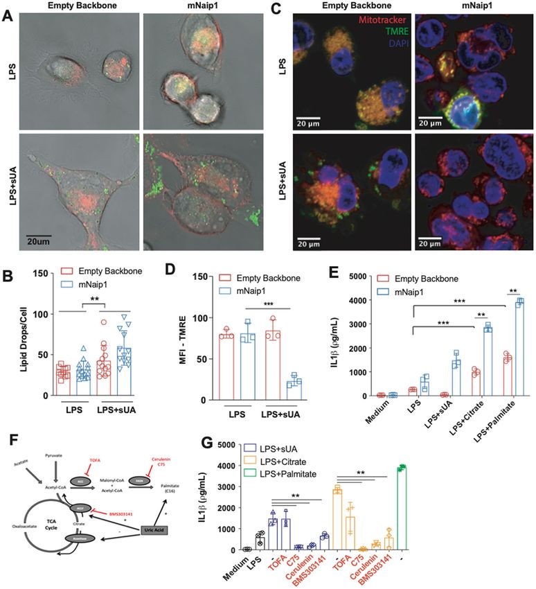
Braga et al. Cell Death and Disease (2021)12:158                                                                Page 2 of 14

caspases10,12,13. The inflammasome is a cytosolic complex         structural modelling predictions to study the sensing of
activated when a nucleotide-binding domain (NBD/                 sUA by mNaip1. In addition to demonstrating that
NACHT) and leucine-rich repeat (LRR)-containing                  mNaip1 expression leads to the activation in human cells
receptor (NLR) senses PAMPs or DAMPs14. NLRs belong              upon sUA stimulation, we found that mNaip1 directly
to a superfamily of innate immune proteins with a very           recognizes sUA. We then hypothesized that Naip may be
conserved structure throughout the phylogeny, from               the lost receptor for UA and, in particular, for sUA.
plants to mammals. In distinct phylogenetic groups, the
number of receptors and paralogous genes differs, possi-         Materials and methods
bly as a consequence of host/pathogen and/or environ-            Soluble UA preparation
mental coevolution15. To date, it has been demonstrated            The medium was prewarmed (37 °C), UA (Ultrapure,
that both soluble UA (sUA)16 and MSU can induce Nlrp3            Sigma; 200 μΜ) was added, and the medium was sterilized
(an NLR containing a pyrin domain as its N-terminal              with 0.20 μm filters. Crystals were not detectable under
domain) oligomerization in mice, while the deposition of         these conditions (polarizing microscopy), nor did they
MSU activates Nlrp3 inflammasome in humans, in the                develop during cell incubation.
context of gout 17.
   Great apes have higher levels of UA in serum                  Reagents
(3.02–6.72 mg/dL, corresponding to 180–400 μΜ) than                Ultrapure LPS (lipopolysaccharide) was obtained from
other animals (18–40 μΜ) and UA crystallization occurs, in       InvivoGen, and nigericin was obtained from Invitrogen.
humans, when the level reaches 6.8 mg/dL in plasma18. This       DRAQ5 was purchased from eBioscience. Anti-GFP
observation is compatible with the absence of uricase (or        monoclonal antibody (GF28R) was purchased from
urate oxidase) activity, the enzyme involved in purine cata-     Thermo Fisher Scientific. CRID3 was purchased from
bolism converting UA into allantoin19,20. The loss of uricase    R&D Systems, and allantoin, urea and palmitate were
at the divergence between great apes and other mammals           purchased from Sigma-Aldrich. TOFA (5-(tetradecyloxy)-
may be related to a survival advantage, as previously hypo-      2-furoic acid) (CAS 54857-86-2) was purchased from
thesized, due to the UA characteristics as a molecule critical   Abcam. C75 (CAS 191282-48-1) was also obtained
for saving energy21; however, this supposition raises a tricky   from Abcam. Cerulenin (17397-89-6) was purchased from
question about the role of a mammalian sensor of UA. We          Sigma-Aldrich, and BMS303141 (CAS 943962-47-8)
hypothesize that, along with uricase loss and a consequent       was purchased from Cayman Chemical. UK5099 CAS
elevation in UA serum levels, humans have lost the sensors       56396-35-1 was obtained from Merck Millipore. Etha-
to recognize “high” levels of sUA.                               nolamine (EA), 11-mercaptoundecanoic acid (11-MUA),
   Among NLRs able to induce inflammasome activation              N-(3 dimethylamino-propyl)-N-ethylcarbodiimide hydro-
in mice and humans, the NLRB subfamily (an NLR con-              chloride (EDC), and N-hydroxysuccinimide (NHS) were
taining a baculovirus inhibitor of apoptosis protein repeat      obtained from Sigma-Aldrich Chemical (St. Louis,
domain as its N-terminal domain) attracted our attention.        MO, USA).
In mice, there are six paralogous genes, namely, Naip1–6,
and four functional receptors (Naip1, 2, 5 and 6), while in      Macrophages obtainment
humans, only one orthologous gene, Naip, has been                  Primary murine macrophages were generated from bone
found22. Despite the differences in the amino acid content       marrow. Briefly, bone marrow-derived cells were filtered
among Naip proteins, both mice and human receptors               through sterile polystyrene syringes (70–100 µm) and divi-
have been described to play role in the host defence             ded among 10 Petri dishes (100 × 20 mm2; BD, Franklin
against pathogens23,24. Murine (m) Naip1 and Naip2 are           Lakes, USA) supplemented with murine macrophage CSF
critical for the detection of needle and rod proteins,           (20 ng/mL; R&D Systems, Minneapolis, MN, USA) diluted
respectively25–27. Naip5 is crucial for the cytosolic            in high-glucose Dulbecco’s modified Eagle’s medium
recognition of flagellin28, the major protein component of        (Invitrogen, USA) in the presence of 5% foetal bovine serum
the bacterial flagellum. Similar to Naip5, human Naip             for 7 days. All procedures with mice were approved by the
(hNaip) can bind bacterial flagellin29 and activate the           local ethics committees at the University of São Paulo
Nlrc4 inflammasome. In this scenario, Naip acts as a              (Document 45/2009). All experiments were performed in
ligand sensor, and Nlrc4 is critical for inflammasome             accordance with relevant guidelines and regulations.
assembling and inflammasome-dependent cell death,                   Human and rhesus macaque (Macaca mulatta) mac-
known as pyroptosis30. Considering the differences in            rophages were generated from circulating monocytes. A
NLRB orthologous genes among different species and the           centrifugation gradient was used to obtain human and
lack of endogenous ligands found for NLRB to date, in            monkey peripheral blood mononuclear cells in Ficoll-
this study, we performed transcriptome- and proteome-            Paque (Dominique Dutscher). Subsequently, the periph-
wide analyses in addition to interaction investigations and      eral blood mononuclear cells were centrifuged in 51%

Official journal of the Cell Death Differentiation Association
Braga et al. Cell Death and Disease (2021)12:158                                                                Page 3 of 14

phosphate-buffered saline-diluted Percoll (GE Healthcare        empty vector GFP+ control cells (n = 6) using TRIzol
Life Sciences), and the monocytes (>70% purity) were            reagent according to the manufacturer’s instructions
allowed to adhere to six-well plates for 4 h. After removal     (Thermo Fisher Scientific, Waltham, MA, USA). The
of nonadherent cells by washing the plates, the monocytes       integrity of the total RNA was checked using a Bioana-
were differentiated into macrophages in RPMI medium             lyzer 2100 (Agilent Technologies, CA, USA) with an RNA
(Gibco, Grand Island, NY, USA) supplemented with 10%            Nano 6000 kit. RNA purity and quantity were measured
foetal calf serum (Gibco) plus antibiotics and antimycotics     using a NanoDrop 1000 spectrophotometer and Qubit
(100 U/mL penicillin, 100 g/mL streptomycin and 25 g/           RNA HS fluorescence kit (Thermo Fisher), respectively.
mL amphotericin; Gibco) in the presence of human                The total RNA concentration of each sample (n = 12) was
macrophage CSF (25 ng/mL; R&D Systems, Minneapolis,             adjusted to 1 µg and used for poly(A) messenger RNA
MN, USA) for 5 days. All procedures with rhesus maca-           (mRNA) enrichment and strand-specific RNA-Seq (RNA
ques were approved by the Ethics Committee on Animal            sequencing) library preparation with a TruSeq Stranded
Use of the Butantan Institute (CEUAIB) (number                  mRNA kit (Illumina, San Diego, CA, USA). Quality con-
9376040717). Leucocyte concentrates were obtained after         trol of the prepared libraries was performed using an
plasmapheresis at the Blood Bank Service of the “Hospital       Agilent Bioanalyzer DNA 1000 kit and Qubit DNA HS
das Clinicas” in Sao Paulo (SP, Brazil), and they were used     fluorescence kit. The libraries were pooled and then
for peripheral blood monocyte isolation. All volunteers         sequenced at CEFAP-USP (Sao Paulo, Brazil) on an Illu-
signed informed consent in compliance with the respec-          mina NextSeq platform in 75-bp paired-end mode using a
tive Institutional Ethics Committee. We performed power         high output kit.
and sample size calculations to determine how large the
sample needed to be to obtain 90% power when com-               RNA-Seq data analysis
paring a categorical variable among groups. Experiments           Before read mapping, clean reads were selected after
were performed in duplicate or triplicate, and at least two     preprocessing with Trimmomatic33 to remove adapter and
independent tests were performed for each assay. The            poly(N) sequences. After cleaning, the quality of reads was
data are described in terms of the means and s.e.m. unless      checked by the FastQC tool and then aligned to the human
specified in the figure legend.                                   genome (GRCh38/hg38) using the HISAT2 aligner (V2-
                                                                2.0.0)34 considering strandness. The overall mapping quality
Viral transduction                                              and uniformity of the read coverage on exons were checked
  Plasmids for GFP-tagged mNaip1 (#60200), Naip5                by the RSeQC tool to ensure good RNA integrity and
(#60205), Naip6 (#60202), Nlrc4 (#60199) and empty              reproducible RNA-Seq. The StringTie (v.1.3.4)35 and Ball-
vector (#60206) were purchased from Addgene (Water-             gown36 algorithms were applied to identify significantly
town, MA, USA). Transformed bacteria with the GFP-              differentially expressed genes (q value  1 and q value
colour-tagged sequences with standard cloning techni-           < 0.05) and downregulated genes (FC < 1 and q value
ques. Briefly, THP1 cells (ATCC, TIB202) were retro-             < 0.05). High-throughput sequence data were uploaded as
virally transduced with constructs for the indicated            supplemental material.
plasmids. After retroviral transduction, the cells were
cytometrically sorted with cells with similar levels of GFP     Proteomics analysis
or turquoise expression. Nlrp3-deficient (Nlrp3−/−)                Protein solutions were quantified by fluorometry using a
macrophages have been previously described31, and Nlrp3         Qubit® protein assay kit. From these solutions, an
deletion from THP1 cells was performed according to the         equivalent aliquot of 50 μg of protein was transferred to
procedure described by Schmid-Burgk et al.32.                   0.5 mL tubes and dried. The protein pellets were sus-
                                                                pended in 6 M urea in aqueous solution (25 μL). The same
Cytokine profile                                                 volume of reducing reagent plus 10 mM dithiothreitol was
  Cell lysates were maintained in RIPA buffer with pro-         added, and the samples were reduced for 60 min at room
tease inhibitors at −80 °C until dosing. IL-1β protein was      temperature. Subsequently, 50 μL of an alkylation solu-
measured using IL-1β (R&D Systems, Minneapolis, MN,             tion, 100 mM iodoacetamide, was added, and the samples
USA) according to the manufacturer’s instructions.              were alkylated for another 60 min at 54 °C in the dark.
                                                                Next, 1 M dithiothreitol (1 μL) was added and allowed to
RNA extraction, library construction and sequencing             react with the remaining iodoacetamide. Finally, 100 μL of
  Total RNA was extracted from GFP-sorted THP1 cells            ice-cold trypsin solution at a 1:50 (trypsin:protein) ratio
containing the lentiviral vector NAIP1 (n = 6) or the           was added to the samples, followed by incubation for 16 h

Official journal of the Cell Death Differentiation Association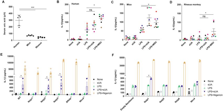
Braga et al. Cell Death and Disease (2021)12:158                                                                 Page 4 of 14

at 37 °C. Following digestion, the reaction was stopped by       until system stabilization. CCCP (carbonyl cyanide p-tri-
adding 10% formic acid (5 μL). The samples were then             fluoromethoxyphenylhydrazone) was used at a final con-
desalted using ZipTips® and maintained at −20 °C until           centration of 5 mM and injected with sodium pyruvate
analysis.                                                        (Sigma) at a final concentration of 5 mM. Oligomycin and
   Peptides were analysed by online nanoflow LC-MS (liquid        antimycin A were used at final concentrations of 1 and
chromatography-mass spectrometry) in an EASY-nLC II              10 μg/mL, respectively. Rotenone was used at a con-
system (Thermo Scientific) connected to an LTQ-Orbitrap           centration of 1 μM. All respiratory modulators were used
Velos instrument (Thermo Scientific) and a Proxeon                at ideal concentrations titrated during preliminary
nanoelectrospray ion source. The peptides were separated         experiments (not shown). A typical oxygen consumption
on an analytical EASY column (10 cm, ID 75 μm, 3 μm, C18;        ratio (OCR) chart is displayed, where OCR represents the
Thermo Scientific) previously trapped in a precolumn EASY         percentage of basal respiration.
column (2 cm, ID100 μm, 5 μm, C18; Thermo Scientific).
Tryptic digested peptides were separated using a 60 min          Western blotting
linear gradient of 0–60% buffer B (acetonitrile in 0.1% formic     Proteins derived from cell lysates were separated on
acid) at a 300 nL/min flow rate. The LTQ-Orbitrap Velos           sodium dodecyl sulfate-polyacrylamide gel electrophor-
mass spectrometer was operated in positive ion mode using        esis gels (4–12%) (Novex, Invitrogen) with 2-(N-mor-
DDA (data-dependent acquisition) mode. Full mass spec-           pholino) ethanesulfonic acid buffer (Novex, Invitrogen) at
trometric (MS) scans were performed with 60,000 resolu-          150 V from 60 to 90 min. The proteins were transferred
tion, and the m/z range for the MS scans was 400–1200. The       onto polyvinylidene difluoride membranes (Millipore,
minimum signal threshold was 15,000 counts, and for              Temecula, CA, USA), which had been pretreated for
dynamic exclusion, it was considered a one repeat count          1–2 min with methanol, at 32 V for 90 min in buffer
with a duration of 30 s. To discriminate the charge state of     containing 10% Tris-glycine and 15% methanol. The
the peptides, charge state screening was enabled, and ions       membranes were blocked with 3% bovine serum albumin
with an unassigned charge state or singly charged ions were      (BSA) containing Tris buffer in saline solution (TBS) for
rejected.                                                        60 min. After blocking, the membranes were incubated
   The MS/MS spectra from each LC-MS/MS run were                 with specific primary antibodies in TBS containing 3%
compared against five different databases with two dis-           BSA and 0.1% Tween-20 overnight. The following pri-
tinct search engines, an in-house program and Proteome           mary antibodies were used: goat anti-mouse Naip1 (sc-
Discoverer 1.4 software (Thermo, USA). The search cri-           11067, 1:200; Santa Cruz Biotechnology, Santa Cruz, CA,
teria were as follows: full tryptic specificity was required,     USA), rabbit anti-mouse NLRP3 (mAb #15101, 1:1000;
two missed cleavages were allowed, carbamidomethyla-             Cell Signalling Technology, Danvers, MA, USA), anti-
tion (C) was set as the fixed modification, oxidation (M)          β-actin mouse (1:1000, Li-COR Bioscience, Lincoln, NE,
was set as the variable modification, precursor ion mass          USA) and anti-IL-1β from an R&D ELISA kit as a
tolerance was set at 10 p.p.m. for all the MS spectra            detection antibody (1:1000). The membranes were
acquired with the Orbitrap mass analyser, and the frag-          washed and incubated with secondary antibodies (IRDye
ment ion mass tolerance was set at 0.6 Da for all the            800CW or IrDye 680RD, LI-COR Biosciences, 1:20,000)
MS2 spectra acquired. All covariates were log-                   for 60 min. After washing, the stained cells were visualized
transformed before statistical analysis was performed.           on an Odyssey imaging system. The staining intensity was
All analyses were performed using STRING software and            quantified using the Fiji/ImageJ software.
UniProt for protein–protein interactions, identification,
and statistics. P ≤ 0.05 was considered significant.              Confocal imaging
                                                                   Confocal laser scanning microscopy was performed
Oxygen consumption rates                                         with a Leica TCS SP5 SMD confocal system (Leica
  An hour before the oxygen consumption measure-                 Microsystems). The images were captured using a single z
ments, the cell medium was replaced by assay medium              step, and the emitted fluorescence was detected by scan-
(2 mM glucose, 0.8 mM Mg2+, 1.8 mM Ca2+, 143 mM                  ned detectors at 490–520 and 575–605 nm and emission
NaCl, 5.4 mM KCl, 0.91 mM NaH2PO4 and 15 mg/mL                   filters. Predefined settings for the laser power and detector
Phenol red) and incubated for 60 min at 37 °C (no CO2)           gain were used for all experiments. Microphotographs
before loading into a Seahorse Bioscience XF96 extra-            were analysed using LAS AF version 2.2.1 (Leica Micro-
cellular analyser. During the 60-min period, the ports of        systems) or Volocity 6.01 software.
the cartridge containing the oxygen probes were loaded
with the compounds to be injected during the assay               Quartz crystal microbalance (QCM)
(75 μL/port), and the cartridge was calibrated. Basal              The interaction of the Naip1 protein with sUA was
respiration was recorded for 30 min at 4-min intervals           analysed in a QCM device (Stanford Research Systems

Official journal of the Cell Death Differentiation Association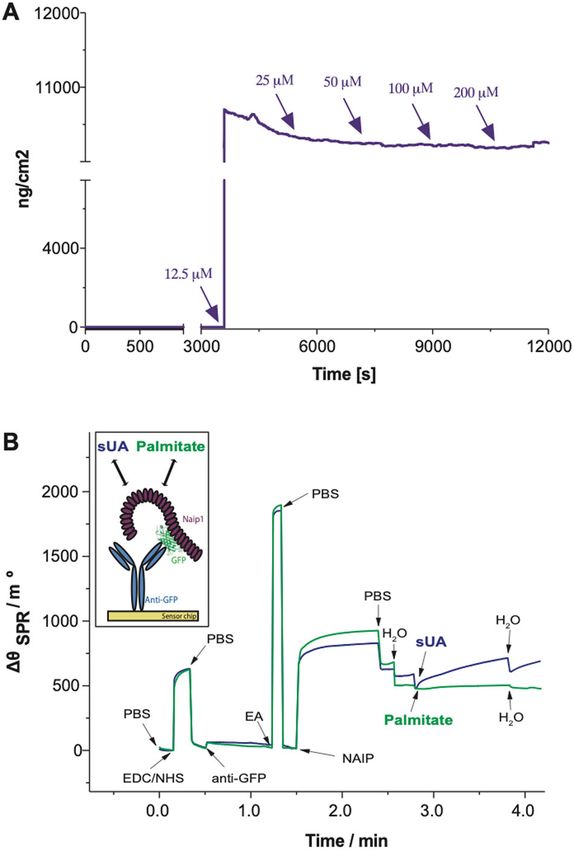
Braga et al. Cell Death and Disease (2021)12:158                                                               Page 5 of 14

(SRS)). A plasmid carrying GFP-tagged Naip1 was virally         MUA (1.0 mmol/L). After the formation of the film, the
transduced into THP1 cells, and cell lysates were used to       gold surface (11-MUA/Au) was copiously washed with
bind the Naip1 protein to anti-GFP previously immobi-           ethanol and water and dried under an N2(g) flow. All the
lized onto gold quartz crystals (SRS, 5 MHz). Prior to          steps described above were performed ex situ. In the next
antibody immobilization, the gold crystals were immersed        step, the functionalized SPR sensor chip was immediately
in piranha solution 1:3H2O2/H2SO4 for 15 min, washed            inserted into the SPR instrument, and the measurements
twice with absolute ethanol for 5 min, washed three times       were obtained in real time. The terminal carboxyl groups
with ultrapure water for 5 min and dried with a gentle          of 11-MUA were activated in a PBS (phosphate-buffered
flow of nitrogen. Then, the gold crystals were incubated         saline) buffer solution (10 mmol/L at pH 7.4) containing
with EDC (100 mmol/L) and NHS (150 mmol/L), and                 EDC (100 mmol/L) and NHS (150 mmol/L) for ~10 min to
finally, 200 μL of 20 μg/mL anti-GFP (MA5-15256,                 allow the formation of NHS-ester groups. This strategy
Thermo Fisher Scientific) diluted in ultrapure water was         was used to allow covalent binding of anti-GFP (20 μg/mL)
deposited over each crystal for 16 h at 4 °C in a humid         onto the gold (11-MUA/Au) of the SAM. Next, successive
chamber. The crystals were rinsed in three sequential           aliquots of the buffer solution were added to remove the
ultrapure water baths, dried at 22 °C and blocked with 1%       excess molecules on the surface. Then, the immobilization
BSA solution for 1 h at 37 °C. After incubation, all sample     of the anti-GFP was monitored via the SPR technique for
chips were washed and dried as described above. Then,           ~45 min. This step was followed by a washing step using
200 μL of cell lysate was deposited onto the crystals for       the successive addition of buffer solution. To prevent
16 h at 4 °C in a humid chamber. The crystals were placed       nonspecific binding, after the immobilization of anti-GFP
in a QCM flow chamber apparatus connected to a syringe           onto the 11-MUA/Au surface (SAM/Au), the unbound
pump with a 100 μL/min flow rate (KD Scientific). An              reactive ester groups were deactivated by a PBS buffer
initial hydration step with 500 μL of ultrapure water was       solution containing EA (1.0 mol/L at pH 8.5) for ~5 min.
performed, and 500 μL of each sUA solution sample (12.5,        Successive additions of the buffer solution then removed
25, 50, 100 and 200 μΜ) was injected in individual              the excess unbound EA molecules. After successfully
experiments. Each experiment was performed in tripli-           characterizing the immobilization and blocking steps, the
cate. The results are expressed as the average value.           interaction between the anti-GFP and Naip1 protein
                                                                (GFP-tagged) was evaluated. Cell lysates derived from
Surface plasmon resonance (SPR)-based immunosensor              GFP-tagged THP1 cells expressing mNaip1 were used to
development                                                     immobilize Naip1. After characterizing the interaction
  An Autolab Sprit instrument (Eco Chemie B.V., The             between anti-GFP and Naip1 protein, an aqueous solution
Netherlands), which reveals the phenomenon of atte-             of sUA was added to evaluate the interaction of sUA with
nuated total internal reflection (Kretschmann configura-          Naip1 protein. As a control for this assay, the interaction
tion) in operation mode38, was employed for the SPR             of palmitate with the Naip1 protein was also evaluated.
analysis. This SPR instrument was equipped with a glass
prism (BK7), a planar gold SPR sensor chip, and two             Protein structure analysis
measurement channels (channels 1 and 2). For the mea-             Homology models were obtained using MODELLER
surements, a laser diode with a wavelength fixed at              v.9.1840 with the 4KXF structure as a template41. To fix
670 nm and a photodiode detector was employed. In               residues with incompatible torsion angles, the target
terms of functionality, changes near the metal/environ-         proteins were repaired using RepairPDB42, and Chi-
ment interface promote a change in the resonance con-           maera43 was used to add hydrogen atoms and charges
ditions of the system, resulting in a shift in the θSPR. In     where appropriate. Surface electrostatic potentials were
this sense, SPR techniques enable information on bio-           calculated using the AMBER force field implemented for
molecular interactions to be obtained in real time.             APBS44 and input files that were converted with the
  The experiments were performed as demonstrated by             PDB2PQR package45. For each NAIP structure, blind
Souto et al.39. Prior to gold surface functionalization, the    docking was performed using SwissDock46 with UA in its
SPR sensor chip was cleaned in piranha solution (1:3            ionized form (urate) as a ligand (ZINC AC: 2041003).
mixture of 30% H2O2 to concentrated H2SO4) for ~1 min,          During docking, the surface of both proteins was scanned
followed by immersion of the substrate in acetone (5 min)       for putative binding pockets with >250 iterations. Several
and then in isopropyl alcohol (5 min). Next, the SPR            low-energy ligand clusters with similar binding modes
sensor chip was washed with deionized water and dried           (poses) were found. All poses with ΔG < −6 kcal/mol
under a pure N2(g) flow. The functionalization of the gold       were considered in further analyses, and those showing
surface was performed through the formation of a self-          the lowest energies were selected as the best repre-
assembled monolayer (SAM), which was obtained after a           sentatives of the binding between urate and both human
24 h incubation in an ethanol solution consisting of 11-        and mNaip.

Official journal of the Cell Death Differentiation Association
Braga et al. Cell Death and Disease (2021)12:158 Page 6 of 14
Statistics unrelated healthy adult human donors (n = 5), C57Bl/6
Experiments were performed in duplicate or triplicate, mice (n = 5), and old-world monkeys (rhesus macaques; n
and at least two independent tests were performed for = 5). As expected, the human’s samples exhibited an average
each assay. The data are described in terms of the means blood UA concentration of 295 μΜ, 4- and 7-fold greater
and s.e.m. unless specified in the figure legend. Differ- than the mice and rhesus macaques UA levels, respectively
ences between groups were compared using analysis of (Fig. 1A). Then, we stimulated murine LPS-primed BMDMs
variance (with Tukey’s post-test) and Student’s t test. and human and rhesus LPS-primed monocyte-derived
Significant differences were regarded as p < 0.05, p < 0.01 macrophages with 200 μΜ sUA. The human cells did not
or p < 0.001, according to the figure. All statistical analyses produce IL-1β after sUA stimulus when compared to the
were performed using GraphPad Prism 6.01 (La Jolla, CA, level produced by LPS-primed cells (Fig. 1B). On the other
USA). Animals were allocated into groups by similar age hand, murine BMDMs showed increased IL-1β production
and sex. Cells were allocated into groups and treated with after sUA stimulation compared to the BMDMs primed
plasmids carrying different constructs as specified in the with LPS (Fig. 1C). Surprisingly, despite 200 μΜ being
figure legends. In addition, cells were treated with dif- supraphysiological for rhesus macaques, the macrophages of
ferent compounds to trigger or inhibit an inflammasome- these primates did not show increased IL-1β production
mediated response, as stated in the figure legends. No after the sUA stimulus (Fig. 1D). Ischaemic tissues con-
masking or blinding was used for group allocations. sistently overproduce UA that triggers immune cell func-
tions47. Both sUA stimulation and hypoxic conditions led to
Results increased Naip1 mRNA expression levels in the mouse
Naip1 is involved in the sUA response BMDMs (a 5- and 15-fold increase, respectively) but did not
To assess the difference in serum basal levels of UA affect Naip5 (Sup. Fig. 1A, B). In addition, BMDMs derived
among species, we initially measured the UA levels of from Naip1−/− and ΔNaip−/− mice did not show increased
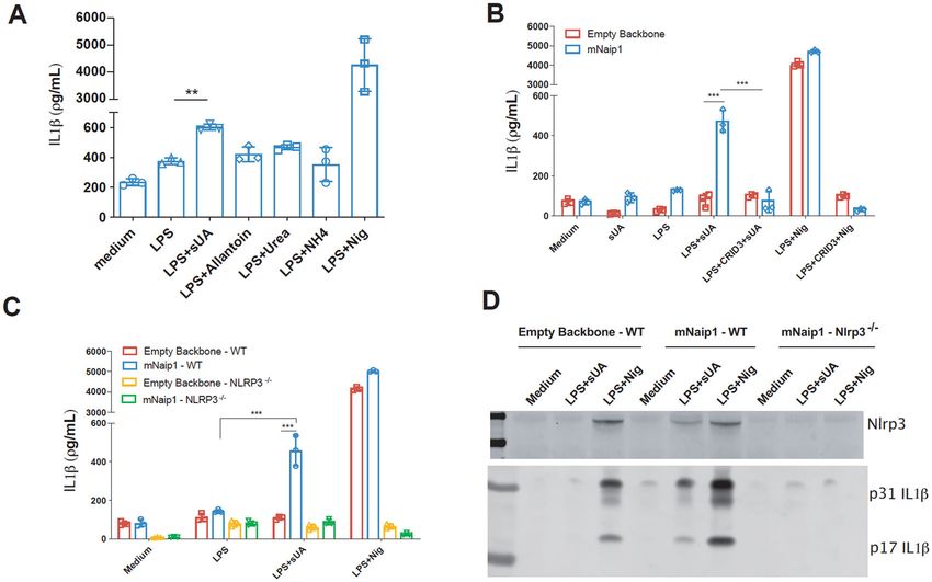
Fig. 1 Human cells do not produce IL-1β upon sUA + LPS stimulus, unless they express mNaip1. A Uric acid levels measured in the serum of
humans, mice and rhesus monkeys. IL-1β Elisa of B monocyte-derived macrophages collected from healthy people, C murine bone marrow-derived
macrophages and D monocyte-derived macrophages collected from rhesus monkeys, all stimulated under different conditions. In B–D, LPS was
added for 1 h at 100 ng/mL and the media were posteriorly changed. MSU (100 μg/mL) and sUA (200 μΜ) were added for 6 h. Each coloured dot
represents a different individual. E IL-1β Elisa of BMDM derived from Naip1−/−, Naip2−/−, Naip5−/−, ΔNaip−/− and Nlrc4−/− mice under different
stimulus. F IL-1β Elisa of human THP1 cells virally transduced with plasmids carrying Naip1, Naip5, Naip6, Nlrc4 and empty vector using lentivirus
constructs and posteriorly stimulated under different conditions. In E, F, LPS was added for 1 h at 100 ng/mL and the media were posteriorly
changed. sUA (200 μΜ) was added for 6 h and nigericin (10 μΜ) was added for 90 min. In A, n = 5 for each analysed species. Triangle refers to
human’s sample, Circle refers to murine’s sample and Square refers to rhesus’ sample. In B, C, we collected cells from eight different individuals; in D,
we collected cells from seven different individuals. In E, F, data are plotted as the median of a triplicate of three to four independent experiments. *p
< 0.05 and ***p < 0.001; n.s. not significant.
Official journal of the Cell Death Differentiation AssociationBraga et al. Cell Death and Disease (2021)12:158 Page 7 of 14
Fig. 2 Naip1 and NLRP3 are required for LPS-primed THP1 cells to produce IL-1β upon sUA. A IL-1β Elisa of mNaip1-transduced LPS-primed
THP1 cells, stimulated with the products of uric acid degradation allantoin, urea and ammonium, the control non-treated cells (Medium) and LPS-
primed treated with nigericin. B IL-1β Elisa of THP1 cells virally transduced with an empty backbone or with mNaip1 after 1 h pretreatment with LPS
(1 µg/mL), 6 h treatment with sUA (200 μM), 30 min treatment with nigericin or control non-treated cells (Medium). Some groups were pretreated
with the Nlrp3 inhibitor CRID3 at 1 μΜ 30 min before LPS priming. C IL-1β Elisa of WT THP1 and Nlrp3−/− THP1 cells virally transduced with an empty
backbone or with mNaip1 after 1 h pretreatment with LPS, 6 h treatment with sUA, 30 min treatment with nigericin or control non-treated cells
(Medium). D IL-1β and Nlrp3 western blotting images of WT THP1 and Nlrp3−/− THP1 cells virally transduced with an empty backbone or with
mNaip1 in control non-treated condition (Medium), or LPS-primed and treated with sUA for 6 h, or with nigericin for 30 min. In A blue bars represent
the levels of produced IL1beta (pg/mL), analysed by Elisa. In D, data are representative of three independent experiments. All experiments were
performed three different times and data are plotted as a median of a triplicate. **p < 0.01, and ***p < 0.001.
IL-1β production upon sUA stimulation compared to the gene and the colour-tagged sequences. In this setup, the
amount produced by the LPS-primed macrophages (Fig. 1E). overexpressed Naip1 protein is not colour-tagged, which
On the other hand, Naip2−/− and Naip5−/− cells behaved as may lead to some data being misinterpreted. Initially, the
wild-type macrophages, and despite the variation within ability of PMA-activated, LPS-primed and mNaip1-
groups, there were no differences in IL-1β production expressing THP1 cells to produce IL-1β upon exposure
between the BMDMs from Nlrc4−/− animals after sUA to UA degradation products, that is, allantoin, urea and
stimulus and the LPS-primed cells from Nlrc4−/− mice (Fig. ammonium were investigated. None of the products was
1E). To confirm the role of the mNaip platform in the sUA able to induce IL-1β production, in contrast to the effect
response, we virally transduced human THP1 cells with of sUA (Fig. 2A). The role of Nlrp3 in sUA sensing, as
mNaip1, mNaip5, mNaip6, mNlrc4, and an empty backbone previously stated16, was further investigated. To this end,
vector. Our data demonstrate that phorbol myristate acetate we initially evaluated IL-1β production upon sUA sti-
(PMA)-activated and LPS-primed THP1 cells produced IL- mulus of THP1 cells virally transduced with either
1β after sUA stimulus only after mNaip1 transduction but mNaip1 or the empty backbone stimulated in the pre-
not mNaip5, mNaip6, mNlrc4 or the control empty vector sence or absence of an Nlrp3 inhibitor, CRID3 (1 μΜ)48.
(Fig. 1F). Such data point to mNaip1 as a target gene The IL-1β levels were reduced in the cells pretreated with
involved in the sUA response. CRID3 (Fig. 2B). IL-1β production was also evaluated in
THP1 cells after Nlrp3 gene deletion by CRISPR-Cas9. It
IL-1β production in human cells is dependent on Nlrp3 and was confirmed that IL-1β production is dependent on
mNaip1 Nlrp3 activation (Fig. 2B–D), because Nlrp3-deleted cells
The Naip1-carrying plasmid was modified by adding a exhibited decreased levels of IL-1β upon sUA stimulation
“self-cleaving” 2A (T2A) sequence between the Naip1 (Fig. 2C, D). To investigate the interaction between Naip1
Official journal of the Cell Death Differentiation AssociationBraga et al. Cell Death and Disease (2021)12:158 Page 8 of 14
and Nlrp3, immunoprecipitation assays with THP1 cell stimulus were compared (Sup. Fig. 4B). Among the 44
lysates using both Nlrp3 and Naip1 as targets were per- proteins expressed only in mNaip1-expressing cells and
formed (Sup. Fig. 2). However, the targets were found not expressed in control cells, we highlighted 30 proteins,
only in the whole-cell lysates, not the immunoprecipitates, including some related to the immune response, such as
suggesting that Nlrp3 and Naip1 may not directly interact. thymopoietin (A0A024RBH7), CD99 (A8MQT7), stress-
Altogether, these data indicate that the observed IL-1β associated endoplasmic reticulum protein (Q9Y6X1) and
production followed by sUA stimulation requires both lysosome-associated membrane glycoprotein 2 (H0YCG2)
Naip1 and Nlrp3 inflammasome platforms. (Sup. Fig. 4B). In addition, the upregulation of mito-
chondrial citrate synthase (F8VRP1), amino acid trans-
Naip1 triggers enhanced immune responses and altered porter (M0R106), mitochondrial glutamate carrier 1
cellular metabolite levels in response to sUA (Q9H936), inorganic pyrophosphatase (Q15181), α-N-
To clarify the mNaip1 influence on sUA sensing, RNA- acetylgalactosaminidase (P17050), phosphoinositide
Seq analysis of mNaip1- and backbone-transduced phospholipase C (B7Z5V4) and acyl-CoA dehydrogenase
THP1 cells was performed. The presence of Naip1 isoform 4 (A0A0S2Z3A5), which are all associated with
affects gene transcription, which was evident when the cellular metabolism, were observed (Sup. Fig. 4B). We
gene expression differences are presented in a volcano highlight 60 proteins among 342 that were expressed only
plot (Sup. Fig. 3A). The unsupervised hierarchical clus- in the GFP+ control cells, including transforming growth
tering of 8000 genes (Sup. Table 1) and the top 20 most factor-beta-induced protein (H0Y8L3), solute carrier
downregulated and most upregulated genes, as deter- family 25-member 4 isoform 3 (A0A0S2Z359) and
mined by the means of the centred logarithm of FPKM vimentin (B0YJC4) (Sup. Fig. 4B and Sup. Table 04).
(fragments per kilobase of exon model per million reads Notably, 84% of the analysed proteins assessed in the
mapped) values in the six replicates of each experimental comparison between the two different cell groups sti-
group, demonstrated that sUA-stimulated macrophages mulated by sUA were detected in the RNA-Seq data.
globally reprogramme transcriptional responses after However, only 2.8% of the expressed proteins corre-
mNaip1 is expressed (Sup. Fig. 3B). A detailed inspection sponded to differentially expressed genes (data not
of the most highly differentially expressed genes revealed shown). We built a STRING network of only the proteins
that ccl2, pik3cd, nck2, tab1 and fgfr1 were expressed expressed in mNaip1-expressing cells but not in GFP+
more strongly in mNaip1-expressing GFP-tagged cells control cells, both stimulated by sUA (Sup. Fig. 4C), and
stimulated with sUA than in GFP-transduced (control) the results showed that they were involved in upregulated
cells stimulated identically (Sup. Fig. 3C). The functional metabolism-related pathways, especially pathways related
annotation enrichment analyses of KEGG and PANTHER to lipid metabolism. These data demonstrate that, in
pathways demonstrated that PMA-activated and LPS- addition to the immune response, mNaip1 is associated
primed THP1 cells expressing mNaip1 led to a shift with altered cellular metabolite content in LPS-primed
towards increased inflammation-, cancer- and infection- macrophages stimulated with sUA.
related signalling pathways (Sup. Fig. 3D). The enrich-
ment term analysis using genes that were upregulated in Naip1 activation may be potentiated after the elevation of
the mNaip1-expressing macrophages revealed a more the cellular content of total lipids
detailed vision of the KEGG pathway enrichment network UA is also described as increasing the amount of tri-
(Sup. Fig. 3E). Together, these analyses showed that glycerides accumulated in hepatic cells49. Hence, we
upregulated genes were associated with processes investigated lipid drop formation in LPS-primed
involved in cytoskeleton regulation, adherent junctions, THP1 cells stimulated with sUA. An increase in the cel-
proteoglycans in cancer, bacterial infection and invasion lular content of lipids was observed after sUA stimulation
and the immune response (Sup. Fig. 3E). These data but in a mNaip1-independent manner (Fig. 3A, B).
demonstrate that mNaip1 triggers enhanced immune Alterations in metabolite content also lead to changes in
responses after sensing sUA. mitochondrial activity; once these plastic organelles sense
In addition to gene expression profiling, we performed a cellular metabolites, oxygen and nutrients, they play
proteomic analysis to identify differentially expressed central roles as sources of energy and ROS50. Changes in
proteins following sUA stimulation in both control (GFP- mitochondrial membrane potential in live cells upon sUA
transduced) and mNaip1-expressing LPS-primed stimulation were therefore measured (Fig. 3C, D). Despite
THP1 cells, and here, we highlight the proteins found only no differences in mitochondrial area stained with Mito-
upon exposure to the sUA stimulus in orange and the Tracker, a reduced mitochondrial membrane potential
proteins expressed only under LPS-primed conditions in was observed, as indicated by failure to load the positively
yellow (Sup. Fig. 4A, Sup. Table 02 and Sup. Table 03). charged mitochondrial indicator TMRE in mNaip1-
Moreover, the two different cell groups under sUA expressing cells stimulated with sUA compared to that
Official journal of the Cell Death Differentiation AssociationBraga et al. Cell Death and Disease (2021)12:158 Page 9 of 14 Fig. 3 Naip1 activation upon sUA stimulus may be potentiated after the elevation of the cellular content of neutral lipid. A Representative images of THP1 cells transduced with an empty backbone or mNaip1 at LPS-primed condition or LPS-primed and stimulated with sUA for 6 h. The membrane is in red and lipid droplets are stained for LD540, in green. B Quantification of lipid droplets per cell. C Representative images of empty backbone- and mNaip1-transduced cells primed with LPS or LPS-primed and sUA-stimulated stained with MitoTracker (red), tetramethylrhodamine ethyl ester (TMRE) (green) and DAPI (blue). The bars in each image represent 20 μm D TMRE quantification, indicating polarized mitochondria of the experiments in panel (C). E IL-1β Elisa of empty backbone- (red bars) and mNaip1- (blue bars) transduced cells, both at non-stimulated (Medium) condition or LPS-primed and stimulated for 6 h with sUA, citrate or palmitate. F Schematic representation of the TCA cycle and the fatty acid synthesis pathway given emphasis to the inhibitors and stimulus used in panel (J). G IL-1β Elisa of mNaip1-transduced and LPS-primed cells, stimulated for 6 h with sUA (200 μΜ), citrate (5 mΜ) or palmitate (100 μΜ), in the presence or absence of ATP citrate lyase inhibitor (BMS303141 at 25 μΜ), acetyl-CoA carboxylase inhibitor (TOFA at 10 μg/mL), or fatty acid synthase inhibitors (C75 at 50 μΜ or cerulenin at 5 μg/mL). In A, B, data are representative of three independent experiments and n = 12. In D, data are plotted as a median of ten different micrography fields of three independent experiments. In E, F, the experiments were performed three different times and n = 3. **p < 0.01 and ***p < 0.001. in the LPS-primed cells (Fig. 3C, D). We next measured independent manner. The mitochondrial pyruvate carrier the OCR of THP1 cells virally transduced with an empty inhibitor UK5099 (100 μΜ) was used to evaluate ATP backbone or mNaip1, both LPS-primed, treated or not consumption derived from fatty acids indirectly. Here, the with sUA. sUA increased the OCR but in a Naip1- OCR increase was also reversed by UK5099 pretreatment Official journal of the Cell Death Differentiation Association
Braga et al. Cell Death and Disease (2021)12:158 Page 10 of 14
in both cell types (Sup. Fig. 5A, B). Altogether, these data
indicate that upon sUA stimulation, mNaip1 expression
alters the cellular fatty acid content and reduces the
number of active mitochondria in cells.
We next investigated whether the elevation of fatty acid
synthesis could trigger IL-1β production. For this assess-
ment, the levels of IL-1β in the supernatant of the LPS-
primed cells virally transduced with an empty backbone or
mNaip1 were measured after 6 h of incubation with citrate51
and palmitate. Despite significant production of IL-1β in the
empty backbone-transduced cells upon both citrate (5 mΜ)
or palmitate (100 μΜ) stimulation, compared to the LPS-
primed cells, the mNaip1-expressing cells produced even
higher levels of IL-1β than the empty backbone-transduced
cells regardless of the stimuli (Fig. 3E). Moreover, the LPS-
primed mNaip1-expressing cells were stimulated with sUA
or citrate in the presence of the acetyl-CoA carboxylase-α
inhibitor TOFA (10 μg/mL), the phospho-ACLy inhibitor
BMS30314152 (25 μΜ) or the fatty acid synthase inhibitors
C75 (50 μΜ) and cerulenin (5 μg/mL)53, as shown in sche-
matic Fig. 3F. All these inhibitors, except TOFA, led to
decreased levels of IL-1β (Fig. 3G) upon sUA or citrate sti-
mulation. Altogether, our data suggest that sUA leads to
fatty acid synthesis independent of mNaip1 expression.
Saturated fatty acids promote Nlrp3 inflammasome activa-
tion54, especially palmitate55. In addition, we found that
citrate- and palmitate-mediated IL-1β production is poten-
tiated in the presence of Naip1.
Naip1 directly recognizes sUA Fig. 4 QCM monitoring and SPR sensorgram evidencing all steps
In a study investigating the role of different lipids in involved in the detection of the interaction between sUA and
macrophage lipidomics, palmitate presented the most pro- Naip1 protein. A QCM responses over time within sUA injection after
nounced effects56. To investigate whether mNaip1 directly Naip1 immobilization upon anti-GFP adsorption on the gold quartz
crystals surface at 37 °C. The arrows indicate sample injection. B Schematic
senses sUA and/or palmitate, we performed a QCM with
representation of the constructed SPR sensor chip (in the box). Sequential
dissipation (QCM-D) analysis. After an initial immobilizing addition of compounds into the system: addition of the buffer solution
step with anti-GFP, we incubated the sUA solution sample (PBS, 10 mmol/L at pH 7.4); mixture consisting of EDC (150 mmol/L) and
of increasing concentrations (12.5, 25, 50, 100 and 200 μΜ). NHS (150 mmol/L); PBS; immobilization of anti-GFP (10 µg/mL); PBS;
It was observed that mNaip1 directly interacts with sUA. As addition of ethanolamine (EA); addition of cell lysates containing Naip1
protein (2 µg/mol/L). It is possible to observe a very intensive response for
sUA adsorption capacity was reached, the QCM-D fre-
the interaction of the Naip1 protein with anti-GFP; PBS; addition of pure
quency changed maximally, even at the lowest studied H2O; addition of sUA (2 µmol/L, purple line) and palmitate (2 µmol/L,
concentration (Fig. 4A), indicating saturation at lower con- green line). It is possible to observe the significant variation of the SPR
centrations and an increase in the mass adsorbed per area. angle (ΔθSPR) due to the interaction between sUA and Naip protein. In A,
Also, we compared the interaction of mNaip1 stimulated B, data are representative of three independent experiments.
with sUA and with palmitate in real time with an SPR
immunosensor. With SPR-based biosensor technology,
mNaip1 is tethered to the surface of a previously functio- (2 µΜ), which characterizes the interaction between sUA
nalized SPR sensor chip, and the possible ligands are and the mNaip1 protein. In this phase of the study,
introduced in solution, as illustrated in the scheme of Fig. higher concentrations of sUA (12.5–200 µΜ) were also
4B. The SPR curves (sensorgram) obtained in real time for accompanied by significant responses (data not shown). In
all steps involved in the evaluation of the interactions turn, the addition of palmitate at a concentration of 2 µΜ
between sUA and mNaip1 protein (purple curve) and (green curve) and at higher concentrations (12.5–200 µΜ—
between palmitate and the mNaip1 protein (green curve) data not shown) did not trigger a notable response.
are shown in Fig. 4B. Notably, a significant variation in These results suggest that sUA directly binds to the
response (ΔθSPR) was obtained after the addition of sUA mNaip1 protein.
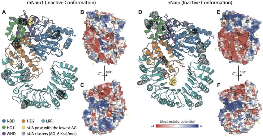
Official journal of the Cell Death Differentiation AssociationBraga et al. Cell Death and Disease (2021)12:158 Page 11 of 14 Fig. 5 Structural analysis of the inactive conformations of hNaip and mNaip1, modelled by homology using an inactive form of Nlrc4 structure (PDB 4KXF) as a template. A Cartoon representation of a mNaip1 (Uniprot: Q9QWK5) homology model. The distinct colours represent functional regions commonly found in proteins of the NLR family (NBD-HD1-WHD-HD2-LRR), coloured as shown in Zhang et al.57. Clusters of uric acid (URC) molecules are shown as black meshes, which represent points on the Naip surface where two or more URC were found to bind, in multiple independent rigid docking simulations. The pose with the lowest ΔG is shown as a yellow sphere representation. B Surface electrostatic potential calculated for mNaip1. Its solvent-accessible surface is shown with a potential gradient ranging from 4 kBT (blue). Yellow arrows highlight URC clusters shown in panel (A). C A 180° rotation of mNaip1 around its Y-axis. D Cartoon representation of a hNaip (Uniprot: Q13075) homology model. E Its surface electrostatic potential. F A 180° rotation of hNaip around its Y-axis. See legend for more details. hNaip and mNaip1 may respond differently to UA binding region of sUA in mNaip1 lies in the NBD As previously studied, for inflammasomes to be domain (ΔG = −7.92 kcal/mol) (Fig. 5A), for hNaip, the formed, NLR proteins, such as Naip, must recognize the ligand most likely exhibits higher affinity for the LRR ligands to be released from their autoinhibited state to domain (ΔG = −8.00 kcal/mol) (Fig. 5D). finally trigger the oligomerization of NLRCs and Considering our experimental results and the homology assemble inflammasome complexes57,58. After model- modelling and molecular docking, we hypothesize that the ling the structures of mNaip1 and hNaip in their inac- surface electrostatics of mNaip1 enables this protein to tive forms, we observed important differences in their recognize sUA and stop its autoinhibition, functions that surface electrostatic properties, which may directly hNaip is unable to perform. With the deletion of the interfere with their ability to recognize specific ligands. uricase gene in the great apes19, throughout evolution, By performing molecular docking, we investigated mutations affecting hNaip surface electrostatics were regions of possible binding of sUA onto the solvent- probably selected to increase its physiological tolerance to accessible surface of both Naip proteins (Fig. 5). More high levels of sUA such that the activation of inflamma- than 250 iterations were performed for each target. somes is prevented, allowing the great apes to benefit Given the ligand-binding geometry of each interaction, from the survival advantages provided by high levels of the docking results were summarized as clusters con- serum UA21. taining one or more ligands with similar binding poses. Figure 5 displays only clusters with a Gibbs free energy Discussion ΔG
Braga et al. Cell Death and Disease (2021)12:158                                                                                Page 12 of 14

activation is sufficient to cause systemic inflammatory dis-          Uricase activity that is missing because of evolutionary
ease with surprising tibiotarsal joint swelling59, the main      process endows UA a puzzling history in the evolution of
joint affected by gout, a disease triggered by the accumula-     humans. Great apes have, in the basal state, high phy-
tion of UA60. This is the first study to postulate that Naip      siological levels of sUA, but human macrophages do not
recognizes a DAMP. Our previous work suggested that sUA          respond to 200 μΜ sUA, an inflammatory-inducing sti-
activates the Nlrp3 inflammasome16; hence, we also inves-         mulus in murine cells. Uricase inhibition therapy and the
tigated the role of Nlrp3 in mNaip1-expressing cells. LPS-       subsequent elevation in serum UA are critical for trig-
primed human THP1 cells produce only the mature form of          gering metabolic syndrome comorbidities in murine
IL-1β upon sUA stimulation when they also express Nlrp3          models66–68. We have demonstrated, on the other hand,
and mNaip1. Most inflammasomes are believed to include            that rhesus macaques, a primate with evolutionarily
only a single NLR, although other NLR-NLR interactions           maintained uricase enzyme activity, have reduced levels of
have been proposed. The interplay between Nlrp3 and Nlrc4        IL-1β production by their monocyte-derived macrophages
reveals an unexpected overlap between inflammasome                following sUA stimulation. It is possible that rhesus
scaffolds previously thought to be distinct61. In addition, it   macaques’ macrophages require higher priming activation
was reported that Nlrc4 can recruit Nlrp3 through its            to induce IL-1β transcription since LPS alone did not
NACHT domain in the context of Salmonella typhimurium            increase IL-1β production in these experiments. More-
infection61. Hence, in addition to the mechanisms by which       over, several cytokines, including IL-1β, circulate at very
sUA activates the mNaip1 inflammasome, it remains to be           low levels in both affected and unaffected rhesus maca-
determined whether mNaip1 interacts with Nlrp3 after sUA         ques in different models of disease69–71. It is also specu-
stimulation, as we observed no clear evidence of Nlrp3 and       lated that the rhesus Naip protein was selected because it
mNaip1 protein interactions.                                     tolerates elevated levels of serum UA.
   In addition to responding to PAMPs and DAMPs, the                In recent years, an understanding of the additional adverse
immune system acts as a signal integrator able to detect         effects of high levels of serum UA has advanced72. Early
disturbances in the cytoplasm related to metabolites, as         scientific literature suggested an association between UA
indicated by recent data. These monitored disruptions are        concentration and the incidence of cardiovascular disease;
termed “homeostasis-altering molecular processes”62. It          specifically, the development of hypertension73, metabolic
has been shown that Nlrp3 inflammasome complex acti-              syndrome74, endothelial dysfunction75 and micro-
vation and posterior caspase-1 and IL-1β production              albuminuria76. Lifestyle and socioeconomic changes over
occur following saturated fatty acid palmitate triggering,       time have resulted in a marked reduction in physical activity
even in human cells63,64. Both QCM and SPR methodol-             and profound dietary changes. These changes correlate with
ogies indicate that the three-dimensional structure of           increased rates of metabolic diseases triggered by overly
mNaip1 provides a great accessible area for interaction          active innate immune functions77, with chronic inflamma-
with sUA and no accessible area for interaction with             tion termed “metaflammation”78,79. Furthermore, multiple
palmitate. Homology modelling and molecular docking              genetic and non-genetic risk and protective factors are also
analysis indicate that the surface electrostatics of mNaip1      thought to contribute to the pathogenesis of metabolic
enable it to recognize sUA by abolishing its autoinhibition      diseases, specifically those related to hyperuricaemic condi-
state, a function that hNaip is unable to perform.               tions. Different states of tolerance to sUA sensing by hNaip
Although we have demonstrated that sUA, but not pal-             may predict the innate immune activation state in the
mitate, is responsible by directly binds to mNaip1, our          context of hyperuricaemia-related diseases. These tolerance
data pointed to increased IL-1β production in the context        differences may be related to polymorphisms in the Naip
of Naip1 expression in human cells following palmitate           gene, changes in the number of gene copies, or even epi-
stimulation. Therefore, further analyses are necessary to        genetic changes; however, a greater investigation is required.
better investigate the mechanisms by which palmitate can         In this sense, in addition to understanding the human evo-
lead to Naip1 permissiveness to sUA.                             lutionary process, investigating which mechanisms mediate
   In addition, the transcriptome and cellular metabolite        the immunomodulatory function of sUA is also essential to
contents were changed in cells upon sUA stimulation, and         better design rational novel anti-inflammatory therapies.
the presence of mNaip1 altered some metabolism-related
enzymes and favoured an increase in immune responses             Acknowledgements
                                                                 We thank Prof. Dario Simoes Zamboni for providing femurs and tibias from WT,
towards sUA. Moreover, we observed that mitochondrial            Naip1−/−, Naip2−/−, Naip5−/−, ΔNaip−/− and Nlrc4−/− mice. We also thank
activity was inhibited in mNaip1-expressing cells stimu-         Uira Souto Melo for all discussions concerning the data presented in the paper.
lated with sUA. This result corroborates the findings of a
study demonstrating that the percentage of TMRE+ cells           Author details
                                                                 1
                                                                  Department of Basic Pathology, Federal University of Parana, Curitiba, PR,
was significantly lower in LPS-primed macrophages sti-            Brazil. 2Department of Immunology, Institute of Biomedical Sciences IV,
mulated with ATP than in control macrophages65.                  University of São Paulo, São Paulo, SP, Brazil. 3Institute of Innate Immunity,

Official journal of the Cell Death Differentiation Association
Braga et al. Cell Death and Disease (2021)12:158 Page 13 of 14
University Hospitals Bonn, Bonn, Germany. 4Department of Physiology and 4. Janeway, C. A. Jr. The priming of helper T cells. Semin. Immunol. 1, 13–20 (1989).
Biophysics, Institute of Biomedical Sciences I, University of Sao Paulo, São 5. Burnet, F. M. The immunological significance of the thymus: an extension of
Paulo, SP, Brazil. 5Department of Microbiology, Institute of Biomedical Sciences the clonal selection theory of immunity. Australas. Ann. Med. 11, 79–91 (1962).
II, University of São Paulo, São Paulo, SP, Brazil. 6Department of Life Sciences, 6. Matzinger, P. The danger model: a renewed sense of self. Science 296, 301–5
Imperial College London, London SW7 2AZ, UK. 7Institute of Chemistry, (2002).
University of Campinas, Campinas, SP, Brazil. 8Department of Chemistry, 7. Matzinger, P. An innate sense of danger. Semin. Immunol. 10, 399–415 (1998).
Federal University of Parana, Curitiba, PR, Brazil. 9Department of Cellular 8. So, A. & Thorens, B. Uric acid transport and disease. J. Clin. Investig. 120, 1791–9
Biology, Federal University of Parana, Curitiba, PR, Brazil. 10Interdisciplinary Post- (2010).
Graduate Program in Health Sciences, Cruzeiro do Sul University, São Paulo, 9. Shi, Y., Evans, J. E. & Rock, K. L. Molecular identification of a danger signal that
Brazil. 11Division of Infectious Diseases and Immunology, Department of alerts the immune system to dying cells. Nature 425, 516–21 (2003).
Medicine, University of Massachusetts Medical School, Worcester, MA 01655, 10. Martinon, F., Petrilli, V., Mayor, A., Tardivel, A. & Tschopp, J. Gout-associated uric
USA. 12Centre for Molecular Inflammation Research (CEMIR), Norwegian acid crystals activate the NALP3 inflammasome. Nature 440, 237–41 (2006).
University of Science and Technology, 7491 Trondheim, Norway. 13Nephrology 11. Compston, A. & Coles, A. Multiple sclerosis. Lancet 359, 1221–31 (2002).
Division, Federal University of São Paulo, São Paulo, SP, Brazil. 14Renal 12. Di Giovine, F. S., Malawista, S. E., Nuki, G. & Duff, G. W. Interleukin 1 (IL 1) as a
Physiopathology Laboratory, Faculty of Medicine, University of São Paulo, São mediator of crystal arthritis. Stimulation of T cell and synovial fibroblast
Paulo, SP, Brazil mitogenesis by urate crystal-induced IL 1. J. Immunol. 138, 3213–8 (1987).
13. Martinon, F., Burns, K. & Tschopp, J. The inflammasome: a molecular platform
Author contributions triggering activation of inflammatory caspases and processing of proIL-beta.
T.T.B. contributed to the design of the study, data collection, data analysis and Mol. Cell 10, 417–26 (2002).
interpretation, drafting the article, critical revision of the article and final 14. Jones, J. D., Vance, R. E. & Dangl, J. L. Intracellular innate immune surveillance
approval of the version to be published. M.R.D., D.M., T.A.d.S., A.F.d.B., M.C.C., M. devices in plants and animals. Science 354, 6316 (2016).
I.H., D.S.d.L., V.N., J.d.F.G., D.E.P.S., T.P., M.L., S.M.P.B. and R.A.d.F. contributed to 15. Mondragón-Palomino, M., Stam, R., John-Arputharaj, A. & Dresselhaus, T.
the design of the study, data collection, data analysis interpretation and final Diversification of defensins and NLRs in Arabidopsis species by different evo-
approval of the manuscript version to be published. A.P. and E.L. contributed lutionary mechanisms. BMC Evol. Biol. 17, 255 (2017).
to drafting the article, critical revision of the article and final approval of the 16. Braga, T. T., Forni, M. F., Correa-Costa, M., Ramos, R. N., Barbuto, J. A. & Branco, P.
version to be published. N.O.S.C. contributed to the design of the study, et al. Soluble uric acid activates the NLRP3 inflammasome. Sci. Rep. 7, 39884
obtaining funding resources and the final approval of the version to be (2017).
published. 17. Kingsbury, S. R., Conaghan, P. G. & McDermott, M. F. The role of the NLRP3
inflammasome in gout. J. Inflamm. Res. 4, 39–49 (2011).
Ethics statement 18. Martillo, M. A., Nazzal, L. & Crittenden, D. B. The crystallization of monosodium
All procedures with mice were performed in accordance with relevant urate. Curr. Rheumatol. Rep. 16, 400 (2014).
guidelines and regulations and approved by the local ethics committees at the 19. Kratzer, J. T., Lanaspa, M. A., Murphy, M. N., Cicerchi, C., Graves, C. L. & Tipton, P.
University of São Paulo (Document 45/2009). All procedures with rhesus A. et al. Evolutionary history and metabolic insights of ancient mammalian
macaques were approved by the Ethics Committee on Animal Use of the uricases. Proc. Natl Acad. Sci. USA 111, 3763–8 (2014).
Butantan Institute (CEUAIB) (number 9376040717). Leucocyte concentrates 20. Kono, H., Chen, C. J., Ontiveros, F. & Rock, K. L. Uric acid promotes an acute
were obtained after plasmapheresis at the Blood Bank Service of the “Hospital inflammatory response to sterile cell death in mice. J. Clin. Investig. 120,
das Clinicas” in Sao Paulo (SP, Brazil), and they were used for peripheral blood 1939–49 (2010).
monocyte isolation. All volunteers signed informed consent in compliance 21. Cicerchi, C., Li, N., Kratzer, J., Garcia, G., Roncal-Jimenez, C. A. & Tanabe, K. et al.
with the respective Institutional Ethics Committee. Uric acid-dependent inhibition of AMP kinase induces hepatic glucose pro-
duction in diabetes and starvation: evolutionary implications of the uricase
loss in hominids. FASEB J. 28, 3339–50 (2014).
Funding statement
22. Hafner, S. & Weitzman, J. Of mice and men-NAIP homologues face Legionella
This study was supported by the São Paulo State Funding Agency (FAPESP)
pneumophila. Microbes Infect. / Inst. Pasteur 14, 1119–22 (2012).
(grants numbers: 2014/06992-8 and 2017/05264-7), Conselho Nacional de
23. Diez, E., Lee, S. H., Gauthier, S., Yaraghi, Z., Tremblay, M. & Vidal, S. et al. Birc1e is
Desenvolvimento Científico e Tecnológico (CNPq) and in part by the
the gene within the Lgn1 locus associated with resistance to Legionella
Coordenação de Aperfeiçoamento de Pessoal de Nível Superior—Brasil
pneumophila. Nat. Genet. 33, 55–60 (2003).
(CAPES) Financial Code 001.
24. Wright, E. K., Goodart, S. A., Growney, J. D., Hadinoto, V., Endrizzi, M. G. & Long,
E. M. et al. Naip5 affects host susceptibility to the intracellular pathogen
Conflict of interest Legionella pneumophila. Curr. Biol. 13, 27–36 (2003).
The authors declare that they have no conflict of interest. 25. Miao, E. A., Mao, D. P., Yudkovsky, N., Bonneau, R., Lorang, C. G. & Warren, S. E.
et al. Innate immune detection of the type III secretion apparatus through the
Publisher’s note NLRC4 inflammasome. Proc. Natl Acad. Sci. USA 107, 3076–80 (2010).
Springer Nature remains neutral with regard to jurisdictional claims in 26. Rayamajhi, M., Zak, D. E., Chavarria-Smith, J., Vance, R. E. & Miao, E. A. Cutting
published maps and institutional affiliations. edge: mouse NAIP1 detects the type III secretion system needle protein. J.
Immunol. 191, 3986–9 (2013).
Supplementary information The online version contains supplementary 27. Zhao, Y., Yang, J., Shi, J., Gong, Y. N., Lu, Q. & Xu, H. et al. The NLRC4
material available at https://doi.org/10.1038/s41419-021-03445-w. inflammasome receptors for bacterial flagellin and type III secretion apparatus.
Nature 477, 596–600 (2011).
Received: 17 November 2020 Revised: 11 January 2021 Accepted: 15 28. Ren, T., Zamboni, D. S., Roy, C. R., Dietrich, W. F. & Vance, R. E. Flagellin-deficient
January 2021 Legionella mutants evade caspase-1- and Naip5-mediated macrophage
immunity. PLoS Pathog. 2, e18 (2006).
29. Katagiri, N., Shobuike, T., Chang, B., Kukita, A. & Miyamoto, H. The human
apoptosis inhibitor NAIP induces pyroptosis in macrophages infected with
Legionella pneumophila. Microbes Infect. 14, 1123–32 (2012).
References 30. Zamboni, D. S., Kobayashi, K. S., Kohlsdorf, T., Ogura, Y., Long, E. M. & Vance, R.
1. Land, W. G. How evolution tells us to induce allotolerance. Exp. Clin. Transplant. E. et al. The Birc1e cytosolic pattern-recognition receptor contributes to the
13, 46–54 (2015). detection and control of Legionella pneumophila infection. Nat. Immunol. 7,
2. Bottomly, K. & Janeway, C. A. Jr. Antigen presentation by B cells. Nature 337, 318–25 (2006).
24 (1989). 31. Hornung, V., Bauernfeind, F., Halle, A., Samstad, E. O., Kono, H. & Rock, K. L. et al.
3. Janeway, C. A. Jr. Approaching the asymptote? Evolution and revolution in Silica crystals and aluminum salts activate the NALP3 inflammasome through
immunology. Cold Spring Harb. Symp. Quant. Biol. 54, 1–13 (1989). phagosomal destabilization. Nat. Immunol. 9, 847–56 (2008).
Official journal of the Cell Death Differentiation AssociationYou can also read