Sleep Disorders in Patients With Craniopharyngioma: A Physiopathological and Practical Update - IRIS ...
←
→
Page content transcription
If your browser does not render page correctly, please read the page content below
REVIEW
                                                                                                                                                  published: 09 February 2022
                                                                                                                                              doi: 10.3389/fneur.2021.817257

                                               Sleep Disorders in Patients With
                                               Craniopharyngioma: A
                                               Physiopathological and Practical
                                               Update
                                               Andrea Romigi 1† , Tiziana Feola 1,2† , Simone Cappellano 1 , Michelangelo De Angelis 1 ,
                                               Giacomo Pio 1 , Marco Caccamo 1 , Federica Testa 1 , Giuseppe Vitrani 1 , Diego Centonze 1 ,
                                               Claudio Colonnese 1 , Vincenzo Esposito 1,3 and Marie-Lise Jaffrain-Rea 1,4*
                                               1
                                                Neuromed Institute, Istituto di Ricovero e Cura a Carattere Scientifico, Pozzilli, Italy, 2 Department of Experimental Medicine,
                                               Sapienza University of Rome, Rome, Italy, 3 Human Neurosciences, Sapienza University of Rome, Rome, Italy, 4 Department
                          Edited by:           of Biotechnological and Applied Clinical Sciences, University of L’Aquila, L’Aquila, Italy
                        Songbai Gui,
     Capital Medical University, China
                                               Sleep disorders (SDs) represent an important issue in patients with craniopharyngioma
                        Reviewed by:
                         Eun Yeon Joo,         (CP). Nearly 70% of these patients complain of sleep-wake cycle alterations and/or
            Sungkyunkwan University,           excessive diurnal somnolence due to sleep-related breathing disorders, such as
                           South Korea
                    Marco Carotenuto,          obstructive sleep apnea (OSA) and/or central hypersomnia, including secondary
          University of Campania Luigi         narcolepsy. SDs may severely reduce quality of life, increase disease-related
                          Vanvitelli, Italy
                                               cardiorespiratory and cardiovascular morbidity, and finally play a major role in increased
                  *Correspondence:
                                               long-term mortality reported on patients with CP. A major risk factor for SDs is
              Marie-Lise Jaffrain-Rea
            marielise.jaffrain@univaq.it       represented by the hypothalamic syndrome, which may develop because of direct
     † These                                   hypothalamic damage by the tumor itself and/or complications of the treatments,
               authors have contributed
                    equally to this work       neurosurgery and/or radiotherapy, and typically includes permanent neuroendocrine
                                               dysfunctions, morbid obesity, and secondary metabolic disorders. Despite increasing
                   Specialty section:
                                               attention to SDs in the general population, and in particular to OSA as a risk factor for
         This article was submitted to
   Neuro-Oncology and Neurosurgical            cardio-metabolic diseases and excessive daytime somnolence, sleep evaluation is still
                             Oncology,         not routinely proposed to patients with CP. Hence, SDs are often underdiagnosed and
               a section of the journal
                Frontiers in Neurology
                                               undertreated. The aim of this paper is to update current knowledge of the pathogenesis
       Received: 17 November 2021
                                               and prevalence of SDs in patients with CP and propose practical algorithms for their
       Accepted: 28 December 2021              evaluation and management in clinical practice. Particular attention is paid to screening
        Published: 09 February 2022
                                               and diagnostic tools for appropriate characterization of SDs, identification of risk factors,
                             Citation:
                                               and potential role of hypothalamic sparing surgery in the prevention of morbid obesity
  Romigi A, Feola T, Cappellano S, De
      Angelis M, Pio G, Caccamo M,             and SDs. Available tools in sleep medicine, including lifestyle interventions, drugs, and
       Testa F, Vitrani G, Centonze D,         respiratory devices, are discussed, as well as the importance of optimal hormone
        Colonnese C, Esposito V and
       Jaffrain-Rea M-L (2022) Sleep
                                               replacement and metabolic interventions. Current limits in the diagnosis and treatment
            Disorders in Patients With         of SDs in patients with CP and possible future avenues for research agenda are
                Craniopharyngioma: A
                                               also considered.
     Physiopathological and Practical
   Update. Front. Neurol. 12:817257.           Keywords: craniopharyngioma, sleep disorder, hypothalamic syndrome, hypothalamic obesity, obstructive sleep
    doi: 10.3389/fneur.2021.817257             apnea, hypersomnia, narcolepsy, circadian rythm disorders

Frontiers in Neurology | www.frontiersin.org                                           1                                           February 2022 | Volume 12 | Article 817257
Romigi et al.                                                                                              Sleep Disorders in Craniopharyngioma

INTRODUCTION                                                            muscles; (4) suboptimal endocrine treatment; (5) fatigue, and
                                                                        psychosocial disorders (14). On the other hand, SDs contribute
Craniopharyngiomas (CPs) are rare benign parasellar tumors              to and aggravate metabolic and cardiovascular co-morbidities.
derived from Rathke’s pouch rests, and are classified into two          In fact, SDs increase the risk of insulin-resistance, obesity,
subtypes (1). Most are adamantinomatous, typically presenting           and diabetes mellitus (DM) (15), and intermittent hypoxemia
as mixed solid/cystic tumors with frequent calcifications, driven       in OSA is an independent risk factor for cardiovascular and
by somatic beta-catenin mutations, whereas papillary CPs are            cardiorespiratory mortality (16). SDs may also represent a risk
suprasellar, mostly solid, tumors with frequent BRAF mutations          factor for neurocognitive decline (17) and cancer (18).
(2). CPs have a bimodal age distribution, with peaks of incidence          Nonetheless, SDs remain largely underdiagnosed in clinical
occurring in pediatric (5–14 years, adamantinomatous) and adult         practice, which may be explained by the complex clinical
patients (50–74 years, both) (1, 2). Despite benign histology           management of patients with CP and insufficient awareness or
and high overall survival (>90% in childhood-onset CP), the             access to centers for sleep medicine. However, no guidelines
standardized mortality rate in patients with CP has been variably       are available for the diagnosis and management of SDs in such
estimated from 2.88 to 9.28, with a 3- to 19-fold increase in           patients. Because increasing attention is currently being given to
cardiovascular mortality compared to the general population             sleep health in the general population because of relevant health
(3). Prognosis may vary according to tumor characteristics and          and socioeconomic consequences (e.g., reduced performance
treatment, secondary co-morbidities, and childhood vs. adult            at work, driving safety, and social relationship), we wished to
onset of the disease (1). Neuroendocrine dysfunctions include           review the current knowledge of SDs in patients with CP and
partial or complete hypopituitarism, hyperprolactinemia, and            propose, based on our multidisciplinary experience, practical
diabetes insipidus (DI). The most dramatic complication is              algorithms for the screening, diagnosis, and management of such
the development of a hypothalamic syndrome (HS), which is               conditions. Current limits and future therapeutic options will
typically associated with neuroendocrine disorders and includes         also be discussed.
neurocognitive changes (4) morbid hypothalamic obesity (HO)
and related systemic complications (5), a variety of sleep
disorders (SDs) including sleep-related breathing disorders             CLASSIFICATION AND PATHOGENESIS OF
(SBDs), central hypersomnia and abnormal wake-sleep circadian           SLEEP DISORDERS IN PATIENTS WITH CP
rhythms (6, 7), and less commonly abnormalities of thirst and
central temperature or cardiovascular regulation. Hypothalamic          Sleep disorders (SDs) are classified according to the third edition
damage may severely impair the quality of life (QoL) of patients        of the International Classification of Sleep Disorders (ICSD-3) of
and has an impact on long-term mortality (8). The extension             the American Academy of Sleep Medicine (19), which identifies
and localization of hypothalamic injury due to the tumor itself,        seven major categories of disorders: insomnia disorders, SBDs,
neurosurgery, and, in some cases, radiotherapy, contribute to           central disorders of hypersomnolence, circadian rhythm sleep-
the timing and severity of HS (5, 9). The optimal treatment for         wake disorders (CRSWDs), sleep-related movement disorders,
CP and related complications remains difficult and relies on a          parasomnias, and other SDs. The most frequently reported SDs
multidisciplinary approach, with increasing attention being paid        in CP are EDS (11, 20), central hypersomnia and secondary
in the last decades to the prevention of hypothalamic damage            narcolepsy (13, 14, 21–27), SBDs (7, 24), and CRSWD (28–30),
during surgery (2, 10).                                                 which require appropriate characterization and understanding of
    Overall, SDs have received more attention in pediatric than         their underlying causes. Since clinical pictures may be complex
in adult patients with CP, who include long-term survivors of           and different elements may co-exist in the same patient, this
childhood-onset CP and patients with an adult-onset disease.            may be achieved through a specific expertise in sleep diseases.
In pediatric cohorts, SDs have been heterogeneously reported            Although the real prevalence of SDs before and after CP
as daytime sleepiness/hypersomnia, sleep disturbances such as           surgery/radiotherapy should be clarified, Mandrell et al. (13)
difficulty to fall asleep or waking up during the night, and            showed that 45% of a large sample of pediatric patients with
variably evaluated by self-assessment—specific questionnaires           CP were affected by hypersomnia due to a medical disorder and
(11), items as a part of QoL assessment (12)—and more recently          35% by narcolepsy, and that the main predictor of sleepiness was
by means of sleep medicine tools aiming to better define                obesity. In addition, 80% of this cohort complained of EDS at
entities such as SBDs, like obstructive sleep apnea (OSA) (7) or        diagnosis or after neurosurgery. This is in keeping with previous
secondary narcolepsy (13). Within the methodological limits and         studies reporting that children and adolescents with CP complain
heterogeneity of reported studies, the prevalence of SDs and/or         of somnolence, fatigue, and sleep–wake disruption, which persist
excessive daytime sleepiness (EDS) approaches 70–80% (11, 13),          in 65–80% of the cases after treatment (20, 31). Similar data
with an adult prevalence of OSA around 40% (6). A common                were reported in unselected adults with CP, with a prevalence of
observation is the higher prevalence of SDs in the presence             SDs around 70% (10). Of note, EDS may reduce work/education
of hypothalamic involvement, with a bi-directional interplay            performance in 43% of patients with CP (6).
between obesity and SDs. Indeed, SDs recognize a multifactorial             From a physiopathological point of view, sleep can be altered
pathogenesis: (1) a strict relationship with HO; (2) damage             because of tumor growth toward structures involved in the
to the hypothalamic nuclei regulating sleep, wakefulness and            control of sleep and wake, direct or indirect/vascular treatment-
circadian rhythm; (3) dysfunction of the pharyngeal/respiratory         related injury to the same structures, or as a consequence of

Frontiers in Neurology | www.frontiersin.org                        2                                  February 2022 | Volume 12 | Article 817257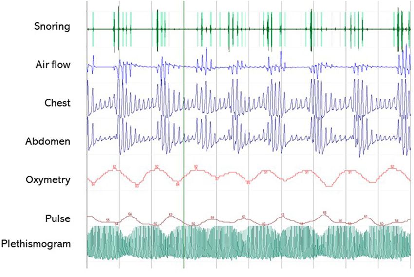
Romigi et al.                                                                                                Sleep Disorders in Craniopharyngioma

HO. Therefore, hypothalamic dysfunction plays an essential role           is characterized by intermittent complete or partial obstruction
in the development of SDs. Damage to the suprachiasmatic                  (obstructive apnea or hypopnea); prolonged partial upper airway
nucleus (SCN), the central biological “master clock,” leads to            obstruction; or both prolonged and intermittent obstructions
abnormal circadian rhythms and sleep-wake cycles. The SCN is              that disrupt normal ventilation during sleep, normal sleep
on the neural way of control of the nocturnal pineal secretion            patterns, or both (at least one obstructive, mixed apnea, or
of melatonin (32). It is composed of 20.000 neurons and                   hypopnea per hour of sleep). The presence of SBD symptoms
glia that change their rate of firing in response to variation            in combination with an AHI of ≥ 1/h has been applied
in light (33) and modulates several processes, such as sleep              to define pediatric OSA in most published studies (44). In
and food intake. Internal rhythm is strictly linked to external           adults, the definition is based on AHI ≥ 5/h, and OSA is
light cycle, and CRSWD develop when misalignment between                  characterized by predominant obstructive respiratory events
light cycle and internal rhythm occurs. Circulating melatonin             (obstructive and mixed apneas, hypopneas, or respiratory
is mostly of central origin, and abnormally low nocturnal                 effort related arousals). Of note, no specific criteria are
melatonin levels have been reported in patients with childhood-           considered for the definition of OSA in the “transitional” age
onset CP in association with daytime somnolence (34) and                  from childhood to adulthood, which represents a significant
disrupted circadian rhythm (29). Similar findings were reported           proportion of adolescent/young adult patients with CP. The
in a population of predominant adult-onset CP in association              clinical presentation of OSA may differ between adult and
with reduced sleep time and efficiency, and a tendency for                pediatric patients. In adults, snoring and breathing irregularity
increased diurnal sleepiness and impaired physical health (30).           in sleep may easily suggest the presence of OSA, whereas in
Of note, chronobiotic effects of melatonin go far above sleep             children EDS is less perceived and may manifest as irritability,
induction and include several systemic effects, leading to                impulsivity, and distractibility (45). EDS manifests only in
endocrine/metabolic dysfunctions in the presence of melatonin             minority of children with OSA (46, 47). OSA is also more
deficiency (32). Most of the master clock genes involved in               common in children with neurological impairment due to
circadian rhythmicity and circadian rhythm integrity are also             hypotonia of pharyngeal muscle or inability to change position
tightly linked to metabolism and weight control (35, 36).                 during sleep (48).
Damage to the lateral hypothalamus, ventrolateral preoptic area,              SDs, in particular SBDs, may also be linked to HO, which in
and median preoptic nucleus may impair the secretion of                   turn could be worsened by disrupted sleep patterns. Damage
hypocretins, also called orexins, which are deficient in narcolepsy       to the ventromedial hypothalamus and arcuate nucleus, which
type 1 (37). Orexin/hypocretin-secreting neurons have broad               regulate hunger, satiety, and energy balance, is considered
projections to the brain and play an essential role in the                as the main determinant of HO (5). Increased energy intake
promotion of wakefulness; their loss induce secondary REM                 and hyperphagia are not sufficient to explain HO, which is
sleep dysregulation, with excessive diurnal somnolence and sleep          also due to imbalance between increased parasympathetic
attacks by abrupt transitions from NREM to REM sleep leading              activity promoting hyperinsulinemia, reduced sympathetic
to narcolepsy (38). Additional manifestations of narcolepsy (i.e.,        activity leading to a reduced energy expenditure, and reduced
cataplexy, hypnagogic hallucinations, and sleep paralysis), an            daily activities because of somnolence, neurological sequelae
expression of fast intrusion of REM sleep or REM atonia,                  such as visual loss, and psychological distress (5). Somnolence
are less frequent in patients affected by secondary narcolepsy            itself contributes to lower energy expenditure and increases
due to suprasellar tumors (39). Almost 38%, 15%, and 7% of                appetite, leading to weight increase (49). Rapid and severe
patients with CP may develop cataplexy, sleep paralysis, and              weight gain, which is typically maximal during the first 12
hallucinations, respectively (39).                                        months following surgery, aggravates psychological distress,
                                                                          inactivity, and deleterious food intake, sustaining a dramatic
Sleep-Related Breathing Disorders                                         vicious cycle in patients with CP, although BMI tends to stabilize
Sleep-related breathing disorders (SBDs) are the most common              later on. OSA has been reported in 5–46% of patients with
SDs among children and adolescents (40, 41), with 1–4% of                 CP (6, 7, 11, 13, 14, 23, 29, 50), depending on demographic
unselected children suffering from OSA, and rising up to 13–              characteristics (Table 1). Obesity is a well-known risk factor for
60% in obese children (42). SBDs are also highly prevalent in             OSA in the general population, and no significant difference was
adults. OSA can be recognized by polysomnography (PSG) or                 found in the prevalence of OSA between adults with CP (46%)
home sleep apnea test (HSAT), based on the Apnea/Hypopnea                 and matched overweight and obese controls (61%) (6). However,
Index (AHI, expressed in events/h). According to the Wisconsin            BMI did not correlate with either the AHI or Epworth Sleepiness
Sleep Cohort, ∼13% of men and 6% of women have moderate-                  Scale (ESS), and diurnal somnolence was higher in adult patients
to-severe sleep apnea (AHI>15/h), and 14% of men and                      with CP than in obese controls (71.5 vs. 17%), confirming that
5% of women have AHI ≥ 5/h plus symptoms of daytime                       OSA is only one of several causes of somnolence in these patients,
sleepiness, both increasing with age and body mass index                  and that obesity alone does not explain the prevalence of OSA
(BMI) (43). As a consequence, these estimates have grown                  (6). Compared with adolescent obese controls, obese adolescents
substantially over the last two decades, largely because of               with CP fall asleep quicker (lower sleep onset latency), tend to
the rising obesity epidemic (43). Adult and pediatric patients            sleep longer (trend toward higher total sleep time), and show
show different presentation, diagnostic criteria, course, and             more severe oxygen desaturation and present more severe AHI
complications. According to the ICDS-3 (19), pediatric OSA                and central apnea index (7). Since unspecified diagnostic criteria

Frontiers in Neurology | www.frontiersin.org                          3                                 February 2022 | Volume 12 | Article 817257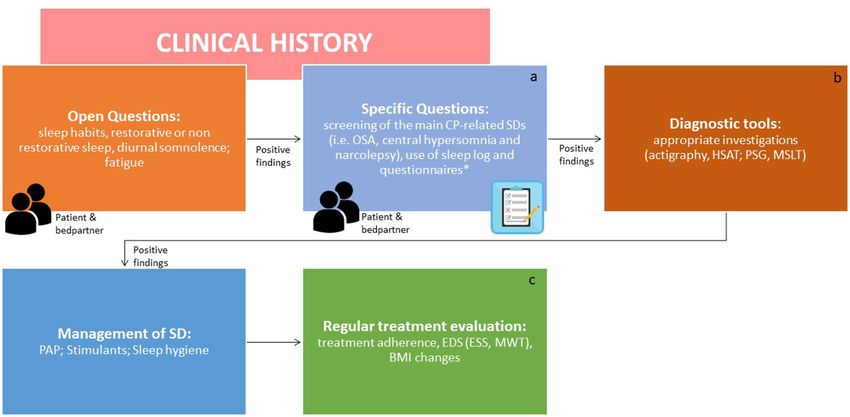
Romigi et al.                                                                                                                             Sleep Disorders in Craniopharyngioma

TABLE 1 | Sleep-related breathing disorders in patients with craniopharyngioma.

References             Patients         Study design                     Prevalence                       Age               Diagnostic criteria                      Diagnostic
                       (n)                                                                                                                                           tool

Snow et al. (23)       5                CP                               2/5 (40%)                        11–19 yrs         Not reported                             PSG
Lipton et al. (29)     3                C selected                       3/3 (100%)                       15–22 yrs         Not reported                             PSG
                                        hypersomnolent patients
O’Gorman et al.        15               CS C (obese CP vs.               7/13 normal-mild (53.8%)         10–21 yrs         Mild OSA AHI 1.5–5/h                     PSG
(7)                                     obese controls)                                                                     Moderate OSA AHI 5–10/h
                                                                         2/13 moderate (15.3%)                              Severe OSA AHI >10/h
                                                                         4/13 severe (30.6%)                                Abnormal CAI >1/h
Crowley et al. (6)     28               C P (obese CP vs. obese          11/28 (39.2%)                    16–67 yrs         AHI ≥ 5/h                                PSG
                                        controls)
Manley et al. (11)     28               RU                               3/7 (42%) (2/3 OSA and           Pediatric         Not reported                             PSG
                                                                         CSA)                             and Adult
Mandrell et al.        110              CS CO U                          5/98 (5.1%)                      Pediatric         AHI ≥ 2/h for pediatric patients         PSG
(13)                                                                                                      and Adult         AHI ≥ 5/h for adult patients
Niel et al. (50)       50               UP                               2/10 (20%)                       3–20 yrs          AHI ≥ 5/h                                PSG

CS, cross sectional; C, controlled; U, uncontrolled; CO, consecutive; P, prospective; R, retrospective; OSA, obstructive sleep apnea; CSA, central sleep apnea; AHI, apnea hypopnea
index (events/h); CAI, central apnea index.

or adult criteria were applied in many pediatric patients with                              related to hypothalamic involvement (31). However, M-ESS may
CP, the pediatric prevalence of OSA may be underestimated                                   not be sensitive enough to screen pediatric patients for EDS
(11, 23, 29, 50).                                                                           (58), so complete sleep evaluation is often recommended (25).
                                                                                            In survivors of childhood brain tumors, hypersomnia/narcolepsy
Central Hypersomnias                                                                        was diagnosed on a median of 6.1 years from diagnosis and 4.7
The second main group of SDs in patients with CP is                                         years from cranial irradiation, and tumor location and radiation
represented by central hypersomnias, which can be recognized                                therapy were potential risk factors (26). Stimulants improved
on PSG. Central hypersomnias are characterized by severe EDS,                               wakefulness and school performance (26). Table 2 summarizes
despite normal quality and timing of nocturnal sleep. ICSD-3                                central hypersomnias and EDS observed in patients with CP.
distinguishes three main subtypes: narcolepsy type 1, narcolepsy
type 2, and idiopathic hypersomnia (19). Secondary narcolepsy                               Circadian Rhythm Sleep-Wake Disorders
and cataplexy are rare disorders described as a consequence of                              CRSWDs are characterized by alterations of the circadian time-
lesions of lateral hypothalamus orexinergic neurons (13, 24, 26,                            keeping system or misalignment of the endogenous circadian
39, 51–54). Narcolepsy is rare and characterized by EDS and REM                             rhythm and the external environment (19), associated with sleep-
sleep dysregulation manifesting as sleep paralysis, cataplexy, and                          wake disturbances (EDS or insomnia) and distress (19). ICSD-
hypnagogic and hypnopompic hallucinations (19). The diagnosis                               3 distinguishes different types of CRSWDs: delayed sleep-wake
is based on clinical symptoms and a mean sleep latency of ≤                                 phase disorder, advanced sleep-wake phase disorder, irregular
8 min with two or more sleep onset REM periods (SOREMPs)                                    sleep-wake rhythm disorder, non-24-h sleep-wake rhythm
on a Multiple Sleep Latency Test (MSLT) or low hypocretin-                                  disorder, shift work disorder, jet lag disorder, and circadian
1 concentration in the cerebrospinal fluid (CSF) (≤110 pg/ml)                               sleep-wake disorder not otherwise specified (19). Patients with
(19). Cataplexy is defined as more than one episode of generally                            CP show frequent disruption of sleep-wake cycle and circadian
brief (< 2 min), usually sudden bilateral and symmetrical loss                              rhythm (20, 27–30), typically caused by involvement of the
of muscle tone with retained consciousness. The episodes are                                hypothalamic SCN and alterations in melatonin transmission
induced by strong emotions, usually positive, with almost all                               (62, 63). Melatonin can be measured in the peripheral blood
patients reporting some episodes induced by emotions associated                             to diagnose clock disruption, but this requires serial blood
with laughter (19). Narcolepsy is strongly associated with obesity                          samples, and several variables may interfere with correct
(24) and other SDs like REM sleep behavior disorders (55),                                  result interpretation (33), preventing routine use in clinical
periodic leg movements during sleep and restless leg sleep                                  practice. Sleep logs, prolonged sleep-wake cycle monitoring by
syndrome (56), and OSA (57). Jacola et al. (31) assessed EDS                                actigraphy, and circadian variations of salivary melatonin by
in a pediatric CP cohort using the Modified Epworth Sleepiness                              dim-light melatonin onset are useful tools to establish a CRSWD
Scale (M-ESS) and MSLT. M-ESS is a quick screening tool derived                             diagnosis (64). Patients with CP lacking midnight melatonin
from ESS to identify EDS in children by assessing their likelihood                          peak had impaired sleep quality, increased EDS, and more
to fall asleep in different everyday situations (from 0, which is                           general and mental fatigue (30). Obesity and EDS were also
low probability to fall asleep, to 3, which is high probability                             linked to low midnight-morning melatonin concentration (20).
to fall asleep). A cutoff of 10 is indicative for EDS (58). EDS                             In addition, sleep fragmentation and EDS are frequently reported
was identified by M-ESS in 76% of pediatric CP and strictly                                 in CP with a circadian profile characterized by early morning

Frontiers in Neurology | www.frontiersin.org                                            4                                           February 2022 | Volume 12 | Article 817257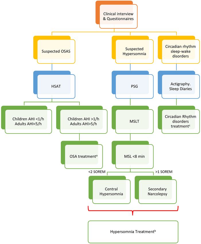
Romigi et al.                                                                                                                              Sleep Disorders in Craniopharyngioma

TABLE 2 | Excessive daytime somnolence and secondary narcolepsy in patients with craniopharyngioma.

References                 Patients (n)       SD                    Study design          Prevalence          Age                      Diagnostic criteria            Diagnostic
                                                                                                                                                                      tools

Snow et al. (23)           5 (3 CF)           Daytime sleepiness    CP                    5/5 (100%)          11–15 yrs                ESS > 12                       ESS, MSLT
Poretti et al. (59)        21                 Daytime sleepiness    PU                    6/21 (28.5%)        Pediatric                ESS > 10                       ESS
Müller et al. (60)         79                 Daytime sleepiness    CP                    28/79 (35.4%)       Pediatric and Adult      ESS > 10                       ESS
van der Klaauw et          27                 Daytime sleepiness    CP                    9/27 (33%)          Adult                    ESS > 10                       ESS
al. (61)
Lipton et al. (29)         3                  Daytime Sleepiness    C (selected           3/42 (7.14%)        17–22 yrs                Self-Reported                  Actigraphy
                                                                    hypersomnolent
                                                                    patients)
Crowley et al. (6)         28                 Daytime Sleepiness    C P (obese CP         20/28 (71.4%)       16–67 yrs                ESS > 10                       ESS
                                                                    vs. obese
                                                                    controls)
Manley et al. (11)         28                 Daytime Sleepiness    RU                    19/28 (67.8%)       Pediatric and Adult      Self-Reported                  Self-
                                                                                                                                                                      Reported
Mandrell et al. (13)       110                Hypersomnia           CS CO U               39/86 (45.3%)       Pediatric and Adult      Tanner Prepubescent            PSG; MSLT
                                                                                                                                       MSL ≤ 15; Tanner
                                                                                                                                       pubescent MSL ≤ 10
                                              Narcolepsy                                  30/86 (34.8%)                                Tanner Prepubescent            PSG; MSLT
                                                                                                                                       MSL ≤ 15; Tanner
                                                                                                                                       pubescent MSL ≤ 10
                                                                                                                                       AND ≥ 2 SOREMPs

CS, cross sectional; C, controlled; U, uncontrolled; P, prospective; CO, consecutive; R, retrospective; ESS, Epworth sleepiness scale; PSG, polysomnography; MSLT, multiple sleep
latency test; SOREMPs, sleep onset REM periods.

TABLE 3 | Sleep-wake cycle alterations in patients with craniopharyngioma.

References                                Patients (n)      Study design           Prevalence               Age            Sleep findings                     Diagnostic tools

Lipton et al.         Sleep-Wake          3                 C (selected            3/3 mild OSA             17–22 yrs      (1) Irregular bed time;            Actigraphy and
(29)                  cycle alterations                     hypersomnolent                                                 (2) Frequent night-time            Melatonin plasma
                                                            patients)                                                      activity;                          dosage vs. levels in
                      Melatonin                                                                                            (3) Inappropriate daytime          Historical controls
                      deficiency                                                                                           episodes of rest;
                                                                                                                           (4) Low melatonin level in
                                                                                                                           patients compared to
                                                                                                                           controls
Pickering et          Melatonin           15                C                      Normal melatonin         18–70 yrs      (1) Unchanged sleep onset          Sleep Log, PSQI,
al. (30)              deficiency                                                   profile and no                          (2) Wake up 1 h earlier            ESS, SF-36, MFI
                      Increased sleep                                              sleep alterations                       (3) Higher global score in         Saliva melatonin
                                                                                   (6/14)                                  PSQI (impaired sleep quality,      dosage, blood
                      Latency                                                      Absent mid night                        increased sleep latency and        cortisol dosage
                                                                                   peak of melatonin                       increased daytime
                                                                                   and impaired sleep                      dysfunction)
                                                                                   quality; EDS and                        (4) Lower melatonin
                                                                                   fatigue (6/14)                          (5) Low midnight melatonin
                                                                                   Phase-shifted                           associated with increased
                                                                                   peak and no sleep                       daytime sleepiness
                                                                                   alterations (2/14)

C, controlled; OSA, obstructive sleep apnea; EDS, excessive daytime somnolence; PSQI, Pittsburgh sleep quality index; ESS, Epworth sleepiness scale; SF-36, short form health survey;
MFI, multidimensional fatigue inventory.

awakening, followed by napping during the afternoon (11, 30,                                 DIAGNOSIS OF SLEEP DISORDERS IN
58). Actigraphy may help to recognize different patterns of SDs                              PATIENTS WITH CP
and is a reliable tool to estimate total sleep time, sleep latency,
sleep onset latency, sleep efficiency, awakenings, and wakefulness                           In order to appropriately evaluate patients with CP for SDs,
after sleep onset (65). Different hypothalamic lesions in obese                              it is crucial to investigate suggesting symptoms such EDS,
children with CP may induce shorter (7) or longer sleep onset                                non-restorative sleep, and fatigue. Physicians should obtain
latency (30). The main CRSWDs in patients with CP are reported                               a comprehensive clinical history on sleep behaviors, sleep
in Table 3.                                                                                  hygiene from patients, and bed partners, even in the absence

Frontiers in Neurology | www.frontiersin.org                                             5                                            February 2022 | Volume 12 | Article 817257
Romigi et al. Sleep Disorders in Craniopharyngioma
FIGURE 1 | Algorithm for the screening and identification of sleep disorders in patients with craniopharyngiomas. ICSD-3, International classification of sleep
disorders version 3; CP, craniopharyngioma; SDs, sleep disorders; OSAS, obstructive sleep apnea syndrome; HSAT, home sleep apnea test; PSG, polysomnography;
MSLT, multiple sleep latency test; CRSWD, circadian rhythm sleep-wake disorders. Endocrinologists and neurosurgeons should be involved in the screening step. The
second step should be performed by a sleep specialist. a Diaries can help to obtain clinical points in a standardized manner. *The use of formal screening
questionnaires for sleep disorders is advisable [i.e., STOP BANG for sleep apnea, Pittsburgh Sleep Questionnaire Index (PSQI) for SDs, Epworth Sleepiness Scale for
EDS, Morningness-Eveningness Questionnaire to identify chronotype]. b SDs should be managed as per overall guidelines. c The effects of treatments should be
regularly evaluated [adherence to PAP, EDS by ESS score or Maintenance Wakefulness Test (MWT) together with multidisciplinary evaluation of obesity and related
cardiometabolic complications as well as appropriate hormone replacement, where present. In particular, body mass index (BMI) should be noticed at each visit].
of evident sleep complaints. Open questions regarding non- current definition and diagnostic criteria of the most relevant SDs
restorative sleep, history of observed snoring, apneas, obesity, reported in patients with CP.
and EDS should raise the suspicion of comorbid OSA and
lead to further investigations. Special care should be paid to
inattention, hyperactivity, high blood pressure, enuresis, and PREVENTION AND TREATMENT OF SLEEP
failure to thrive that are commonly reported in pediatric OSA DISORDERS IN PATIENTS WITH
(44). In addition, some questionnaires may help to suspect SBDs, CRANIOPHARYNGIOMA
EDS, and CRSWDs. Although specific guidelines for SDs in CP
and other suprasellar tumors are lacking, the general indications A multidisciplinary approach is crucial to target several factors
for diagnostic tests should be applied based on clinical suspicion: involved in the onset and progression of SDs in patients with
HSAT for SBDs/OSA, PSG for hypersomnia and MSLT for CP. The choice of a safe neurosurgical approach is the first step
narcolepsy and other central hypersomnias, and actigraphy for in the prevention of hypothalamic damage, and optimization
suspected SDs and more specifically CRSWDs and insomnia (19). of hormone replacement therapy is necessary in patients with
A clinical algorithm aimed to suspect SDs in patients with hypopituitarism and/or DI to correct their potential contribution
CP and summarize their approach and follow-up according to to SDs. In the presence of HS, complementary strategies should
current sleep medicine guidelines (19) is proposed in Figure 1. be put in place to simultaneously address sleep health (direct
Based on clinical suspicion, another algorithm is proposed in strategies) and HO (indirect strategies). We will, therefore,
Figure 2 to guide the diagnosis and treatment of the most analyze risk factors for the development of HS and SDs, and focus
frequently reported SDs. Examples of OSA, central hypersomnia on their prevention and treatment, pointing out the potential
and CRSWD observed in adult patients with CP in our institution benefits of sleep medicine in such patients.
and diagnosed by HSAT, PSG and actigraphy, respectively, are
shown in Figures 3–5. Of note, all these patients had a supra- Neurosurgical Treatment
and retro-sellar extension at pre-operative magnetic resonance The optimal treatment of patients with CP is still a matter of
imaging (MRI). Table 4 provides a glossary and abbreviation list debate due to difficulty in finding effective balance between an
of sleep terms reported in our study, and Table 5 reports the aggressive approach aiming for complete resection to prevent
Frontiers in Neurology | www.frontiersin.org 6 February 2022 | Volume 12 | Article 817257Romigi et al. Sleep Disorders in Craniopharyngioma FIGURE 2 | Algorithm for the management of sleep disorders in patients with CP. PSG, polysomnography; OSAS, Obstructive Sleep Apnea (OSA) syndrome; HSAT, home sleep apnea test; AHI, apnea-hypopnea index per hour of sleep; MSLT, multiple sleep latency test; MSL, mean sleep latency; a The management of OSA should include weight loss, avoidance of alcoholic intake and smoking, sleep hygiene, and positional therapy. Positive Airway Pressure (PAP) is considered first-line treatment. Oral appliances may be suggested for mild to moderate OSA and surgery to correct anatomic obstructions (66). b The treatment of central hypersomnias and secondary narcolepsy should include cognitive behavioral therapy (CBT) and approved stimulants (i.e., modafinil, pitolisant, solriamfetol, and sodium oxybate) (67). c Sleep hygiene, CBT, and short-term pharmacologic approach should be considered for insomnia and CRSWD (68). recurrences, and a more conservative approach aiming to CPs may occasionally extend to the anterior, middle, or reduce the risk of post-operative complications and long-term posterior fossa, rarely they are completely situated within sequelae. The surgical approach itself has also significantly the 3rd ventricle, and hydrocephalus may be present more evolved in the last decades. Controversies about surgical frequently in children than in adults (1). Radical resection has objectives and techniques are related to the complexity of the long been considered as the therapy of choice at any age for anatomical location and extension of CP, which may arise the primary treatment of CP, and several open transcranial anywhere along the craniopharyngeal duct. Nearly 95% have microsurgical (TC) approaches have been developed, offering a suprasellar component, up to 75% are intra-suprasellar and uni- or bilateral access to the tumor. TC surgery allows for good only a minority are purely intrasellar (
Romigi et al.                                                                                                                  Sleep Disorders in Craniopharyngioma

 FIGURE 3 | A 5-min segment from home sleep apnea test (HSAT) in the diagnosis of sleep-related breath disorders in a 51-year old male patient with CP. The patient
 was operated on for a huge supra- and retrosellar craniopharyngioma with hydrocephalus and ataxia, achieving complete resection of an adamantinomatous lesion.
 He developed post-operative diabetes insipidus and partial hypopituitarism, and had severe weight gain (+50 kg) with snoring and markedly excessive daytime
 somnolence (EDS), confirmed by a high ESS score (16/24). HSAT confirmed the presence of severe OSA syndrome (AHI 58.8/h), characterized by several obstructive
 apneas. PAP treatment induced the disappearance of EDS (ESS score 7/24). Overall, the patient was very compliant to lifestyle interventions and endocrinological
 management, and significant weight loss (−30 kg) was also achieved.

and optic nerve and vascular manipulation, and increased                             present as multilobulated mixed solid/cystic intra/suprasellar
resection rates have been associated with increased morbidity                        masses, with frequent calcifications (90%) on X-ray or
and mortality, in particular with neuroendocrine dysfunction                         computerized tomography (CT). The solid component is
and HO (1–3). Exposure of infra-chiasmatic, retrosellar, and                         unevenly hypointense in T2 on MRI and, together with the
interpeduncular extension of some midline tumors may also be                         peripheral component of the cysts, shows irregular contrast
limited. Conversely, microsurgical transsphenoidal approaches                        enhancement. Cystic components are hypointense on T1,
provide limited exposure and maneuverability in the suprasellar                      and their intensity in T2 depends on their protein content.
space and may cause CSF leak. In the last 15 years, the                              Papillary CPs are suprasellar and devoid of calcifications. Of
development of expanded endoscopic endonasal approaches                              note, perilesional edema in FLAIR may hardly be distinguished
(EEAs) in skull base surgery has changed the approach to CP                          from the lesion when it infiltrates the chiasm, hypothalamus,
(69–72). Although initially limited to resection of intrasellar                      or mammillary bodies. Identifying the hypothalamus and
tumors, with increasing experience and improved technology,                          mammillary bodies is essential, although controversies remain
EEAs are being increasingly used for suprasellar CP, and the                         about the relative impact of pre-operative hypothalamic
incidence of CSF leaks has been reduced by the use of multilayer                     involvement (HI) itself and surgical strategies on long-term post-
reconstruction techniques (69, 70). In experienced hands, EEAs                       operative outcome, including post-operative HO. Sainte-Rose
may now be proposed for the treatment of midline infra-                              et al. (75), Van Gompel et al. (76), and Muller et al. (77) have
chiasmatic and suprasellar CP, and, in some cases extended to                        proposed different neuroradiological classifications of CP to
CP of the 3rd ventricle (70). A recent consensus statement of                        define the grading of HI. The prognostic value of HI according
the European Association of Neurosurgical Societies (EANS),                          to either classification, together with additional characteristics
skull base section, recommends surgery in tertiary referral centers                  such as unidentified pituitary stalk, retrochiasmatic extension,
and further supports the role of EEAs as suitable to most adult                      and peri-tumoral edema, has been confirmed in a multifactorial
CP (71). For anatomical reasons, endonasal surgery has been                          analysis of risk factors for the development of HS and morbid
considered difficult in the pediatric population, but the place of                   HO (78).
EAAs in the treatment of pediatric CP may progressively increase                        Differences in CP management worldwide and across the
with improved technology (73).                                                       last decades have been discussed in details elsewhere (2). As
   Preoperative neuroradiological imaging is essential for the                       a general rule, experienced neurosurgeons currently advise
diagnosis and surgical treatment of CPs, and their characteristics                   gross total resection where presumably safe, and conservative
have been well-described (74). Adamantinomatous CP typically                         approaches with subtotal tumor removal in the presence of risk

Frontiers in Neurology | www.frontiersin.org                                     8                                        February 2022 | Volume 12 | Article 817257
Romigi et al.                                                                                                                    Sleep Disorders in Craniopharyngioma

 FIGURE 4 | Examples of MSLT in the diagnosis of central hypersomnias in two female patients with CP. Patient 1 (A–D). Pre-operative evaluation of a 44-year-old
 woman who presented with spontaneous hypothalamic syndrome with severe weight gain (+30 kg) associated with headache, secondary amenorrhea, asthenia,
 insomnia, and diurnal somnolence. Contrast-enhanced T1-weighted magnetic resonance imaging (MRI) revealed a huge solid and cystic suprasellar lesion [(A),
 coronal view] with posterior extension [(B), sagittal view]. MSLT showed a 30-s epoch of NREM sleep (N2) (C) with hypnogram confirming severe excessive daytime
 somnolence (mean sleep latency 4.2 min) without sleep-onset REM in 5 of 5 nap periods (D). A diagnosis of central hypersomnia was made. Patient 2 (E–H).
 Post-operative evaluation of a 52-year-old woman affected by complex post-operative sleep disorders accompanied by diabetes insipidus, pan-hypopituitarism,
 ongoing severe weight gain (+7 kg before surgery, +30 kg after surgery) and asthenia. Preoperative contrast-enhanced T1-weighted MRI showed a huge solid and
 cystic suprasellar lesion [(E), coronal view] with posterior extension [(F), sagittal view]. Excessive daytime somnolence persisted on continuous PAP for documented
 post-operative OSA (data not shown), and MSLT was recently proposed. The MSLT showed a 30-s epoch of REM sleep (G), with hypnogram confirming severe
 excessive daytime somnolence (mean sleep latency 2.3 min) with sleep-onset REM in 2 of 5 nap periods [(H), see blue arrows]. A diagnosis of secondary narcolepsy
 was made, and a stimulant oral agent (modafinil) was started. In both patients, complete tumor resection was achieved, and pathological examination revealed
 adamantinomatous (patient 1) and papillary (patient 2) craniopharyngiomas. ROC, right oculogram; LOC, left oculogram; M1 and M2 reference electrodes placed on
 the mastoid process; Chin, Chin electromyogram.

Frontiers in Neurology | www.frontiersin.org                                      9                                         February 2022 | Volume 12 | Article 817257
Romigi et al.                                                                                                                    Sleep Disorders in Craniopharyngioma

 FIGURE 5 | Example of circadian sleep-wake alteration evaluation by actigraphy. A 75-year-old female patient came to our observation because of headache and
 visual loss in the context of recent and rapidly worsening neurological symptoms consisting of insomnia, excessive daytime somnolence, cognitive impairment,
 reduced appetite, and weight loss. No poliurodyspia was present, and basal pituitary function and electrolytes were normal. Contrast-enhanced T1-weighted MRI
 revealed a mixed cystic and solid tumor consistent with suprasellar craniopharyngioma (A) with retrosellar extension (B). Sleep-wake patterns are displayed for
 individual days on actigraphy (C): vertical black bars and the red line under each day indicate movement, and the absence of black bars indicates supposed sleeping
 periods. The blue band designates the sleep period. The actigram shows frequent nighttime activity, severe insomnia, sleep fragmentation, and frequent short diurnal
 naps. The patient is currently awaiting surgery.

factors for significant post-operative morbidity. Interestingly,                      significantly prevent weight gain (79). Similar findings arise
according to a meta-analysis performed on adult patients with                         from a pediatric meta-data analysis in the United Kingdom
CP, conservative surgery itself was associated with reduced                           (80), pointing out unresolved issues in the identification of
risk of post-operative hypopituitarism and DI but did not                             individual risk factors and personalized surgery. An algorithm

Frontiers in Neurology | www.frontiersin.org                                     10                                         February 2022 | Volume 12 | Article 817257
Romigi et al.                                                                                                                       Sleep Disorders in Craniopharyngioma

TABLE 4 | Glossary of sleep terms included in the review.

Term                             Abbreviation     Definitions

Actigraphy                                        A non-invasive technique that measures physical activity levels of a subject by means of a wristwatch-like
                                                  motion-sensing device that can be worn for prolonged periods of time. Its use is considered useful to diagnose
                                                  CRSWDs, insomnia and other sleep disorders (i.e., OSA, restless legs syndrome)
Apnea-Hypopnea index             AHI              A diagnostic tool for determining the presence and severity of OSA. It represents the average number of apneas and
                                                  hypopneas by hour during sleep
Circadian rhythm sleep           CRSWDs           Chronic or recurrent patterns of sleep-wake rhythm disruption primarily caused by an alteration in the endogenous
wake disorders                                    circadian timing system or misalignment between the endogenous circadian rhythm and the sleep-wake schedule
Cognitive behavioral therapy     CBTi             A short, structured, and evidence-based approach to improve symptoms of insomnia, by identifying and replacing
of insomnia                                       thoughts and behaviors that cause or worsen sleep problems
Epworth sleepiness scale         ESS              A subjective questionnaire to measure daytime sleepiness in the past month
Home sleep apnea test            HSAT             An alternative simplified medical test for the diagnosis of OSA in uncomplicated adults presenting with signs and
                                                  symptoms that indicate an increased risk of moderate to severe OSA. It does not include electroencephalography,
                                                  electrooculogram, and electromyography
Maintenance wakefulness          MWT              An objective measure of daytime vigilance that is used to quantify changes in the ability to stay awake
test
Morningness–Eveningness          MEQ              A self-assessment tool that can provide details regarding an individual’s subjective timing preferences
questionnaire
Multiple sleep latency test      MSLT             An objective measure of daytime sleepiness that is used to measure physiological sleep tendency in the absence of
                                                  alerting factors among 5 diurnal naps. MSL (mean sleep latency) is the mean of each sleep latency
Obstructive sleep apnea          OSA              Obstructive sleep apnea (OSA) is a sleep-related breathing disorder that involves a decrease or complete halt in airflow
                                                  despite an ongoing effort to breathe
Pittsburgh sleep quality         PSQI             A self-rated, subjective, questionnaire to evaluate sleep quality, and disturbances over a 1-month time interval
index
Polysomnography                  PSG              A comprehensive sleep study including electroencephalography, electrooculogram, chin and leg electromyography,
                                                  body position, airflow, respiratory movement, oxygen saturation. PSG is considered the “gold standard” of sleep study
Positive airway pressure         PAP              PAP is the first-choice treatment for OSA involving devices to maintain upper airway patency by increasing the upper
                                                  airway pressure
Sleep onset REM periods          SOREMPs          REM sleep period occurring ≤15 min after the onset of sleep on an overnight PSG or MSLT
Stop-BANG questionnaire          Stop-BANG        An easy to use, concise, effective, and reliable OSA screening tool including Snoring, Tiredness, Observed aPnea, high
                                                  BP, BMI, Age, Neck circumference, and male Gender

for risk-adapted hypothalamic-sparing surgery (HSS) was first                           and still depended on tumor volume and pre-operative BMI
proposed by Sainte-Rose and Puget in pediatric CP, limiting                             (83). SDs were not addressed in a large series of CP managed
the indications for extensive surgical resection (ESR) to CP                            by HSS (81, 82). However, encouraging results were reported
presenting with no HI or with hypothalamic compression                                  about potential improvement of sleep-wake cycle and body
without invasion (75). In their experience, patients receiving risk-                    core temperature in a small series of CP of the 3rd ventricle
adapted HSS had similar relapse and progression rates, with a                           managed by EEA (84). The recent consensus statement on the
significant benefit in weight gain, when compared with their                            surgical treatment of adult CP by EANS skull base section
historical cohort of patients with CP treated by ESR (morbid                            suggests to systematically evaluate pre-operative hypothalamic
obesity 28 vs. 54% and normal post-operative BMI at last follow-                        function (regulation of weight, body temperature, and sleep-wake
up 38 vs. 17%, respectively) (81). Although validation of this                          cycles) and encourages further attention to their post-operative
single-center observation in a prospective multicenter setting is                       evolution in the future (71).
still missing, additional studies have been conducted to evaluate                          A drawback of conservative approaches to CP is their potential
the outcome of HSS in patients with CP. In a recent retrospective                       to recur, and multiple recurrences are a serious concern after
analysis of the German multicenter KRANIOPHARYNGIOMA                                    incomplete tumor resection, especially in children. The best
2007 cohort, neither pre-operative HI nor anterior hypothalamic                         results in terms of progression-free survival after conservative
surgical injury, but posterior hypothalamic surgical injury was                         surgery have been reported in association with post-operative
significantly associated with increased risk of obesity and lower                       radiotherapy and are similar to those obtained by gross total
QoL (82). Hence, the authors proposed that HSS should                                   resection. However, side effects of irradiation are delayed and
particularly point to a “posterior HSS.” However, the potential                         may include hypothalamic complications (9). Among modern
benefits of less aggressive surgical approaches to CP on SDs have                       techniques, proton therapy may take an increasing place in
deserved less attention than HO. Compared to TC surgery, EEA                            the treatment of residual or recurrent CP because of the
was recently reported to be associated with lower incidence of                          dosimetric characteristics of protons and limited off-target
DI, but post-operative weight gain was not significantly lower                          toxicity (2, 85).

Frontiers in Neurology | www.frontiersin.org                                       11                                         February 2022 | Volume 12 | Article 817257
TABLE 5 | Classification and definition of sleep disorders of interest in patients with craniopharyngioma (19).
Frontiers in Neurology | www.frontiersin.org
Romigi et al.
Term (abbreviation) Definition Diagnostic criteria (ICSD-3)
Central disorders of A group of disorders in which the primary Narcolepsy type 1
hypersomnolence complaint is daytime sleepiness not caused by Criteria A and B must be met
disturbed nocturnal sleep or misaligned A. The patient has daily periods of irrepressible need to sleep or daytime lapses into sleep occurring for at least 3 months B. The presence of
circadian rhythms. Other sleep disorders may one or both of the following:
be present, but they must be adequately 1. Cataplexy (defined as more than one episode of generally brief (1/3 of mean values obtained in normal subjects with the same standardized assay
E. The hypersomnolence and/or MSLT findings are not explained more clearly by other causes such as insufficient sleep, obstructive sleep
apnea, delayed sleep phase disorder or the effect of medication or substances or their withdrawal
Hypersomnia due to medical disorders Criteria A–D must be met
A. The patient has daily periods of irrepressible need to sleep or daytime lapses into sleep occurring for at least 3 months
12
B. The daytime sleepiness occurs as a consequence of a significant underlying medical or neurological condition
C. If an MSLT is performed, the mean sleep latency is ≤ 8 min, and fewer than two sleep onset REM periods (SOREMPs) are observed
D. The symptoms are not better explained by another untreated sleep disorder, a mental disorder, or the effects of medications or drugs. (a) If
criteria for narcolepsy are fulfilled, a diagnosis of narcolepsy type 1 or type 2 due to a medical condition should be used rather than
hypersomnia due to a medical condition; (b) In patients with severe neurological or medical disorders in whom it is not possible or desirable to
perform sleep studies, the diagnosis can be made by clinical criteria
Circadian rhythm Chronic or recurrent patterns of sleep-wake General criteria for circadian rhythm sleep–wake disorder Criteria A–C must be met
sleep wake disorders rhythm disruption primarily caused by an A. A chronic or recurrent pattern of sleep–wake rhythm disruption due primarily to alteration of the endogenous circadian timing system or
(CRSWDs) alteration in the endogenous circadian timing misalignment between the endogenous circadian rhythm and the sleep–wake schedule desired or required by an individual’s physical environment
system or misalignment between the or social/work schedules
endogenous circadian rhythm and the B. The circadian rhythm disruption leads to insomnia symptoms, excessive sleepiness or both
sleep-wake schedule. This group includes 1. C. The sleep and wake disturbances cause clinically significant distress or impairment in mental, physical, social, occupational, educational, or
Delayed sleep–wake phase disorder; 2. other important areas of functioning
February 2022 | Volume 12 | Article 817257
Advanced sleep–wake phase disorder; 3.
Irregular sleep–wake rhythm disorder; 4.
Sleep Disorders in Craniopharyngioma
Non-24 h sleep-wake rhythm disorder; 5. Shift
work disorder; 6. Jet lag disorder; 7. Circadian
sleep–wake disorder not otherwise specified
Insomnia A persistent difficulty with sleep initiation, Chronic Insomnia
duration, consolidation, or quality that occurs Criteria A–F must be met
despite adequate opportunity and A. The patient reports, or the patient’s parent or caregiver observes, one or more of the following:
circumstances for sleep, and results in some 1. Difficulty initiating sleep
form of daytime impairment
(Continued)TABLE 5 | Continued
Frontiers in Neurology | www.frontiersin.org
Romigi et al.
Term (abbreviation) Definition Diagnostic criteria (ICSD-3)
2. Difficulty maintaining sleep
3. Waking up earlier than desired
4. Resistance to going to bed on appropriate schedule
5. Difficulty sleeping without parent or caregiver intervention
B. The patient reports, or the patient’s parent or caregiver observes, one or more of the following related to the nighttime sleep
difficulty:
1. Fatigue/malaise
2. Attention, concentration or memory impairment
3. Impaired social, family, occupational, or academic performance
4. Mood disturbance/irritability
5. Daytime sleepiness
6. Behavioral problems (e.g., hyperactivity, impulsivity, aggression)
7. Reduced motivation/energy/initiative
8. Proneness for errors/accidents
9. Concerns about or dissatisfaction with sleep
C. The reported sleep/wake complaints cannot be explained purely by inadequate opportunity (i.e., enough time is allotted for
sleep) or inadequate circumstances (i.e., the environment is safe, dark, quiet, and comfortable) for sleep
D. The sleep disturbance and associated daytime symptoms occur at least three times per week E. The sleep disturbance and
associated daytime symptoms have been present for at least 3 months
F. The sleep/wake difficulty is not explained more clearly by another sleep disorder
Sleep-Related A range of conditions characterized by OSA (ADULT)
breathing disorders abnormal breathing during sleep; in many (A and B) or C satisfy the criteria
(SDBs) cases this is associated with narrowing or
13
A. The presence of one or more of the following:
obstruction of the upper airway (pharynx). The The patient complains of sleepiness, non-restorative sleep, fatigue or insomnia symptoms
disordered breathing ranges from intermittent, The patient wakes with breath holding, gasping or choking
partial obstruction of the airway without sleep The bed partner or other observer reports habitual snoring, breathing interruptions or both during the patient’s sleep
disturbance (snoring) to frequent apneas The patient has been diagnosed with hypertension, a mood disorder, cognitive dysfunction, coronary artery disease, stroke, congestive heart
associated with repetitive hypoxaemia and failure, atrial fibrillation, or type 2 diabetes mellitus
arousals leading to sleep disruption and B. Polysomnography (PSG) or HSAT (Home Sleep Apnea Test) demonstrates:
daytime sleepiness. This group includes Five or more predominantly obstructive respiratory events [obstructive and mixed apneas, hypopneas or respiratory effort-related arousals
obstructive sleep apnea (OSA) syndrome, (RERAs)] per hour of sleep during a PSG or per hour of monitoring (HSAT)
central sleep apnea disorders, sleep-related or
hypoventilation disorders and sleep-related C. PSG or HSAT demonstrates:
hypoxaemia disorders. OSA is a sleep disorder Fifteen or more predominantly obstructive respiratory events (apneas, hypopnoeas, or RERAs) per hour of sleep during a PSG or per hour of
involving cessation or significant decrease in monitoring (HSAT)
airflow in the presence of breathing effort
February 2022 | Volume 12 | Article 817257
OSA (PEDIATRIC)
Sleep Disorders in Craniopharyngioma
Criteria A and B must be met
The presence of one or more of the following:
1. Snoring
2. Labored, paradoxical, or obstructed breathing during the child’s sleep
3. Sleepiness, hyperactivity, behavioral problems, or learning problems
PSG demonstrates one or more of the following:
1. One or more obstructive apneas, mixed apneas, or hypopneas, per hour of sleep
2. A pattern of obstructive hypoventilation, defined as at least 25% of total sleep time with hypercapnia (PaCO2 > 50 mm Hg) in association
with one or more of the following: (a) Snoring, (b) Flattening of the inspiratory nasal pressure waveform, (c) Paradoxical thoracoabdominal
motion
ICSD-3: International Classification of Sleep Disorders - Third Edition (19).Romigi et al. Sleep Disorders in Craniopharyngioma
Endocrine-Metabolic Treatments and insufficiency being lower in patients with corticotroph deficiency
Lifestyle Interventions because of a typically preserved mineral corticoid function,
Although lifestyle and endocrinological/metabolic interventions it may also occur and requires appropriate education of
are not resolutive in patients with HS, they still play a patients and their families. Because any available replacement
fundamental role in the control of homeostasis, optimization therapy is unable to reproduce the physiological rhythm
of SD treatment, and prevention of long-term cardio- of cortisol secretion, patients are frequently exposed to
metabolic/vascular complications. The essential role of supraphysiological evening levels of cortisol, which may impact
short-term post-operative management of endocrine deficiencies on sleep quality, sleep latency, and daytime functioning (93,
and sodium/water imbalance is beyond the scope of this 94). It may be, therefore, be useful to prefer modified-release
article. It should be remembered, however, that excessive hydrocortisone to a standard replacement therapy with twice or
post-operative fluctuations in serum osmolarity due to DI, trice daily oral hydrocortisone. Modified-release hydrocortisone
SIADH, and/or salt wasting syndrome should be avoided, as replacement therapy best approaches physiological cortisol
excessively rapid corrections of hyponatremia may lead to secretion and may have a favorable impact on body weight
irreversible neurological damage (86, 87). Patients should be control, metabolism, immune function, and QoL (95). The single
clearly informed on the potential risks, clinical manifestations morning administration of modified-release hydrocortisone may
and timing of neuroendocrine alterations and HS following also improve patient compliance in the setting of multiple
surgery, and the need for long-term endocrinological follow-up. treatments for hormone replacement and/or associated co-
Guidelines on the treatment of single or multiple pituitary morbidities. We, therefore, suggest, where available, to consider
deficiencies are currently available (88), and we will focus on modified-released hydrocortisone in the long-term treatment of
the benefits and potential risks of hormone replacement therapy patients with complex CP, including those with SDs.
on SDs.
Thyroid Hormone Replacement Therapy
Hormone Replacement Therapy
Thyroid function and sleep also have a bidirectional relationship,
The relationship between hormone replacement and sleep health
influencing each other through the circadian clock (96).
is complex and bidirectional. Optimal hormone replacement
Hypothyroidism results in poor sleep quality and architecture
therapy may have a beneficial effect on muscle function,
(96), and may trigger or worsen preexisting OSA (97). This
including upper airway dilator muscles, body composition,
may occur as a consequence of impaired neural response to
and metabolism, as well as fatigue and mood. Conversely,
hypoxemia and hypercapnia, increased airway resistance due
overtreatment maybe deleterious for sleep, and worsens OSA
to mucoprotein deposition, increase in BMI, and changes
or sleep-wake cycle and circadian rhythm. Even in the
in upper airway muscle activity (96). Conversely, over-
presence of undamaged hypothalamic-pituitary connections,
replacement may favor insomnia, increase oxygen consumption,
sleep fragmentation and reduction in slow wave sleep impair
and impact on muscle and cardiovascular function as observed
circadian pituitary hormone regulation, in particular ACTH and
in hyperthyroidism (98). Therefore, optimizing thyroxine
GH (89). Therefore, fine-tuning of hormone replacement is
replacement therapy may contribute to improve sleep quality and
needed to optimize metabolic, cardiovascular, and sleep issues in
architecture in patients with CP.
patients with CP.
Diabetes Insipidus Testosterone Replacement Therapy
Diabetes insipidus (DI) may be transient or life-long and Because testosterone replacement therapy has pleiotropic
requires desmopressin replacement therapy according to benefits in hypogonadal men, including improvement in
current guidelines (87, 88). Uncontrolled DI is characterized fatigue, lean mass, and hemoglobin concentration, it should be
by polyurodipsia (>50 mL/kg of body weight/24 h) with considered in hypogonadal male patients with CP according to
nycturia and deleterious consequences on sleep quality, current guidelines (88). The association between testosterone
increasing daytime sleepiness and fatigue (88, 90). Conversely, replacement and OSA is controversial and has not been
overtreatment results in hyponatremia and related neurological specifically addressed in patients with CP, but the Endocrine
complications (86). A minority of CP patients with hypothalamic Society recommends against testosterone replacement in patients
injury present an impaired sense of thirst, which should be with severe untreated OSA (99). In a large retrospective study,
promptly recognized by systematic evaluation of water balance. an elevated risk of OSA among testosterone users compared
Adipsia complicates clinical management, reduces QoL (91, 92), with controls was observed (100). Potential mechanisms
and exposes patients to the risk of severe and potentially fatal include the impact of androgens on muscle contraction and
dehydration (92). Patients and their families should be informed neuromuscular control of upper airway muscles; increase in
on such risks and the potential recovery of DI to optimize oxygen consumption leading to hypoxia, and changes in neural
desmopressin treatment. response to hypoxemia and hypercapnia (100). Thus, clinicians
should be careful in prescribing testosterone replacement
Glucocorticoid Replacement Therapy to patients CP and untreated OSA, and reevaluation during
Patients affected by either primary or secondary glucocorticoid ventilation treatment may be useful. Of note, obesity in itself is
deficiency complain of fatigue and impaired QoL with reduced frequently accompanied by functional hypogonadism. Because
daytime activities (93). Despite the risk of acute adrenal the cardiovascular risks and benefits of testosterone replacement
Frontiers in Neurology | www.frontiersin.org 14 February 2022 | Volume 12 | Article 817257You can also read