Surgery for Peripheral Nerve Sheath Tumours of the Buttocks, Legs and Feet in 90 Patients With Neurofibromatosis Type
←
→
Page content transcription
If your browser does not render page correctly, please read the page content below
in vivo 35: 889-905 (2021)
doi:10.21873/invivo.12329

 Surgery for Peripheral Nerve Sheath Tumours of the Buttocks,
  Legs and Feet in 90 Patients With Neurofibromatosis Type 1
                                      REINHARD E. FRIEDRICH and CAGLAYAN T. TUZCU

                   Department of Oral and Craniomaxillofacial Surgery, Eppendorf University Hospital,
                                      University of Hamburg, Hamburg, Germany

Abstract. Background/Aim: Neurofibromatosis type 1 (NF1)           cutaneous neurofibroma (CNF) (9). This type of neurofibroma
is an autosomal dominant tumour predisposition syndrome            causes primarily aesthetic problems due to the large number
that can cause plexiform neurofibromas (PNFs). This study          of mushroom-like tumours arising in the skin (10, 11).
examines the surgical procedures that have been performed             In contrast, another type of nerve sheath tumour in
on large PNFs of the lower extremities. Patients and               patients with NF1 arises from larger nerve trunks or covers
Methods: Surgical procedures on the lower extremity                large areas of the skin and invades the subcutaneous tissue
performed on 90 patients with NF1 with PNFs were                   (12, 13). In addition to the aesthetic impairment, these
evaluated. The topography of the tumours was classified            tumours often also result in functional deficits (14-16). The
according to dermatomes and functional units. Results: A           tumours are very likely congenital in origin and are termed
total of 243 surgical interventions on the regions of interest     ‘plexiform neurofibromas’ (PNFs) (12, 13), which constitute
were performed. Neurological complications were rarely             one out of six clinical findings allowing the diagnosis of NF1
noted and usually occurred temporarily. There was no               (11). The term is derived from characteristic histological
preference for dermatomes affected by PNF. The proportion          findings (17). PNF is a characteristic tumour in NF1, which
of patients with malignant peripheral nerve sheath tumours         is identified in about 30% of patients. Indeed, sporadic PNF
(MPNSTs) in this group was 4/90 (4.4%). Conclusion: PNFs           is a very rare diagnosis. A finding of sporadic PNF without
often require repeated local interventions to achieve the          further diagnostically relevant features of NF1 may represent
treatment goal. Local tumour recurrences are to be expected        the so-called segmental type of the disease (18-22). In a
even after extensive tumour reduction. Rapid tumour growth         large, unicentric study on the surgical treatment of PNSTs,
combined with new pain sensations can be signs of a MPNST.         benign, sporadic neurofibromas were the largest diagnostic
                                                                   group (21). Therefore, in order to characterize a study group
The autosomal dominant transmitted hereditary disorder,            for the treatment of PNSTs, information on the genetic
neurofibromatosis type 1 (NF1), develops with relative             background of the tumour disease is important. PNF is
frequency in humans (1-5). NF1 is a tumour predisposition          regarded as a precancerous lesion, potentially giving rise to
syndrome (6, 7), with individuals affected carrying a              a malignant peripheral nerve sheath tumour (MPNST) (21-
substantial risk of developing tumours of neurogenic origin        26). NF1 is the most common inheritable disease with a
(1), in particular peripheral nerve sheath tumours (PNSTs).        predisposition to cancer development.
The tumours are usually large in number (8) and termed                Surgery is the most effective measure to treat patients with
neurofibromas (9). The most frequent PNST in NF1 is                PNFs (25-30); however, due to the large extent of many PNFs,
                                                                   complete resection of the lesions is hardly feasible (30). PNFs
                                                                   are slowly growing lesions that can develop significant
                                                                   increases in volume (31). A particularly striking shape of PNF
This article is freely accessible online.                          growth is the tumour-associated enlargement of an extremity
                                                                   or parts of it. This phenotype has been described as
Correspondence to: Reinhard E. Friedrich, Department of Oral and   ‘elephantiasis neuro(fibro)matosa’ (32, 33). Alternatively,
Craniomaxillofacial Surgery, Eppendorf University Hospital,        enormous neurofibromatous tumours are also termed ‘massive
University of Hamburg, Martinistraße 52, 20246 Hamburg,
                                                                   soft tissue neurofibromas’ (17). This tumour type appears to
Germany. Tel: +49 40741053259, e-mail: rfriedrich@uke.de
                                                                   be restricted to generalized NF1 (17, 34-42) and is often
Key Words: Neurofibromatosis type 1, buttocks – surgery, lower     highly vascularized (43). The massive tumour rarely develops
extremity – surgery, foot – surgery, plexiform neurofibroma,       into a MPNST (17, 44, 45). On the other hand, the large
MPNST, peripheral nerve sheath tumour.                             volume of tumours is the prerequisite for unnoticed growth of

                                                                                                                              889
in vivo 35: 889-905 (2021)

Figure 1. Classification of dermatomes. Image adapted from Keegan and Garett 1948 (83), supplemented by a classification of the region of interest
defined by functional levels (horizontal lines). The abbreviations indicate the assignment of the skin areas to the respective spinal nerves. T: thoracic;
L: lumbar; S: sacral; C: coccygeal.

malignant tumours. PNF in association with NF1 may develop                     the procedure and diagnosis. Of these 235 patients with NF1
with variable extension and size in the buttocks, legs and feet                operated on for NF1-associated pathologies of the leg, foot, buttocks
(17). Many reports have been published on the diagnosis and                    or inguinal area, a total of 90 patients with the plexiform type were
                                                                               selected and included in the study. In the remaining 145 patients who
treatment of PNF in NF1 and have been presented as case
                                                                               were not operated on for a PNF, the main reasons for surgery were
reports or clinical studies (46-79). The aim of this study is to               excision of (multiple) CNFs or iliac crest biopsy/graft associated
contribute to the classification of surgical interventions for                 aesthetic and reconstructive measures. All paper files were
PNFs in the region of the buttocks, legs and feet, by analysing                subsequently ordered and checked, digital files examined, digital
clinical data. This information may contribute to improving                    photographs viewed and photos on celluloid (slides) screened.
the treatment process in affected patients.                                        One patient had changed their sex from male to female. Since the
                                                                               patient was last operated on as a man with regard to PNF treatment
                                                                               and gender transformation took place later, the patient was included
Patients and Methods                                                           as a ‘male’ in the statistical evaluation. Another patient had
                                                                               developed squamous cell carcinoma of the sole of the foot. This
Patient population and study material. The archive of surgical                 patient had no PNFs in the affected region of the body and was
reports of the senior author was searched for the operation reports            excluded from further evaluation (82).
of patients with neurofibromatosis undergoing surgery on the                       The following data were collected from these 90 patients for further
buttocks and lower extremities. All these patients have been operated          calculations: i) date of birth, ii) gender, iii) affected body side (right,
on by the senior author in the Department of Oral and                          left or bilateral), iv) neurofibroma surgery in other parts of the body, v)
Craniomaxillofacial Surgery, Eppendorf University Hospital in                  diagnosis       of     malignancy     (MPNST),         vi)    elephantiasis
Hamburg, Germany. The surgical interventions took place between                neuro(fibro)matosa, vii) age at the first surgical intervention in the
the spring of 1990 and April 2017. Patients were excluded if they              hospital, viii) date of surgery, ix) duration of operation, x) number of
had been affected by NF2 or schwannomatosis (11). The patients                 interventions in the region of interest per individual, xi) recording dates
participating in this retrospective study all fulfilled the diagnostic         for inpatient stay and discharge (duration of stay in hospital), xii)
criteria for NF1 (80, 81). The first sighting of files provided 235            pathological findings, xiii) blood transfusion(s) and xiv) complications
patients with appropriate information on the timing and location of            related to surgery (Tables I and II). The external size of the lesions was

890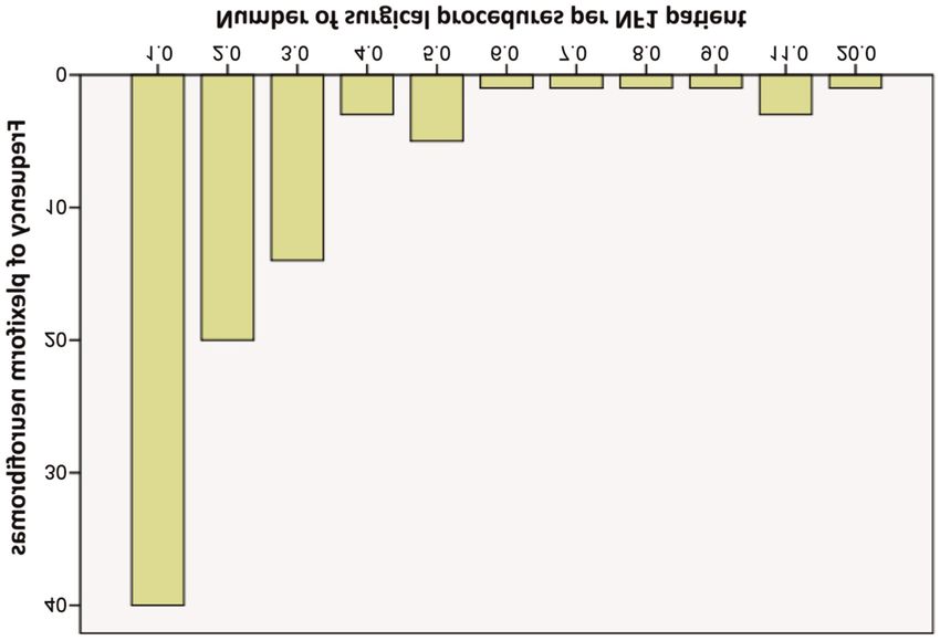
Friedrich and Tuzcu: Surgery for Plexiform Neurofibroma of Lower Extremity in NF1

Table I. Classification of complications related to surgical intervention.   Table II. Classification of histological findings of tumours.

Code                                                Definition               Code                                   Definition

0                                              No complication               1                         Plexiform-diffuse neurofibroma
1                                             Regular rebleeding             2                          Dermal-diffuse neurofibroma
2                                            Wound healing disorder          3                     Malignant peripheral nerve sheath tumour
3                                             Hematoma drainage              4                                     Lipoma
4                                              Wound infection               5                             Atypical neurofibroma
5                                              Revision surgery              6              Plexiform neurofibroma+schwannoma in neurofibroma
                                                                             7                   No pathological examination report available

determined on the basis of operation reports and photo documentations.       Table III. Number of surgeries for NF1 patients with plexiform
Reports of diagnostic imaging [magnetic resonance imaging (MRI) and          neurofibromas.
computed tomography (CT)] were used to further specify the findings.
                                                                             Number of surgical procedures                N                  Percent (%)
Diagnostic subdivision of the regions into anatomical and functional
units. The distribution pattern of dermatomes used for this study was        1                                           40                     44.4
according to Keegan and Garett (83) (Figure 1). Dermatomes are               2                                           20                     22.2
classified according to their sensitive radicular innervation. However,      3                                           14                     15.6
                                                                             4                                            3                      3.3
we knew from previous investigations (45) that assignment of tumour
                                                                             5                                            5                      5.6
extension to dermatomes can cause considerable inadequacy in the
                                                                             6                                            1                      1.1
graphical representation of tumour spread, because firstly, the tumours
                                                                             7                                            1                      1.1
may spread out over a wide area and often grow horizontally, which           8                                            1                      1.1
means that they develop perpendicular to the dermatome of the                9                                            1                      1.1
extremity. Secondly, the dermatomes in the regions of interest are of        11                                           3                      3.3
very different sizes, so no estimation about the area and size of the        20                                           1                      1.1
tumour is possible with reference to the assignment to a dermatome.          Total                                       90                    100.0
Thirdly, due to the original segmental structure of the dermatomes,
some dermatomes are localized close together and narrowly limited,           N: Number of patients.
so tumours in this region very easily infiltrate several dermatomes,
even if they are small in size. In contrast, tumours of the same size
affect only one or a few dermatomes elsewhere (and then dermatomes
may be only partially tumorous). Despite these evident weaknesses            according to both dermatome and level, then numerically coded for
in the classification of tumour extension according to dermatomes,           further data processing. The data concerning surgical complications
this determination method was used to obtain an initial estimate of          and histological findings were further differentiated and transformed
the distribution and extension of the lesion in the region of interest.      into numerical codes (Tables I and II). Subsequently, the results
The measurements can provide some information on the treatment               were summarized in an Excel sheet and evaluated using software
needs of the patients.                                                       SPSS™ (Version 24) (IBM, Armonk, NY, USA) and STATA™
    Since a descriptive anatomical classification according to the           (Version 14) (StataCorp, College Station, TX, USA) for statistical
affected skin area is obviously of limited value for determining the         computing. The chi square test was used to determine differences,
extension of tumours, a functional classification, according to the          and regression equations were used to uncover correlations. A p-
vertical level of the limb, was chosen as an alternative. This               Value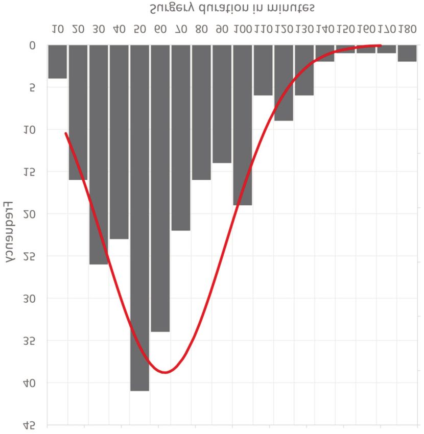
in vivo 35: 889-905 (2021)

Figure 2. Graphical description of number of surgeries for NF1 patients with plexiform neurofibromas.

Results

Gender. Thirty affected men and 60 affected women were
recorded and statistically evaluated.

Age at the first operation. The age range was between 1 and
61 years [male: 1-61 years (yrs), mean±SD=31.57±18.97 yrs;
female: 5-60 yrs, mean±SD=26.35±12.14 yrs].

Body side. The location of the nerve sheath tumours was
recorded with reference to body side. In some patients, the
predominantly unilaterally growing PNF exceeded beyond the
midline of the body, preferentially identified in lesions arising
in the buttocks. This finding was considered a bilateral spread
of the PNF. Thirty-eight patients (42.2%) were affected on
their right side, and 32 patients (35.6%) were affected on their
left side. There were 20 patients (22.2%) with a continuous
spread of the lesion to both halves of the body.

Number of surgical interventions per patient. Most patients
were operated on 1-3 times but exceptions were not                       Figure 3. Duration of plexiform neurofibroma surgeries. (Mean±SD:
uncommon. On average, a patient experienced surgical                     63.45±33.096; N=243).
procedures in the region of interest about 2.7 times. Forty
patients (44.4%) had surgery only once. Table III and Figure
2 show the distribution of number of procedures and                      Blood transfusion. Most patients were treated without the
patients (minimum=1, maximum=20, mean±SD=2.69±2.93,                      need for blood substitution [86 patients (95.6%)]. Only four
median=2).                                                               patients (4.4%) needed this measure. Out of 243 surgical

892
Friedrich and Tuzcu: Surgery for Plexiform Neurofibroma of Lower Extremity in NF1
Table IV. Type 1 neurofibromatosis patients affected by complications Table V. Type 1 neurofibromatosis patients affected by complications
that occurred during surgery or post-surgery following debulking that occurred during surgery or post-surgery following debulking
procedures for large plexiform neurofibroma. Complication rate in procedures for large plexiform neurofibroma. Complication rate in
relation to the number of patients (N). relation to the number of interventions (N).
Complication Code N Percentage (%) Complication Code N Percentage (%)
No complication 0 63 70 No complication 0 182 74.9
Regular oozing bleeding 1 16 17.8 Regular oozing bleeding 1 37 15.2
(Penrose drain) (Penrose drain)
Wound healing disorder 2 4 4.4 Wound healing disorder 2 14 5.8
Hematoma drainage 3 3 3.3 Hematoma drainage 3 5 2.1
Wound infection 4 2 2.2 Wound infection 4 3 1.2
Revision surgery 5 1 1.1 Revision surgery 5 1 0.4
Permanent motor nerve damage 6 1 1.1 Permanent motor nerve damage 6 1 0.4
Total 90 100.0 Total 243 100.0
interventions, blood transfusions were performed 18 times, PNFs that were distributed deep in the body and continued to
corresponding to 7.41% of all surgical procedures [no increase in number and size over time (follow-up: 20 years).
transfusion in 225 procedures (92.59%)]. It is noticeable that Some of these nodular neurofibromas were removed and
a small group of patients (extensive tumours, necrotizing identified as PNFs. During this long follow-up period, the
MPNST) more often required a blood transfusion, while the patient also developed few CNFs at different body sites.
majority of patients did not need this measure.
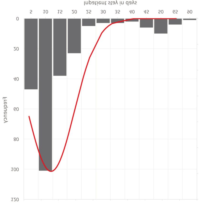
Hospitalization. In the total of 243 surgeries, the shortest
Duration of surgical procedures. A single procedure lasted hospital stay was 3 days and the longest hospital stay was 88
63.45 minutes on average (minimum: 10 min, maximum: 180 days. The arithmetic mean was 15.72 days (SD=13.51;
min, median±SD=56±33.1 min, n=243 procedures) (Figure 3). median=11 days) (Figure 5).
Complications. Table IV shows the relationship between Elephantiasis neuro(fibro)matosa. Thirty patients were
complications in terms of number of patients (n=90). No classified as suffering from elephantiasis neuro(fibro)matosis.
complications were recorded during the treatment of 63 Only the diagnoses from the written paper documents and
patients (70%). Sixteen patients (17.8%) had some oozing digital files were considered. In comparison to photographs
blood, 4 patients (4.4%) had wound healing disorders, while showing findings prior to the intervention and during the
in 3 patients (3.3%) a hematoma had to be drained and two surgical procedure, the term describes tumours spreading like
patients (2.2%) had a wound infection. Only one patient lumpy masses, extending from one or more body levels and
(1.1%) had a revision surgery. A total of 182 of all operations having caused a noticeable deformation of the body region.
(74.9%) took place without complications. The term is an imprecisely used qualitative judgment, which
Out of a total of 243 procedures, 37 (15.2%) were is mainly used for diffuse PNFs. A more precise description
associated with regular oozing bleeding. Of these, 14 of the structure of the body region (e.g., asymmetry of the
operations (5.8%) were associated with a wound healing skeleton) is not associated with the metaphorical terminus.
disorder, 5 (2.1%) were caused by a need for hematoma Elephantiasis in connection with the planning of the
drainage, 3 (1.2%) were caused by wound infection and one procedure addresses a large, spreading tumour, however,
(0.4%) was a revision surgery (Table V). In one case, the without further specification of the finding. The somewhat
peroneal nerve was damaged as a result of the resection of a more abstract term ‘massive soft tissue tumour’ (17) fully
large, nodular PNF of the buttocks. The tumour had grown corresponds to the descriptive term ‘elephantiasis’.
within a short period of time, causing considerable pain,
causing inability to take a pain-free sitting position, Pathology. Table VI indicates all pathological findings of
impairment of driving a car and restricted movement of the patients with NF1. As already detailed, the pathological
affected leg. The operative site shows the large tumour with findings were divided into six different diagnoses, of which
peripheral nerve growing into the lesion (Figure 4A). The case PNF was the most frequently diagnosed type in 43 patients
is unusual in that the patient with genetically confirmed NF1 (47.8%), followed by dermal diffuse neurofibroma in 24
was missing all the cutaneous stigmata of the disease at the patients (26.7%), and MPNST in four patients (4.4%). Four
time of tumour resection. The patient had developed multiple other patients (4.4%) had atypical neurofibroma, one tumour
893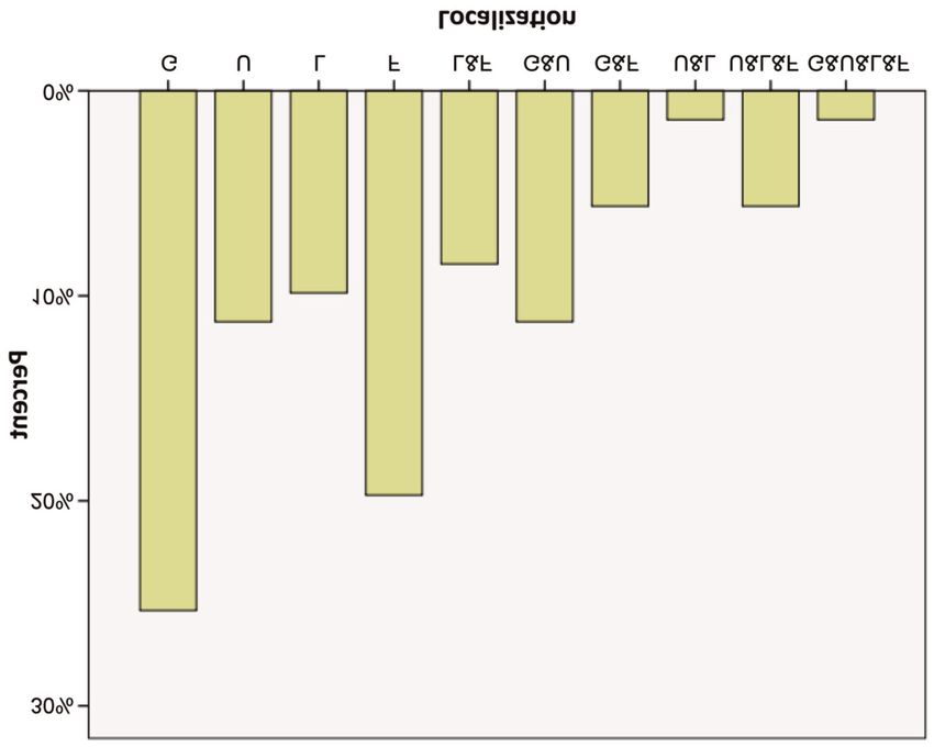
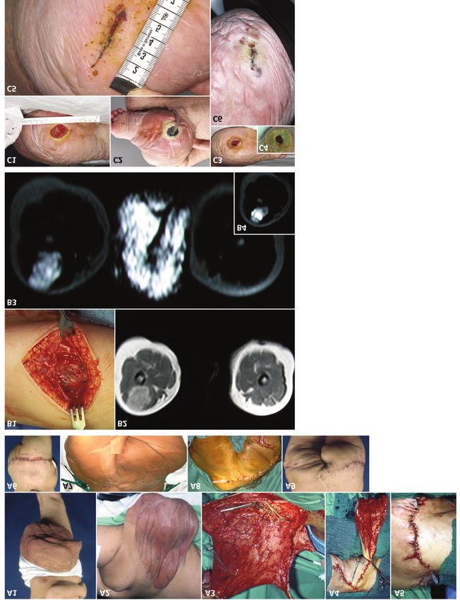
in vivo 35: 889-905 (2021)

Figure 4. Plexiform neurofibroma phenotypes. A1-A5: Painful, rapidly growing tumour of the buttocks. A1-A2: MRI shows nodular mass of right side
of buttocks with inhomogeneous internal enhancements (arrows) of the tumour in axial and coronal sections. A3: The nodular tumour was resected
in toto. A4: A nerve inserts into the tumour, in which the strand-like structure (arrow) cannot be further identified. A5: The incised tumour is solid
and shows inhomogeneities in the tissue distribution, but no visible necrosis. B1: Small-size plexiform neurofibroma of the leg. B2: Large tumour
resection specimen of the leg. B1 is a 2×2 cm neurofibroma taken from the right dorsal lower leg and B2 is about a 30×15 cm piece of tissue that
was excised from the left dorsal thigh. Both specimens were histologically diagnosed with diffuse plexiform neurofibroma (WHO grade 1).

was a lipoma (1.1%), four patients (4.4%) had a PNF with a                   histological diagnosis has no correlation to the macroscopic
schwannoma in the neurofibroma and in 10 patients (11.1%),                   extent of the tumour but to the texture of the tissue. The
the pathological findings were inconclusive or missing.                      comparison of visible tumour size and histological tumour
   The histological finding of PNFs differs from the clinical                diagnosis shows that the independence of both
use of the term PNF in the sense of describing a large                       characteristics has a significant impact on the topographical
tumour mass with diffuse infiltration of the skin,                           scale used in the evaluation (i.e., the dermatome infiltrated
subcutaneous tissue and possibly the muscles. Therefore, the                 by the tumour). Here are photographs of two surgical
histological findings were evaluated. This shows that there                  specimens of two different patients that are very different in
are clear differences between the clinical assessment of the                 size to illustrate the divergence of macroscopic and
lesion and the histological findings. Furthermore, the                       microscopic aspects (Figure 4B).

894
Friedrich and Tuzcu: Surgery for Plexiform Neurofibroma of Lower Extremity in NF1

Table VI. Histology of peripheral nerve sheath tumours of NF1 patients   Table VII. Total number of individual dermatomes from operations of
(Institutes of Pathology and Neuropathology, UKE).                       NF1 patients with plexiform neurofibroma.

Pathology                          Code        N       Percentage (%)    Summary of all regions          N (Dermatomes)      Percentage (%)

Plexiform-diffuse neurofibroma       1         43            47.8        Not affected                         3,872               61.3
Dermal-diffuse neurofibroma          2         24            26.7        Affected                             1,536               24.3
MPNST                                3          4             4.4        Not adequately depicted               910                14.4
Lipoma                               4          1             1.1         on photograph
Atypical neurofibroma                5          4             4.4        Total                                6,318               100.0
Plexiform neurofibroma and           6          4             4.4
 schwannoma in neurofibroma
No pathological examination          7         10            11.1
 report available
Total                                          90          100.0

MPNST: Malignant peripheral nerve sheath tumours; N: number of           number of dermatomes and indication for blood transfusion were
patients.                                                                associated and had a statistically significant impact on surgical
                                                                         treatment (Tables XI, XII, XIII). The indication for blood
                                                                         transfusion influenced the duration of surgery. For example, if
                                                                         there was a blood transfusion, the surgical procedure lasted, on
Affected dermatomes of the whole group. A total of 6,318                 average, 36 minutes shorter than if there was no transfusion. This
individual dermatomes in the various body regions were                   correlation shows that the surgical concept aimed to ensure local
counted and evaluated (Table VII). In 3,872 dermatomes, the              wound control and adapt the surgical measure accordingly,
corresponding areas were not affected by a PNF (61.3%).                  especially through repeated tumour reductions, to allow for
The number of affected dermatomes was 1,536 (24.3%)                      easier control.
(Tables VIII, IX, X). However, in 910 dermatomes (14.4%),                   The number of dermatomes had an influence on the
no corresponding tumour localization could be made due to                duration of surgery (Figure 3). For example, with each
insufficient visual representation of the tumour region on               dermatome, the duration of surgery increased on average by
photographs. The dermatomes in the buttock area were                     1.7 minutes. This proved a linear relationship between the
largely equally affected (Tables VIII and Table IX, Figure 6).           area and the operating time. No other factors had a
   The segmental structure of the skin innervation was precisely         significant effect on the duration of surgery (p>0.05).
mapped (85, 86), but the individual sensitive innervation fields
showed very large differences. Tumour extension hardly fit into          Malignancy (MPNST). A total of four patients were
the skin regions assigning the developmental history of the              diagnosed with MPNST. Of these four patients, the legs of
peripheral nervous system in terms of affected dermatomes.               three patients were amputated during the course of disease.
Overall, the frequency proportions of the dermatomes of the              Two of these patients survived. In all four patients (4.4%),
investigated body regions affected by a PNF vary only within             an extensive PNF had transformed to malignancy (MPNST).
a narrow range. The investigation revealed a largely uniform             In 86 patients (95.6%), there was no malignancy in the
distribution of tumour spread in relation to the dermatomes, and,        region of interest. Two patients have developed a MPNST
thus, the random distribution of somatic mutations that cause            later during the course after reducing a PNF in the study
the development of the tumours.                                          area. One patient came to the hospital with an already
                                                                         confirmed diagnosis of MPNST. One child, who was
Levels. The classification of the tumour locations according to          diagnosed with MPNST for the first time and was not
level also showed the frequent need for the treatment of tumours         subjected to further surgical measures following diagnostic
in the gluteal region (about 40%, Figure 7). On the other hand,          exploration, underwent multiple rounds of chemotherapy
locations, such as the foot and upper leg were also very often           and died 2 years later with a diagnosis of tumour
affected when all patients with tumours from this region were            progression at the age of 5 years.
included (Table X). All in all, the classification of tumours by
level showed the well-known invasive growth, which can be                Neurofibromas in other parts of the body. Of the 90 patients
descriptively assigned to a specific region of the body.                 examined, 52 (57.8%) had neurofibromas manifested not
                                                                         only in the lower extremities but also in other parts of the
                                                                         body. Large PNFs growing in continuity from the lower
Correlation analysis. The number of affected dermatomes varied           trunk to the gluteal region were included in the ‘gluteal’
considerably (minimum: 1, maximum: 26; mean±SD=5.03±4.85,                region and thus not considered for registration of tumours
median: 4). However, correlation analyses revealed that the              in other body regions. Neurofibromas in other parts of the

                                                                                                                                          895
in vivo 35: 889-905 (2021)
Table VIII. Number of not-affected dermatomes in NF1 patients subjected Table IX. Number of affected dermatomes in NF1 patients subjected to
to surgical procedures of the gluteal region, lower extremity and foot. surgical procedures of the gluteal region, lower extremity and foot.
Not affected dermatome N (Dermatomes) Percentage (%) Affected dermatome N (Dermatomes) Percentage (%)
GL1 147 3.80 GL1 61 3.97
GL2 157 4.05 GL2 51 3.32
GL3 161 4.16 GL3 47 3.06
GL4 152 3.93 GL4 56 3.65
GL5 141 3.64 GL5 67 4.36
GS1 126 3.25 GS1 82 5.34
GS2 121 3.13 GS2 87 5.66
GS3 147 3.80 GS3 61 3.97
GS4 154 3.98 GS4 54 3.52
GS5 157 4.05 GS5 51 3.32
UL1 157 4.05 UL1 51 3.32
UL2 154 3.98 UL2 54 3.52
UL3 155 4.00 UL3 53 3.45
UL4 159 4.11 UL4 49 3.19
UL5 157 4.05 UL5 51 3.32
US1 155 4.00 US1 53 3.45
US2 148 3.82 US2 60 3.91
LL3 164 4.24 LL3 44 2.86
LL4 148 3.82 LL4 60 3.91
LL5 137 3.54 LL5 71 4.62
LS1 134 3.46 LS1 74 4.82
LS2 143 3.69 LS2 65 4.23
FL4 154 3.98 FL4 54 3.52
FL5 137 3.54 FL5 71 4.62
FS1 149 3.85 FS1 59 3.84
FS2 158 4.08 FS2 50 3.26
Total 3,872 100.00 Total 1,536 100.00
Alphanumeric coding of dermatomes: First code digit: G: gluteal; U: upper Alphanumeric coding of dermatomes: First code digit: G: gluteal; U: upper
leg; L: lower leg; F: foot; Second code digit: L: lumbar; S: sacral; Third leg; L: lower leg; F: foot; Second code digit: L: lumbar: S: sacral; Third
code digit: Numbering refers to the spinal nerve of the vertebral segment. code digit: Numbering refers to the spinal nerve of the vertebral segment.
body were registered irrespective of neurofibroma type. Age. The average age of men at the first operation is
Surgery for PNFs in the buttocks or legs was the only approximately 31.6 years and of women 26.4 years. The age
surgical procedure for treatment of PNST in 38 patients difference is probably due to the earlier assessment of
(42.2%). Figure 8 illustrates some findings and surgical women and that an attempt at treatment is preferable to a
procedures. wait-and-see attitude. However, these assessments should be
checked by analysis of the age structure of patients with this
Discussion diagnosis, phenotype and treatment request from other
centres. The age structure of surgically treated patients with
The study presents the clinical data of long-term care for NF1 changes significantly when analyses for the treatment
patients with NF1 who have been surgically treated for of bone asymmetries of the extremities and pseudarthroses
extensive tumours of the buttocks, legs and feet (30). The are presented, in particular in children (27, 29) and in studies
data can be used to orient treatment plans for patients with on MPNST in NF1 (25).
this phenotype.
Topography. The findings disclose the irrelevance of body side
Gender. The motivation for choosing surgical measures for in the development of PNFs. PNF can arise bilaterally in an
patients was either the clarification of the biological individual. In these cases, the buttocks usually were affected
characteristics of the lesion or the functional and aesthetic and the extension of the tumours to both branches of the trunk
improvements of the tumorous region. The literature on was not symmetrical. In fact, a symmetrical expansion of the
surgery in NF1 gives no evidence of gender dimorphism of tumours could only be assumed if the tumours had developed
PNFs arising in the lower extremity and buttocks. relatively narrowly around the median sagittal plane and were
896Friedrich and Tuzcu: Surgery for Plexiform Neurofibroma of Lower Extremity in NF1
Table X. Frequency of NF1 patients by localization of the plexiform type
of neurofibromatosis type 1 affected topographic areas (levels) and level
combinations (buttocks, legs and feet).
Frequency Percentage Valid
(patients) (%) percentage (%)
Valid
Gluteal 18 20.0 25.4
Upper leg 8 8.9 11.3
Lower leg 7 7.8 9.9
Foot 14 15.6 19.7
Lower leg and foot 6 6.7 8.5
Gluteal and upper leg 8 8.9 11.3
Gluteal and foot 4 4.4 5.6
Upper leg and foot 1 1.1 1.4
Upper leg and lower leg 4 4.4 5.6
and foot
Gluteal, upper leg, lower 1 1.1 1.4
leg and foot
Total 71 78.9 100.0
Missing 19 21.1
Total 90 100.0
Figure 5. Duration of hospital length of stay following plexiform
neurofibromas surgery.
of small size. Neurofibromas in the area of the anal cleft or in
the area of the mons pubis that overlap on both halves of the
body were also evaluated as arising on both sides. Bilateral Histological diagnosis: Nomenclature. The macroscopic
PNFs occurring separately on both sides of the lower body finding of a PNF with extensive lobular spread in the skin
region (legs) were rarely recognized. and subcutaneous tissue may differ from the histological
When discussing the laterality of the neoplasm, it is assessment (17). Histological diagnosis may be a diffuse
necessary to point out that the surgical interventions were PNF. A diffuse neurofibroma with a rather superficial spread
predominantly in diffuse PNF tumour type. As is well known, potentially will be easy to treat in the case of limited
there are patients with NF1 with nodular PNFs affecting a body extension. Therefore, the practitioner must be aware that the
part or even the whole body. These tumours develop deep in histological diagnosis is different from the clinical estimation
the body, arise in larger nerves or plexus, do not have to grow in some cases. Tumour size does not play any role in the
into a disfiguring tumour mass and may completely escape histological diagnosis of PNF (67); however, the histological
view from the outside (15, 16). However, this nodular type of finding, especially in the case of extensive tumour masses,
tumour can occur singularly or as multiple findings within is always the inductive conclusion, which infers from
larger diffuse PNFs (45). The alternative term for the NF1- individual specimens to the characteristic of the totality of
specific tumour spread is massive soft tissue neurofibroma the lesion (45).
(17), which is the object of this investigation. It has been The majority of PNFs presented here corresponds to
explicitly pointed out that all variants of PNSTs can be detected plexiform and diffuse PNFs in the microscopic assessment.
in this type of tumour (17); however, it was emphasized that Topographically, the tumours have either grown to a variable
the development of a MPNST is rarely observed with this extent in the skin and subcutaneous tissue or have expanded
manifestation. On the other hand, two out of four MPNSTs in diffusely and invaded into the deeper layers of the organs.
this study developed in massive soft tissue tumours. The transition from diffuse PNF to elephantiasis
The MRI-based differentiation of large PNSTs neuro(fibroma)matosa is gradual. Local hyperplasia of the
(collectively referred to as ‘plexiform’) into those with long bones with the consequence of asymmetrical extremities
superficial, nodular or invasive growth patterns, is a helpful is a finding that occurs especially in diffuse PNFs.
imaging classification for assessing tumour biology and
planning surgical measures (15, 16, 45). Despite this, the Histological diagnosis: Schwannoma in NF1. In syndromic
image-based morphological classification has no correlation diseases, schwannomas predominantly occur in NF2. If the
to the histological findings, in particular to subtyping of diagnosis of NF2 is excluded, multiple tumours of this type
diffuse PNF. in one individual make the diagnosis of schwannomatosis
897in vivo 35: 889-905 (2021)
Figure 6. Scatter diagram for the number of dermatomes with respect to the number of operations.
likely (81); however, the majority of schwannomas are Blood transfusion, resection planning. The portion of
sporadic cases. Schwannomatous parts within a neurogenic patients who had blood transfusions to compensate for blood
tumour are well recognized in histopathology (17). This loss is comparatively low. One reason for the rare indication
finding is also known in NF1-associated neurofibromas (84). for blood transfusion is the adaptation of the surgical
On the other hand, Riccardi is of the opinion that no procedure to the conditions of the surgical unit. The goal in
schwannomatous parts can arise in peripheral nerve sheath all cases was to achieve primary wound care. For this reason,
tumours of NF1 patients (4). skin flaps were lifted which, with sufficient mobilization and
careful haemostasis of the donor region, enabled coverage of
Elephantiasis neuro(fibro)matosa. Unfortunately, there is no the primary defect. Free grafts were never used for wound
precise and uniform definition for the traditional term closure after debulking procedures for PNFs (73).
elephantiasis neuro(fibro)matosa (32-34, 36-38, 48, 55, 62). Vascularized transplants were successfully used to cover
The definition and assignment of the term depends on the defects in debulking procedures of NF1-associated PNFs
practitioner and their own estimation of the lesion’s size. A (73). The risk of malignant tumour in a body region of the
clear definition of which anatomical structures are affected patient with NF1 also applies to the transplanted segment
(85, 86), in particular with regards to whether the bone is (87). The advantage of using local flaps for wound closure
affected, is an important consideration in diagnostics and is that wound areas, which often bleed quite heavily, can be
therapy, but is not included in the designation ‘elephant-like’ compressed and closed very quickly. The disadvantage of the
lesion. For good reasons, the term is rejected as outdated, procedure is the often smaller resection area adapted to the
even for some time. However, the alternative description of local conditions. However, the chosen treatment regimen
the phenotype as massive soft tissue neurofibroma (17) reduces the mean treatment time. In addition, the length of
conveys little diagnostic clarity and does not indicate the hospital stay does not differ significantly from other
morbidity of the lesion to the patient, which usually interventions in the region in which regional flaps were used
comprises much more than a neurogenic tumour, and does instead of tumour-invaded tissue for wound coverage. This
not indicate the biological potential of the tumour (39, 40, assessment of the indication for transfusion in PNFs is
43, 51, 63, 76). consistent with a recently published study on this topic (77).
898Friedrich and Tuzcu: Surgery for Plexiform Neurofibroma of Lower Extremity in NF1
Figure 7. Bar graph indicating the frequency of the plexiform neurofibroma affected levels and level combinations. G: gluteal region; U: upper
leg; L: lower leg; F: foot.
Malignancy (MPNST). The percentage of patients with Cross-sectional imaging techniques can assist in the
MPNST was relatively low. In two patients, exploration of diagnosis (44, 66, 68, 70). In individual cases, amputation
the tumour region (PNFs of the entire leg) in the thigh was can be avoided in patients with NF1 with a MPNST of the
suspected for a MPNST. The diagnosis was confirmed extremities (78).
histologically. One patient survived this tumour after
amputation and has been tumour-free for more than 10 years. Duration of surgery. The duration of the surgical measures
The second patient was a boy of three at the time of indicates that these interventions can be easily integrated into
diagnosis. The exploration of the limb was suspicious for a the daily schedule of a surgical unit. However, for specially
MPNST. The biopsy confirmed the finding. The child was selected, locally very extensive tumours (79) or surgical
treated exclusively with chemotherapy and died with concepts with interventions that affect larger anatomical units
evidence of tumour metastasis. An essential characteristic of (64), far longer treatment times must be planned and are not
the study group is the indication for the operative measure covered in this time regime. Alternative surgical techniques
for aesthetic or functional reasons. The data do not contradict require a separate categorization of the duration of the
the relatively high risk of the patient with NF1 suffering operation in relation to the size, extent, and type of PNF (64,
from a MPNST (23-26). Rather, the data reflect the need of 65). Embolization of nutrient vessels has been recommended
many patients to receive improvement in their physical as a means to reduce blood loss during surgery. We did not
conditions by correcting the often disfiguring and function- use this intervention as a presurgical treatment due to the
impairing lesions. In patients with NF1, the most important unforeseeable impact of the measure on wound healing.
clinical parameters that alert the clinician to check the patient
for a MPNST are unusually rapid tumour growth and local Complications. The complication rate is relatively low, in
pain (4, 81). Of course, these findings are not limited to particular concerning the management of bleeding. However,
NF1-associated tumours, but because of their, often, multiple deep suturing of the resection region prior to ablative
tumour burden, patients with NF1 may be less alert to measures is no guarantee of undisturbed wound healing or
changes in their body’s palpable findings and their general the safe prevention of bleeding during and after the
self-assessment. operation. Wound healing in the PNF region is delayed.
899in vivo 35: 889-905 (2021)
Table XI. Linear mixed model to study impact factors on the duration Table XII. Examination of various criteria in comparison to the number
of surgical intervention with patients as random factors. of dermatomes.
p-Value Statistically Criterion p-Value
(Significance) calculated value
Gender 0.005
Female vs. male 0.943 0.45 Body side 0.503
Age at first surgical intervention 0.919 0.02 Age at first surgical intervention 0.597
Number of surgical interventions 0.605 –0.48 Number of surgical interventions 0.000
Body side 0.617 *
Number of affected dermatomes 0.014 1.7 Dependent variable: number of dermatomes.
TransfusionsFriedrich and Tuzcu: Surgery for Plexiform Neurofibroma of Lower Extremity in NF1
Figure 8. Case survey of three NF1 patients (A, B, C). A1-A9) Patient with extensive tumour masses of the buttocks on both sides. A3-A5) Tumour
reduction and coverage of the wound surface with Z-flaps (here: right side). A6) Regular wound healing. A7-A9) Corrective intervention fifteen years
later due to recurrent tumour growth and sagging of the soft tumour masses caudally. B1-B4) Thigh tumour in a small child with NF1. B1) The
massive tumour had grown firmly attached to the surrounding connective tissue. B2-B4) Magnetic resonance imaging shows the continuous transition
of the lesion into the skin layers and the inhomogeneous structure of the lesion. The diagnosis of the lesion was malignant peripheral nerve sheath
tumour. C1-C6) Impaired wound healing following tumour reduction of the lower leg alio loco. The ulcer did not heal and enlarged in diameter due
to functional stress on the foot. C2-C5) Negative pressure conditioning of the wound was carried out over several weeks. With this measure, the defect
in the sole of the foot was considerably reduced (C3-C4), so that the wound could definitely be closed by approximating the skin edges (C5-C6).
901in vivo 35: 889-905 (2021)
problem of finding only insufficiently suitable footwear was Conflicts of Interest
described very often in the medical reports. The size and
distribution pattern of the tumours in relation to the The Authors declare no conflicts of interest regarding the
dermatomes indicate the need for treatment. publication of this manuscript.
All the patients in this study met the diagnostic criteria for
NF1 (81). Patients with mosaic formation of NF1 were excluded Authors’ Contributions
from the evaluation based on clinical records. It is currently
REF treated the patients, designed the study, evaluated the
assumed that the proportion of patients with mosaic type NF1 is documents and wrote the manuscript. CTT contributed to the
well below 10%. The mosaic NF1 phenotype is usually milder, analysis of data, designed parts of the graphic work, and was
and the need for treating PNFs may be less frequent than in involved in the corrections of the manuscript. Both Authors have
patients with the complete NF1 phenotype. However, recent approved the final release of the manuscript for publication.
studies show that epidemiological assessments of the prevalence
of the NF1 mosaic status probably need to be checked, because Acknowledgements
patients with NF1 mosaic status can escape discovery, despite
very precise examination techniques (88). However, these The Authors would like to thank Prof. C. Hagel, MD, Institute of
Neuropathology, UKE, for his expertise in diagnosing NF1-
considerations so far have no impact on surgical treatment
associated nerve sheath tumours.
planning concerning PNFs arising in the region of interest.
On the horizon of current pharmacological therapy
concepts, the option to reduce tumour volume for hitherto
References
untreatable patients with very extensive PNFs has appeared 1 Huson SM: Neurofibromatosis 1. A clinical and genetic overview.
(89, 90). A permanent medication could cause previously In: Huson SM and Hughes RAC (eds): The Neurofibromatoses.
untreatable tumours to reach a size that can be surgically Chapman and Hall, London, pp. 160-203, 1994.
treated or at least medication can contribute to reducing the 2 Huson SM, Clark D, Compston DAS and Harper PS: A genetic
extent of surgical treatment. These current developments study of von Recklinghausen neurofibromatosis in South East
may change the spectrum of surgical treatment fields in NF1 Wales. I: Prevalence, fitness, mutation rate and effect of parental
in the near future. Whether these potential expansions of transmission on severity. J Med Genet 26(11): 704-711, 1989.
PMID: 2511318. DOI: 10.1136/jmg.26.11.704
therapeutic spectrum will also apply to the treatment of
3 McGaughran JM, Harris DI, Donnai D, Teare D, MacLeod R,
mosaic NF1 (91) and NF1-associated peripheral nerve sheath Westerbeck R, Kingston H, Super M, Harris R and Evans DG: A
tumours with currently unknown biological potential is the clinical study of type 1 neurofibromatosis in North West England.
subject of ongoing research (90). J Med Genet 36(3): 197-203, 1999. PMID: 10204844.
In conclusion, the surgical treatment of patients with NF1 4 Riccardi VM: Neurofibromatosis. Phenotype, Natural History,
with extensive, disfiguring PNFs can alleviate tumour and Pathogenesis. Second Edition, The Johns Hopkins
symptoms by debulking procedures to adjust the outer shape University Press; Baltimore and London, pp. 30-31, 66, 108-118,
224-250, 1992.
of the body region to its natural appearance in many cases.
5 Lammert M, Friedman JM, Kluwe L and Mautner VF:
However, NF1 is a chronically progressive disease, so relapses Prevalence of neurofibromatosis 1 in German children at
have to be expected. The proportion of patients with NF1 with elementary school enrollment. Arch Dermatol 141(1): 71-74,
PNFs developing MPNST is relatively low in this study. 2005. PMID: 15655144.
However, the patient selection of this examination is biased 6 Xu GF, O’Connell P, Viskochil D, Cawthon R, Robertson M,
by the predominant patient desire to improve shape and Culver M, Dunn D, Stevens J, Gesteland R, White R and Weiss R:
function in the body area. A surgical treatment concept for The neurofibromatosis type 1 gene encodes a protein related to
GAP. Cell 62(3): 599-608, 1990. PMID: 2116237.
extensive nerve sheath tumours that is adapted to the
7 Legius E, Marchuk DA, Collins FS and Glover TW: Somatic
individual situation can make a significant contribution to the deletion of the neurofibromatosis type 1 gene in a
symptomatic improvement of the patient suffering from neurofibrosarcoma supports a tumour suppressor gene hypothesis.
disfiguring neoplasms. Nat Genet 3(1): 122-126, 1993. PMID: 8499945. DOI:
10.1038/ng0293-122
Conference Presentation 8 Smith RW: A treatise on the pathology, diagnosis, and treatment of
neuroma. Hodges and Smith, Dublin (1849), reprinted in: Clin
The results of this study were presented in part in oral form (REF) Orthop 245: 3-9, 1989. PMID: 2502348.
on the occasion of the first “Chirurgisches Forum Neurofibromatosen 9 von Recklinghausen FD: Über die multiplen Fibrome der Haut und
Tübingen (CFNT)” connected with the 21st meeting of ihre Beziehung zu den multiplen Neuromen. Verlag August
“Arbeitsgemeinschaft Neurofibromatosen,” Tübingen, Germany, 1- Hirschwald, Berlin, 1882.
2 December 2017 and as an oral and poster presentation (REF) on 10 Tilesius von Tilenau WG: Historia pathologica singularis cutis
the occasion of “Joint Global Neurofibromatosis Conference”, Paris, turpitudinus. Jo Godofredi Rheinhardi viri 50 Annorum, S.L.
2-6 November 2018. Crusins, Leipzig, 1793.
902Friedrich and Tuzcu: Surgery for Plexiform Neurofibroma of Lower Extremity in NF1
11 Ferner RE and Gutmann DH: Neurofibromatosis type 1 (NF1): PMID: 2119249. DOI: 10.1002/1097-
diagnosis and management. Handb Clin Neurol 115: 939-955, 2013. 0142(19900915)66:63.0.co;2-r
PMID: 23931823. DOI: 10.1016/B978-0-444-52902-2.00053-9 26 Valeyrie-Allanore L, Ismaïli N, Bastuji-Garin S, Zeller J, Wechsler
12 Bruns P: Das Ranken-Neurom. Arch Pathol Anat Physiol 50: 80- J, Revuz J and Wolkenstein P: Symptoms associated with
108, 1870. DOI: 10.1007/BF01941152 malignancy of peripheral nerve sheath tumours: a retrospective
13 Friedrich H, Gilsbach J, Mennel HD and Schumacher M: study of 69 patients with neurofibromatosis 1. Br J Dermatol
Rankenneurom – sog. Plexiformes Neurofibrom im Caudasack. 153(1): 79-82, 2005. PMID: 16029330. DOI: 10.1111/
[Knotted neurinoma or plexiform neurofibroma in the cauda j.1365-2133.2005.06558.x
equina]. Minim Invasive Neurosurg (Neurochirurgia (Stuttg)) 21(4): 27 Canavese F and Krajbich JI: Resection of plexiform neurofibromas
135-138, 1978. PMID: 673106. DOI: 10.1055/s-0028-1090336 in children with neurofibromatosis type 1. J Pediatr Orthop 31(3):
14 Nguyen R, Kluwe L, Fuensterer C, Kentsch M, Friedrich RE and 303-311, 2011. PMID: 21415691. DOI: 10.1097/BPO.
Mautner VF: Plexiform neurofibromas in children with 0b013e31820cad77
neurofibromatosis type 1: frequency and associated clinical 28 Ball JR and Biggs MT: Operative steps in management of benign
deficits. J Pediatr 159(4): 652-655.e2, 2011. PMID: 21621223. nerve sheath tumours. Neurosurg Focus 22(6): E7, 2007. PMID:
DOI: 10.1016/j.jpeds.2011.04.008 17613224. DOI: 10.3171/foc.2007.22.6.8
15: Friedrich RE, Korf B, Fünsterer C and Mautner VF: Growth type of 29 Needle MN, Cnaan A, Dattilo J, Chatten J, Phillips PC, Shochat S,
plexiform neurofibromas in NF1 determined on magnetic resonance Sutton LN, Vaughan SN, Zackai EH, Zhao H and Molloy PT:
images. Anticancer Res 23(2A): 949-952, 2003. PMID: 12820328. Prognostic signs in the surgical management of plexiform
16 Mautner VF, Hartmann M, Kluwe L, Friedrich RE and Fünsterer neurofibroma: the Children’s Hospital of Philadelphia experience,
C: MRI growth patterns of plexiform neurofibromas in patients 1974-1994. J Pediatr 131(5): 678-682, 1997. PMID: 9403645.
with neurofibromatosis type 1. Neuroradiology 48(3): 160-165, DOI: 10.1016/s0022-3476(97)70092-1
2006. PMID: 1643271. DOI: 10.1007/s00234-005-0033-4 30 Friedrich RE, Wening JV, Meenen N, Hellner D, Mautner VF and
17 Scheithauer BW, Woodruff JM and Erlandson RA: Tumours of the Schmelzle R: Interdisziplinär durchgeführte plastisch-
peripheral nervous system. Washington DC: American Registry of rekonstruktive Operationen elephantiasisartiger plexiformer
Pathology (Atlas of Tumour Pathology (AFIP), Third series, Neurofibrome des Stammes und der Extremitäten bei
fascicle 24), pp. 177-218, 303-356, 385-404, 1999. Neurofibromatose Typ 1. In: Schmelzle R and Bschorer R (eds)
18 Gyedu A: Giant solitary neurofibroma in the gluteal area of a Plastische und Wiederherstellungschirurgie (Verhandlungen der 32.
patient without neurofibromatosis. East Afr Med J 89(1): 34-36, Tagung der Deutschen Gesellschaft für Plastische und
2012. PMID: 26845809. Wiederherstellungschirurgie, Hamburg, 12-15.10.1994) Uni-Med
19 Jank S, Raubenheimer EJ, Bouckaert MR, Obrist P, Bodner G, Verlag, Lorch/Württemberg and Bremen, pp. 870-878, 1996.
Rudisch A, Baldissera I, Wimmer K and Strobl H: Intraorbital 31 Gross AM, Singh G, Akshintala S, Baldwin A, Dombi E, Ukwuani
plexiform neurofibroma in an NF-1-negative patient. S, Goodwin A, Liewehr DJ, Steinberg SM and Widemann BC:
Dentomaxillofac Radiol 36(4): 240-244, 2007. PMID: 17536094. Association of plexiform neurofibroma volume changes and
DOI: 10.1259/dmfr/83834938 development of clinical morbidities in neurofibromatosis 1. Neuro
20 Listernick R, Mancini AJ and Charrow J: Segmental Oncol 20(12): 1643-1651, 2018. PMID: 29718344. DOI:
neurofibromatosis in childhood. Am J Med Genet A 121(2): 132- 10.1093/neuonc/noy067
135, 2003. PMID: 12910491. 32 Hourani R, Rizk T, Kung S and Boudghène F: Elephantiasis
21 Kim DH, Murovic JA, Tiel RL, Moes G and Kline DG: A series neuromatosa in neurofibromatis type I. MRI findings with review
of 397 peripheral neural sheath tumours: 30-year experience at of the literature. J Neuroradiol 33(1): 62-66, 2006. PMID:
Louisiana State University Health Sciences Center. J Neurosurg 16528208. DOI: 10.1016/s0150-9861(06)77230-3
102(2): 246-255, 2005. PMID: 15739552. DOI: 10.3171/ 33 Ponti G, Pellacani G, Martorana D, Mandel VD, Loschi P, Pollio
jns.2005.102.2.0246 A, Pecchi A, Dealis C, Seidenari S and Tomasi A: Giant
22 Beert E, Brems H, Renard M, Ferreiro JF, Melotte C, Thoelen R, elephantiasis neuromatosa in the setting of neurofibromatosis type
De Wever I, Sciot R, Legius E and Debiec-Rychter M: Biallelic 1: A case report. Oncol Lett 11(6): 3709-3714, 2016. PMID:
inactivation of NF1 in a sporadic plexiform neurofibroma. Genes 27284375. DOI: 10.3892/ol.2016.4469
Chromosomes Cancer 51(9): 852-857, 2012. PMID: 22585738. 34 Blitz NM, Hutchinson B and Grabowski MV: Pedal plexiform
DOI: 10.1002/gcc.21969 neurofibroma: review of the literature and case report. J Foot Ankle
23 Gottfried ON, Viskochil DH and Couldwell WT: Neurofibromatosis Surg 41(2): 117-124, 2002. PMID: 11995832. DOI: 10.1016/
Type 1 and tumourigenesis: molecular mechanisms and therapeutic s1067-2516(02)80036-9
implications. Neurosurg Focus 28(1): E8, 2010. PMID: 20043723. 35 Pu LL and Vasconez HC: Large recurrent plexiform neurofibroma
DOI: 10.3171/2009.11.FOCUS09221 of the foot and ankle. Microsurgery 24(1): 67-71, 2004. PMID:
24 Reilly KM, Kim A, Blakely J, Ferner RE, Gutmann DH, Legius E, 14748029. DOI: 10.1002/micr.10201
Miettinen MM, Randall RL, Ratner N, Jumbé NL, Bakker A, 36 Ross AL, Panthaki Z and Levi AD: Surgical management of a giant
Viskochil D, Widemann BC and Stewart DR: Neurofibromatosis plexiform neurofibroma of the lower extremity. World Neurosurg
type 1-associated MPNST state of the science: Outlining a research 75(5-6): 754-757, 2011. PMID: 21704948. DOI: 10.1016/j.wneu.
agenda for the future. J Natl Cancer Inst 109(8): djx124, 2017. 2010.09.030
PMID: 29117388. DOI: 10.1093/jnci/djx124 37 Fijałkowska M and Antoszewski B: Treatment of patients with
25 Hruban RH, Shiu MH, Senie RT and Woodruff JM: Malignant giant tumours in the course of Recklinghausen disease - own
peripheral nerve sheath tumours of the buttock and lower experience. Pol Przegl Chir 84(12): 632-637, 2012. PMID:
extremity. A study of 43 cases. Cancer 66(6): 1253-1265, 1990. 23399630. DOI: 10.2478/v10035-012-0104-2
903in vivo 35: 889-905 (2021) 38 Cavallaro G, Pedullà G, Crocetti D, D’Ermo G, Giustini S, Calvieri 52 Matsubara J, Ban I, Nakata Y, Hirai M, Kawai S and Shionoya S and De Toma G: Vacuum-assisted closure treatment of leg skin S: Spontaneous rupture of a sural artery in a patient with necrosis after angiographic embolization of a giant plexiform neurofibromatosis von Recklinghausen. Vasa 5(4): 352-354, neurofibroma. G Chir 33(6-7): 239-242, 2012. PMID: 22958807. 1976. PMID: 827136. 39 Nishida Y, Tsukushi S, Urakawa H, Arai E, Kozawa E and 53 Serdoz LL, Lepow GM and Lepow RS: Neurofibromatosis: a Ishiguro N: Lower leg compartment syndrome in case history. J Am Podiatry Assoc 68(11): 770-772, 1978. PMID: neurofibromatosis 1 patient with plexiform neurofibroma: a case 100542. DOI: 10.7547/87507315-68-11-770 report of aneurysm rupture. Ann Vasc Surg 28(4): 1035.e5-9, 54 Minton DK: Plexiform neurofibroma in von Recklinghausen’s 2014. PMID: 24556179. DOI: 10.1016/j.avsg.2013.05.018 disease: a case report. J Foot Surg 19(3): 139-141, 1980. PMID: 40 DeFazio MV, Ter Louw RP, Attinger CE and Barbour JR: 6790599. Management of advanced plexiform neurofibromatosis of the 55 Harris MC and Sorto LA: Plexiform neurofibroma: a case foot presenting with skeletal deformation and intractable pain: presentation. J Foot Surg 20(3): 124-126, 1981. PMID: 6792259. an indication for proximal amputation. Foot (Edinb) 25(1): 30- 56 Harris WC Jr, Alpert WJ and Marcinko DE: Elephantiasis 35, 2015. PMID: 25496857. DOI: 10.1016/j.foot.2014.11.001 neuromatosa in von Recklinghausen’s disease. A review and case 41 Zhou J, Li M, Luo C, He Q, Yin Z, Peng H, Chen Z, Chen J, report. J Am Podiatry Assoc 72(2): 70-72, 1982. PMID: Zhong S and Huiqing X: Giant neurofibroma in the right lower 6802894. DOI: 10.7547/87507315-72-2-70 limb of a 26-year-old woman: report of a case. Int Surg 97(1): 57 Kodama K, Sakurai T, Okada N and Gen E: von Recklinghausen’s 71-77, 2012. DOI: 10.9738/CC2.1 disease and malignant changes in three patients. Jpn J Surg 12(5): 42 Shen XQ, Shen H, Wu SC, Lv Y, Lu H and Lin XJ: Surgically 362-367, 1982. PMID: 6815357. DOI: 10.1007/BF02469636 treated solitary giant gluteal and retroperitoneal neurofibroma: a 58 Cohen MD: Neurofibromatosis manifested in the foot. Literature case report. World J Surg Oncol 14: 125, 2016. PMID: review and case presentation. J Am Podiatry Assoc 74(3): 143- 27122017. DOI: 10.1186/s12957-016-0880-y 146, 1984. PMID: 6421921. DOI: 10.7547/87507315-74-3-143 43 von Campe A, Omaren H, Troeger M and Meuli-Simmen C: [A 59 Daimaru Y, Hashimoto H and Enjoji M: Malignant “triton” case of nerve angiomatosis associated with neurofibromatosis tumours: a clinicopathologic and immunohistochemical study of type I]. Handchir Mikrochir Plast Chir 446(6): 379-380, 2012. nine cases. Hum Pathol 15(8): 768-778, 1984. PMID: 6235165. PMID: 22945613. DOI: 10.1055/s-0032-1321780 DOI: 10.1016/s0046-8177(84)80169-0 44 Derlin T, Tornquist K, Münster S, Apostolova I, Hagel C, 60 Gazivoda PL, Hart TJ and Wolf JA: Surgical management of Friedrich RE, Wedegärtner U and Mautner VF: Comparative plantar von Recklinghausen neurofibroma. J Foot Surg 27(1): effectiveness of 18F-FDG PET/CT versus whole-body MRI for 52-56, 1988. PMID: 3126222. detection of malignant peripheral nerve sheath tumours in 61 Michelson JD and Sinclair M: Sarcomatous degeneration of neurofibromatosis type 1. Clin Nucl Med 38(1): e19-e25, 2013. neurofibromatosis presenting in the foot. Foot Ankle Int 15(7): PMID: 23242059. DOI:10.1097/RLU.0b013e318266ce84 400-403, 1994. PMID: 7951977. DOI: 10.1177/1071100794 45 Friedrich RE and Diekmeier C: Peripheral nerve sheath tumours 01500710 of the upper extremity and hand in patients with 62 Zachos M, Parkin PC, Babyn PS and Chait P: Neurofibromatosis neurofibromatosis type 1: Topography of tumours and evaluation type 1 vasculopathy associated with lower limb hypoplasia. of surgical treatment in 62 patients. GMS Interdiscip Plast Pediatrics 100(3 Pt 1): 395-398. PMID: 9282714. DOI: 10.1542/ Reconstr Surg DGPW 6: Doc15, 2017. PMID: 29214122. DOI: peds.100.3.395 10.3205/iprs000117 63 Stevens KJ, Ludman CN, Sully L and Preston BJ: Magnetic 46 Herzog EG: Neurofibroma of the sole in a case of von resonance imaging of elephantiasis neuromatosa. Skeletal Radiol Recklinghausen’s disease. J Bone Joint Surg Br 31B(2): 227, 27(12): 696-701, 1998. PMID: 9921933. DOI: 10.1007/ 1949. PMID: 18150537. s002560050462 47 Jäger M: [Congenital circumscribed gigantism of the hand and 64 Inoguchi H, Mii S, Sakata H, Orita H and Mori A: Critical limb foot. Differential diagnosis, case history, operative therapy]. ischemia in a patient with von Recklinghausen’s Arch Orthop Unfallchir 61(2): 151-183, 1967. PMID: 4969759. neurofibromatosis. Report of a case. J Cardiovasc Surg (Torino) DOI: 10.1007/BF00418813 41(4): 627-629, 2000. PMID: 11052296. 48 Guilleminet M, Creyssel J, de Mourgues G and Fischer L: [Von 65 Thomas J: Adjunctive tumescent technique in massive Recklinghausen’s neurofibromatosis. Congenital hypertrophy of resections. Aesthetic Plast Surg 25(5): 343-346, 2001. PMID: the lower limb in childhood and spontaneous luxation of the 11692247. DOI: 10.1007/s002660010146 homolateral hip in adult age]. Presse Med 78(28): 1269-1271, 66 Babovic S, Bite U, Karnes PS and Babovic-Vuksanovic D: 1970. PMID: 4987607. Liposuction: a less invasive surgical method of debulking 49 Nixon HH and Scobie WG: Congenital lipomatosis: a report of plexiform neurofibromas. Dermatol Surg 29(7): 785-787, 2003. four cases. J Pediatr Surg 6(6): 742-745, 1971. PMID: 5002165. PMID: 12828709. DOI: 10.1046/j.1524-4725.2003.29199.x DOI: 10.1016/0022-3468(71)90855-4 67 Mautner VF, Friedrich RE, von Deimling A, Hagel C, Korf B, 50 Galinski AW: Neurofibromatosis - von Recklinghausen’s disease Knöfel MT, Wenzel R and Fünsterer C: Malignant peripheral in podiatric medicine. A review and report of two cases. J Am nerve sheath tumours in neurofibromatosis type 1: MRI supports Podiatry Assoc 64(2): 87-91, 1971. PMID: 4204900. DOI: the diagnosis of malignant plexiform neurofibroma. 10.7547/87507315-64-2-87 Neuroradiology 45(9): 618-625, 2003. PMID: 12898075. DOI: 51 Berlin SJ, Donick II, Block LD and Costa AJ: Nerve tumours of 10.1007/s00234-003-0964-6 the foot: diagnosis and treatment. J Am Podiatry Assoc 65(2): 68 Friedrich RE, Schmelzle R, Hartmann M, Fünsterer C and 157-166, 1975. PMID: 807612. DOI: 10.7547/87507315-65-2-157 Mautner VF: Resection of small plexiform neurofibromas in 904
You can also read