The experience in reconstructing of the head of Elasmotherium (Rhinocerotidae) - Journals of KMK Scientific Press Ltd
←
→
Page content transcription
If your browser does not render page correctly, please read the page content below
Russian J. Theriol. 20(2): 173–182                                  © RUSSIAN JOURNAL OF THERIOLOGY, 2021

                     The experience in reconstructing of the head
                          of Elasmotherium (Rhinocerotidae)
                 Vadim V. Titov*, Vera S. Baigusheva & Roman S. Uchytel’
   ABSTRACT. We have reconstructed Elasmotherium’s head based on complete intact skulls morphology
   analysis. The bony protuberance on the frontal bone was covered with a horny substance that protected the
   dome’s relatively thin bones. The keratinized cover grew from the base, clearly visible in the lower part of
   the bony dome, and its top was displaced dorso-aborally. The dome’s inner surface was an overgrown nasal
   cavity and served to intensify sense of smell, and, possibly, enhance sounds emitted. A relatively small narrow
   terminal horn-like cornified pad was attached at nasal and intermaxillary bones’ end, it served to loosen and
   dig up soil for lants’ succulent underground parts searching. Powerful muscles were especially prominent
   on the neck, they used to carry out lateral and dorsolateral movements of the head.

   How to cite this article: Titov V.V., Baigusheva V.S., Uchytel’ R.S. 2021. The experience in reconstructing of
   the head of Elasmotherium (Rhinocerotidae) // Russian J. Theriol. Vol.20. No.2. P.173–182. doi: 10.15298/
   rusjtheriol.20.2.06

   KEY WORDS: Elasmotherium, Pleistocene, appearance reconstruction, skull, muscles, habit of life.

   Vadim V. Titov [vvtitov@yandex.ru], Southern Scientific Centre RAS, Chekhov str. 41, Rostov-on-Don 344006, Russia;
   Southern Federal university, Rostov-on-Don, Russia; Vera S. Baigusheva, [paleorostov@yandex.ru], Azov Historical,
   Archaeological and Paleontological Museum-Reserve, Moskovskaya str. 38/40, Azov 346780, Russia; Roman S. Uchitel’,
   [roman.uchytel@gmail.com], Prehistoric Fauna Studio, Klochkivska str., 148 A, Kharkiv 61145, Ukraine.

                                Опыт реконструкции головы
                               Elasmotherium (Rhinocerotidae)
                            В.В. Титов*, В.С. Байгушева, Р.С. Учитель
   РЕЗЮМЕ. На основании анализа морфологии нескольких целых черепов Elasmotherium sibiricum
   выполнена реконструкция головы эласмотерия. Вздутие лобных костей было покрыто роговым
   веществом, защищавшим относительно тонкие кости купола. Роговой слой нарастал от основания,
   хорошо заметного в нижней части костного купола, и его верхний конец был смещён дорзо-аборально.
   Внутренняя поверхность купола являлась разросшейся носовой полостью и служила обострению
   обоняния, и, возможно, усилению издаваемых звуков. На конце носовых и межчелюстных костей
   крепилась некрупная узкая терминальная рогоподобная ороговевшая подушка, служившая для рыхле-
   ния и раскапывания почвы в поисках сочных подземных частей растений. На шее особо выделялись
   мощные мышцы, осуществляющие латеральные и дорзо-латеральные движения головы.

   КЛЮЧЕВЫЕ СЛОВА: Elasmotherium, плейстоцен, реконструкция внешнего вида, череп, мышцы,
   образ жизни.

   Introduction                                              contradict each other. The history of the transformation of
                                                             views on the appearance of Elasmotherium is described
     The rhinoceros Elasmotherium is one of the most         in sufficient detail in a number of works (Teryaev,
enigmatic Eurasian Pleistocene large ungulate. Due           1948; Mazza & Azzarolli, 1993; Zhegallo et al., 2005;
to the peculiar structure of the skull and the dome-         Shvyreva, 2016). The lifestyle of Elasmotherium was
shaped protuberance in the frontal region of the skull,      associated with movement in open steppe landscapes
it is sometimes called the “dome-forehead rhinoceros”        (Flerov, 1953; Svistun, 1973; Shvyreva, 2016) or with a
(Teryaev, 1948). Since there are no analogues of this        semi-aquatic lifestyle in near-water stations and overflow
animal in the modern fauna, there are a number of            lands covered with dense near-water vegetation such as
reconstructions of this animal appearance, which often       reed and rush (Teryaev, 1948). Almost all researchers
                                                             hold to the point of view of the elasmotherium’s nutrition
   * Corresponding author
174                                                 Vadim V. Titov et al.

in the lower vegetation layer based on the angle of the          rhinoceros long ago. This happened presumably in the
plane of the occipital bone with the line of the skull base,     Early Oligocene (Antoine, 2002; Shvyreva, 2016) or
which exceeds 90º and usually amounts to 105–115º                even in the Eocene (Kosintsev et al., 2019). During their
(Shvyreva, 2016). The woolly rhinoceros Coelodonta               independent evolution, Elasmotheriinae have acquired a
antiquitatis Blumenbach, 1799 and the modern white               number of morphological features that distinguish them
rhinoceros Ceratotherium simum (Burchell, 1817) have             from other representatives of Rhinocerotidae. The genus
similar indicators of this character, indicating a low           Elasmotherium probably appeared in the late Pliocene.
position of the head in relation to the body.                        Elasmotherium remains are known within a rather
    There is no concurrent view about Elasmotherium’s            vast territory — from the North-Western Black Sea
feeding’s nature. Brandt (1878a, b) believed that                Region to Eastern Asia (Kožamkulova, 1981; Shvyreva,
Elasmotherium were typical herbivores. Some modern               2016). The most finds of E. sibiricum comes from Western
researchers suggest that the features of the teeth of these      Siberia and the Northern Caspian Region. However,
animals indicate that they were typical grazers and that         most often, the skulls’ cerebral parts are in museum’s
the conversion to a browser type of diet could lead to their     collections. Due to the dome bone’s thinness, the skull
death (Rivals et al., 2020). According to our preliminary        often broke at level of its posterior edge at frontal and
investigations of enamel microwear of E. caucasicum and          parietal bones’ junction. There are few complete skulls
E. sibiricum the characters of teeth wearing falls within        of this animal. There are no known intact skulls of older
the upper limits of variability in herbivores and is close       members of the genus (Baigusheva et al., 2018). Finds of
to mixed feeders animals consuming various types of              incomplete skulls of E. cf. caucasicum (locality Tokmak,
vegetation (Baigusheva et al., 2011). However, this type         Zaporozhye Region, Ukraine; collection of the National
of dietary analysis does not indicate the specific type of       Science and Natural History Museum of the National
consumed vegetation. It only determines the amount of            Academy of Sciences of Ukraine, Kiev; Svistun, 1973)
abrasive material and the vegetation layer on which the          and E. caucasicum Borissiak, 1912 (locality Sinyaya
animals ate during the period prior to death (Solounias &        Balka, Taman Peninsula, Russia; collection of the Azov
Semprebon, 2002). According to these data, a significant         Museum-Reserve) suggest that there are no significant
amount of abrasive material (sand) was present in the food       differences in the general morphology of the skull
of the elasmotheriums, more than that of the most part of        comparing with that of E. sibiricum.
grazers. This indirectly testify to the possibility of feeding       The Elasmotheriini skull, described from the Late
of Elasmotherium by the underground parts of plants, too.        Miocene locality Dingbian (Shaanxi, NW China),
    Teryaev (1948), supposing a “hippopotamus” way               as a holotype of the new taxon «Elasmotherium
of life of humpbacks, believed that the basis of their           primigenium» (Sun et al., 2021), is not considered in this
food was lush greenery and rootstocks of aquatic and             article. In our opinion, the taxonomic definition of this
near-water plants. Zhegallo and coauthors (Zhegallo &            find is premature. At the same time, it should be noted
Noskova, 2001; Zhegallo et al., 2005) developed this             that the Late Miocene and Pliocene representatives of
idea, considering the steppe landscape zone with intra-          the Tribe (genus Sinotherium) had, in general, similar
zonal near-water biotopes of river floodplains, meadows,         characteristics of the skull (Deng et al., 2013), which we
and overgrown ponds to be the habitat of Elasmotherium.          draw attention to in this work.
According to these authors, these forage lands used by
the Elasmotherium as preferred feeding places for the
underground parts of aquatic and semi-aquatic plants.               Materials
We adhere to the repeatedly expressed point of view
that a number of morphological features (wedge-shaped                The base for the reconstruction were the almost
structure of the skull with keratinization at the very end       complete skulls of males E. sibiricum from the Lower
of the snout, large anterior outgrowths of the orbits,           Volga Region (Sarepta locality, Volgograd Region,
hypsodont teeth, a highly developed sense of smell, sig-         Russia; collection of the Natural History Museum (NHM),
nificant development of the lateral muscles of the neck,         London, PV M 12429; Antoine, 2002), Novouzensk
etc.) in conjunction with limbs adapted for movement             (Saratov District, Bolshoy Uzen’ River, collection of the
on a solid substrate allowed Elasmotherium to actively           Saint Petersburg State Mining Institute (MM) No.57/357
feed by underground parts of plants in the zone of open          and No.66/357), and Western Kazakhstan (Akzhar
and semi-open landscapes (Flerov, 1953; Titov, 2008;             River, Akmolinsk District, Kazakhstan; collection of
Shvyreva, 2016). The study of stable isotopes δ13C and           the Museum of Zoology of Kazakhtsan (MZK), Almaty;
δ15N from the remains of Elasmotherium sibiricum Fish-           Tleuberdina & Nazymbetova, 2010), on which the nasal
er, 1808 showed that these animals’ diet was markedly            and premaxillary bones have been preserved (Figs. 1, 2).
different from other Pleistocene rhinoceroses, but it does       We also used skulls with varying degrees of damage, but
not exclude underground non-photosynthetic parts of              they allowed to restore most of the head’s morphology.
plants. It turned out that the values of stable isotopes         They originate from Stavropol Region (Zelenokumsk;
δ13C and δ15N in E. sibiricum are most similar to those          collection of the Stavropol State Museum-Reserve,
in Saiga antelope (Kosintsev et al., 2019).                      Stavropol, No.19907; Shvyreva, 2016), the Lower Volga
    The origin of the Elasmotherium is rather ancient.           Region (collection of the Paleontological institute RAS,
The Elasmotheriinae lineage separated from the other             Moscow (PIN); male and female), in particular from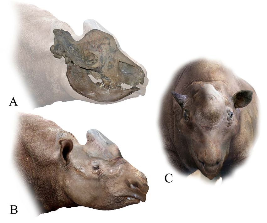
The head of Elasmotherium                                              175

Fig. 1. Skull of Elasmotherium sibiricum from Western Kazakhstan, Akzhar River, Akmola Region, coll. MZK. A — lateral
view, B — dorsal view, C — rostral view.

Sarepta (collection of the Zoological Institute RAS            descending below the level of the parietal bones. A solid
(ZIN), Saint Petersburg, No.10792), Verkhniy Kolyshley         bony septum divides the nasal cavity and the dome cavity
(Saratov Regional Local History Museum No. 8470),              into the right and left parts. The thickness of this septum
and Western Kazakhstan (Atyrau (formerly Guryev);              in the front reaches 32–33 mm, in the posterior direction
collection of the V.I. Vernadsky State Geological              it becomes thinner and in the skull’s dome it is 9.6 mm
Museum, Moscow (GGM) No.32-261 / PV-167).                      thick (in specimen from Atyrau).
    Due to the deficiency of data on detail investigation of       One of the most important distinguishing features
topographic anatomy of head and neck of rhinoceroses,          of the elasmotheriums’ skulls is the presence of a bony
we used the muscles’ terminology of this body’s part from      protuberance on the frontal bone, which occupies almost
the horse’s anatomy (Popesko, 1961). The measurement           the entire width of the dorsal part of the skull’s facial
were taken by Guerin (1980) and Shvyreva (2016).               part (except eye orbits). The dome is analogous to the
                                                               back horn's basis of the woolly rhinoceros. The size and
   Description                                                 shape of the bony protuberance are variable depending
                                                               on both sexual and age-related dimorphism. The type of
    A detailed description of the Elasmotherium skulls         the roughness of this part of the elasmotherium’s skulls
and vertebrae is given in some papers (for example,            is very variable. This may indicate differences in the
Svistun, 1973; Shvyreva, 2016). In this work, we focus         shape of the horn structure. The rugosity surface has
on those parts of the skull that allow us to reconstruct       hummocky profile with relatively uniform distribution of
the features of the elasmotherium head.                        bone tubercles (features by Hieronymus et al., 2009). An
    Skulls are large and elongated. The length of the          evident ring-shaped distribution of rugose’s elements at
skulls of E. sibiricum males (from the localities of Sarep-    the bone dome is not observed. Neurovascular foramens
ta, Zelenokumsk, Atyrau, Akzhar) reaches 86–89 cm.             on the dome surface are no detected. The transition of ru-
The ratio of skull width to length is less than 50%. The       gosity to adjacent bone surfaces is inequality at different
width at lateral occipital tubers reaches 237–380 mm,          part of the dome. At the anterior and lateral surfaces of a
and the width of a skull at the level of mastoid bones —       dome it has raised edge, and it is smooth at the posterior
298–486 mm (Shvyreva, 2016; our data). The facial part         part on the transition to parietal bones. This feature is
of the skull is longer than the cerebral one. The nasal        also variable. For example, at the skull from Akzhar this
bones are long, narrow, tapering anteriorly and rostrally      raised edges are obviously developed, but at the most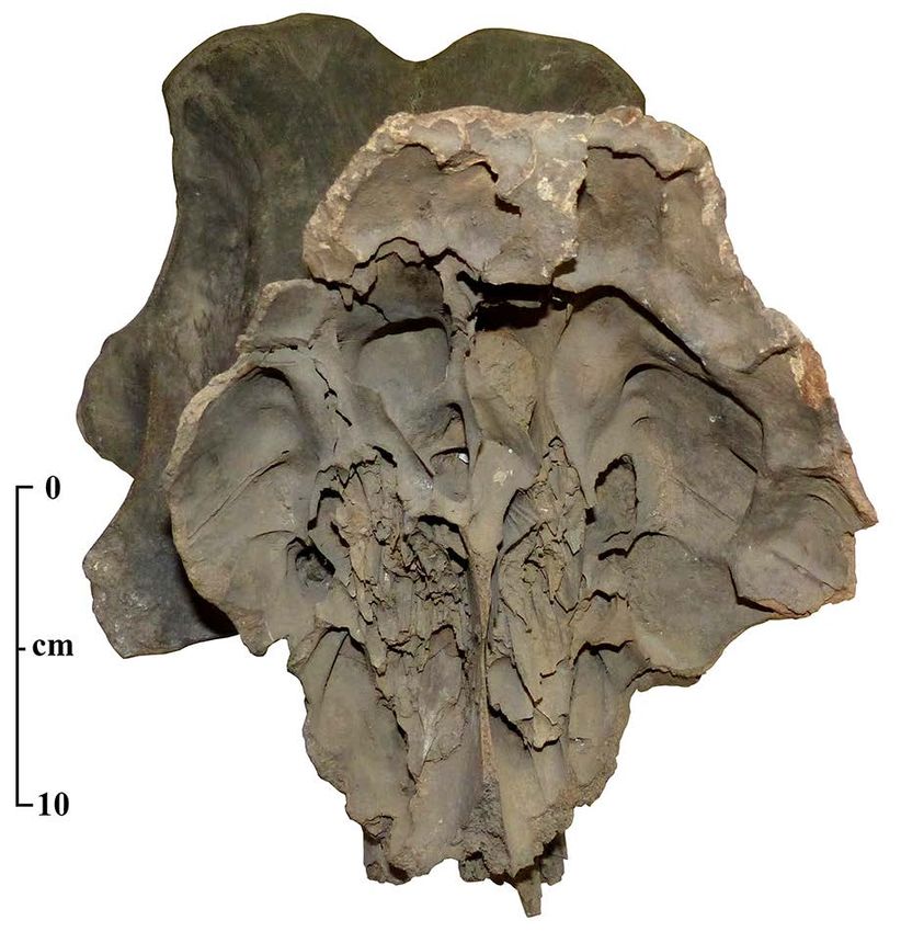
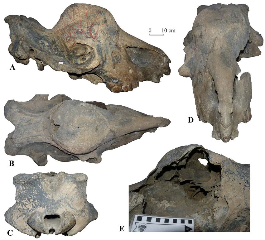
176                                               Vadim V. Titov et al.

Fig. 2. Skull of Elasmotherium sibiricum from Western Kazakhstan, Atyrau, coll. GGM No.32-261/PV-167. A — lateral view,
B — dorsal view, C — posterior view, D — anterior view, E — dome’s inner part.

part of specimens (from Atyrau and Lower Volga River               The bony protuberance’s diameter at the base ranges
Region’s localities) it is weakly marked. On some finds        from 25 to 35 cm (depending on the skull’s size). The
(for example, on specimen from Akzhar), the roughness          thickness of the frontal bone of the dome is 8–9 mm
is very markable, while on other skulls, even with large       from anterior, 6–13.5 mm from the lateral (on average,
protuberances, which are attributed to males, the surface      8.5 mm) and 5–16 mm from posterior sides. Only when
has moderate irregularities. On the female skull from the      the frontal bone passes to the nasal, lacrimal and parietal
PIN collection only slight roughness is visible on the         bones its thickness increases.
dome. The roughness on the anterior surface of the dome            There are grooves sulci on lateral and partially on
is less developed than on the dorsal and posterior parts.      dorsal and posterior surfaces of the dome, it allows to
In many specimens, the lower border of the roughness is        restore the largest arteries of the domed protuberance
clearly visible, it is slightly higher than the dome’s base    on the skull. In whole, nevrovascular grooves are sparse
in anterior and lateral sides, and in the posterior part       and its orientation is anastomosing. The main blood
extends to the anterior portion of the parietal bones. In      supply to this part of the skull was carried out by the
some finds diagonal grooves are clearly visible on the         dorsal nasal artery arteria dorsalis nasi, which is divided
aboral part of the place of attachment of the horn struc-      into branches — the anterior ramus anterior and the
ture in the region of the border of the frontal and parietal   posterior ramus posterior at the orbit’s upper edge level.
bones, they converge medially with the posterior ends.         The anterior one approaches the dome’s anterior base.
The head of Elasmotherium                                                   177

A vessel branche forward from it, going to the posterior
edge of the nasal notch. Posterior branch a. dorsalis
nasi is subdivided into 3–6 large arteries (their number
may vary in different individuals) feeding the lateral,
dorsal and posterior parts of the dome. Grooves from
large vessels are noted not on all specimens at the top
of the protuberance.
    The rostral part of the nasal and premaxillary bones
has a well-pronounced roughness, often equipped with
a rounded hook-shaped outgrowth. The width of the
anterior part of nasal bones is rather small and reaches
48–49 mm. The shape of this platform for the attachment
of the horn-like cornified pad is very variable on all
5 known finds with this part of the skull preserved
(Fig. 3). It indicates the variability of the nasal terminal
horn structure. The nasomaxillary notche incisura
nasomaxillaris are high and deep, it indicates a presence
of wide and mobile nostrils.
    The partially destroyed lateral wall of the dome
on some finds (in particular, on the skull from Atyrau)
makes it possible to clearly see a structure of the nasal
cavity and the cavity of the dome (Fig. 2E). Inside the
domed protuberance, there are cellular thin-walled
bony outgrowths formed by the frontal concha sinuses           Fig. 3. The shape of the rostral part of the elasmotheriums
conchalis dorsalis and the maxillary sinuses sinus             skulls: A — Atyrau (coll. GGM No. 32-261 / PV-167), B —
maxillaries in anterior, dorsal and lateral parts, as well     Novouzensk (coll. MM No.57/357), C — Novouzensk (coll.
as by the labyrinth of the ethmoid bone ethmoturbinalia        MM No.66/357); D — Akzhar (coll. MZK), E — Sarepta (coll.
in the posterior one (Figs. 4, 5). They form a single          NHM No. PV M-12429).
wide-meshed structure, which significantly increased
the surface of the greatly enlarged nasal cavity and,
accordingly, the mucous membranes. It should be noted
that these internal outgrowths did not form a continuous
structure with internal septa, which could increase the
strength of the bony dome, as can be observed, for
example, in the cavities of the cerebral part of the skull
of elephants.
    The eye orbit is limited by well-defined ridges of
frontal, lacrimal and zygomatic bones at the front,
above and below. Especially the orbit’s anterior sides
are limited by powerful outgrowths. The orbit is not
closed from behind.
    The parietal bones have a noticeable concavity and
rise considerably towards the occipital ridge. The lateral
parietal ridges are well expressed. They are almost
parallel to each other in the anterior half, but diverge       Fig. 4. Sagittal section of the skull of Elasmotherium sibiricum
significantly in the transition to the occipital part.         from the coll. ZIN (by Shvyreva, 2016).
    The occipital part is low and wide. The upper part
of the occipital bone bifurcates, forming two powerful
lateral occipital tubers, which hang over the occipital
plane and extend beyond the level of the occipital             has transverse processes up to 30 cm long, and has a
condyles. The incisures in the occipital ridge is deep.        range of wings up to 70 cm and exceeds the width of the
On its upper edge, the place of nuchal ligament’s              skull. An atlas is aproximately twice wider the width of
attachment is well expressed. There is a well-defined          the skull at lateral occipital tubers and mastoid bones.
roughness for a muscle attachment on the occipital             The second vertebra (epistropheus) has an elongated
bone’s nuchal surfaces. The mastoid processes of               body with a well-defined keel on the ventral surface,
the temporal bone processus mastoideus are highly              on the sides of which there are deep depressions. Other
developed and often protrude beyond the level of the           cervical vertebrae have high spinous processes, the length
zygomatic arch and orbit.                                      of which increases from the third to the seventh. Long
    The cervical vertebrae of Elasmotherium have a             narrow flattened lateral processes are distinguished on
number of structural features. The first of them (atlas)       the sixth cervical vertebra (Shvyreva, 2016).
178 Vadim V. Titov et al.
of the dorsal raphe of the strongly developed nuchal
occipital ligament ligamentum nuchae. The medial fibers
of the trapezius muscle and the intertwined tendons of
the splenius muscle of the head musculus splenius capitis
and the rhomboid minor m. rhomboideus are attached
to it. The nuchal ligament helps to hold the heavy head
and less actively use the muscles of the neck and back,
connecting the head’s back and the spinous processes of
all cervical vertebrae. It is required for running animals,
and accommodates the lowered head in the grazing
position. Bundles of the splenius muscle m. splenius
capitis extend from the tendinous bundles of the nuchal
funiculus attached to the nuchal surfaces under the
occipital lateral tubers.
The occipital bone’s nuchal surfaces, on which
the roughness is expressed, are also the place of
m. semispinalis capitis head semispinal muscle’s
attachment, which connects the skull with the transverse
processes of three-four posterior cervical vertebrae and
five anterior thoracic vertebrae. The upper fasciculus
of splenius muscle m. splenius capitis is attached to the
well-developed lateral tubers of the occipital crest. It
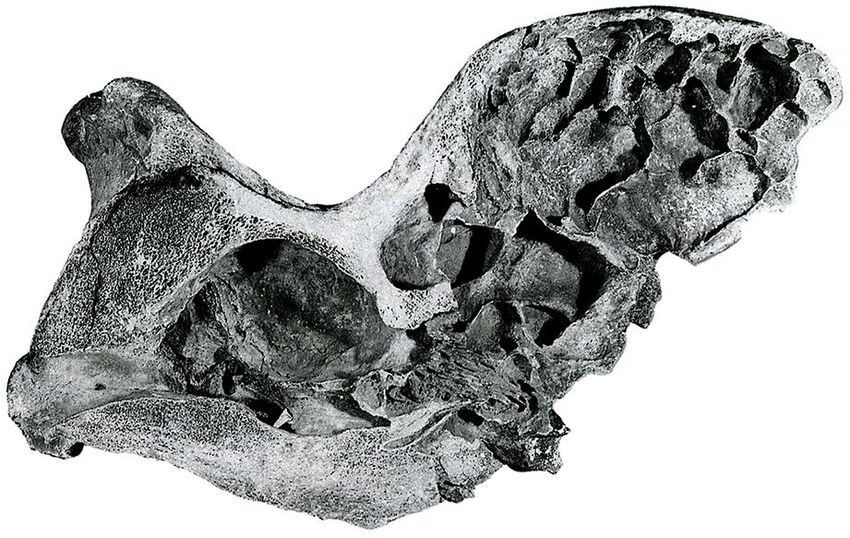
Fig. 5. Transversal section of the skull of subadult individual
connects the occiput with the transverse processes of
of Elasmotherium sibiricum at the level of the posterior part of
the first five cervical vertebrae, including the alas wings.
the bony dome, coll. Saratov Regional Local History Museum
Here, the cranial oblique muscle of head m. obliquus
No. 8470.
capitis cranialis fasten, which originates at the cranial
edge of the atlas wing. It is possible that the semispinal
muscles of the head m. semispinalis capitis, partially
Discussion attached to the lateral parts of the occipital crest, too.
These portions of the muscles facilitated to the spinal
The whole skulls, used for the Elasmotherium head’s column upper part extension and also pulled the head
external appearance reconstruction, the accumulated data back, keeping it in the overturned position.
and conclusions of other researchers, makes it possible Elasmotherium has well-defined mastoid processes of
to make some changes to the existing reconstructions. the temporal bone processus mastoideus. They suppose
The point of view of the present paper’s authors a significant development of dorsal band of cervical
largely coincides with the views of Flerov (1953) muscles: one of the tendinous branches of the longissimus
and I.A. Dubrovo and V.D. Kolganоv (exposition muscle of a head m. longissimus capitis as well as the head
of PIN; Zhegallo et al., 2005), on the habitus of this part of the splenius muscle m. splenius, which go to the
animal. We consider a number of ideas of V.A. Teryaev transverse processes of the cervical vertebrae, and also
and V.A. Vatagin (by Teryaev, 1948) concerning participates in head’s lateral movements.
Elasmotherium head’s structural features are deserved In general, the well-developed lateral muscles of the
attention. neck medial and lateral groups allowed the animal to
perform head’s powerful movements in the lateral and
The muscular system dorsolateral directions in addition to the conventional
The skull’s morphology and large muscles insertions dorsoventral ones. In elasmotheriums there is an increase
on the skull allow us to restore the head and neck’s in the arm of the interaction lever of the first cervical
large muscles’ development. Reconstruction of muscles vertebra and the skull (occipital tubers and mastoid
entire set development degree in this part of the body bones) in the lateral plane compared to other rhinos. For
is not possible at this stage of the study. Dividing of the example, the width of the atlas of Coelodonta antiquitatis
elasmotheriums’ occipital ridge and separation of the is 319–391 cm (Garutt, 1998), which is on average almost
nuchal areas laterally causes some different development twice less than that of Elasmotherium sibiricum.
of the ligaments and muscles connecting the skull with
the cervical and anterior thoracic vertebrae. Keratoid covering of the dome
Some semblance of a bifurcated occipital ridge into We think, that the Elasmotherium’s dome was
two lateral tubers in Elasmotherium is observed in wild covered with a keratoid substance that protects this
boars, they often break surface layer of the greensward thin-walled part of the skull, which is vitally important
when searching food. On pigs’ skulls, the lateral parts for animals.
of occipital ridge are also deflected backward. The well- To search for an analogy of the characteristics of
pronounced rugosity in the upper part of the incisure in this structure at Elasmotherium, it is logical to look for
the occiput of Elasmotherium indicates the attachment it among other rhinos. In modern representatives of theThe head of Elasmotherium 179
Rhinocerotidae, horns are a cornified papillary epidermal active blood supply of the terminal part in area of core
appendages. It is strongly convergent with similar tissues, intensive growth. The burr represented by a tubercles’
such as ungulate hoof wall and bovid artiodactyls horns ring on the core, is located at the place of the keratoid
(Hieronymus et al., 2006). As mentioned above, the substance formation. This burr development is more
divergence of the lineage of Elasmotheriinae and other noticeable on adult males’ horncores. It is poorly
Rhinocerotidae occurred at the end of the Paleogene developed in young males and females. With age, a
(30–40 million years ago). In Rhinocerotinae, the first number of vascular apertures in a core decreases in the
appearance of derived dermal support of the epidermal proximal part of a horn. In very old animals’ bone cores’
horns is attributed only to the early Miocene (16–20 Ma) growth in length stops and their partial resorption occurs.
(Hieronymus, 2009). Therefore, it is possible that the A number of vascular apertures decreases, and the holes
nature of the keratinized cover of the skull’s dome of and channels in the core are closed. The keratoid substance
Elasmotherium differed from that of other rhinos. after the stopping of growth on the horn surface, begins to
According to Hieronymus (2009) the presence of an chap and peel off, almost does not regenerate. The horn
annular (ring-shaped) distribution of rugose bone is one sheaths in bovids does not correspond to the bone cores
of the main indicators of a keratinized horns’ presence in length. For example, bison’s horny sheaths are longer
on the skull’s bones of rhinos. However, our analysis of than their horncores. The ones of adult animals can be
the character of rugosities at the places of attachment longer on average by 1/3 of their bony base (Sokolov,
of the horns on the skulls of C. antiquitatis shows that 1979: pp. 31–32).
the obvious ring character is not always observed, The presence of varying degrees of rugosity’s rate
especially on the same of females and young animals. on the surface of the bone’s dome in Elasmotherium
Here we can observe rather the radial ordering of bone indicates an individual, age and sexual variability of
exostoses, which often coincides with the presence of the cornified pad. We can assume that in young, very
bilateral symmetry of the pattern. In general, at known old individuals and females, the dome was covered with
taxa of rhinos with a clear presence of nasal epidermal a relatively thin keratin layer. It is logical to assume a
horns there is a compact area on the nasal bones with greater degree of development of this structure in adult
a relatively ordered arrangement of elements and often males. The presence of diagonal grooves on the aboral
with annular rugosities. Horn’s basis is a fairly dense part of the rugosity surface, partially extending from the
layer of bone. The more developed horn correlates with back of the dome-shaped protuberance to the parietal
more pronounced relief of the rugosity (for example, as bones, may indicate that the upper end of the more
in the male of woolly rhinoceros). developed pad was displaced dorso-aborally. Indirect
There are differences in the places of Elasmotheriums’ evidence of the presence of a relatively thin keratin
"horns" attachment and horned rhinoceros. The absence layer of the dome is a trace of intravital damage on the
of a clear annular or radial structure of rugosity on the anterio-lateral surface of the dome at the skull from Atyrau
elasmotherium skull, together with rather thin walls of (Fig. 1). This hole with traces of overgrowth is interpreted
the dome, indicates the absence of a epidermal horn on as a injury’s consequence (Zhegallo et al., 2005).
frontal bones. It should be noted that the presence of
individual variability of the of rugosity’s appearance Bone dome
and its certain orientation, together with a highly The Elasmotherium dome surface is covered
developed circulatory system, indicates that an epidermal with roughness. But the degree of its intensity can
integumentary pad but not dermal armor was appeared vary individually, as, for example, at the attachment
here. A comparative similarity of the rugose structure is points of horns in woolly rhinoceros. But, at the same
observed on the frontal boss of african buffalo Syncerus time, the degree of development of this roughness in
caffer, and muskox Ovibos moschatus. In both cases, Elasmotherium is never as strong as, for example, in
the bone that supports the heavily cornified pad of woolly rhinoceros. The dome cavities, being a widening
epidermis is highly vascular and have an uneven surface of the nasal cavity, in addition to enhancing the sense
(Hieronymus et al., 2009). The difference is observed of smell (as pointed out by many researchers of
in the presence of rounded knobs on the skull bones of Elasmotherium) could also function as sounds amplifier.
Elasmotherium, but not depressions. We observe that the dome greatest development occurs
Some analogy of the growth and development of for adult males. On young animals’ skulls and on the
such a structure can be found in bovids. It is known skull from the collection of the PIN, which is attributed
that in Bovidae, for example, in representatives of the to the female (Zhegallo et al., 2005; Shvyreva, 2016),
genus Bison, horny sheaths grow on horncores (bony the dome cavity is not so hypertrophied. Probably, a
outgrowths of the frontal bones) gradually, shifting from relatively small widening of this cavity was enough
the base to the end of the horn. It is known that in modern for elasmotherium for normal life. For males, to attract
rhinos, horn also grows from base (Hieronymus, 2006; females and control of the territory, it was necessary to
Hieronymus et al., 2006). For this purpose, furrows reproduce louder sounds, which were amplified with the
and ridges are formed on them, along which the sheaths help of a volumetric dome. In addition to protecting the
growing at the base is shifted. Numerous blood vessels thin-walled bone dome from mechanical influences, the
pass through the cavities in the horncore. They go out and overgrown keratoid pad in Elasmotherium could also
lie in the canals on the distal half of the horn, providing serve to protect the neck from the attack of predators.180 Vadim V. Titov et al.
Rostral part of the skull more researchers adhere to the point of view that
The presence of a solid internasal septum in underground parts of plants were the nutrition basis for
Elasmotherium suggests significant loads at the muzzle Elasmotherium. It is a food resource poorly consumed
end. Despite the fact that recent rhinos are also capable by other animal species. In contrast to the opinion of
of producing significant effects with the nasal horns, their Teryaev (1948) and Zhegallo et al. (2005), we believe
internasal septum remains cartilaginous throughout their that these animals harvested bulbs, tubers, or rootstocks
life (Garutt, 1998). A similar development of internasal not so much in shallow parts of water bodies as in other
septum is characteristic for woolly rhinoceros, which had biocenoses. Steppes, forests and meadows are the phyto-
a large anterior horn, used as well as for shoveling snow communities rich by ephemeroids (perennial herbaceous
and an upper layer of greensward. At the anterior end of plants with underground succulent organs). Probably,
the nasal and intermaxillary bones of the Elasmotherium, seasonality in the change of places for obtaining food
there are clearly visible thickenings with a rough surface, was characteristic for elasmotheriums. It is also possible
which suggest the attachment of small and narrow horn that the diet of these animals included aboveground parts
formations to them. Taking into account the supposed of herbaceous plants and shrubs in smaller quantity.
active use of the end of the muzzle for raking and digging Based on the characteristics noted, the head
a soil to find underground parts of plants, it is possible of Elasmotherium was reconstructed (Fig. 6). The
that a horn was used for this purpose. In Teryaev (1948), skulls and mandibles of E. sibiricum were taken as a
on reconstructions the nasal horn is displaced dorsally basis. The pelage, which was present on the head of
rather high, like for recent rhinos. Brandt (1878b) representatives of this species most likely, was not taken
suggested the presence of a small nasal horn, located into account in the figures presented in order to detail
at the muzzle tip and representing a low keratoid plate. some morphological features. Taking into account that
However, on the Rashevsky’s engraving, supervised the roughness is much more developed on the posterior
by Brandt, such a horn almost did not discernible. The part of the bony dome and on the adjacent parts of the
authors know about five skulls with intact rostral part parietal bones, and the posterior portion of the blood
(Fig. 3), their analysis allows us to clarify the anterior vessels on the dome is more pronounced, we assume
horn-like conformation’s position. The development of that the keratoid substance was more developed precisely
variable in shape thickenings and rounded hook-shaped on the posterior half of the dome-shaped protuberance.
bony outgrowths on the rostral part of the intermaxillary Probably, the uneven growth of the keratoid substance
bones suggests the location here of a small terminal horn- led to the displacement of the apex of the horny sheaths
like structure. The nasal bones tip, slightly protruding in the dorsal-aboral direction.
above the intermaxillary bones, probably acted as an
upper stop for this “horn”. Conclusions
There are no annular distribution of rugose bone on
the anterior part of the nasal bones and on the terminal Representatives of the genus Elasmotherium have
part of the internasal septum. This indicates the absence no analogues in the modern fauna. Elasmotheriinae
of true epidermal horns. Roughness on the anterior part of separated from other branches of the Rhinocerotidae
the nasal bones indicates the presence of a cornified pad family more than 30 million years ago. During this period
or cornified sheath, as well as for a keratinized covering of evolution they acquired a number of morphological
of the dome at the frontal bones. Taking into account and physiological features. Therefore, during the
the different degree of the rugouse area development reconstructing the external appearance of elasmotherium,
and the presence of bony “hooks” on the anterior edge one cannot fully rely on that of recent rhinoceroses
of the internasal septum, we can assume the variable of or other well-studied fossil taxa (for example, woolly
shape and size of this terminal cornified pad, regardless rhinoceros). Both the late Middle Pleistocene Siberian
of gender. The presence of pronounced bony “hooks” elasmotheriums and Early Pleistocene Caucasian ones
(for example, on the skull from Atyrau) suggests that had a wedge-shaped form of the anterior part of the
they were a support for the horn-like structure of the skull with a small "horn" at the very end of the muzzle,
cornified pad in adult individuals. well-defined anterior outgrowths of the orbits, hypsodont
The presence of roughness on the nasal bone anterior teeth, large olfactory lobes of the brain, hypertrophic
part and the rostral part of the internasal septum also nasal cavities, a highly developed sense of smell, and
suggests the presence of a movable fleshy upper lip, significant development of the lateral muscles of the
which is necessary for digging out tubers and rootstocks. neck. We adhere to the point of view of researchers
who believed that the characteristics of the skulls and
Food base necks of elasmotheriums indicate adaptations to digging
Teeth of elasmotheriums have a sidewall hypsodonty out from the soil and feeding by underground parts of
with the highest crown height among ungulates. On plants. Bulbs, tubers, and rootstocks of perennial plants in
the permanent teeth of Elasmotherium sibiricum roots steppes, meadows, and forest areas probably constituted
did not formed at all, even on premolars, and in the a significant part of the diet of these large animals, which
Early Pleistocene E. caucasicum roots on M2 and have occupied an unoccupied ecological niche. This
M3 are unknown also. Such hypsodont teeth suggests allowed the Elasmotherium to exist from the end of
intake of a highly abrasive food. Recently, more and the Pliocene to the late Pleistocene without any specialThe head of Elasmotherium 181
Fig. 6. Reconstruction of the head of male of Elasmotherium. Author R.S. Uchitel’.
morphological rearrangements. Late Elasmotherium the bones surface contributed to the growth and partial
differed from the early Pleistocene forms only by slightly renewal of this formation. The different degrees of
smaller sizes, minor changes in the dentition, and, most roughness intensity on the surface of the dome indicates
likely, in the presence of a coat. that the keratoid cover was growing to a greater extent
One of the main distinguishing features of these for males, while this process was slower for females,
distinctive rhinos is a dome-shaped bony protuberance juvenile and old individuals.
on the frontal bones, which causes major controversy A solid internasal septum indicates a significant load
among scientists and restorers. The thin walls of the bony on the end of the muzzle. The presence of outgrowths
dome in anterior, lateral and dorsal sides have an average and roughness on the rostral parts of the nasal and
thickness of about 1 cm. The wide-meshed structure intermaxillary bones suggests a presence of a terminal
inside the dome, formed by overgrown thin-walled horn-like structure of cornified pad and a fleshy upper
bony outgrowths of the frontal concha and maxillary lip, which participated in the process of the soil hoeing
sinuses, as well as the labyrinth of the ethmoid bone is and selecting of food objects.
not a structure of strengthening of the dome strength. The significant development of the atlas transverse
This makes it possible to assume that the dome was processes, the high spinous processes of other cervical
not a place of attachment of a large horn or any other vertebrae, as well as the massive mastoid processes of the
structure, but was covered with a relatively thin keratoid temporal bone, the bifurcation of the occipital crest into
substance, which mainly performs a passive protective two lateral halves indicate a presence of powerful lateral
function for fragile bones. The dome inner surface was neck muscles that carry out lateral and dorsolateral head
an overgrown nasal cavity, which served to intensify a movements in addition to the conventional dorsoventral
sense of smell, and, possibly, to amplify sounds emitted. ones. The development of such a muscular system
The cornified pad grew from the clearly visible base in confirms the adaptability of Elasmotherium to raking
the lower part of the bony dome, and its upper end was the upper soil layer.
displaced dorso-aborally. The blood vessels located on182 Vadim V. Titov et al.
ACKNOWLEDGEMENTS. The authors are grateful Kosintsev P., Mitchell K.J., Devièse Th., Plicht J. van der,
to the staff of the V.I. Vernadsky State Geological Kuitems M., Petrova E., Tikhonov A., Higham Th.,
Museum (I.A. Starodubtseva), Institute of Zoology Comeskey D., Turney C., Cooper A., Kolfschoten Th. van,
of Ministry of Education and Science of Republic of Stuart A.J. & Lister A.M. 2019. Evolution and extinction
Kazakhstan (P.A. Tleuberdina), and Saratov Regional of the giant rhinoceros Elasmotherium sibiricum sheds
Local History Museum (A.V. Biriukov) for the light on late Quaternary megafaunal extinctions // Nature
opportunity to work with the material. We are thankful for Ecology & Evolution. Vol.3. P.31–38.
Dr. P.-O. Antoine and anonymous reviewer for the helpful Kožamkulova B.S. 1981. Elasmotherium sibiricurn und sem
remarks and recommendations for the improvement of Verbreitungsgebiet auf dem Territorium der UdSSR //
the manuscript. The study was supported by the Russian Quartärpaläontologie. No.4. P.85–91.
Science Foundation, project No. 16-17-10170-P. Mazza P. & Azzarolli A. 1993. Ethological inferences on
Pleistocene rhinoceroses of Europe // Rendiconti Lincei.
Scienze Fisiche e Naturali. S.9. Vol.4. P.127–137.
References Popesko P. 1961. [Atlas of Topographic Anatomy of Farm
Antoine P.O. 2002. Phylogénie et évolution des Elasmotheriina Animals. Vol.1. Head and Neck]. Bratislava: Slovak
(Mammalia, Rhinocerotidae) // Mémoires du Muséum Agricultural Literature Publishing House. 215 p. [in Russian].
National d’Histoire Naturelle. Vol.188. P.1–359. Rivals F., Prilepskaya N.E., Belyaev R.I. & Pervushov E.M.
Baigusheva V.S., Timonina G.I. & Titov V.V. 2011. [Some 2020. Dramatic change in the diet of a late Pleistocene
characteristics of the functioning and change of teeth of the Elasmotherium population during its last days of life:
Caucasian elasmotherium Elasmotherium caucasicum] // Implications for its catastrophic mortality in the Saratov
[Zoological Research for 20 years of Independence of the region of Russia // Palaeogeography, Palaeoclimatology,
Republic of Kazakhstan. Materials of International Confer- Palaeoecology. Vol.556. P.e109898.
ence]. Almaty: Institute of Zoology. P.304–305 [in Russian]. Sokolov V.E. (ed.). 1979. [European bison. Morphology,
Baigusheva V.S., Titov V.V. & Timonina G.I. 2018. [Problems of systematic, evolution, ecology]. Moscow: Nauka. 496 p.
species diagnosis of elasmotheriums (Rhinocerotidae, Elas- [in Russian].
motheriinae)] // [Fundamental and Applied Paleontology. Ma- Solounias N. & Semprebon G. 2002. Advances in the
terials of 64 Session of Paleontological Society of RAS]. Saint reconstruction of ungulate ecomorphology with application
Petersbourg: Kartofabrika VSEGEI. P.171–173 [in Russian]. to early fossil equids // American Museum Novitates.
Brandt A.F. 1878a. Milleilungen uber die Gattung No.3366. P.1–49.
Elasmotherium besonders den Schaedelbau derselben // Shvyreva A.K. 2016. [Elasmotheriums of Pleistocene of Eurasia].
Mémoires de l’Académie Impériale des Scences de St.- Stavropol: Pechatniy Dvor Publishing. 218 p. [in Russian].
Pétersbourg, VII series. Vol.26. No.6. P.1–36. Svistun V.I. 1973. [The skull of Caucasian elasmotherium
Brandt A. 1878b. [Elasmotherium (fossil rhinoceros)] // Niva. (Elasmotherium caucasicum Boriss.) from Late Pliocene
No.23. P.411–415 [in Russian]. deposits of Zaporozh’e Region] // Vestnik Zoologii. No.2.
Deng T., Wang S. & Hou S. 2013. A bizarre tandem-horned P.53–60 [in Russian].
elasmothere rhino from the Late Miocene of northwestern Sun D., Deng T. & Jiangzuo Q. 2021. The most primitive
China and origin of the true elasmothere // Chinese Science Elasmotherium (Perissodactyla, Rhinocerotidae) from the
Bulletin. No.58. P.1811–1817. Late Miocene of northern China // Historical biology, DOI:
Flerov K.K. 1953. [Unicorn — elasmotherium] // Priroda. 10.1080/08912963.2021.1907368
No.9. P.110–112 [in Russian]. Teryaev V.A. 1948. [Geological position of dome-forehead
Garutt N.V. 1998. [Wolly Rhinoceros (Morphology, Systematic, rhinoceros (elasmotherium)] // Sovetskaya Geologiya.
Geological Significance)]. PhD Thesis. Saint Petersburg: No.34. P.81–89 [in Russian].
G.V. Plekhanov Saint Petersburg State institute of mines. Titov V.V. 2008. [Late Pliocene Large Mammals from
247 p. [in Russian]. Northeastern Sea of Azov Region]. Rostov-on-Don:
Guerin C. 1980. Les rhinoceros (Mammalia, Perissodactyla) du Southern Scientific Centre RAS Publishing. 262 p. [in
miocene terminal au pleistocene superieur en Europe Occi- Russian, with English summary].
dentale. Comparaison avec les especes actuelles // Documents Tleuberdina P. & Nazymbetova G. 2010. Distribution of
des Laboratories de Geologie Lyon. No.79. Fasc.1. P.1–421. Elasmotherium in Kazakhstan // Quaternary Stratigraphy
Hieronymus T.L. 2009. Biological Sciences Osteological and Paleontology of the Southern Russia: Connections
Correlates of Cephalic Skin Structures in Amniota: between Europe, Africa and Asia. Abstracts of 2010
Documenting the Evolution of Display and Feeding Annual Meeting INQUA-SEQS. Rostov-on-Don: Southern
Structures with Fossil Data. PhD Thesis. Athens: College Scientific Centre RAS Publishing. P.171–173.
of Arts and Sciences of Ohio University, USA. 254 p. Zhegallo V.I. & Noskova Y.G. 2001. [Several morphological
Hieronymus T.L., Witmer L.M. & Ridgely R.C. 2006. and functional features of skeleton of rhinoceros
Structure of white rhinoceros (Ceratotherium simum) horn Elasmotherium and its ecological interpretation] // Bulleten
investigated by X-ray computed tomography and histology Moskovskogo Obshchestva Ispytateley Prirody, Otdel
with implications for growth and external form // Journal Geologicheskiy. Vol.76. No.6. P.63–69 [in Russian].
of Morphology. Vol.267. P.1172–1176. Zhegallo V., Kalandadze N., Shapovalov A., Bessudnova Z.,
Hieronymus T.L., Witmer L.M., Tanke D.H. & Currie Ph.J. Noskova N. & Tesakova E. 2005. On the fossil rhinoceros
2009. The facial integument of centrosaurine ceratopsids: Elasmotherium (including the collections of the Russian
morphological and histological correlates of novel skin Academy of Sciences) // Cranium. Vol.22. No.1. P.1–40.
structures // The Anatomical Record. Vol.292. P.1370–1396.You can also read