Tumor Microenvironment of Esophageal Cancer - MDPI
←
→
Page content transcription
If your browser does not render page correctly, please read the page content below
cancers
Review
Tumor Microenvironment of Esophageal Cancer
Lars M. Schiffmann † , Patrick S. Plum † , Hans F. Fuchs                          , Benjamin Babic         , Christiane J. Bruns
and Thomas Schmidt *

                                             Department of General, Visceral, Cancer and Transplantation Surgery, Faculty of Medicine with University
                                             Hospital Cologne, University of Cologne, 50931 Cologne, Germany; lars.schiffmann@uk-koeln.de (L.M.S.);
                                             patrick.plum@uk-koeln.de (P.S.P.); Hans.fuchs@uk-koeln.de (H.F.F.); benjamin.babic@uk-koeln.de (B.B.);
                                             christiane.bruns@uk-koeln.de (C.J.B.)
                                             * Correspondence: thomas.schmidt1@uk-koeln.de; Tel.: +49-221-478-4804; Fax: +49-221-478-6258
                                             † These authors contributed equally.

                                             Simple Summary: Esophageal cancer is one of the top ten most deadly cancers. Even when di-
                                             agnosed in a curable stage, patients prognosis poor. One of the parameters that is very relevant
                                             for long-term survival is response to radio(chemo)therapy prior surgery. Complete response rates
                                             are between 24 and 50 percent. This puts more than a half of every esophageal cancer patient that
                                             is diagnosed in a non-metastasized stage at high risk of recurrence. To improve response rates of
                                             treatment regimens prior curative surgery is, therefore, a major challenge in treating esophageal
                                             cancer. Not only the response of the cancer cell itself to cancer therapy is determining patients’ fate.
                                             Cells around the tumor cells called the tumor microenvironment that together with the cancer cell
                                             constitute a malignant tumor are also involved in tumor progression and therapy response. This re-
                                             view depicts the most important parts of the esophageal cancer microenvironment, evaluates chances
                                             and challenges of current already established therapeutic concepts that target this microenvironment.
                                   It furthermore elucidates specific pathways that are potential valuable targets in the future.


Citation: Schiffmann, L.M.; Plum,
                                             Abstract: Esophageal cancer is among the top ten most deadly cancers worldwide with adenocarcino-
P.S.; Fuchs, H.F.; Babic, B.; Bruns, C.J.;
                                             mas of the esophagus showing increasing incidences over the last years. The prognosis is determined
Schmidt, T. Tumor Microenvironment
                                             by tumor stage at diagnosis and in locally advanced stages by response to (radio-)chemotherapy fol-
of Esophageal Cancer. Cancers 2021,
                                             lowed by radical surgery. Less than a third of patients with esophageal adenocarcinomas completely
13, 4678. https://doi.org/10.3390/
cancers13184678                              respond to neoadjuvant therapies which urgently asks for further strategies to improve these rates.
                                             Aiming at the tumor microenvironment with novel targeted therapies can be one strategy to achieve
Academic Editor: Alexander Nikitin           this goal. This review connects experimental, translational, and clinical findings on each component
                                             of the esophageal cancer tumor microenvironment involving tumor angiogenesis, tumor-infiltrating
Received: 24 August 2021                     immune cells, such as macrophages, T-cells, myeloid-derived suppressor cells, and cancer-associated
Accepted: 16 September 2021                  fibroblasts. The review evaluates the current state of already approved concepts and depicts novel
Published: 18 September 2021                 potentially targetable pathways related to esophageal cancer tumor microenvironment.

Publisher’s Note: MDPI stays neutral         Keywords: esophageal cancer; esophageal adenocarcinoma; esophageal squamous cell cancer;
with regard to jurisdictional claims in      tumor microenvironment; cancer-associated fibroblasts; tumor angiogenesis; tumor-associated
published maps and institutional affil-
                                             macrophages; immunotherapy
iations.

                                             1. Introduction
Copyright: © 2021 by the authors.
                                                   Esophageal cancer is among the top ten most common and most deadly cancers
Licensee MDPI, Basel, Switzerland.
                                             worldwide [1,2]. Concerning the western world, the incidence of esophageal squamous
This article is an open access article
                                             cell cancer (ESCC) is decreasing while the occurrence of esophageal adenocarcinomas
distributed under the terms and
conditions of the Creative Commons
                                             including adenocarcinomas of the esophagogastric junction (EAC) is rapidly increasing,
Attribution (CC BY) license (https://
                                             which overall significantly accelerated the incidence of the disease. Mostly detected at
creativecommons.org/licenses/by/
                                             locally advanced or already metastasized stages the prognosis is still detrimental in many
4.0/).                                       cases though great advances in (multimodal) treatment strategies have been achieved over

Cancers 2021, 13, 4678. https://doi.org/10.3390/cancers13184678                                                 https://www.mdpi.com/journal/cancers
Cancers 2021, 13, 4678                                                                                         2 of 14

                         the last decades. Detected in a non-metastasized stage, the multimodal treatment consists
                         of perioperative chemotherapy or neoadjuvant radiation and a radical oncologic surgical
                         therapy, except for early stage pT1 cases (which qualify for either endoscopic resection or
                         primary surgery). The overall survival in large randomized controlled trials examining the
                         value of neoadjuvant radiation before surgery or perioperative chemotherapy improved
                         to 24–50 months in the groups of multimodally treated patients, which lead to the im-
                         plementation of these treatment strategies in international and national guidelines [3,4].
                         Pathological complete response rates to chemoradiotherapy were 23% for EAC patients
                         and 49% for ESCC patients in the CROSS trial and 16% in the FLOT4 trial in adenocar-
                         cinomas of the esophagogastric junction [3,5]. Results from these studies showed that
                         response to neoadjuvant treatment is highly relevant for overall survival with complete
                         responders showing a 5-year survival rate of over 70% in contrast to poor responders
                         with at least 50% residual tumor in the specimen having a 5-year survival rate of around
                         20% [4]. Patients with adenocarcinoma of the esophagogastric junction were found to
                         have a significantly longer disease-free survival after pathological complete response after
                         preoperative chemotherapy [6].
                              This shows that one major challenge in the treatment of esophageal cancer at least
                         for locally advanced stages that are treated with curative intent is the improvement of
                         response rates. Although one strategy was the implementation of more intense conven-
                         tional chemotherapeutic regimens as in the FLOT protocol [7,8], another is to develop
                         new targeted therapies that complement conventional therapy [9,10]. To do so a deep
                         mechanistic understanding of each specific tumor entity is essential.
                              A malignant tumor is composed not only of tumor cells but to a large part of adjacent
                         non-malignant cells, the so-called tumor stroma. The interaction between tumor cells and
                         stromal cells, between stromal cells among themselves and between cells from different
                         areas of tumor tissues is described as the tumor microenvironment [11]. This article aims
                         to give an overview of the tumor microenvironment in esophageal cancer with a specific
                         focus on the mechanistic backgrounds that give the rationale for tumor microenvironment
                         targeted therapies.

                         2. Endothelial Cells and Tumor Angiogenesis
                               When a malignant lesion reaches a critical size, diffusion does not suffice to reach
                         the center of the tumor with oxygen and nutrients. The tumor core becomes hypoxic
                         which leads to the stabilization of hypoxia-inducible factor-1α (HIF-1α), a transcription
                         factor that then further induces the upregulation of several pro-angiogenic genes encoding
                         for cytokines and growth factors that mediate an angiogenic response. This process has
                         been described as the angiogenic switch. Until now vascular endothelial growth factor
                         (VEGF) is considered to be the most potent pro-angiogenic factor. VEGF-receptors, mainly
                         VEGFR2, with their downstream signaling pathways mediate proliferation, migration,
                         and cell survival in endothelial cells under physiologic and pathologic conditions [12].
                         Many drugs that target pro-angiogenic signaling cues have been developed in the last
                         2 decades with bevacizumab (bev) a monoclonal antibody against VEGF being the most
                         prominent one. Bevacizumab prolonged progression-free survival and overall survival in
                         patients with metastatic colorectal cancer in combination with conventional chemotherapy
                         compared to chemotherapy alone [13]. Bevacizumab became standard of care in many
                         tumor entities mostly in a palliative setting, but also in neoadjuvant treatment strategies,
                         e.g., for the multimodal treatment of ovarian cancer.
                               Experimentally in mice, anti-angiogenic therapies can impressively reduce tumor
                         vascularization, thereby ‘starving’ tumors to death, following Judah Folkman’s hypothesis,
                         which stimulated the field of angiogenesis research in the 1970s.
                               On a tumor histological and cellular level experimental work [14–17] could demon-
                         strate the mechanisms underlying the efficacy of anti-VEGF therapies is more complex than
                         initially believed. Anti-angiogenic therapy in patients does indeed reduce the number of
                         tumor vessels [18] but also reduces the tumor interstitial fluid pressure making the tumor
Cancers 2021, 13, 4678 3 of 14
more sensitive for drug uptake. The beneficial effect on tumor interstitial fluid is believed
to result from improved blood vessel structure and function due to VEGF withdrawal. This
phenomenon was summarized by Rakesh Jain as the vascular normalization theory [19].
In esophageal cancer, VEGF seems to play a pivotal role in tumor progression similar
to other entities. Shimada et al. examined VEGF serum levels of patients with esophageal
squamous cell cancer (ESCC) and found that serum levels were correlated with increased
tumor stage and prognosis [20]. Interestingly, high serum VEGF levels predicted poor
response to radiochemotherapy. These data suggest a strong rationale to pharmacologically
deplete serum VEGF levels in these patients; further translational and clinical research
should be done to evaluate anti-angiogenic treatment in combination with conventional
radiochemotherapy in this particular tumor entity. Surprisingly not a single clinical trial
involving anti-angiogenic treatment was conducted in ESCC following these promising
initial translational data. However, in esophageal adenocarcinoma and carcinoma of
the esophagogastric junction intense effort has been done to evaluate anti-angiogenic
treatments [21].
First, bevacizumab was tested in an international multi-center randomized trial (AV-
AGAST). This study compared capecitabine-cisplatin chemotherapy in conjunction with
bevacizumab as first-line treatment in patients with advanced gastric cancer [22]. Though
the study did not reach its primary endpoint, which was an improvement in overall sur-
vival, the addition of bevacizumab to capecitabine-cisplatin significantly improved median
progression-free survival (6.7 months vs. 5.3 months; hazard ratio, 0.80; p < 0.0037) and
overall response rate (46.0% vs. 37.4%; p = 0.0315). Overall, the AVAGAST trial underscored
the role of VEGF signaling and its therapeutic potential in advanced gastric cancer and
carcinomas of the esophagogastric junction, the latter representing 45 from 200 analyzed
patients in Europe [22]. Preplanned subgroup analysis within the AVAGAST trial sug-
gested that Asian patients do not benefit from the addition of bevacizumab to conventional
chemotherapy. This was confirmed by the AVATAR study which showed no effect of beva-
cizumab when added to capecitabine-cisplatin therapy compared to capecitabine-cisplatin
therapy plus placebo in Chinese patients [23]. Accordingly, there are potential ethical
differences in the sensitivity towards VEGF-blockade which might be a result of differences
concerning tumor immunological gene expression signature [24]. It is further notewor-
thy that both, in the AVAGAST trial and the AVATAR study the majority of included
patients suffered from gastric cancer with significantly fewer patients with carcinomas of
the esophagogastric junction being randomized. This is a potential bias in both studies
which probably leads to the underestimation of beneficial effects of VEGF inhibition in
esophagogastric junction tumors.
Accordingly, it was not surprising that bevacizumab failed to improve survival out-
comes in patients with resectable esophagogastric adenocarcinoma patients that were
randomized to receive peri-operative epirubicin, cisplatin, and capecitabine chemotherapy
or chemotherapy plus bevacizumab prior to surgery [25]. The patients included in this
study consisted of around a third of patients with gastric cancer supporting the hypothesis
that tumor location has potential relevance for the efficacy of VEGF inhibition in gastro-
esophageal adenocarcinoma. These results claim for further examination as subgroup
analysis concerning tumor location was inconsistent in between different types of Siewert
tumors with GEJ type III tumors showing a beneficial outcome after chemotherapy plus
bevacizumab and GEJ Type I and II tumors showing no difference [25].
In parallel ramucirumab, a fully humanized monoclonal antibody that counteracts
VEGF binding do VEGF-R2, entered clinical trials. The REGARD study investigating
the effect of ramucirumab as a monotherapy compared to placebo in previously treated
patients with advanced gastric cancer or adenocarcinomas of the esophagogastric junction
that showed progressive disease after first-line treatment. Ramucirumab significantly
prolonged overall survival in these patients and, therefore, is the first and until now only
anti-angiogenic agent that prolonged survival administered as a single agent without addi-
tional chemotherapy [26]. Additionally, ramucirumab was evaluated in a large multi-centerCancers 2021, 13, 4678 4 of 14
randomized phase 3 trial as second-line treatment in combination with paclitaxel [27].
Results from the RAINBOW study demonstrated an improvement in overall survival in
patients treated with ramucirumab plus paclitaxel compared to placebo plus paclitaxel
(median 9.6 months vs. 7.4 months; hazard ratio, 0.807; p = 0.017). These results together
lead to the implementation of ramucirumab in national and international guidelines for
the second-line treatment of advanced gastric and esophagogastric junctional adenocarci-
nomas [28].
One of the major unanswered questions or challenges in the field of tumor-related
angiogenesis research is, however, to identify mechanisms of response or resistance and to
develop suitable biomarkers based on these findings.
Tremendous efforts have been spent on uncovering mechanisms of escape and resis-
tance to anti-angiogenic treatments involving cellular, non-cellular, stromal, and tumor cell
inert mechanisms in other tumor entities than esophageal cancer [18,29–35]. To our knowl-
edge, no experimental work has been done specifically on esophageal or esophagogastric
cancer which might simply be related to the fact that there is a lack of suitable animal
models for this disease, especially for esophageal cancers. On a translational basis, serum
samples from the AVAGAST trial were evaluated for the potential role of angiopoetin-2
as a predictive biomarker for bevacizumab-containing therapies [36]. This hypothesis
was generated based on positive findings in colorectal cancer patients [37]. Other than
in colorectal cancer patients, angiopoietin-2 had no value in response to bevacizumab
concerning survival. However, high serum angiopoietin-2 levels are associated with a poor
survival outcome in patients randomized for the AVAGAST trial which again emphasizes
the role of angiogenesis in esophagogastric junction tumors [36,38]. Further research on
other angiogenesis mediating signaling pathways is needed to develop alternative or
complementary approaches to pharmacologically target tumor angiogenesis in esophageal
cancer [39].
In summary, inhibition of tumor angiogenesis with ramucirumab in (distal) esophageal
cancer is a valuable option in a palliative setting in combination with chemotherapy or is
administered as a single agent. The fact that ramucirumab is potently prolonging overall
survival, which bevacizumab was not able to achieve in clinical trials, points towards a
very interesting connection that warrants further investigation. Bevacizumab eliminates
serum and tissue VEGF by binding the growth factor itself. In theory, this does not only
inhibit VEGF effects that are mediated via VEGF-R2 on endothelial cells (Figure 1) but
also inhibits other signals that are mediated by other VEGF receptors, such as VEGF-R1
on endothelial cells, or other cells of the tumor microenvironment like tumor-infiltrating
immune cells like macrophages or monocytes. For example, VEGF-R1 on macrophages
was recently found as a crucial player in metastasis and progression. A high percentage of
VEGF-R1 positive immune cells within colorectal metastasis predicts worse outcomes in
patients [35]. It is known from endothelial cell sprout differentiation that VEGF-R2 and
VEGF-R1 are somehow reciprocally regulated meaning that VEGF-R1 signaling can limit
VEGF-R2 signaling by acting as a decoy receptor. Eliminating VEGF from the system could
interact with these processes in ways that are context dependently not always beneficial or
complex to predict in every oncological situation. These thoughts are certainly hypothetical
but might explain the differences in the efficacy of bevacizumab and ramucirumab.
Another open question is whether other promising novel anti-angiogenic approaches
that have been explored preclinically over the past decades are potent and tolerable enough
to enter the clinical application. Especially, concepts that target endothelial metabolism
produce promising experimental results but have to be further evaluated in early phase
clinical trials [40–45].Cancers 2021,
Cancers 13,13,4678
2021, x FOR PEER REVIEW 5 of 15 5 of 14
Figure
Figure 1. Scheme
1. Scheme depicting
depicting the angiogenic
the angiogenic switch. A
switch. A malignant malignant
tumor tumor secretes pro-angiogenic
secretes pro-angiogenic
growth
growth factors
factors thatthat stimulate
stimulate angiogenesis
angiogenesis by on
by acting acting on endothelial
endothelial cells with cells with pro-migratory,
pro-migratory, pro- prolifer-
liferative,
ative, andandanti-apoptotic,
anti-apoptotic, pro-survival
pro-survivalsignals.
signals.
Another open question is whether other promising novel anti-angiogenic approaches
3. Cancer-Associated Fibroblasts
that have been explored preclinically over the past decades are potent and tolerable
enoughFibroblasts
to enter the are cellsapplication.
clinical with spindle-formed morphology
Especially, concepts which
that target are easyme-
endothelial to identify but
are quite
tabolism indistinctly
produce promisingdefined on a molecular
experimental basis.
results but have to This makes
be further it difficult
evaluated to define these
in early
cellsclinical
phase in comparison to macrophages or endothelial cells for example. They are present
trials [40–45].
in every organ differing significantly in a site-specific manner [46]. Tumor- or cancer-
3.associated
Cancer-Associated Fibroblasts
fibroblasts (CAFs) show a highly activated state that is induced by growth
factors with transformingspindle-formed
Fibroblasts are cells with growth factormorphology
(TGF) as the which mostare prominent
easy to identifyonebutand expresses
are quite indistinctly defined on a molecular basis. This makes it difficult to define these
the highly contractable α smooth muscle actin (αSMA; also known as ACTA2) representing
cells in comparison to macrophages or endothelial cells for example. They are present in
a so-called “myofibroblastic” phenotype [46,47]. Additionally, this heterogenous intra-
every organ differing significantly in a site-specific manner [46]. Tumor- or cancer-associ-
tumoral
ated subpopulation
fibroblasts (CAFs) showcan promote
a highly angiogenesis
activated state that isby the production
induced of VEGF [48] and
by growth factors
with transforming growth factor (TGF) as the most prominent one and expresseshypoxic
are involved in metabolic reprogramming, as well as resistance towards the stress in
tumor
highly tissues [49].
contractable α smooth muscle actin (αSMA; also known as ACTA2) representing a
Since
so-called there is still phenotype
“myofibroblastic” a lack of [46,47].
specificAdditionally,
CAF markers during the clinical
this heterogenous intra-tu- routine, the
moral subpopulation differentiation
histopathological can promote angiogenesis
is done bybytheirthe production of VEGF [48]
typical spindle-like and and
shape are the absence
involved
of “classical” endothelial, epithelial, or leukocyte markers. Interestingly, thetu-
in metabolic reprogramming, as well as resistance towards hypoxic stress in exact origin of
mor tissues [49].
CAFs is also an ongoing debate. Most of CAFs are supposed to differentiate from normal
Since there is still a lack of specific CAF markers during the clinical routine, the his-
local fibroblasts at the tumor site during a process called “stromagenesis” resembling that
topathological differentiation is done by their typical spindle-like shape and the absence
ofthe cellularendothelial,
“classical” dysfunction not only
epithelial, or affects
leukocytethemarkers.
tumor cells but alsothe
Interestingly, theexact
stroma in which these
origin
cells are embedded [47,50]. However, CAFs may also develop
of CAFs is also an ongoing debate. Most of CAFs are supposed to differentiate from nor- from bone marrow-derived
mesenchymal
mal local fibroblasts stem
at the cells
tumor (MSCs) since
site during experimental
a process in vivo data resembling
called “stromagenesis” suggest the ability of
MSCs
that to change
the cellular into CAFs
dysfunction [51].affects
not only Astonishingly, evenbut
the tumor cells adipocytes
also the stromamight be able to convert
in which
these cells are embedded [47,50]. However, CAFs may also develop from
into CAFs as this phenomenon has been described before [52]. Other cell types from which bone marrow-
derived
CAFs mesenchymal
may derive are stem cells (MSCs)
pericytes since experimental
or endothelial in vivo
cells [47]. data suggest the abil-
Epithelial-to-mesenchymal (EMT)
ity of MSCs to change into CAFs [51]. Astonishingly, even adipocytes might be able to
transition from tumor cells into the activated CAFs has been discussed, too.
convert into CAFs as this phenomenon has been described before [52]. Other cell types
So far, only little is known about the molecular function of CAFs within both EAC and
from which CAFs may derive are pericytes or endothelial cells [47]. Epithelial-to-mesen-
ESCC.
chymal (EMT)However, there
transition fromis tumor
more cells
and into
more theevidence that these
activated CAFs cells
has been may play
discussed, too.a crucial role
within
So far, only little is known about the molecular function of CAFs within both EACSchoppmann
the patients’ prognosis reflecting unfavorable tumor biology. In 2013,
and colleagues
and ESCC. However, first described
there is more andthe more
negative prognostic
evidence that theserelevance of CAFs
cells may play within a cohort
a crucial
of 200 EAC patients being associated with worse tumor stage, as well as a higher rate
role within the patients’ prognosis reflecting unfavorable tumor biology. In 2013,
Schoppmann and colleagues
of nodal metastasis [53]. first described
Especially the negative
those CAFs with prognostic relevance of
myofibroblastic CAFs
phenotype seem to
within a cohort of 200 EAC patients being associated with
predict a poor outcome as patients with αSMA-positive CAFs show impairedworse tumor stage, as well as a postsurgical
higher rate of nodal metastasis [53]. Especially those CAFs with myofibroblastic pheno-
survival [54]. Interestingly, Hanley and coworkers could demonstrate that the underlying
type seem to predict a poor outcome as patients with αSMA-positive CAFs show impaired
fibroblast-to-myofibroblast transdifferentiation was depending on intracellular reactive
oxygen species generated by NOX4 and that pharmacological blockage of this enzyme
caused decreased αSMA, inhibited positive myofibroblastic CAF formation, and slower
tumor growth in both in vitro and in vivo models [55]. Thus, this targeted pharmacological
stroma manipulation might reveal novel therapeutic options in cancer treatment.
Different CAF populations within EC seem to underlie selection processes, such as
therapeutic pressure. In a mixed study cohort of both EC entities (including EAC and
ESCC), SPARC-positive CAFs were enriched after neoadjuvant chemotherapy comparedCancers 2021, 13, 4678 6 of 14
to decreased numbers of COL11A1-positive CAFs [56]. In this way, certain subclones
might serve as putative targets for upcoming therapeutic approaches or as makers for
treatment prediction in EC of both histopathological subtypes. Additionally, patients
with a high intra-tumoral stroma ratio show lower to no response towards neoadjuvant
chemoradiation compared to patients with only low stroma expression [57]. On the other
hand, neoadjuvant therapy itself may induce CAF-depending resistance since it has been
reported that chemoradiation in EAC patients might stimulate increasing autocrine TGF-β
production within the epithelial tumor cells resulting in a higher rate of EMT [58].
Recently, it has been reported that inhibition of Vimentin and Nf-κB as relevant medi-
ators for carcinogenesis within the myofibroblasts of a Barrett’s esophagus mouse model
prohibits the progression into dysplastic epithelium [59]. Blocking the interleukin-6 (IL-6)
crosstalk between CAFs and epithelial tumor cells negatively affects tumor growth in vitro
as IL-6 might mediate the EMT in both subtypes, EAC and ESCC [60,61] via autocrine and
paracrine secretion of this cytokine. Several molecular pathways have been suggested to be
involved in EMT including the PTEN/Akt and MEK/Erk or FOXO1/TGFβ1signaling and
CXCL1 secretion [62–64] as putative mechanisms for CAF-associated chemoresistance es-
pecially in ESCC. In addition, microRNAs (miRs) such as miR-27 seem to be able activating
CAFs in a TGF-β depending manner [65].
Another relevant feature of CAFs is their ability to remodel the extracellular matrix
stiffness within the tumor microenvironment [66]. Interacting with matrix metallopro-
teinases (MMPs) (e.g., MMP-2, -3, -7, and -9) and influencing the collagen fiber content
within the tumor tissue, this subgroup of cells directly affects such crucial aspects as tumor
formation, progression, or metastasis [67]. Recently, it has been shown that metastasis-
associated fibroblasts (MAFs) lead to stiffening of the extracellular matrix within hepatic
metastases of colorectal cancer, causing increased angiogenesis and anti-angiogenic therapy
resistance [18]. Interestingly, renin-angiotensin (RAAS) inhibition reduced MAF activity
and, therefore, impaired the stiffness within the metastasis supporting the results of anti-
angiogenic therapy in vivo [18]. However, it is unclear if those results can be transferred
to esophageal cancer since ambiguous data have been published. On the one hand, RAS
factors, such as angiotensin-converting enzyme (ACE) and the angiotensin II subtype
1 receptor (AT1R), seem to be upregulated in Barrett’s esophagus [68] but, on the other
hand, no prognostic effects of RAS-inhibition have been found in both ESCC and EAC so
far [69].
Although we do not fully understand all interactions of CAFs with other cellular
subpopulations within the intra-tumoral environment, it becomes more and more obvious
Cancers 2021, 13, x FOR PEER REVIEW 7 of 15
that stroma-specific manipulation might be a novel therapeutic approach for treatment of
ESCC and EAC in the future (Figure 2).
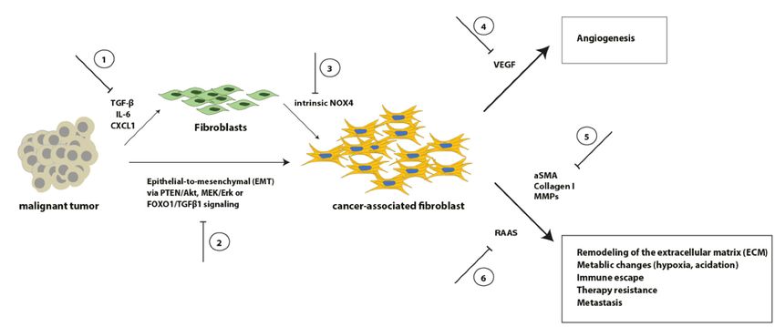
Figure 2. Schematic overview of selected signaling pathways involved in the transformation from fibroblasts into CAFs,
Figure 2. Schematic overview of selected signaling pathways involved in the transformation from
as well as representative CAF effects on crucial tumor aspects. Inhibition of these processes (1–6) might result in novel
fibroblasts
therapeutic into CAFs, as well as representative CAF effects on crucial tumor aspects. Inhibition of
options.
these processes (1–6) might result in novel therapeutic options.
4. Tumor-associated Macrophages
Infiltration of tumor-associated immune cells is one of the hallmarks of cancer [70].
A significant amount of these cells is represented by macrophages; accordingly, nearly
every solid tumor is heavily infiltrated by macrophages [71–74]. To understand the role
of macrophages in tumors it is helpful to look at how macrophages function in woundCancers 2021, 13, 4678 7 of 14
4. Tumor-associated Macrophages
Infiltration of tumor-associated immune cells is one of the hallmarks of cancer [70].
A significant amount of these cells is represented by macrophages; accordingly, nearly
every solid tumor is heavily infiltrated by macrophages [71–74]. To understand the role of
macrophages in tumors it is helpful to look at how macrophages function in wound healing.
In the early phase of a wound, macrophages clear the wound of debris and bacteria and
recruit other immune cells to help repair the damaged tissue. These tasks are summarized
under the term ‘pro-inflammatory’. Next, macrophages help to rebuild the tissue by foster-
ing angiogenesis and re-epithelization by producing granulation tissue. In the last phase of
wound healing, they help to limit immune responses of other cells, remodel the tissue, and
clear apoptotic cells. For these different processes, macrophages need different states of
activation termed polarization [75]. These different polarization states are simplified in M1
or M2 macrophages. This nomenclature is based on inflammation and immunity also used
for tumor-associated macrophages often with the addition ‘M1/M2-like’. M1-macrophages
are believed to be more in a pro-inflammatory state, while M2-like macrophages are
immune-suppressive, at the same time pro-angiogenic, and express metalloproteases to
degrade basement membranes and other extracellular matrix structures to foster invasion
and migration [76,77]. Polarization states can be determined by distinct chemokine ex-
pression and immune receptor expression patterns [78,79] triggered by a chaotic milieu
of an exponentially expanding malignant lesion. The functional and structural abnormal
vascular system that lacks a hierarchic architecture does not provide proper delivery of
oxygen and nutrients and shows insufficient clearance of metabolic waste and carbonic
dioxide which leads to a hostile environment where normoxic, hypoxic, and necrotic tissue
is in the constant remodeling process. Dying cells secrete chemokines which lead together
with that harsh environment to the recruitment of macrophages, that similar to the early
phase of a wound start to (try to) repair ‘the wound that never heals’ [80].
Accumulation of tumor-associated macrophages in human solid tumors is correlated
in most but not every entity with a poor prognosis [55,56]. In some entities, e.g., colorectal
cancer the implication of intra-tumoural immune cells seems to change throughout disease
progression [31,35,81,82]. Tumor macrophages in most advanced cancers execute key func-
tions of tumor progression by secreting growth factors that directly stimulate tumor cell
proliferation and survival, by fostering angiogenesis by secreting pro-angiogenic cytokines
and producing extracellular components of angiogenesis like, e.g., collagen IV, by promot-
ing tumor cell invasion via degradation of basement membranes and other extracellular
matrix by metalloproteinases and by mediating adaptive immunity via immunomodulatory
or immunosuppressive stimuli [70,73].
Li et al. published a meta-analysis concerning the impact of tumor-associated macrophages
in esophageal cancer. The studies included in this analysis were exclusively from Asia
and reported on ESCC, besides one study from the US [83]. The meta-analysis found
infiltration with M2-macrophages as significantly relevant for overall survival. Infiltration
with M2-macrophages contributed to poor survival and increased TNM stage in ESCC.
Interestingly, high infiltration of M2-macrophages to ESCC is also associated with poor
prognosis after and poor pathological response to neoadjuvant treatment [84].
EAC is a disease that is at least initially driven by chronic inflammation, due to
reflux disease with metaplasia of the distal esophagus with a significant upregulation
of inflammatory cytokines which can influence the prognosis [85]. It is not surprising
that immune cells are deeply involved in promoting malignant progression [86]. In EAC,
M2-macrophage infiltration, specifically, a high M2/M1-like ratio was accompanied by
poor prognosis [87]. Interestingly, this was only relevant in treatment naïve patients and
the observed role of macrophages was not detectable after neoadjuvant treatment.
Nevertheless, both in EAC and ESCC tumor-infiltrating macrophages seem to play a
pivotal role in malignant progression and therapy resistance [88] and represent a potentially
valuable therapeutic target to further increase pathological response and overall survival.
Though, targeting macrophages has not entered clinical practice as rapidly as other targetedCancers 2021, 13, 4678 8 of 14
concepts due to the complexity and diversity of TAM function and phenotype. Table 1
gives an overview of recent Phase I trials evaluating macrophage targeted therapies.
Table 1. Overview of macrophage targeting therapies in clinical Phase I trials. Please note that the last column indicates
inclusion or eligibility of esophagogastric cancers or ESCC (+) or not (−).
Stage Towards Clinical
Drug Targeted Mechanism Including EAC/ESCC
Application, Reference
Carlumab CCL2 Inhibition Phase I [89] +
Vanucizumab VEGF/ANG-2 Inhibition Phase I, NCT02665416 −
CP-870,893 CD40 Agonism Phase I [90] −
AMG820 CSF-1R Inhibition Phase I [91] −
LY3022855 CSF-1R Inhibition Phase I NCT02718911 +
Modified vitamin-D-binding protein
EF-022 (Efranat) Phase I NCT02052492 +
(macrophage-activating factor)
PLX7486 CSF-1R Inhibition Phase I NCT01804530 +
5. T-Cells and Myeloid-Derived Suppressor Cells, Immunotherapy
In addition to monocytes, mast cells, myeloid progenitors, and macrophages, T-cells
compose a significant part of the tumor immune cell infiltrate in most solid tumors. CD-
8+ cytotoxic T-lymphocytes, CD4+ Th 1 helper T cells, and natural killer cells are critical
players in eliminating malignant cells in the healthy human organism which has widely
been demonstrated in mice models where mice lacking these cells or subsets of them
have a significantly higher susceptibility to develop malignancies [70,92]. Tumors develop
effective strategies to avoid such elimination by the immune system. To regain effective
T cell-mediated anti-tumor activity became one of the most applied targeted therapy
concepts of modern cancer treatment namely checkpoint inhibition. Programmed cell
death protein 1 (PD-1) is a so-called immune checkpoint protein expressed on the cell
surfaces of lymphocytes. Tumor cells express programmed cell death ligand 1 (PD-L1)
which mediates prevention of cytotoxic anti-tumor t cell activity by inducing apoptosis
in antigen-specific T cells and by interfering with regulatory T cells [93,94]. Drugs that
prevent PD-1/PD-L1 interaction, e.g., pembrolizumab, a humanized monoclonal anti-PD-1
antibody, were developed with highly promising results in several tumor entities.
In ESCC, PD-L1 expression by tumor cells is an independent prognostic factor pre-
dicting worse outcomes in PD-L1 positive patients [95]. In line with this, the Keynote-181
study, a randomized phase III trial involving over 600 patients with esophageal cancer
including a mixed cohort of both, ESCC and EAC patients showed that pembrolizumab
lead to a significant survival benefit compared to chemotherapy (investigator’s choice
of paclitaxel, docetaxel, or irinotecan) in patients with ESCC. Interestingly, patients with
EAC showed no survival benefit [96]. These results were in line with both, the Keynote-
061 trial, where Pembrolizumab failed to show any effect in gastro-esophageal junction
adenocarcinoma [97], and the Attraction-3 study, where Nivolumab, another anti-PD-1
antibody, significantly prolonged overall survival compared to chemotherapy in ESCC
patients [98]. These large clinical trials show that in ESCC immune checkpoint blockade is
a valuable treatment approach that is as a single-agent even superior to chemotherapy in
a palliative setting, but in EAC these drugs still have to show efficacy. Whether and how
these differences are determined by the cancer cell itself or complex microenvironmental
cues that manipulate the immune response beyond PD-L1/PD-1 signaling has to be further
elucidated. That the latter is a likely scenario is supported by findings that high amounts
of intratumoral CD8+ T cells have been shown to be associated with prolonged survival in
both ESCC and EAC [99]. Another study demonstrated that high abundance of CD8+ T
cells was accompanied by high PD-L1 expression and that both factors were beneficial for
patients survival in esophagogastric junction and gastric adenocarcinomas [100]. A factorCancers 2021, 13, 4678 9 of 14
independent of PD-L1/PD-1 expression status might be the abundance and activity of
myeloid-derived suppressor cells (MDSCs) that are known to critically interfere with adap-
tive anti-cancer immune responses in several ways [101–103]. High infiltration counts of
MDSCs are associated with detrimental outcome parameters in esophageal cancer patients’
and MDSCs promote esophageal cancer growth in experimental disease models [104,105].
A recent work has done great efforts to characterize the immune-suppressive landscape in
esophageal cancer at single-cell resolution by transcriptome analysis of tumor-infiltrating
immune cells [106]. Unfortunately, this work was limited to ESCC, similar data are ur-
gently needed for EAC patients. This would potentially clarify the differential response
to immunotherapy between EAC and ESCC patients. This is highly clinically relevant to
improve and individualize this therapeutic concept in the future. Furthermore, results
from trials that incorporate immunotherapy into neoadjuvant regimens in esophageal or
esophagogastric junction cancer (NCT04159974, NCT03421288) are eagerly awaited to clar-
ify safety and efficacy and whether immunotherapy can improve response to neoadjuvant
treatment, which is highly relevant for overall survival, particularly in EAC.
6. Conclusions
During recent decades a considerable amount of knowledge concerning different
cellular and extracellular compartments within the tumor microenvironment of esophageal
cancer has been gained. Instead of merely focusing on the epithelial tumor cells, it becomes
more and more obvious to consider other intra-tumoral cell populations, as well as multiple
interactions between these populations.
Based on this knowledge targeted therapies have been developed that mostly in
conjunction with conventional chemotherapy aim to advance treatment efficacy. Although
significant improvement has been reached, treatment responses and overall survival in
esophageal cancer patients is still poor. Major challenges remain in further improving
established therapeutic concepts and (re-)evaluating them in certain clinical situations as,
e.g., ramucirumab in the neoadjuvant setting.
Another very interesting question is the differences in sensitivity of EAC and ESCC
towards immunotherapy. To uncover potential causes might elicit new modes of resistance
and chances for adjustments to checkpoint inhibition in esophageal cancer.
Finding suitable biomarkers of response and resistance is a highly relevant challenge.
This is true for every oncologic treatment concept, but particular for targeted therapies due
to the high cost for health care systems and society. Very few markers exist or proceeded
to daily clinical practice in solid tumors which highlights the urgent need for further
research here.
The vaguest but probably also the most exciting challenge is to explore novel concepts
based on molecular findings regarding the regulation of the tumor microenvironment.
Thorough basic and translational research that also reports potential risks of potential novel
targeted therapies is required.
Author Contributions: Conceptualization, L.M.S., P.S.P. and T.S.; writing—original draft preparation,
L.M.S. and P.S.P.; writing—review and editing, L.M.S., P.S.P., B.B., H.F.F., C.J.B., T.S.; visualization,
L.M.S. and P.S.P. All authors have read and agreed to the published version of the manuscript.
Funding: This research received no external funding.
Conflicts of Interest: The authors declare no conflict of interest.
References
1. Lagergren, J.; Smyth, E.; Cunningham, D.; Lagergren, P. Oesophageal cancer. Lancet 2017, 390, 2383–2396. [CrossRef]
2. Fitzmaurice, C.; Dicker, D.; Pain, A.; Hamavid, H.; Moradi-Lakeh, M.; MacIntyre, M.F.; Allen, C.; Hansen, G.; Woodbrook, R.;
Wolfe, C.; et al. The Global Burden of Cancer 2013. JAMA Oncol. 2015, 1, 505–527. [CrossRef]Cancers 2021, 13, 4678 10 of 14
3. Al-Batran, S.-E.; Hofheinz, R.D.; Pauligk, C.; Kopp, H.-G.; Haag, G.M.; Luley, K.B.; Meiler, J.; Homann, N.; Lorenzen, S.;
Schmalenberg, H.; et al. Histopathological regression after neoadjuvant docetaxel, oxaliplatin, fluorouracil, and leucovorin
versus epirubicin, cisplatin, and fluorouracil or capecitabine in patients with resectable gastric or gastro-oesophageal junction
adenocarcinoma (FLOT4-AIO): Results from the phase 2 part of a multicentre, open-label, randomised phase 2/3 trial. Lancet
Oncol. 2016, 17, 1697–1708. [CrossRef] [PubMed]
4. Al-Batran, S.-E.; Homann, N.; Pauligk, C.; Goetze, T.O.; Meiler, J.; Kasper, S.; Kopp, H.-G.; Mayer, F.; Haag, G.M.; Luley, K.; et al.
Perioperative chemotherapy with fluorouracil plus leucovorin, oxaliplatin, and docetaxel versus fluorouracil or capecitabine
plus cisplatin and epirubicin for locally advanced, resectable gastric or gastro-oesophageal junction adenocarcinoma (FLOT4): A
randomised, phase 2/3 trial. Lancet 2019, 393, 1948–1957. [CrossRef]
5. Shapiro, J.; van Lanschot, J.J.B.; Hulshof, M.C.C.M.; van Hagen, P.; van Berge Henegouwen, M.I.; Wijnhoven, B.P.L.; van
Laarhoven, H.W.M.; Nieuwenhuijzen, G.A.P.; Hospers, G.A.P.; Bonenkamp, J.J.; et al. CROSS study group Neoadjuvant
chemoradiotherapy plus surgery versus surgery alone for oesophageal or junctional cancer (CROSS): Long-term results of a
randomised controlled trial. Lancet Oncol. 2015, 16, 1090–1098. [CrossRef]
6. Lorenzen, S.; Thuss-Patience, P.; Al-Batran, S.E.; Lordick, F.; Haller, B.; Schuster, T.; Pauligk, C.; Luley, K.; Bichev, D.; Schumacher,
G.; et al. Impact of pathologic complete response on disease-free survival in patients with esophagogastric adenocarcinoma
receiving preoperative docetaxel-based chemotherapy. Ann. Oncol. 2013, 24, 2068–2073. [CrossRef] [PubMed]
7. Mönig, S.P.; Schiffmann, L.M. Resection of advanced esophagogastric adenocarcinoma: Extended indications. Chirurg 2016, 87,
398–405. [CrossRef]
8. Homann, N.; Pauligk, C.; Luley, K.; Kraus, T.W.; Bruch, H.-P.; Atmaca, A.; Noack, F.; Altmannsberger, H.-M.; Jäger, E.; Al-Batran,
S.-E. Pathological complete remission in patients with oesophagogastric cancer receiving preoperative 5-fluorouracil, oxaliplatin
and docetaxel. Int. J. Cancer 2011, 130, 1706–1713. [CrossRef] [PubMed]
9. Yang, Y.-M.; Hong, P.; Xu, W.W.; He, Q.-Y.; Li, B. Advances in targeted therapy for esophageal cancer. Signal Transduct. Target.
Ther. 2020, 5, 229. [CrossRef]
10. Vivaldi, C.; Catanese, S.; Massa, V.; Pecora, I.; Salani, F.; Santi, S.; Lencioni, M.; Vasile, E.; Falcone, A.; Fornaro, L. Immune
Checkpoint Inhibitors in Esophageal Cancers: Are We Finally Finding the Right Path in the Mist? Int. J. Mol. Sci. 2020, 21, 1658.
[CrossRef]
11. Quail, D.F.; Joyce, J.A. Microenvironmental regulation of tumor progression and metastasis. Nat. Med. 2013, 19, 1423–1437.
[CrossRef]
12. Carmeliet, P. Angiogenesis in life, disease and medicine. Nature 2005, 438, 932–936. [CrossRef] [PubMed]
13. Hurwitz, H.; Fehrenbacher, L.; Novotny, W.; Cartwright, T.; Hainsworth, J.; Heim, W.; Berlin, J.; Baron, A.; Griffing, S.; Holmgren,
E.; et al. Bevacizumab plus Irinotecan, Fluorouracil, and Leucovorin for Metastatic Colorectal Cancer. N. Engl. J. Med. 2004, 350,
2335–2342. [CrossRef]
14. Goel, S.; Wong, A.H.-K.; Jain, R.K. Vascular Normalization as a Therapeutic Strategy for Malignant and Nonmalignant Disease.
Cold Spring Harb. Perspect. Med. 2012, 2, a006486. [CrossRef] [PubMed]
15. Jain, R.K.; Duda, D.G.; Clark, J.W.; Loeffler, J.S. Lessons from phase III clinical trials on anti-VEGF therapy for cancer. Nat. Clin.
Pr. Oncol. 2006, 3, 24–40. [CrossRef]
16. Fukumura, D.; Jain, R.K. Tumor microenvironment abnormalities: Causes, consequences, and strategies to normalize. J. Cell.
Biochem. 2007, 101, 937–949. [CrossRef] [PubMed]
17. Winkler, F.; Kozin, S.V.; Tong, R.T.; Chae, S.-S.; Booth, M.F.; Garkavtsev, I.; Xu, L.; Hicklin, D.J.; Fukumura, D.; di Tomaso, E.; et al.
Kinetics of vascular normalization by VEGFR2 blockade governs brain tumor response to radiation. Cancer Cell 2004, 6, 553–563.
[CrossRef] [PubMed]
18. Shen, Y.; Wang, X.; Lu, J.; Salfenmoser, M.; Wirsik, N.M.; Schleussner, N.; Imle, A.; Valls, A.F.; Radhakrishnan, P.; Liang, J.; et al.
Reduction of Liver Metastasis Stiffness Improves Response to Bevacizumab in Metastatic Colorectal Cancer. Cancer Cell 2020, 37,
800–817. [CrossRef]
19. Jain, R.K. Normalization of Tumor Vasculature: An Emerging Concept in Antiangiogenic Therapy. Science 2005, 307, 58–62.
[CrossRef]
20. Shimada, H.; Takeda, A.; Nabeya, Y.; Okazumi, S.-I.; Matsubara, H.; Funami, Y.; Hayashi, H.; Gunji, Y.; Kobayashi, S.; Suzuki, T.;
et al. Clinical significance of serum vascular endothelial growth factor in esophageal squamous cell carcinoma. Cancer 2001, 92,
663–669. [CrossRef]
21. Nienhüser, H.; Schmidt, T. Angiogenesis and Anti-Angiogenic Therapy in Gastric Cancer. Int. J. Mol. Sci. 2017, 19, 43. [CrossRef]
22. Ohtsu, A.; Shah, M.A.; Van Cutsem, E.; Rha, S.Y.; Sawaki, A.; Park, S.R.; Lim, H.Y.; Yamada, Y.; Wu, J.; Langer, B.; et al.
Bevacizumab in Combination with Chemotherapy As First-Line Therapy in Advanced Gastric Cancer: A Randomized, Double-
Blind, Placebo-Controlled Phase III Study. J. Clin. Oncol. 2011, 29, 3968–3976. [CrossRef]
23. Shen, L.; Li, J.; Xu, J.; Pan, H.; Dai, G.; Qin, S.; Wang, L.; Wang, J.; Yang, Z.; Shu, Y.; et al. Bevacizumab plus capecitabine
and cisplatin in Chinese patients with inoperable locally advanced or metastatic gastric or gastroesophageal junction cancer:
Randomized, double-blind, phase III study (AVATAR study). Gastric Cancer 2015, 18, 168–176. [CrossRef]
24. Lin, S.J.; Gagnon-Bartsch, J.A.; Tan, I.B.; Earle, S.; Ruff, L.; Pettinger, K.; Ylstra, B.; Van Grieken, N.; Rha, S.Y.; Chung, H.C.; et al.
Signatures of tumour immunity distinguish Asian and non-Asian gastric adenocarcinomas. Gut 2015, 64, 1721–1731. [CrossRef]Cancers 2021, 13, 4678 11 of 14
25. Cunningham, D.; Stenning, S.P.; Smyth, E.C.; Okines, A.F.; Allum, W.H.; Rowley, S.; Stevenson, L.; Grabsch, H.I.; Alderson, D.;
Crosby, T.; et al. Peri-operative chemotherapy with or without bevacizumab in operable oesophagogastric adenocarcinoma (UK
Medical Research Council ST03): Primary analysis results of a multicentre, open-label, randomised phase 2–3 trial. Lancet Oncol.
2017, 18, 357–370. [CrossRef]
26. Fuchs, C.S.; Tomasek, J.; Yong, C.J.; Dumitru, F.; Passalacqua, R.; Goswami, C.; Safran, H.; Santos, L.V.D.; Aprile, G.; Ferry,
D.R.; et al. REGARD Trial Investigators Ramucirumab monotherapy for previously treated advanced gastric or gastro-oesophageal
junction adenocarcinoma (REGARD): An international, randomised, multicentre, placebo-controlled, phase 3 trial. Lancet 2014,
383, 31–39. [CrossRef]
27. Wilke, H.; Muro, K.; Van Cutsem, E.; Oh, S.-C.; Bodoky, G.; Shimada, Y.; Hironaka, S.; Sugimoto, N.; Lipatov, O.; Kim, T.-
Y.; et al. RAINBOW Study Group Ramucirumab plus paclitaxel versus placebo plus paclitaxel in patients with previously treated
advanced gastric or gastro-oesophageal junction adenocarcinoma (RAINBOW): A double-blind, randomised phase 3 trial. Lancet
Oncol. 2014, 15, 1224–1235. [CrossRef]
28. Smyth, E.C.; Verheij, M.; Allum, W.; Cunningham, D.; Cervantes, A.; Arnold, D. ESMO Guidelines Committee Gastric cancer:
ESMO Clinical Practice Guidelines for diagnosis, treatment and follow-up. Ann. Oncol. 2016, 27, v38–v49. [CrossRef] [PubMed]
29. Schiffmann, L.M.; Brunold, M.; Liwschitz, M.; Goede, V.; Loges, S.; Wroblewski, M.; Quaas, A.; Alakus, H.; Stippel, D.; Bruns,
C.J.; et al. A combination of low-dose bevacizumab and imatinib enhances vascular normalisation without inducing extracellular
matrix deposition. Br. J. Cancer 2017, 116, 600–608. [CrossRef]
30. Coutelle, O.; Schiffmann, L.M.; Liwschitz, M.; Brunold, M.; Goede, V.; Hallek, M.; Kashkar, H.; Hacker, U.T. Dual targeting of
Angiopoetin-2 and VEGF potentiates effective vascular normalisation without inducing empty basement membrane sleeves in
xenograft tumours. Br. J. Cancer 2015, 112, 495–503. [CrossRef] [PubMed]
31. Schiffmann, L.M.; Fritsch, M.; Gebauer, F.; Günther, S.D.; Stair, N.R.; Seeger, J.M.; Thangarajah, F.; Dieplinger, G.; Bludau, M.;
Alakus, H.; et al. Tumour-infiltrating neutrophils counteract anti-VEGF therapy in metastatic colorectal cancer. Br. J. Cancer 2019,
120, 69–78. [CrossRef]
32. Bergers, G.; Hanahan, D. Modes of resistance to anti-angiogenic therapy. Nat. Rev. Cancer 2008, 8, 592–603. [CrossRef] [PubMed]
33. Ebos, J.; Lee, C.R.; Cruz-Munoz, W.; Bjarnason, G.A.; Christensen, J.G.; Kerbel, R.S. Accelerated Metastasis after Short-Term
Treatment with a Potent Inhibitor of Tumor Angiogenesis. Cancer Cell 2009, 15, 232–239. [CrossRef] [PubMed]
34. Pàez-Ribes, M.; Allen, E.; Hudock, J.; Takeda, T.; Okuyama, H.; Viñals, F.; Inoue, M.; Bergers, G.; Hanahan, D.; Casanovas, O.
Antiangiogenic Therapy Elicits Malignant Progression of Tumors to Increased Local Invasion and Distant Metastasis. Cancer Cell
2009, 15, 220–231. [CrossRef]
35. Valls, A.F.; Knipper, K.; Giannakouri, E.; Sarachaga, V.; Hinterkopf, S.; Wuehrl, M.; Shen, Y.; Radhakrishnan, P.; Klose, J.; Ulrich,
A.; et al. VEGFR1 + Metastasis–Associated Macrophages Contribute to Metastatic Angiogenesis and Influence Colorectal Cancer
Patient Outcome. Clin. Cancer Res. 2019, 25, 5674–5685. [CrossRef] [PubMed]
36. Hacker, U.T.; Escalona-Espinosa, L.; Consalvo, N.; Goede, V.; Schiffmann, L.; Scherer, S.J.; Hedge, P.; Van Cutsem, E.; Coutelle, O.;
Buning, H. Evaluation of Angiopoietin-2 as a biomarker in gastric cancer: Results from the randomised phase III AVAGAST trial.
Br. J. Cancer 2016, 114, 855–862. [CrossRef]
37. Goede, V.; Coutelle, O.; Neuneier, J.; Reinacher-Schick, A.; Schnell, R.; Koslowsky, T.C.; Weihrauch, M.R.; Cremer, B.; Kashkar, H.;
Odenthal, M.; et al. Identification of serum angiopoietin-2 as a biomarker for clinical outcome of colorectal cancer patients treated
with bevacizumab-containing therapy. Br. J. Cancer 2010, 103, 1407–1414. [CrossRef]
38. Dreikhausen, L.; Blank, S.; Sisic, L.; Heger, U.; Weichert, W.; Jäger, D.; Bruckner, T.; Giese, N.; Grenacher, L.; Falk, C.; et al.
Association of angiogenic factors with prognosis in esophageal cancer. BMC Cancer 2015, 15, 121. [CrossRef]
39. Nienhüser, H.; Crnovrsanin, N.; Nerz, D.; Heckler, M.; Sisic, L.; Lasitschka, F.; Schneider, M.; Schmidt, T. Expression of Angiogenic
Proteins in Tumor and Stroma Affects Survival in Patients with Gastric Cancer. J. Surg. Res. 2020, 255, 172–180. [CrossRef]
40. Cantelmo, A.R.; Conradi, L.-C.; Brajic, A.; Goveia, J.; Kalucka, J.; Pircher, A.; Chaturvedi, P.; Hol, J.; Thienpont, B.; Teuwen,
L.-A.; et al. Inhibition of the Glycolytic Activator PFKFB3 in Endothelium Induces Tumor Vessel Normalization, Impairs
Metastasis, and Improves Chemotherapy. Cancer Cell 2016, 30, 968–985. [CrossRef]
41. Conradi, L.-C.; Brajic, A.; Cantelmo, A.R.; Bouché, A.; Kalucka, J.; Pircher, A.; Brüning, U.; Teuwen, L.-A.; Vinckier, S.; Ghesquière,
B.; et al. Tumor vessel disintegration by maximum tolerable PFKFB3 blockade. Angiogenesis 2017, 20, 599–613. [CrossRef]
42. Schiffmann, L.M.; Werthenbach, J.P.; Heintges-Kleinhofer, F.; Seeger, J.M.; Fritsch, M.; Günther, S.D.; Willenborg, S.; Brodesser, S.;
Lucas, C.; Jüngst, C.; et al. Mitochondrial respiration controls neoangiogenesis during wound healing and tumour growth. Nat.
Commun. 2020, 11, 1231. [CrossRef]
43. Coutelle, O.; Hornig-Do, H.; Witt, A.; Andree, M.; Schiffmann, L.M.; Piekarek, M.; Brinkmann, K.; Seeger, J.M.; Liwschitz, M.;
Miwa, S.; et al. Embelin inhibits endothelial mitochondrial respiration and impairs neoangiogenesis during tumor growth and
wound healing. EMBO Mol. Med. 2014, 6, 624–639. [CrossRef]
44. Rohlenova, K.; Veys, K.; Miranda-Santos, I.; De Bock, K.; Carmeliet, P. Endothelial Cell Metabolism in Health and Disease. Trends
Cell Biol. 2018, 28, 224–236. [CrossRef] [PubMed]
45. Eelen, G.; Treps, L.; Li, X.; Carmeliet, P. Basic and Therapeutic Aspects of Angiogenesis Updated. Circ. Res. 2020, 127, 310–329.
[CrossRef] [PubMed]
46. Kalluri, R.; Zeisberg, M. Fibroblasts in cancer. Nat. Rev. Cancer 2006, 6, 392–401. [CrossRef]Cancers 2021, 13, 4678 12 of 14
47. Sahai, E.; Astsaturov, I.; Cukierman, E.; DeNardo, D.G.; Egeblad, M.; Evans, R.M.; Fearon, D.; Greten, F.R.; Hingorani, S.R.;
Hunter, T.; et al. A framework for advancing our understanding of cancer-associated fibroblasts. Nat. Rev. Cancer 2020, 20,
174–186. [CrossRef] [PubMed]
48. Fukumura, D.; Xavier, R.; Sugiura, T.; Chen, Y.; Park, E.-C.; Lu, N.; Selig, M.; Nielsen, G.; Taksir, T.; Jain, R.K.; et al. Tumor
Induction of VEGF Promoter Activity in Stromal Cells. Cell 1998, 94, 715–725. [CrossRef]
49. Manousopoulou, A.; Hayden, A.; Mellone, M.; Baquero, D.G.; White, C.; Noble, F.; Lopez, M.; Thomas, G.J.; Underwood, T.J.;
Garbis, S.D. Quantitative proteomic profiling of primary cancer-associated fibroblasts in oesophageal adenocarcinoma. Br. J.
Cancer 2018, 118, 1200–1207. [CrossRef]
50. Arina, A.; Idel, C.; Hyjek, E.M.; Alegre, M.-L.; Wang, Y.; Bindokas, V.P.; Weichselbaum, R.R.; Schreiber, H. Tumor-associated
fibroblasts predominantly come from local and not circulating precursors. Proc. Natl. Acad. Sci. USA 2016, 113, 7551. [CrossRef]
51. Raz, Y.; Cohen, N.; Shani, O.; Bell, R.E.; Novitskiy, S.V.; Abramovitz, L.; Levy, C.; Milyavsky, M.; Leider-Trejo, L.; Moses, H.L.;
et al. Bone marrow–derived fibroblasts are a functionally distinct stromal cell population in breast cancer. J. Exp. Med. 2018, 215,
3075–3093. [CrossRef]
52. Dirat, B.; Bochet, L.; Dabek, M.; Daviaud, D.; Dauvillier, S.; Majed, B.; Wang, Y.Y.; Meulle, A.; Salles, B.; Le Gonidec, S.; et al.
Cancer-Associated Adipocytes Exhibit an Activated Phenotype and Contribute to Breast Cancer Invasion. Cancer Res. 2011, 71,
2455–2465. [CrossRef]
53. Schoppmann, S.F.; Jesch, B.; Riegler, M.F.; Maroske, F.; Schwameis, K.; Jomrich, G.; Birner, P. Podoplanin expressing cancer
associated fibroblasts are associated with unfavourable prognosis in adenocarcinoma of the esophagus. Clin. Exp. Metastasis 2012,
30, 441–446. [CrossRef]
54. Underwood, T.; Hayden, A.L.; Derouet, M.; Garcia, E.; Noble, F.; White, M.; Thirdborough, S.; Mead, A.; Clemons, N.; Mellone,
M.; et al. Cancer-associated fibroblasts predict poor outcome and promote periostin-dependent invasion in oesophageal
adenocarcinoma. J. Pathol. 2015, 235, 466–477. [CrossRef]
55. Hanley, C.J.; Mellone, M.; Ford, K.; Thirdborough, S.M.; Mellows, T.; Frampton, S.J.; Smith, D.M.; Harden, E.; Szyndralewiez, C.;
Bullock, M.; et al. Targeting the Myofibroblastic Cancer-Associated Fibroblast Phenotype through Inhibition of NOX4. J. Natl.
Cancer Inst. 2018, 110, 109–120. [CrossRef] [PubMed]
56. Galván, J.A.; Wiprächtiger, J.; Slotta-Huspenina, J.; Feith, M.; Ott, K.; Kröll, D.; Seiler, C.A.; Langer, R. Immunohistochemical
analysis of the expression of cancer-associated fibroblast markers in esophageal cancer with and without neoadjuvant therapy.
Virchows Archiv 2019, 476, 725–734. [CrossRef] [PubMed]
57. van Pelt, G.; Krol, J.; Lips, I.; Peters, F.; van Klaveren, D.; Boonstra, J.; de Steur, W.; Tollenaar, R.; Sarasqueta, A.F.; Mesker, W.; et al.
The value of tumor-stroma ratio as predictor of pathologic response after neoadjuvant chemoradiotherapy in esophageal cancer.
Clin. Transl. Radiat. Oncol. 2020, 20, 39–44. [CrossRef]
58. Steins, A.; Ebbing, E.A.; Creemers, A.; Van Der Zalm, A.P.; Jibodh, R.A.; Waasdorp, C.; Meijer, S.; Van Delden, O.M.; Krishnadath,
K.K.; Hulshof, M.C.; et al. Chemoradiation induces epithelial-to-mesenchymal transition in esophageal adenocarcinoma. Int. J.
Cancer 2019, 145, 2792–2803. [CrossRef] [PubMed]
59. Anand, A.; Fang, H.-Y.; Mohammad-Shahi, D.; Ingermann, J.; Baumeister, T.; Strangmann, J.; Schmid, R.M.; Wang, T.C.; Quante,
M. Elimination of NF-κB signaling in Vimentin+ stromal cells attenuates tumorigenesis in a mouse model of Barrett’s Esophagus.
Carcinogenesis 2021, 42, 405–413. [CrossRef] [PubMed]
60. Ebbing, E.A.; van der Zalm, A.P.; Steins, A.; Creemers, A.; Hermsen, S.; Rentenaar, R.; Klein, M.; Waasdorp, C.; Hooijer, G.K.J.;
Meijer, S.; et al. Stromal-derived interleukin 6 drives epithelial-to-mesenchymal transition and therapy resistance in esophageal
adenocarcinoma. Proc. Natl. Acad. Sci. USA 2019, 116, 2237–2242. [CrossRef]
61. Karakasheva, T.A.; Lin, E.W.; Tang, Q.; Qiao, E.; Waldron, T.J.; Soni, M.; Klein-Szanto, A.J.; Sahu, V.; Basu, D.; Ohashi, S.; et al.
IL-6 Mediates Cross-Talk between Tumor Cells and Activated Fibroblasts in the Tumor Microenvironment. Cancer Res. 2018, 78,
4957–4970. [CrossRef]
62. Zhang, H.; Yue, J.; Jiang, Z.; Zhou, R.; Xie, R.; Xu, Y.; Wu, S. CAF-secreted CXCL1 conferred radioresistance by regulating DNA
damage response in a ROS-dependent manner in esophageal squamous cell carcinoma. Cell Death Dis. 2017, 8, e2790. [CrossRef]
[PubMed]
63. Zhang, H.; Xie, C.; Yue, J.; Jiang, Z.; Zhou, R.; Xie, R.; Wang, Y.; Wu, S. Cancer-associated fibroblasts mediated chemoresistance by
a FOXO1/TGFβ1 signaling loop in esophageal squamous cell carcinoma. Mol. Carcinog. 2017, 56, 1150–1163. [CrossRef]
64. Higashino, N.; Koma, Y.-I.; Hosono, M.; Takase, N.; Okamoto, M.; Kodaira, H.; Nishio, M.; Shigeoka, M.; Kakeji, Y.; Yokozaki, H.
Fibroblast activation protein-positive fibroblasts promote tumor progression through secretion of CCL2 and interleukin-6 in
esophageal squamous cell carcinoma. Lab. Investig. 2019, 99, 777–792. [CrossRef] [PubMed]
65. Tanaka, K.; Miyata, H.; Sugimura, K.; Fukuda, S.; Kanemura, T.; Yamashita, K.; Miyazaki, Y.; Takahashi, T.; Kurokawa, Y.;
Yamasaki, M.; et al. miR-27 is associated with chemoresistance in esophageal cancer through transformation of normal fibroblasts
to cancer-associated fibroblasts. Carcinogenesis 2015, 36, 894–903. [CrossRef]
66. Han, P.; Cao, P.; Hu, S.; Kong, K.; Deng, Y.; Zhao, B.; Li, F. Esophageal Microenvironment: From Precursor Microenvironment to
Premetastatic Niche. Cancer Manag. Res. 2020, 12, 5857–5879. [CrossRef]
67. Palumbo, J.A.; Da Costa, N.M.; Pontes, B.; De Oliveira, F.L.; Codeço, M.L.; Pinto, L.F.R.; Nasciutti, L.E. Esophageal Cancer
Development: Crucial Clues Arising from the Extracellular Matrix. Cells 2020, 9, 455. [CrossRef]You can also read