Virtual cleaning and unwrapping of non-invasively digitized soiled bamboo scrolls
←
→
Page content transcription
If your browser does not render page correctly, please read the page content below
www.nature.com/scientificreports

                        OPEN       Virtual cleaning and unwrapping
                                   of non-invasively digitized soiled
                                   bamboo scrolls
Received: 29 November 2018         Daniel Stromer1, Vincent Christlein            1
                                                                                      , Xiaolin Huang   2
                                                                                                            , Patrick Zippert3, Tino Hausotte      3

Accepted: 24 January 2019          & Andreas Maier 1
Published: xx xx xxxx
                                   In ancient China, symbols and drawings captured on bamboo and wooden slips were used as main
                                   communication media. Those documents are very precious for cultural heritage and research, but
                                   due to aging processes, the discovered pieces are sometimes in a poor condition and contaminated
                                   by soil. Manual cleaning of excavated slips is a demanding and time-consuming task in which writings
                                   can be accidentally deleted. To counter this, we propose a novel approach based on conventional 3-D
                                   X-ray computed tomography to digitize such historical documents without before manual cleaning.
                                   By applying a virtual cleaning and unwrapping algorithm, the entire scroll surface is remapped into 2-D
                                   such that the hidden content becomes readable. We show that the technique also works for heavily
                                   soiled scrolls, enabling an investigation of the content by the naked eye without the need for manual
                                   labor. This digitization also allows for recovery of potentially erased writings and reconstruction of the
                                   original spatial information.

                                   From the Shang dynasty (商朝, 17 century B. C.) to the Jin dynasty (两晋, after 300 A. D.), the main media in
                                   China was not paper, writings and drawings were instead captured from top to bottom on bamboo or wooden
                                   slips called 简牍1. The individual slips, ranging from 20 up to 70, were bound together and rolled up to a scroll
                                   forming a chapter of a book. Bamboo was mainly used as writing material because it was abundant everywhere
                                   throughout the ancient empire and cheaper than the alternative silk. Depending on the dynasty, symbols were
                                   brushed with ink onto the slips or carved into the surface2. For brushing, carbon-based inks made of lampblack or
                                   soot were produced and formed to inksticks that could then be liquefied again by adding water and rubbing it on
                                   inkstone3. After the invention of paper and its rapid dissemination across the continent during the Han dynasty
                                   (汉朝), bamboo and wooden slips were gradually replaced. Nowadays, such scrolls and slips are a valuable source
                                   to increase knowledge of the life of the past.
                                       In the last decades, some major bamboo scroll collections were discovered across China. The Yinqueshan Han
                                   Slips (银雀山汉简) were found near Linyi (临沂) in the province of Shandong, where construction workers ran-
                                   domly unearthed the Yinqueshan Han tombs4. Around 38 of the discovered slips (1,887 out of total 4,942) were
                                   heavily damaged such that a digitization was hardly possible5. One of the most important Chinese archaeological
                                   discoveries in the last decade was the unearthing of the Liye Qin Slips (里耶秦简) near Liye (里耶), Hunan. More
                                   than 37000 pieces with around 200000 recorded characters were found containing a wide range of document
                                   types6. In terms of numbers, this is not the biggest discovery, but the content of some slips is of very high value.
                                   A very famous piece is the world’s oldest example of a multiplication table7. The latest significant discovery of
                                   bamboo slips was the tomb of Marquis of Haihun (海昏侯). It was discovered 2011 in the Jiangxi province and
                                   contained about 5000 slips, bearing for example the ‘Book of medicine’ (医书), the ‘Analects of Confucius’ (论语)
                                   or the ‘Classics of filial piety’ (孝经)8.
                                       One basic challenge comes with all those discovered historical slips. Due to their age, the slips are in differ-
                                   ent stages of decay and intact scrolls are found only very rarely9. The cleaning process can be very challenging
                                   for a conservator because the writing media, as well as the ink and the carvings, can suffer from used chemicals

                                   1
                                     Pattern Recognition Lab, Computer Science, Friedrich-Alexander-Universität Erlangen-Nürnberg, Erlangen,
                                   91058, Germany. 2Institute of Image Processing and Pattern Recognition, Shanghai Jiao Tong University, Shanghai,
                                   200240, P.R. China. 3Institute of Manufacturing Metrology, Mechanical Engineering, Friedrich-Alexander-Universität
                                   Erlangen-Nürnberg, Erlangen, 91052, Germany. Correspondence and requests for materials should be addressed to
                                   D.S. (email: daniel.stromer@fau.de)

        Scientific Reports |   (2019) 9:2311 | https://doi.org/10.1038/s41598-019-39447-0                                                              1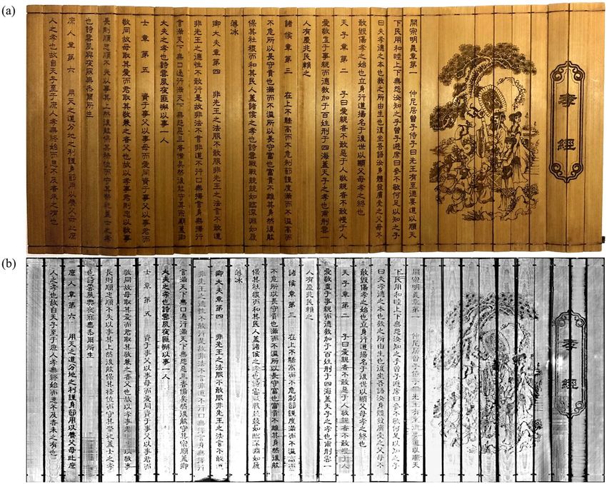
www.nature.com/scientificreports/

                           Figure 1. (a) Photograph of exemplary bamboo scrolls located at the Changsha Bamboo Slips Museum,
                           Changsha, Hunan. (b) Used bamboo scroll covered and filled with potting soil and packed in a plastic bag for
                           the experiments. The carvings were contaminated by pressing the soil onto the surface to create the worst case
                           scenario. (c) The 3-D X-ray CT measurement setup: The X-ray source and the flat panel detector are fixed while
                           the object rotates around the z-axis. The object is completely covered by the X-rays emitted to the detector such
                           that it can be fully visualized in the reconstructed volume. The scroll is placed upright such that a constant ray
                           length through the object is given aiming to reduce artifacts.

                           and human imprudence. Figure 1(a) shows a photograph of discovered and cleaned bamboo scrolls located at
                           the Changsha Bamboo Slips Museum (长沙简牍博物馆). The reconstruction of the slips is also very challeng-
                           ing because the strings can be rotten, hydrated, soiled and the slips can be positioned arbitrarily. To reveal the
                           writings of those soiled slips, conservators clean the fragments in a meticulous process. Cleaning and reading
                           unearthed documents by hand is not only a very time-consuming process, furthermore, changing the preserving
                           environment (e. g., brushing off the soils) exposes the slips directly to the atmosphere and they may oxidize rap-
                           idly. A preceding non-invasive digitization capable of capturing a digital volume of the entire unprocessed fragile
                           object can help to preserve the valuable content forever without destroying the original structure.
                               In 2016, Redo-Sanchez et al.10 used 3-D Terahertz (THz) imaging to read through a stack of paper with prom-
                           ising results. This technique does not expose ionizing radiation to an object, however, it is only well suited for
                           homogeneous materials. As the structure of bamboo scrolls, and especially of surrounding soil or sand, is highly
                           inhomogeneous, the material damping dramatically increases, resulting in a total reflection of the emitted THz
                           waves in the top layers of the object. Furthermore, humidity causes a total reflection of THz waves and as the
                           samples can be highly hydrated, the approach is not suited for prompt digitization in most cases. Given these
                           circumstances, only the surface and a small area underneath can be imaged such that this approach is not suitable
                           for a soiled scroll. However, if only individual soiled slips have to be investigated, this technique is promising for
                           imaging the content underneath the soil layers.
                               In 2015, Mocella et al.11 presented a proof-of-concept where they utilized X-ray phase-contrast imaging with
                           Synchrotron radiation for the digitization of a Herculaneum scroll sample. Subsequently, Bukreeva et al.12 devel-
                           oped an image processing method for virtually unrolling the scroll data. Phase-contrast imaging has also been
                           used for different archaeological purposes, such as scanning cultural heritages13–15. Although this technique
                           shows very good results, it is neither widely accessible nor mobile. The discoveries would have to be carried to
                           measurement centers which is costly due to security and insurance reasons.
                               Previous works on cellulose or collagen-based writing material show that conventional 3-D X-ray computed
                           tomography (CT) systems are well suited for non-invasive imaging when the ink’s X-ray material absorption
                           differs from that of the writing material16–19. The increased attenuation is recognizable within the X-ray volume20
                           and applying page segmentation and 2-D mapping algorithms21 enable a non-invasive digitization of fragile man-
                           uscripts. A disadvantage of this method is the applied X-ray radiation which could accelerate the aging process
                           of cellulose when performing a scan22,23. However, the impact of this X-ray radiation is still very limited24 and
                           through simulations, the dose can also be kept to a minimum25. Immel et al. showed experiments on sub-fossils
                           and provide a guideline for X-ray imaging such specimens26. Based on an object’s dimension, 3-D X-ray CT
                           reaches resolutions down to 1 μm and even less. Furthermore, this system can easily be carried and mounted at
                           libraries and excavation sites as long as a constant temperature and humidity is given. We showed in an initial
                           proof-of-concept27, that utilizing this technique for the digitization of such documents is promising. However, the
                           data processing was performed manually throughout the entire experiment.
                               For this work, we extended our previous work27 in a number of aspects. For the first time, we describe the
                           entire processing pipeline for the digitization of a wrapped up, heavily contaminated bamboo scroll based on
                           a conventional 3-D X-ray CT scan. First, we discuss an appropriate selection of scan parameters based on the
                           material characteristics in detail. Due to corrugated and curved surfaces of the bamboo slips, manual segmen-
                           tation of the scroll is very time-consuming. We address this issue by proposing a predominantly automatic algo-
                           rithmic solution. Initially, contamination is erased by a virtual cleaning algorithm. Next, the scroll gets digitally
                           unwrapped and the 3-D slips are finally processed by a texturing step to receive a 2-D representation readable for
                           the naked eye. We show, that our approach is well suited to image and visualize even heavily soiled slip carvings
                           of two exemplary scrolls. If writings or symbols are erased by manual cleaning, the virtually cleaned volume, and
                           the 2-D mapped image can help conservators to identify such spots and recover the information. Furthermore,

Scientific Reports |   (2019) 9:2311 | https://doi.org/10.1038/s41598-019-39447-0                                                              2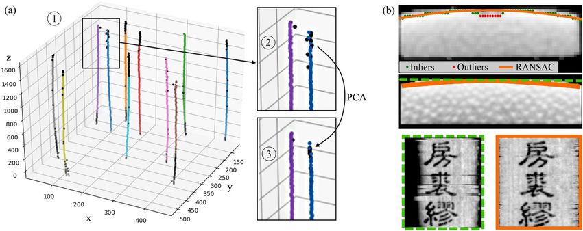
www.nature.com/scientificreports/

                           the volume can be investigated (e. g., by augmented reality) to relocate original slip locations for reconstruction.
                           Based on the scanner and object dimensions, the approach is capable of scanning multiple scrolls at the same
                           time.

                           Material Characteristics of Bamboo Scrolls
                           Obtaining real historical documents is very cost-intensive and hardly possible without conducting initial exper-
                           iments. Thus, two modern bamboo scrolls bought in Beijing, China, were used. The goal was the investigation
                           of heavily soiled documents. Therefore, the use of replicas is a reasonable way without the need to wait for an
                           excavation or artificially soiling valuable documents. The scrolls are made of bamboo and consist of 32 individual
                           slips, each having a size of 1.2 cm × 15 cm × 0.3 cm (length × width × depth). Rolled up, each scroll has a diameter
                           of about 5.50 cm. The slips have carvings with the depth ranging from 0.10.3 representing either Chinese writings
                           or drawings. The carvings are located on the ‘recto’ surface of the scroll whereas the surface without carvings is
                           called ‘verso’. Scroll A includes a total of 584 Chinese characters denoting traditional Chinese surnames. Scroll B’s
                           467 characters show the Confucian classic treatise ‘Classic of Filial Piety’ (孝经). The font size ranges from 3 to
                           6 mm. The ancient scrolls are mostly discovered underground and tend to be contaminated. Therefore, we heavily
                           contaminated the scrolls by potting soil (the used soil also included minerals such as Magnesium or Calcium). We
                           demonstrate that our method still works in the worst case scenario by pressing the soil onto the surface such that
                           the carvings are completely filled. The scrolls were then packed in plastic bags to prevent the scanner from contam-
                           ination and finally, the bags were completely filled with potting soil. Figure 1(b) shows the described setup of Scroll
                           A. Potting soil, consisting of a mix of cellulose (C6H10O5) and minerals, has a density range of 0.25 to 0.75 g/cm3,
                           where the minerals have a rather high density compared to the cellulose particles. For manufacturing scrolls, the
                           bamboo species Phyllostachys was a commonly used material due to its hardness grade. Raw bamboo mainly con-
                           sists of cellulose, hemicellulose, lignin, ash, and other extractives28,29. A density distribution profile study of this
                           species revealed a decreasing density from the external to the internal surface, which is caused by the different tis-
                           sue structures30. Although the bulk density of the tested Phyllostachys bamboo was about 0.60 g/cm3, the outer sur-
                           face reached densities of more than 1.40 g/cm3, whereas the internal surface density dropped to roughly 0.20 g/cm3.

                           Methods
                           Volumetric scanning and reconstruction. In an initial experiment, we showed that industrial 3-D X-ray
                           CT is well suited for non-invasive digitization of the given scroll samples27. Although the used scroll sample was
                           heavily contaminated by soil, the carvings could be identified in the output by the naked eye. All scans were per-
                           formed with a commonly used cone beam geometry micro-CT system (ZEISS METROTOM 1500) consisting of
                           an X-ray source (type: micro-focus tube, focal spot size: >0.007), a flat-panel detector (model: PerkinElmer XRD
                           1620 xN CS, number of pixels: 2048 × 2048, pixel size: 200 × 200 μm2), and a turntable as shown in Fig. 1(c) 31.
                           The target material of the built-in X-ray tube was tungsten with the characteristic X-ray spectrum: kα1 = 59.3 keV,
                           Kα2 = 58 keV, Kβ1,3,5 = 67.2 keV, Kβ2,4 = 69.1 kev32. The turntable rotates around 360 to acquire a set of projection
                           images from which a 3-D volume is reconstructed. round-mode = places,round-precision = 2 The main reason
                           that this concept leads to a readable digitization is an appropriate scan trajectory and parameter selection. The
                           source-detector distance of the setup was 1377.28 mm and the source-object distance was set to 507.44 mm with
                           a half fan angle of 8.4°. The maximum field-of-view of the scanner is 305 × 260 mm3 (diameter × height) which is
                           sufficient for the given scroll. When it comes to larger documents, the system provides an image field extension
                           by turntable manipulations increasing the field-of-view to 570 × 550 mm3 (diameter × height), however, the scan
                           time will increase for this setup. Also, detector manipulations could lead to an extended field of view33.
                               When scanning objects with 3-D X-ray CT, the reconstructed volume can suffer from artifacts when the X-ray
                           beam length through an object is of high variation34. In our case, the scrolls have an approximately constant
                           diameter in z-direction, and soil not directly touching the outer layers can be removed carefully. Through an
                           upright scroll positioning (cf. Figure 1(c)), the X-rays penetrate nearly constant material thicknesses reducing the
                           reconstruction artifacts to a minimum.
                               To calculate the attenuation of an X-ray beam through a material, the Beer-Lambert law is defined as
                                                                                              μ′
                                                                      I = I0 ⋅ e−μd = I0 ⋅ e− ρ ρd,                                            (1)
                               where I denotes the measured beam intensity at the detector, I0 the incident beam energy of the X-ray source
                           and d the thickness of a material. The X-ray mass attenuation coefficient μ is calculated by μ′ ⋅ ρ−1 with μ′ denot-
                           ing the linear attenuation coefficient of material m with its mass density ρ. The mass attenuation coefficient is a
                           measure of how strong the photons interact with a certain material. In the present case, the object consists of two
                           identical material compositions based on cellulose where only the densities differ. It is assumed that mineral par-
                           ticles are too large to lie in between the carvings as this could not be observed throughout the acquired volumes
                           at any position. Furthermore, the unique structure of bamboo with its decreasing intensities from the recto to the
                           verso surface plays into the X-ray’s hands: when using rather small X-ray energies, the resulting X-ray attenuation
                           of the two components approach a similar or even identical measured attenuation because the photon absorption
                           is rather high for both materials. In contrast, when applying higher X-ray energies, statistically more photons pass
                           through the low-density material than the high-density material. In the given case, where the soil’s and the recto
                           surface’s mean densities differ by approximately a factor of two, higher emitted X-ray energies will lead to
                           improved visibility of the writings as the attenuation of the contamination material in between the very dense
                           carvings vanishes. For our experiments, we selected a tube voltage of 130 kV, a tube current of 1.088 mA, an expo-
                           sure time of 0.50 s and a projection size of 1,800.

Scientific Reports |   (2019) 9:2311 | https://doi.org/10.1038/s41598-019-39447-0                                                                3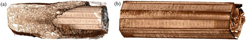
www.nature.com/scientificreports/

                           Figure 2. The proposed digitization pipeline: the X-ray CT scan volume is virtually cleaned by providing a
                           manually extracted template image T. After virtual unwrapping of the cleaned volume, a 2-D texturing process
                           is performed. The result is a 2-D image of the virtually unwrapped scroll that can be read with the naked eye.

                           Figure 3. Illustrated virtual cleaning steps for an exemplary volume slice. (a) The template T is manually
                           extracted by a rectangular selection (orange box) from a central slice. (b) NCC Template matching output for
                           a single angle θ. (c) Output of maximization over all θ NCC Template matching results. In comparison to (b),
                           the slips’ orientation is clearly visible. (d) After smoothing the individual slip center locations (red dots) using
                           RANSAC and PCA, the pixel values within the rectangular template rotated by θ is written to the resulting
                           volume yielding a virtually cleaned scroll.

                               All calculations were performed with the CONRAD framework for cone beam geometries35. For the recon-
                           structions, the commonly used Feldkamp, Davis, Kress (FDK) algorithm was applied consisting of cosine weight-
                           ing, ramp filtering and back projection36. For the 3-D case, the approach extends an exact 2-D reconstruction
                           algorithm for fan-beam projections by adapting the cosine weightings. The FDK algorithm performs well for
                           objects that are cylindrical with a small diameter, however, the output may suffer from cone-beam artifacts as it
                           is rather high37. These artifacts were not observed throughout our experiments. The resulting maximum voxel
                           size for Scroll A was 103 × 103 × 103 and 163 × 163 × 163 for Scroll B. With the used parameters, the scan and
                           reconstruction time was kept relatively low and can be further reduced by decreasing the projection size or using
                           different trajectories (e. g., short scan trajectory)31 or reconstruction algorithms38.

                           Virtual processing of soiled scrolls. After reconstructing the X-ray volume, the digitized content can
                           be investigated. In case of a soiled relic, handling the acquired data is not intuitive as the individual slips can be
                           arbitrarily located with heavy contaminations along their curved surfaces. One approach to gathering the valu-
                           able content from the volume is to manually extract each slip and remove the soil voxel by voxel by hand. The
                           high-resolution volume, however, makes this a very time-consuming issue. In the following section, we propose
                           an image processing pipeline with a minimal amount of user interaction needed resulting in an unwrapped 2-D
                           representation of the scroll’s surface with all writings and drawings readable for the naked eye. The entire digiti-
                           zation pipeline is illustrated in Fig. 2. The unwrapping process can be broken down into three major steps: virtual
                           cleaning, virtual unwrapping, and 2-D texturing.

                           Virtual cleaning. First, the algorithm locates and segments all individual slips of the volume’s scroll resulting
                           in a binary mask where the contamination is removed. This step can be compared with cleaning the scroll in a
                           virtual manner.
                               From a central slice of the soiled volume V, a 2-D template T has to be extracted manually as shown in
                           Fig. 3(a). The orange rectangular selection denotes the template which is the only necessary manual input for the
                           proposed algorithm. As the appearance and shape of all slips are rather constant in z-direction, we apply a nor-
                           malized cross-correlation template matching (NCC) step along that axis. For a 2-D image I, the NCC R at image
                           pixel (x, y) is calculated by evaluating

                                                                         ∑ x ′, y ′(T (x ′ , y ′) ⋅ I (x + x ′ , y + y ′))
                                                       R (x , y ) =                                                                    ,
                                                                                               2                                   2
                                                                      ∑ x ′ , y ′ T (x ′ , y ′) ⋅ ∑ x ′ , y ′ I (x + x ′ , y + y ′)           (2)

Scientific Reports |   (2019) 9:2311 | https://doi.org/10.1038/s41598-019-39447-0                                                                 4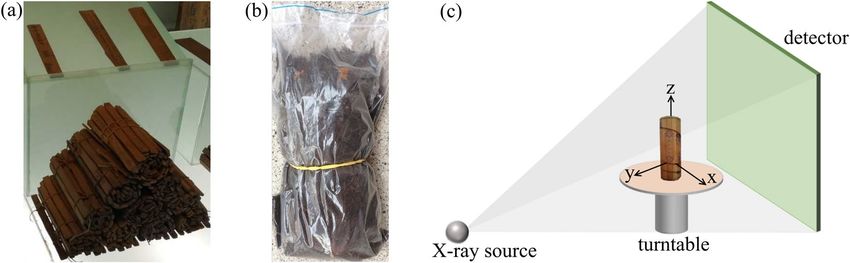
www.nature.com/scientificreports/

                           Figure 4. (a) All class centroids calculated by the k-means algorithm are plotted with different colors (1) and
                           the outliers calculated by RANSAC are marked black. (2) shows a zoom on the box with the black marked
                           outliers which are cropped and (3) the new centroids along the best line fit resulting from the PCA. (b) The top
                           image shows the extracted brightest intensities (green and red points) for each column and a RANSAC curve fit
                           (orange line) through the inlier set (green points). The centered and bottom image show a straight line sampling
                           (green) versus curved sampling (orange). Whereas the straight line sampling shows shadows at the horizontal
                           borders, the curve fit also hits the outer parts of the slips. The row-wise artifacts in the bottom left image are
                           caused by slight misalignment due to missing ECC image alignment.

                           which can be explained by calculating the cross-correlation of the template and the image with subsequent nor-
                           malization by the sum of the squared differences resulting in a 2-D probability map (0: no match, 1: perfect
                           match). Figure 3(a) shows a single template matched image. As the slips can be arbitrarily rotated within the
                           volume, this step is repeated with the template T rotated by various angles θ resulting in a probability map Rθ. For
                           our experiment, the template was rotated with an angular increment of 0.5° in the range of [0°, 360°]. To get a final
                           probability map Rfinal, the maximum value at each pixel (x, y) over all Rθ’s is extracted:
                                                               R final (x , y ) = max θmin≤θ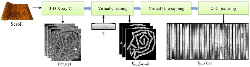
www.nature.com/scientificreports/
Figure 5. (a) Reconstructed and rendered soiled Scroll A volume. The scroll is mostly covered and filled with
soil, however, due to gravity, some slips and their carvings are partly visible. (b) The rendered volume after
virtual cleaning is shown. Nearly all contaminations were removed by the proposed algorithm such that the
individual slips are visible.
alignment technique41. The ECC method estimates the warp matrix between two images by maximizing the cor-
relation coefficient between them. This results in a warp matrix which is applied to the neighboring image. Only
the upper half of the image containing the recto surface of the document was considered because the vascular
bamboo structure could negatively influence the registration results.
The recto and verso layer can be automatically identified by utilizing the material’s density distribution informa-
tion. An increased density results in higher photon absorption and image intensities and when investigating the data,
the recto surface showed increased intensities compared to the verso. Using this information, the recto surface was
detected by searching the maximum values along the depth of the ECC aligned image. The top image of Fig. 4(b)
illustrates this process where green and red dots denote the extracted maxima corresponding to the recto layer.
It can be observed that the recto layer is slightly curved such that straight sampling would neglect the out-
ermost slip areas. Therefore, we apply RANSAC with a second-degree curve fit onto the set of detected maxima
yielding the sampling curve along the surface (Fig. 4(b), orange line). The carvings are correctly detected as out-
lier set (red dots) and omitted from the curve fit. The central and bottom images of Fig. 4(b) show the comparison
of a straight line (dashed green box) versus curved sampling (solid orange box) where the horizontal edges are
shaded due to missed recto voxels. Furthermore, the straight-sampled image (bottom left) shows a slip without
image alignment resulting in slight geometric differences between neighboring rows introducing row-wise arti-
facts (especially visible around the image center). The alignment was deactivated to highlight the importance of
slip-by-slip registration before sampling. This step concludes the proposed algorithm and results in a 2-D rep-
resentation of the unwrapped, cleaned scroll Ifinal where the symbols and drawings are readable by the naked eye.
Results
For evaluating the proposed digitization pipeline, we first scanned and reconstructed the soiled documents. The
scan took about 75 minutes in total for a single scroll. The reconstruction was performed in parallel to the scan
such that the final reconstructed volume was available seconds after the last projection was generated. We initially
cropped the volume to a cuboid including all the relevant information to reduce the large amount of data. Next,
we determined the template image from a central slice of Scroll A and applied the proposed algorithm as illus-
trated in Fig. 2. Throughout the entire pipeline, the algorithms were implemented with CPU multi-threading.
Processing the entire volume took approximately 280 minutes (template matching: 278 min, k-means: 28 s,
RANSAC: 17 s, PCA: 2, 2-D texturing: 2 min). The method’s bottleneck is the template matching process of the
volume V ∈ X×Y×Z with a complexity of approximately O(θXZN2) where N denotes the template’s dimension
(N = 36 × 118, θ = 720, X × Y × Z = 548 × 586 × 1544). In order to reduce the runtime to a minimum, we
increased the angular increment to 1 and downsized the template as well as the xy-slice of the volume by a factor
of 4 for the template matching process. With these adaptations, the runtime decreased to a total of around seven
minutes, which is acceptable and does not influence the output significantly. A GPU-based implementation could
further reduce the runtime.
The rendered raw volume of the 3-D X-ray CT scan can be seen in Fig. 5(a) where due to gravity, the scroll was
partly uncovered. The resulting virtual cleaned volume is shown in Fig. 5(b). Here, the contaminations are mostly
removed and only small areas on the scroll’s surface are still covered. However, this will not affect the final output
and investigations on the slips’ orientation can be applied.
Next, the unwrapping and 2-D texturing steps were performed for receiving a readable version of Scrolls A
content. Figure 6(a) shows a photograph of the unwrapped Scroll A before soiling and wrapping. The virtually
cleaned, unwrapped and 2-D textured image Ifinal of the recto surface is shown in Fig. 6(b). As can be seen, most
of the characters and drawings are readable without application of any post-processing steps. The curved sam-
pling correctly remaps the surface voxels to the 2-D image and all slips appear smooth and in the correct order. To
highlight the X-ray attenuation difference of the same material with varying densities, an exemplary measure-
ment of a single slice was made. Figure 7(a) shows the randomly selected exemplary carving ‘ ’ (translation:
‘positive’). The depth section was extracted at the position of the orange line and shown in Fig. 7(b) (here, the
orange lines correspond to the illustrated layer of Fig. 7(a)). To compare the photon absorption of the different
material densities, four layers were investigated by calculating their mean intensity within the image. These man-
ually segmented layers are shown in Fig. 7(c). The black area corresponds to air with a mean intensity of 3.48. The
dark gray area denotes soil with a mean of 4.47. Brighter spots within this area are minerals with higher attenua-
tion than cellulose. The slip was divided into two areas – the verso and recto layer. The recto surface has a mean
intensity of 5.49 whereas the verso layer reaches 4.95. These measurements were performed on a random basis for
several slices with about the same results. The analysis of the intensities validates the observation that the recto
surface has a higher attenuation than the verso layers such that the recto can be identified automatically. When
Scientific Reports | (2019) 9:2311 | https://doi.org/10.1038/s41598-019-39447-0 6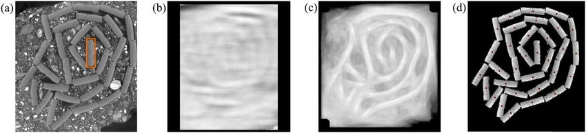
www.nature.com/scientificreports/

                           Figure 6. (a) A photograph of the manually unwrapped original Scroll A. The sample was rolled up, soiled and
                           digitized by the proposed pipeline. (b) The final 2-D output of the reconstructed and processed soiled Scroll A.
                           Although the scroll was heavily contaminated with potting soil, most characters and drawings are clearly visible.

                           Figure 7. (a) Randomly selected carving slice of an extracted slip. At the position of the orange lines, a depth
                           view was extracted shown in (b). The depth view shows the slip surrounded by air and soil. The segmentation
                           map is shown in (c). Recto layer (white), verso layer (light gray), soil (dark gray) and air (black) were manually
                           segmented. An analysis of the mean intensities of the areas reveals that the recto layer’s attenuation differs by
                           29% from the soil’s one.

                           normalizing the mean values according to air, the mean soil X-ray attenuation is 29% higher, whereas the mean
                           recto surface density is even 58% higher. This increase of attenuation leads to the contrast difference between
                           bamboo and carving uncovering the writings and drawings.
                               Finally, we tested the algorithm for Scroll B where the photograph before contamination can be seen in
                           Fig. 8(a). Instead of extracting a template again, the one from Scroll A was used as input, since the scrolls differ
                           only slightly. The results after applying the digitization pipeline are shown in Fig. 8(a). Once more, all writings are
                           readable, however, the drawings appear blurry. This is due to the lower resolution of this scan where the object
                           was placed closer to the X-ray source reducing the magnification. The slips were ordered correctly and were not
                           flipped within the final image.
                               The results show that the non-invasive digitization of excavated, heavily soiled bamboo slips by 3-D X-ray CT is
                           highly promising when the writings and drawings are carved into the bamboo. By using the proposed unwrapping
                           algorithm, a minimal amount of manual effort is needed to receive a readable, digital version of the scrolls content.

                           Discussion
                           In this work, we presented a complete pipeline for a non-invasive digitization of soiled bamboo scrolls composed
                           of individual bamboo slips. Our method may enable libraries and conservators to preserve the structure and writ-
                           ings or drawings of historical bamboo and wooden scrolls. The approach is based on a conventional 3-D X-ray
                           CT scan followed by a fast unwrapping algorithm. Such X-ray systems are widely accessible and more mobile

Scientific Reports |   (2019) 9:2311 | https://doi.org/10.1038/s41598-019-39447-0                                                               7
www.nature.com/scientificreports/
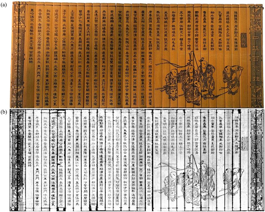
Figure 8. (a) A photograph of the manually unwrapped original Scroll B. Also this sample was rolled up, soiled
and digitized by the proposed pipeline. (b) The final 2-D output of the reconstructed and processed soiled
Scroll B. Although the scroll was heavily contaminated by potting soil, most characters and drawings are clearly
visible.
compared to phase-contrast systems. The scan as well as the proposed algorithm were tuned to address the unique
material characteristics of bamboo. With the user only providing a template image of a single slip, the algorithm’s
output is a 2-D image of the scroll’s recto layer containing the carvings. We could show that even when the docu-
ments were heavily soiled by material of similar chemical structure, their hidden content could be made readable
for the naked eye. With the proposed technique, there is no need to manually clean the scroll before scanning. If
such a scan is performed before the scroll is uncovered by a conservator, writings possibly erased by the cleaning
process are stored and could be recovered instead of being lost forever.
Until now, only carved writings were investigated. Ink brushed onto the surface without being carved into the
surface can only be made readable digitally, if the ink is composed of a material with different X-ray attenuation
than the document media and its surrounding. Also the density difference of cellulose and soil is rather small.
To improve the contrast between document, writings, and soil, phase-contrast and X-ray dark-field imaging can
be used instead of conventional X-ray CT. Both methods are able to reveal unique information about structural
variations at sub-micron scale42–46.
The digital volume generated by 3-D X-ray CT can be very helpful for conservators. Usually, the slips are arbi-
trarily located in the soil due to rotten strings and material decay and only very few cases exist where the entire
scroll was found intact. According to works of E. Giele47,48, determining the original composition of the slips is a
very challenging but highly important task for researchers, who have to mark the excavated slips, draw a location
map, and reconstruct everything within their laboratory. T. Staack9 shows an exemplary drawing. The work also
mentions brushed or knife-cut lines on the verso of slips (‘verso-lines’), which can indicate the original position
of a slip. Furthermore, mirrored writings on the verso were present, imprinted by a touching slip’s recto. When
using the proposed non-invasive CT technique, those imprints could be automatically detected by applying image
transformations and registration approaches. In addition, all information is stored in a digital manner and meth-
ods such as augmented reality or 3-D printing of the virtually cleaned volume can be used to further investigate
the original spatial information. The technique can be extended for all kinds of woodcuts, archaeological findings
or forensics when the material composition differs from the contamination material.
The generated data can be a basis for future algorithms in the field of document processing. Therefore, the
acquired scan data and the code will be made publicly available to enable future research. We will also create a
publicly available database with more scan data, where the data from this work could serve as ground truth seg-
mentation for machine-learning algorithms. All generated data and code from this work will be made available at
https://www5.cs.fau.de/research/data/bamboo-scroll-dataset/.
Scientific Reports | (2019) 9:2311 | https://doi.org/10.1038/s41598-019-39447-0 8www.nature.com/scientificreports/
The proposed work provides a basis for non-invasive digitization of ancient bamboo and wooden scrolls
where the greatest challenge lies in the high variability of the excavated documents and the surrounding soil. For
future work, we plan to investigate real historical slips or scrolls. The success of imaging those excavated scrolls
with the proposed method depends on their state of decay. Fengel et al.49 state that fossilized timbers showed
decreased densities and Hamid et al.50 showed that some bamboo species are more resistant to fungi than others.
Hence, the density of bamboo slips is highly depended on the used bamboo species and the surrounding soil. It
follows that the attenuation values of both materials approach similar levels. The proposed slip detection method
(virtual cleaning) is mainly based on the appearance of the slips. When it comes to differing appearances of slips
in the volume due to deterioration, the proposed algorithm will no longer detect the writing media. In this case,
machine-learning algorithms could help to virtually clean the volume by turning the process around: the con-
servators could scan an amount of surrounding soil from nearby the excavation location. Based on this data, an
algorithm could be trained to learn the soil’s appearance and afterward segment the scroll in the scroll-included
volume.
References
1. Yu, X. Language and state: A theory of the progress of civilization (UPA, 2015).
2. Wu, K. The Chinese book: Its evolution and development. T’ien-hsia Mon. (Shanghai) 3, 25288 (1936).
3. Swider, J. R., Hackley, V. A. & Winter, J. Characterization of Chinese ink in size and surface. J. Cult. Herit. 4, 175–186 (2003).
4. Clements, J. The art of war: a new translation (Hachette UK, 2012).
5. Yates, R. D. The yin-yang texts from yinqueshan: an introduction and partial reconstruction, with notes on their significance in
relation to huang-lao daoism. Early China 19, 75–144 (1994).
6. Chen, S. A summarize of the qin slips collected by yuelu academy. Cul. Relics 3, 010 (2009).
7. Jane, Q. Ancient times table hidden in Chinese bamboo strips, https://www.nature.com/news/ancient-times-table-hidden-in-
chinese-bamboo-strips-1.14482 (2014).
8. Yang, J. & Xu, C. Marquis of Haihun’s tomb of the western Han dynasty in Nanchang, Jiangxi. Chin. Archaeol. 17, 44–59 (2107).
9. Staack, T. The neglected “backyard” of early Chinese manuscripts: How an analysis of the verso of bamboo slips can enable the
reconstruction of a manuscript roll. In Giele, E. & Venture, O. (eds) Proceedings of the Fourth EASCM Workshop in Paris 2012 (2012).
10. Redo-Sanchez, A. et al. Terahertz time-gated spectral imaging for content extraction through layered structures. Nat. Commun. 7,
12665 (2016).
11. Mocella, V., Brun, E., Ferrero, C. & Delattre, D. Revealing letters in rolled herculaneum papyri by X-ray phase-contrast imaging. Nat.
communications 6, 5895 (2015).
12. Bukreeva, I. et al. Virtual unrolling and deciphering of herculaneum papyri by X-ray phase-contrast tomography. Sci. Reports 6,
27227 (2016).
13. Ludwig, V. et al. Non-destructive testing of archaeological findings by grating-based X-ray phase-contrast and dark-field imaging. J.
Imaging 4, 58 (2018).
14. Morigi, M., Casali, F., Bettuzzi, M., Brancaccio, R. & d’Errico, V. Application of X-ray computed tomography to cultural heritage
diagnostics. Appl. Phys. A: Mater. Sci. & Process. 100, 653–661 (2010).
15. Chhem, R. K. & Brothwell, D. R. Paleoradiology: Imaging mummies and fossils (Springer Science & Business Media, 2007).
16. Stromer, D. et al. Browsing through sealed historical manuscripts by using 3-D computed tomography with low-brilliance X-ray
sources. Sci. Reports 8, 15335, https://doi.org/10.1038/s41598-018-33685-4 (2018).
17. Rosin, P. L. et al. Virtual recovery of content from X-ray micro-tomography scans of damaged historic scrolls. Sci. Reports 8, 11901
(2018).
18. Albertin, F. et al. Virtual reading of a large ancient handwritten science book. Microchem. J. 125, 185–189 (2016).
19. Seales, W. B. et al. From damage to discovery via virtual unwrapping: Reading the scroll from En-Gedi. Sci. Adv. 2 (2016).
20. Stromer, D., Christlein, V., Anton, G., Kugler, P. & Maier, A. 3-D reconstruction of historical documents using an X-ray c-arm ct
system. In University, M. (ed.) Proceedings of the 31th conference on Image and Vision Computing New Zealand 2016, 1–6 (2016).
21. Stromer, D., Christlein, V., Schön, T., Holub, W. & Maier, A. Browsing through closed books: fully automatic book page extraction
from a 3-D X-ray ct volume. In IEEE (ed.) The 14th IAPR International Conference on Document Analysis and Recognition, 224–229
(2017).
22. Charlesby, A. The degradation of cellulose by ionizing radiation. J. Poly. Sci. Part A: Polym. Chem. 15, 263–270 (1955).
23. Mantler, M. & Klikovits, J. Analysis of art objects and other delicate samples: Is xrf really nondestructive? Powder Diffr. 19, 16–19
(2004).
24. Mills, D. et al. Apocalypto: revealing the unreadable. In Developments in X-Ray Tomography VIII, vol. 8506, 85060A (International
Society for Optics and Photonics, 2012).
25. Stromer, D. et al. Dose reduction for historical books digitization by 3-D X-ray ct. In of Applied Sciences Upper Austria, U. (ed.)
Proceedings of 8th Conference on Industrial Computed Tomography (iCT 2018), 1–2 (2018).
26. Immel, A. et al. Effect of X-ray irradiation on ancient dna in sub-fossil bones–guidelines for safe X-ray imaging. Sci. reports 6, 32969
(2016).
27. Stromer, D. et al. Non-destructive digitization of soiled historical Chinese bamboo scrolls. In IEEE (ed.) 2018 13th IAPR Workshop
on Document Analysis Systems (DAS 2018), 55–60, https://doi.org/10.1109/DAS.2018.37 (2018).
28. Sharma, B. et al. Chemical composition of processed bamboo for structural applications. Cellul. 25, 3255–3266 (2018).
29. Li, X. Physical, chemical, and mechanical properties of bamboo and its utilization potential for fiberboard manufacturing. Master’s
thesis, Louisiana State University and Agricultural and Mechanical College (2004).
30. Huang, P., Chang, W.-S., Ansell, M. P., Chew, Y. J. & Shea, A. Density distribution profile for internodes and nodes of phyllostachys
edulis (moso bamboo) by computer tomography scanning. Constr. and Build. Mater. 93, 197–204 (2015).
31. Maier, A., Steidl, S., Christlein, V. & Hornegger, J. Medical imaging systems - an introductory guide (Springer International Publishing,
2018).
32. Poludniowski, G. G. Calculation of X-ray spectra emerging from an X-ray tube. part ii. X-ray production and filtration in X-ray
targets. Med. physics 34, 2175–2186 (2007).
33. Stromer, D. et al. Comparison of SART and eTV reconstruction for increased c-arm ct volume coverage by proper detector rotation
in liver imaging. In 2016 IEEE 13th International Symposium on Biomedical Imaging, 589–592 (IEEE, 2016).
34. Stromer, D., Schön, T., Holub, W. & Maier, A. 3-d reconstruction of iron gall ink writings. In Leuven, K. (ed.) Proceedings of 7th
Conference on Industrial Computed Tomography (iCT 2017), 136–137 (2017).
35. Maier, A. et al. CONRAD - a software framework for cone-beam imaging in radiology. Med. Phys. 40, 111914-1–111914-8 (2013).
36. Feldkamp, L., Davis, L. & Kress, J. Practical cone-beam algorithm. J. Opt. Soc. Amer. 1, 612–619 (1984).
37. Scarfe, W. C. & Farman, A. G. What is cone-beam ct and how does it work? Dental Clin. North Am. 52, 707–730 (2008).
38. Huang, Y. et al. Image quality analysis of limited angle tomography using the shift-variant data loss model. In Bildverarbeitung für
die Medizin 2016, 277–282 (Springer, 2016).
Scientific Reports | (2019) 9:2311 | https://doi.org/10.1038/s41598-019-39447-0 9www.nature.com/scientificreports/
39. MacQueen, J. Some methods for classification and analysis of multivariate observations. In Proceedings of the Fifth Berkeley
Symposium on Mathematical Statistics and Probability, vol. 1, 281–297 (Oakland, CA, USA, 1967).
40. Fischler, M. A. & Bolles, R. C. Random sample consensus: A paradigm for model fitting with applications to image analysis and
automated cartography. In Readings in computer vision, 726–740 (Elsevier, 1987).
41. Evangelidis, G. D. & Psarakis, E. Z. Parametric image alignment using enhanced correlation coefficient maximization. IEEE
Transactions on Pattern Analysis Mach. Intell. 30, 1858–1865 (2008).
42. Momose, A., Takeda, T., Itai, Y. & Hirano, K. Phase-contrast X-ray computed tomography for observing biological soft tissues. Nat.
medicine 2, 473–475 (1996).
43. Wilkins, S., Gureyev, T. E., Gao, D., Pogany, A. & Stevenson, A. Phase-contrast imaging using polychromatic hard X-rays. Nat. 384,
335 (1996).
44. Cloetens, P., Barrett, R., Baruchel, J., Guigay, J.-P. & Schlenker, M. Phase objects in synchrotron radiation hard X-ray imaging. J.
Phys. D: Appl. Phys 29, 133 (1996).
45. Pfeiffer, F. et al. Hard-X-ray dark-field imaging using a grating interferometer. Nat. materials 7, 134 (2008).
46. Lak, M. et al. Phase contrast X-ray synchrotron imaging: opening access to fossil inclusions in opaque amber. Microsc. microanalysis
14, 251–259 (2008).
47. Giele, E. Using early Chinese manuscripts as historical source materials. Monum. Serica 51, 409–438 (2003).
48. Giele, E. Excavated manuscripts: Context and methodology. China’s Early Empires: A Re-appraisal 114–34 (2010).
49. Fengel, D. Aging and fossilization of wood and its components. Wood Sci. Technol. 25, 153–177 (1991).
50. Hamid, N. H., Sulaiman, O., Mohammad, A. & Ahmad Ludin, N. The decay resistance and hyphae penetration of bamboo
gigantochloa scortechinii decayed by white and brown rot fungi. Int. J. For. Res. 2012 (2012).
Author Contributions
D.S., X.H. and A.M. conceived the original idea. D.S. and P.Z. prepared the scrolls and performed the X-ray scans.
D.S. developed the image processing pipeline. D.S. and V.C. wrote the main part of the manuscript. A.M., X.H.
and T.H. provided expertise through intense discussions. All authors reviewed the manuscript.
Additional Information
Competing Interests: The authors declare no competing interests.
Publisher’s note: Springer Nature remains neutral with regard to jurisdictional claims in published maps and
institutional affiliations.
Open Access This article is licensed under a Creative Commons Attribution 4.0 International
License, which permits use, sharing, adaptation, distribution and reproduction in any medium or
format, as long as you give appropriate credit to the original author(s) and the source, provide a link to the Cre-
ative Commons license, and indicate if changes were made. The images or other third party material in this
article are included in the article’s Creative Commons license, unless indicated otherwise in a credit line to the
material. If material is not included in the article’s Creative Commons license and your intended use is not per-
mitted by statutory regulation or exceeds the permitted use, you will need to obtain permission directly from the
copyright holder. To view a copy of this license, visit http://creativecommons.org/licenses/by/4.0/.
© The Author(s) 2019
Scientific Reports | (2019) 9:2311 | https://doi.org/10.1038/s41598-019-39447-0 10You can also read