Virtual reality and annotated radiological data as effective and motivating tools to help Social Sciences students learn neuroanatomy
←
→
Page content transcription
If your browser does not render page correctly, please read the page content below
www.nature.com/scientificreports
OPEN Virtual reality and annotated
radiological data as effective
and motivating tools to help
Social Sciences students learn
neuroanatomy
Margot van Deursen1,4, Laura Reuvers1,4, Jacobus Dylan Duits3, Guido de Jong3,
Marianne van den Hurk1 & Dylan Henssen2*
Neuroanatomy as a subject is important to learn, because a good understanding of neuroanatomy
supports the establishment of a correct diagnosis in neurological patients. However, rapid changes
in curricula reduced time assigned to study (neuro)anatomy. Therefore, it is important to find
alternative teaching methods to study the complex three-dimensional structure of the brain. The
aim of this manuscript was to explore the effectiveness of Virtual Reality (VR) in comparison with
Radiological Data (RaD) as suitable learning methods to build knowledge and increase motivation for
learning neuroanatomy. Forty-seven students (mean age of 19.47 ± 0.54 years; 43 females; 4 males)
were included; 23 students comprised the VR group. Both methods showed to improve knowledge
significantly, the improvement between groups was not different. The RaD group showed to have a
significantly higher score on expectancy than students in the VR group. Task value scores regarding
finding a task interesting, useful and fun were found to be significantly different in favor of the VR
group. Consequently, significant higher Motivation scores were found in the VR group. Motivation and
expectancy, however, did not moderate learning results, whereas task value impacted the results in
favour of the VR group. This study concludes that VR and RaD are effective and diverting methods to
learn neuroanatomy, with VR being more motivating than RaD. Future research should investigate
motivation and task value when using VR over a longer period of time.
Over recent years, medical and psychology curricula have changed drastically, resulting in a renewed focus on
communication skills and less time devoted to study anatomy1–4. Especially the subspecialty of neuroanatomy
suffers from these reductions considering that students find it difficult to understand the three-dimensional
(3D) relations of different brain structures5. Traditionally, neuroanatomy in medical and psychology curricula is
learned by lectures and a combination of brain dissection, the use of (plastinated) prosections, plastic anatomical
models and studying neuroanatomical atlases5–10. Furthermore, some faculties have described to use brightly
colored clay to help students learn neuroanatomical structures11,12.
In order to optimize learning during the sparse hours of neuroanatomy education, technological innovations
have been suggested as a valuable addition to other neuroanatomy education tools. These technological innova-
tions include various forms of electronic 3D m odels13–15. Generally, these electronic 3D models are based on
consecutively stacked two-dimensional (2D) data, including radiological imaging data (e.g., magnetic resonance
imaging; MRI, computed tomography; CT). Incorporating screen-based radiological data (RaD) in anatomy
education has been reported frequently in literature.
The use of post-processed data in the form of electronic 3D models comprises various options, including
virtual reality (VR) models. VR was introduced in the 1960’s and is defined as a computer-generated simula-
tion of a 3D environment that can be interacted with in a seemingly real or physical way by the user16. One of
1
Department of Educational Sciences, Faculty of Social Sciences, Radboud University, Nijmegen, The
Netherlands. 2Department of Medical Imaging, Radboud University Medical Center, Nijmegen, The
Netherlands. 3Radboudumc 3D Lab, Radboud University Medical Center, Nijmegen, The Netherlands. 4These
authors contributed equally: Margot van Deursen and Laura Reuvers. *email: dylan.henssen@radboudumc.nl
Scientific Reports | (2021) 11:12843 | https://doi.org/10.1038/s41598-021-92109-y 1
Vol.:(0123456789)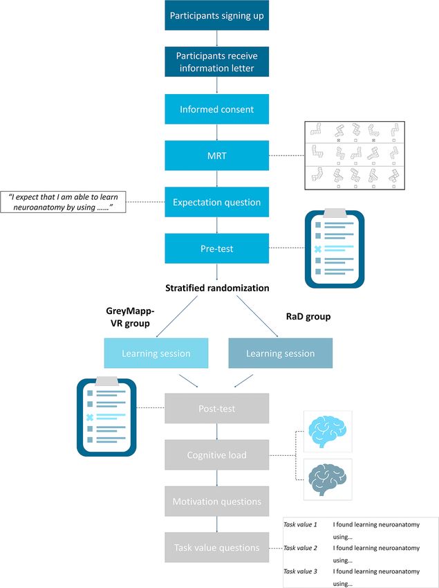
www.nature.com/scientificreports/
the advantages of VR is that it provides the opportunity to practice situations that are potentially dangerous to
practice in real life or cannot be experienced physically (e.g., physical inaccessibility). The use of VR has been
suggested to be a valuable asset for a broad variety of challenges, including treatment of gambling addiction17
and depression18, neurorehabilitation19,20 and pain reduction21. In medical educational research, VR has been
reported to have a positive effect on learning a bilities22–25. In anatomy education, VR environments can be imple-
mented as less expensive and promising alternative to cadaver dissection26. Additionally, VR has been reported
as an effective learning tool, especially for students with lower spatial a bilities25,27,28. Furthermore, the use of VR
in anatomy education promotes intrinsic benefits such as increased learner immersion and e ngagement29–32.
With regard to RaD, most medical curricula are known to use imaging data in their anatomy education
programs. These teaching approaches have steadily developed since the 1970s when the first reports on the
use of X-rays in anatomy education were p ublished33. Nowadays, the vast majority of medical curricula have
integrated some sort of radiological imaging into their anatomy education34–36. Over the years, integration of
clinical images obtained from high-resolution CT and MRI scans has been described in literature as well (for
an overview, see37). The implementation of these imaging data in anatomy education has propelled the develop-
ment of RaD in the form of online radiology atlases with labeled radiology d ata38,39. It has been reported that
the integration of RaD in anatomy education allows students to acquire a spatial understanding of anatomical
structures and their complex relative p ositions40–43. However, the implementation of RaD features in psychology
education remains understudied.
Both the use of RaD and VR with 3D models are considered interactive multimodal learning environments
as at least two different modes are used to represent the learning content44. Multimodal learning environments
can be interactive and non-interactive44. Classification of the different types of interactivity concern dialoguing,
controlling, manipulating, searching and navigating. Both RaD and VR can be considered as manipulating inter-
active modalities as the learner can use the learning tool to zoom in or out, move objects around and interact in
other ways with virtual data44. Next to determining whether a tool serves non-interactive or interactive learning,
a distinction can be made between two views of learning: information acquisition and knowledge construction45.
With information acquisition learning involves adding information to the learner’s memory. The learner’s job is to
receive information. The most fitting learning environment for information acquisition is a non-interactive envi-
ronment. Knowledge construction, on the other hand, involves building a mental representation. The learner’s
job here is to select, organize and integrate new information with existing knowledge. The goal of the instruc-
tion method is to guide the learner to actively fathom the instructional m aterials46. The most fitting learning
environment for knowledge construction is an interactive multimodal learning environment47 as incorporating
interactivity can promote deeper learning from a multimedia explanation if it is done in a theory-based way.
Thereby, both RaD and VR are hypothesized to be effective methods to help students learn (neuro)anatomy.
Interactive multimodal learning environments such as VR and RaD also motivate learners to engage in the
cognitive processes48. Because of this, when assessing the value of a new teaching method, it is important to
consider their effect on student motivation. Moreno and Mayer (2007) support this statement by stating that
motivational factors mediate learning by increasing or decreasing cognitive e ngagement44. Parong and Mayer
reported that a lesson on scientific information in a VR environment was experienced as more enjoyable and
motivating as compared to an equivalent lesson in a PowerPoint s lideshow49. In turn, motivation is known to
result in better quality of l earning50. Individuals’ expectancies for success on a specific task and the value of the
task are other important determinants of their motivation on achievement tasks51. The expectancy-value theory
has been one of the most important views on the nature of achievement motivation. Thereby, a relationship can
be identified between competence beliefs and expectancies for success (for an extensive overview s ee51). However,
to which extend these educational and motivational theories actually play a role in (neuro)anatomy education,
and especially the use of RaD and VR with 3D models, has not been investigated before.
The current study therefore aimed to explore the effectiveness of VR in comparison with RaD as suitable
learning methods to build knowledge. In addition, we aimed to investigate students’ opinions with regard to
task value and level of motivation when learning neuroanatomy by either RaD or VR. The authors hypothesized
that students would develop a superior comprehension of the 3D relationship of the structures of the human
brain when having worked with VR when compared to students who worked with RaD. Also, we believed that
the students who worked with VR would be more engaged when learning neuroanatomy and that this would
increase their level of motivation and positively influence experienced task value. The outcomes of this study
can provide other educators with new insights how to effectively engage students when teaching neuroanatomy.
Also, these outcomes will show teachers and students which technological innovations can be used to yield the
greatest effect with regard to learning neuroanatomy.
Materials and methods
Ethical approval and participants. All methods carried out within this study were in agreement with
the Statement on the Declaration of Helsinki and the Ethical Conduct of Clinical Studies. In addition, the study
details were approved by Ethical Review Board of The Netherlands Association for Medical Education (NVMO)
and registered (NERB dossier number 2018.8.2).
Students from the Faculty of Social Sciences at Radboud University were recruited to participate in this study.
Students participated voluntarily and were recruited by using recruiting announcements, online advertisements
and posters. Students signed up by sending an e-mail to one of the researchers (M.v.D.). Students who reported
to have previously studied neuroanatomy were excluded due to the strong link between anatomy training and
spatial ability (for reviews and meta-analyses s ee52,53). We deliberately chose to include students without an
educational background in natural sciences, technology and mathematical reasoning as they are known to have
Scientific Reports | (2021) 11:12843 | https://doi.org/10.1038/s41598-021-92109-y 2
Vol:.(1234567890)www.nature.com/scientificreports/
inferior spatial and visual skills54. Also, this group of students represents a relatively understudied student popu-
lation who might benefit from the innovations with regard to anatomy education as well as medical s tudents55.
Study design. The study followed a pretest–posttest design. After signing the informed consent forms, par-
ticipants were randomized over two groups. Randomization was carried out following the block randomization
method to result in balanced groups with regard to number of included participants as well as with regard to
gender56. Participants were informed which teaching tool they were going to work with prior to the pre-test. The
first group used GreyMapp-VR to learn neuroanatomy (i.e., the VR-group). The second group learned neuro-
anatomy by using a laptop on which the annotated MRI data of GreyMapp was presented (i.e., the RaD-group).
Participants started the experiment by taking the mental rotations test (MRT) to assess spatial abilities. Then,
expectancy questions were answered by the participants, followed by a pre-test on neuroanatomy. Thereafter,
students were given the opportunity to work with their randomly assigned learning method for a maximum of
five minutes. When participants expressed that they understood the programs that they were supposed to work
with, they received their assignments (on paper or within the VR environment) which they could use to study
neuroanatomy in 30 min. The overall assignment was to study the structures’ names, their three dimensional
characteristics and relationship to the surrounding structures. Participants were free to choose their learning
strategy. After studying, a post-test similar to the pre-test was presented, which was to be followed by questions
assessing task value and motivation (Fig. 1).
All participants were allowed to choose a preferred timeslot with the duration of 90 min to take part in the
experiment. Each participant participated individually under the supervision of two of the investigators (M.v.D.
and L.R.).
Teaching applications. The present study uses an in-house-written application called GreyMapp-VR.
GreyMapp was developed and first launched in 201657. GreyMapp was designed with the aim of helping students
master the 3D anatomy of the human brain in a modern and innovative way. An augmented reality version of
GreyMapp has already been reported on before15 and is now freely available within the App Store (https://apps.
apple.com/nl/app/greymapp/id1533656197?l=en).
GreyMapp-VR allows users to navigate a 3D brain model while being completely embedded in a VR-envi-
ronment. The 3D model contains the ventricular system with the internal capsule and the major structures of the
basal ganglia and the limbic system surrounding it. Source data of GreyMapp was derived from an annotated,
7 T post-mortem MRI scan of the human brain. The annotated source data was used for the RaD application in
this experiment. Methods that were used to acquire the MR data and which were used to construct GreyMapp
have been published by our group before15,57.
GreyMapp-VR was created by an affiliated software engineer using C# within Unity v 2018.4.5F (LTS) (Unity
Technologies ApS, San Francisco, CA). Students were equipped with the HTC Vive Focus Plus (https://enterprise.
vive.com/us/product/focus-plus/, accessed: 30-09-2020). The HTC Vive Focus Plus is a standalone VR headset
with two controllers which support 6 degrees of freedom, indicating that rotation and spatial movements are
tracked whilst using them. In the environment of GreyMapp-VR, the 3D model is positioned on a laboratory
table. On the user’s right-handed-side, a cheat sheet could be found which helped participants to help remember
anatomical orientations and -structures. Participants could walk around the VR-model, in addition to rotate the
model 360 degrees using the controllers. The controllers could also be used to dismantle the VR-model and to
point out different structures. When pointing at a structure, the name of the particular structure would appear.
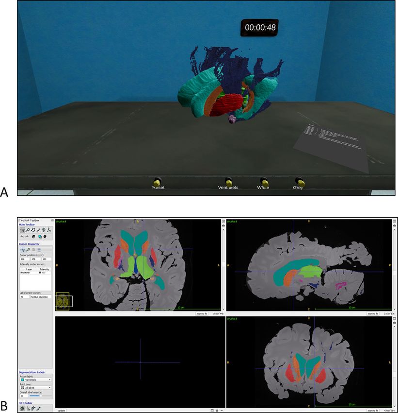
Figure 2A shows a screenshot of GreyMapp-VR.
The control group worked with the annotated MRI scan on which the GreyMapp-project is based. Images and
annotations were shown by use of the software ITK-SNAP, which is a general-purpose interactive tool for image
visualization58. ITK-SNAP provided an environment for visualizing complex 3D imaging data sets and offered
linked visualization of two-dimensional image cross sections (i.e., sagittal-, coronal- and transversal planes).
ITK SNAP allowed participants to work with MR data like radiologists by scrolling through the data using a
cross-hair tool. In Fig. 2B, a screenshot of the built ITK-SNAP module is shown. Again, a cheat sheet could be
found which helped participants to remember anatomical orientations and -structures.
Models used in GreyMapp-VR and in ITK SNAP were color-coded using red–green–blue-alpha (RGBA)
codes that were identical for both teaching applications.
Testing instruments and measures. The pre-test and the post-test were conducted to measure the
knowledge of neuroanatomy. The content and format of the tests was constructed to reflect identical con-
tent. Pre- and posttest each consisted out of two parts. The first part comprised an extended matching test
that included ten questions on spatial relations of different neuroanatomical structures. An example question
is: ‘Which of the abovementioned structures is situated between the nucleus caudate and the fornix?’ (Cor-
rect answer: Lateral Ventricle). Maximum score for this part was 10 points. The second part of the tests was a
multiple-choice test which included 20 questions (maximum score was 20 points). An example question is: ‘The
capsula interna is situated medial/lateral from the putamen’ (Correct answer: Medial). Each question had two
answer options. Tests were designed based on previous empirical experience and scientific papers produced by
our group and others11,15. Based on Bloom’s taxonomy, the tests assessed learning information belonging to the
Application Dimension. Learners were tested whether or not they were capable of implementing abstractions
that were both similar and different from the learning situation59. None of the questions were accompanied by
figures or pictures.
Scientific Reports | (2021) 11:12843 | https://doi.org/10.1038/s41598-021-92109-y 3
Vol.:(0123456789)www.nature.com/scientificreports/
Figure 1. Schematic representation of the study design. MRT Mental rotation test, VR Virtual reality.
Pre-test and post-test were similar testing forms with a maximum score of 20 points each. Correction of
guessing was carried o ut60 for the multiple choice questions to include choice weighting. Scores could not be
lower than 0 points.
To assess variable spatial ability, the mental rotation test was used61, previously validated62 and redrawn by
Peters and c olleagues63. The MRT scores were used to assess whether significant differences existed in spatial
ability. Students were awarded with a point when both of the stimulus figures that match the target figure were
identified correctly. No credit was given for a single correct answer. Maximum MRT-score was 24 points.
The moderator expectancy value was measured with one question and aimed to measure the participants’
expectation on how well they thought they were going to perform using the teaching method. The question for
the participants in the VR group was: ‘I expect that I am able to learn neuroanatomy by using Virtual Reality’.
The question for the participants in the control group was: ‘I expect that I am able to learn neuroanatomy by
using the RaD’. Answers were given on a 5-point Likert scale ranging from ‘I strongly disagree’ to ‘I strongly
agree’. The moderator task value was measured with three questions and aimed to measure how the participants
valued the teaching method that they used. The questions were extracted from the Motivated Strategies for
Learning Questionnaire64 and adapted to fit this study. The answers provided an indication of how participants
Scientific Reports | (2021) 11:12843 | https://doi.org/10.1038/s41598-021-92109-y 4
Vol:.(1234567890)www.nature.com/scientificreports/
Figure 2. Exemplary images of the used teaching applications. (A) VR environment showing the 3D model
in the VR environment (view on the brain model in the VR group. This figure shows the in-house created VR
environment, constructed with Unity software package; https://unity.com). (B) Coronal, sagittal and coronal
sections of the RaD environment (view on the screen in the control group; http://www.itksnap.org)58.
felt about the value of the teaching method by saying how interesting, fun and useful they found the teaching
method (Table 1).
The covariable motivation was measured with one question to investigate how motivated participants felt
when using their teaching method. This question was scored on a scale from 1 (not motivated) to 10 (very
motivated). Cognitive load was assessed by use of a set of questions which have been previously investigated
and reported65,66.
Statistical analysis. The statistical package SPSS Statistics, version 26 (IBM Corp., Armonk, NY) was used
for statistical analyses. The Cronbach’s alpha test was carried out to assess whether (1) the pre-test and post-test
and (2) the experience task questions were internally consistent (i.e., assessing internal validity and reliability).
In addition, McDonald’s omega coefficient was used to assess internal reliability of both tests as well. To use
Scientific Reports | (2021) 11:12843 | https://doi.org/10.1038/s41598-021-92109-y 5
Vol.:(0123456789)www.nature.com/scientificreports/
Experience task value
Task value 1 I found learning neuroanatomy using GreyMapp-VR/RaD interesting
Task value 2 I found learning neuroanatomy using GreyMapp-VR/RaD fun
Task value 3 I found learning neuroanatomy using GreyMapp-VR/RaD useful
Table 1. Questions to measure the experienced task value. Adapted from (Pintrich, 1991)64.
Variable Total group (n = 47) GreyMapp-VR group (n = 23) Control group (n = 24) p-value Cohen’s d
Age 19.47 ± 3.73 19.09 ± 4.58 19.83 ± 2.73 0.504 0.20
4 males 2 males 2 males
Gender 0.965 0.013
43 females 21 females 22 females
42 psychology 20 psychology
3 pedagogical sciences 1 pedagogical sciences 22 psychology
Study 0.407 0.24
1 law 1 law 2 pedagogical sciences
1 sociological sciences 1 sociological sciences
MRT 12.28 ± 2.71 12.70 ± 2.77 11.88 ± 2.64 0.304 0.30
Expectation 3.87 ± 0.61 3.70 ± 0.70 4.04 ± 0.46 0.019 0.58
Pre-test score 2.45 ± 3.27 2.57 ± 3.5 2.33 ± 3.10 0.900 0.04
Post-test score 12.94 ± 6.08 13.04 ± 5.99 12.83 ± 6.3 0.774 0.08
Cognitive load score 6.30 ± 1.50 6.09 ± 1.2 6.50 ± 1.75 0.352 0.30
Motivation 6.70 ± 1.92 7.78 ± 1.20 5.67 ± 1.93 < 0.001 1.31
Task value Interesting 4.06 ± 0.90 4.57 ± 0.51 3.58 ± 0.93 < 0.001 1.30
Task value fun 3.53 ± 1.16 4.26 ± 0.69 2.83 ± 1.09 < 0.001 1.56
Task value useful 4.06 ± 0.79 4.30 ± 0.56 3.8 ± 0.92 < 0.001 0.62
Table 2. Baseline characteristics per group and p-values of the inductive statistics. MRT Mental rotation test,
VR Virtual reality.
McDonald’s omega coefficient, the methodology as described by Hayes et al. and the accompanying software
extension which can be integrated into SPSS Statistics was used67. Internal consistency is generally regarded
acceptable when both Cronbach’s alpha value and McDonald’s omega coefficient was ≥ 0.7.
Statistics for the scores on the pre-test, post-test, mental rotation test, expectation-, task value- and motiva-
tion scores were calculated. Paired and unpaired student’s t-tests were applied to compare mean scores between
the (1) pre-test results; (2) post-test results; (3) MRT scores; (4) expectation scores; (5) task value questions; (6)
motivation scores; and (7) cognitive load questions between the GreyMapp-VR group and the control group.
To analyze whether the GreyMapp-AR group and the control group had different distributions of categorical
parameters (i.e., sex), a chi-squared test was conducted. Repeated-Measures Anova was done to analyze the
effect of the different teaching methods. The variables expectation, task value and motivation were added to a
Repeated Measures analysis with a covariable to investigate whether these had moderating effects. A Bonferroni
post-hoc test was carried out to test for multiple comparisons. Furthermore, correlations between MRT score
and post-test results were investigated by use of the Pearson Correlation Coefficient.
Results
An overview of the included participants of this study is provided in Table 2. In total, 47 students with a mean age
of 19.47 ± 0.54 years were included. Forty-three of the participants (91.5%) identified as female; four participants
identified as male (8.5%). The majority of students (89.4%) studied Psychology; 6.4% of the participants studied
Pedagogical Sciences. One student (2.1%) studied Sociological sciences and one student (2.1%) studied Law. The
used anatomical tests and task value questions were found to be internally consistent with a Cronbach’s alpha of
0.803 and 0.814, respectively and a McDonald’s omega of 0.718 and 0.857, respectively. Corrected mean pre- and
post-test scores, as well as other mean scores can be found in Table 2.
When comparing the students in the GreyMapp-VR group versus the control group, no significant differ-
ences were found in gender-distribution (p = 0.965; Cohen’s d = 0.01; df = 1), study direction (p = 0.407; Cohen’s
d = 0.24; df = 5) and distribution of age (p = 0.504; Cohen’s d = 0.20; F = 0.224; df = 45). No significant differences
were found between the corrected pre- and post-test scores between groups (p = 0.900; Cohen’s d = 0.04; F = 0.043;
df = 45 and p = 0.774; Cohen’s d = 0.08; F = 0.043; df = 45 respectively). No differences were found between groups
with regard to mental rotation test (p = 0.913; Cohen’s d = 0.30; F = 0.012; df = 45).
With regard to expectancy, the RaD group showed to have a significantly higher score than the students in
the VR group (p = 0.019; Cohen’s d = 0.58; F = 5.972; df = 45). Task value scores regarding finding a task inter-
esting, useful and fun were found to be significantly different (all p < 0.001; Cohen’s d = 1.30; F = 4.567; df = 45,
Cohen’s d = 0.62; F = 0.553; df = 45 and Cohen’s d = 1.56; F = 7.053; df = 45 respectively) in favor of the Grey-
Mapp-VR group. Motivation was also found significantly different between groups in favor of the VR group
Scientific Reports | (2021) 11:12843 | https://doi.org/10.1038/s41598-021-92109-y 6
Vol:.(1234567890)www.nature.com/scientificreports/
(p < 0.001; Cohen’s d = 1.31; F = 3.473; df = 45). Cognitive load scores were found not to be different between
groups (p = 0.352; Cohen’s d = 0.30; F = 0.645; df = 45).
Repeated measures ANOVA showed a significant improvement in test results when comparing the pre-test
and post-test results (p < 0.0001; Partial Eta Squared = 0.766; F = 158.76; df = 47). Significant differences were
found when comparing pre-test and post-test results in the VR-group (p < 0.0001; Partial Eta Squared = 0.749;
F = 109.24; df = 22) and control-groups (p < 0.0001; Partial Eta Squared = 0.783; F = 85.52; df = 23). No significant
differences were found in improvement of test scores between groups (p = 0.408; Partial Eta Squared = 0.546;
F = 0.70; df = 47).
To analyse the moderating effect of expectancy, task value and motivation on learning outcomes, a Repeated
Measures ANOVA with covariates was performed. These analyses showed that motivation and expectancy did
not moderate this relationship (p = 0.981; Partial Eta Squared < 0.0001; F = 0.001; df = 44 and p = 0.970; Partial
Eta Squared < 0.0001; F = 0.001; df = 44, respectively). Task value, however, was found to significantly moderate
this relationship (p = 0.001; Partial Eta Squared = 0.070; F = 3.40; df = 45). Sub analysis showed that task value for
finding the methods interesting and useful were significantly impacting the learning effects (p = 0.025; Partial
Eta Squared = 0.112; F = 5.43; df = 43 and p = 0.008; Partial Eta Squared = 0.065; F = 3.01; df = 44, respectively) in
favour of the VR group. Task value for experiencing the methods as fun or entertaining were not significantly
impacting the learning outcomes (p = 0.527; Partial Eta Squared = 0.407; F = 13.74; df = 44). MRT scores and
post-test scores were not correlated (r = 0.179; p = 0.228).
Discussion
This study showed that VR and RaD are two effective methods to help students learn neuroanatomy, which is
in line with the multimodal learning t heory46 as both VR and RaD are considered interactive learning environ-
ments suitable for knowledge building. In terms of knowledge building there were no differences between the
methods which can be explained that both methods were comparable in that the verbal representations and
non-verbal representations were meaningful, closely related in content (coherence principle) and in space and
time (contiguity principle). Both methods offer interactivity and although VR offers more opportunity to interact
with the anatomical structures, this did not seem to add value to the results.
Although it is known that student’s spatial ability influences anatomy learning68, this study did not find a
significant relationship between the learning effect and spatial ability as measured by MRT. Other papers reported
that instruction with desktop rendering was more effective than identical instruction with VR because the infor-
mation in the VR environment created more overload in working memory. This overload in turn distracted the
learners from the essential content44,48,69. However, these findings were not corroborated by the current study.
Other factors for effective learning (i.e., expectancy, motivation and task value) were also included in this
study. Prior to the practical work with the teaching methods, there was a significant difference between the
expectancy rates for students in favor of the RaD group. This is important for learning due to the fact that when
individuals expect that they are going to be successful in performing a task, they will use more advanced cognitive
strategies and be more persistent when performing this task51. However, why expectancy rates were significantly
higher for students working with RaD remains partially elusive. A possible explanation might be that the included
participants felt more comfortable with RaD displayed on a computer screen than with VR models wearing a
VR headset. It is known that junior doctors find working with radiological data exciting when these data are of
sufficient quality70. Whether this also accounts for non-medical students remains unclear. However, after having
worked with either VR or RaD, it was found that VR was valued more as a task for learning neuroanatomy than
using the screen-based RaD assignment. It was also found that the learning effects were significantly impacted by
task value scores regarding finding the learning methods interesting, fun and useful, favoring students working
with VR. This could be explained by the fact that VR is a relatively new and exciting teaching method (i.e., the
novelty effect). In addition, the novelty effect, which is defined as “a person’s subjective first response to (using)
a technological innovation”, plays an important role in the studies that used technological innovations to teach
anatomy71. Previous studies noted that as the novelty effect wears off, users discontinue their use of new technolo-
gies, indicating a loss of i nterest71,72. To find out whether or not the task value will be persistent during a longer
period of time, further research is necessary. With regard to motivation scores, a significant difference was found
in favor of the VR group. In general, when a task is valued more, this has a positive effect on the motivation of
students73. These insights might be important when searching for fitting hands-on neuroanatomy learning tools.
The results from the present study are in agreement with findings of previous s tudies29,44,49. The present
results partially conflict with the results from Kurul et al. (2020) as they found that students in the VR group
significantly improved more than students who learned anatomy by attending a presentation on the subject.
These results suggest that VR systems can be used as an alternative method to anatomy lectures30. An interest-
ing difference in the design of Kurul et al. and the here presented design concerns that the groups of Kurul et al.
compared an interactive multimodal learning environment (i.e., VR) with a non-interactive multimodal learning
environment (i.e., presentation). Therefore, the circumstances are different between groups, which should be
considered a confounding factor in this study. On the other hand, the study of Kurul et al. used learning strategy
as assessed by the Kolb Learning Style Inventory for stratification purposes. The Kolb Learning Style Inventory
was developed by David Kolb in 1976 and divides learning strategies into four stages/categories of learning:
diverger, assimilator, converger, and accommodator74. It has already been suggested that these learning styles
are important when learning with VR75. More specifically, learning with VR showed to elicit significant positive
effects for both assimilator learners and accommodator learners75. In addition, both studies report that, during
the lessons, VR participants were more likely to exhibit adverse effects such as headaches, dizziness or blurred
vision29,30. These adverse events were not reported in the current study.
Scientific Reports | (2021) 11:12843 | https://doi.org/10.1038/s41598-021-92109-y 7
Vol.:(0123456789)www.nature.com/scientificreports/
One of the limitations of the present study concerns the relatively small sample size which complicates the
generalization of the statements made in this study. In addition, the population is skewed with regard to gender
of the participants. According to a review by Severiens and Ten Dam76, no differences in learning styles were
found in the dimensions of Kolb’s learning preferences only that men showed a greater preference than women
for the assimilator and converger learning styles. This indicated that men preferred learning by a concise, logi-
cal approach as well as using problem solving as a learning style (i.e., the abstract conceptualization mode of
learning)76. Another limitation concerns the lack of a third group in which students were taught neuroanatomy
in the traditional fashion. Adding this group to the study protocol would have made it possible to investigate
the differences between learning outcomes when studying with the traditional study materials as compared to
studying with the here presented interactive multimodal learning environments. The lack of this group, however,
has no considerable impact on the conclusions drawn in this paper. Furthermore, pre-test scores were very low
which could be regarded as a limitation and a strength. With such low pre-test scores, a significant improve-
ment in anatomical test scores is more plausible. On the other hand, as students had very limited knowledge
on the subject prior to the experiment, it could be concluded that the exclusion criteria were adhered to. Major
strengths of this paper concern the fact that various covariates were assessed, including spatial ability, expec-
tancy and task value. The covariate “motivation” has been investigated by use of a pragmatic question since no
validated instrument exists to test level of motivation when students work with VR features or other interactive
multimodal learning tools. In addition, the relative robustness of the other used psychometric instruments is a
relative drawback of this type of research.
Although this study has some limitations, the recent study also has some implications for future research. It
has now been shown that students experience a higher task value using VR, suggesting that they find VR more
motivating and interesting, fun and useful than learning using the RaD. However, whether this effect persists
on the long-term remains unknown.
Conclusion
We showed that both VR and RaD are effective interactive multimodal learning tools for teaching non-medical
students neuroanatomy. We found no significantly different test scores between groups of students working with
VR and RaD. It was, however, observed that GreyMapp-VR motivated students more to study neuroanatomy
following the mechanisms as proposed by the expectancy-value theory. Therefore, this study opens doors to
help implement the use of VR in neuroanatomy education. However, whether these effect remain present on
the long-term remains elusive.
Received: 11 February 2021; Accepted: 4 June 2021
References
1. Johnson, E. O., Charchanti, A. V. & Troupis, T. G. Modernization of an anatomy class: From conceptualization to implementation:
A case for integrated multimodal-multidisciplinary teaching. Anat. Sci. Educ. 5, 354–366. https://doi.org/10.1002/ase.1296 (2012).
2. Drake, R. L., McBride, J. M., Lachman, N. & Pawlina, W. Medical education in the anatomical sciences: The winds of change
continue to blow. Anat. Sci. Educ. 2, 253–259. https://doi.org/10.1002/ase.117 (2009).
3. Louw, G., Fizenberg, N. & Carmichael, S. W. The place of anatomy in medical education: AMEE Guide no 41. Med. Teach. 31,
373–386. https://doi.org/10.1080/01421590902825149 (2009).
4. Singh, R. et al. Is the decline of human anatomy hazardous to medical education/profession? A review. Surg. Radiol. Anat. 37,
1257–1265. https://doi.org/10.1007/s00276-015-1507-7 (2015).
5. Javaid, M. A., Chakraborty, S., Cryan, J. F., Schellekens, H. & Toulouse, A. Understanding neurophobia: Reasons behind impaired
understanding and learning of neuroanatomy in cross-disciplinary healthcare students. Anat. Sci. Educ. 11, 81–93. https://doi.
org/10.1002/ase.1711 (2018).
6. Albanese, M. The gross anatomy laboratory: A prototype for simulation-based medical education. Med. Educ. 44, 7–9. https://doi.
org/10.1111/j.1365-2923.2009.03536.x (2010).
7. Arnts, H., Kleinnijenhuis, M., Kooloos, J. G., Schepens-Franke, A. N. & van Walsum, A. M. Combining fiber dissection, plastina-
tion, and tractography for neuroanatomical education: Revealing the cerebellar nuclei and their white matter connections. Anat.
Sci. Educ. 7, 47–55. https://doi.org/10.1002/ase.1385 (2014).
8. Bergman, E. M. Dissecting Anatomy Education in the Medical Curriculum PhD thesis, Maastricht University (2014).
9. Karamaroudis, S., Poulogiannopoulou, E., Sotiropoulos, M. G., Kalantzis, T. & Johnson, E. O. Implementing change in neuro-
anatomy education: Organization, evolution, and assessment of a near-peer teaching program in an undergraduate medical school
in Greece. Anat. Sci. Educ. 13, 694–706. https://doi.org/10.1002/ase.1944 (2020).
10. Moxham, B. J., Plaisant, O. & Pais, D. The teaching of the anatomical sciences. Eur. J. Anat. 19, 215–228 (2015).
11. Kooloos, J. G. M., Schepens-Franke, A. N., Bergman, E. M., Donders, R. A. R. T. & Vorstenbosch, M. A. T. M. Anatomical knowl-
edge gain through a clay-modeling exercise compared to live and video observations. Anat. Sci. Educ. 7, 420–429. https://doi.org/
10.1002/ase.1443 (2014).
12. Estevez, M. E., Lindgren, K. A. & Bergethon, P. R. A novel three-dimensional tool for teaching human neuroanatomy. Anat. Sci.
Educ. 3, 309–317. https://doi.org/10.1002/ase.186 (2010).
13. Pani, J. R., Chariker, J. H. & Naaz, F. Computer-based learning: Interleaving whole and sectional representation of neuroanatomy.
Anat. Sci. Educ. 6, 11–18. https://doi.org/10.1002/ase.1297 (2013).
14. Kucuk, S., Kapakin, S. & Goktas, Y. Learning anatomy via mobile augmented reality: Effects on achievement and cognitive load.
Anat. Sci. Educ. 9, 411–421. https://doi.org/10.1002/ase.1603 (2016).
15. Henssen, D. J. H. A. et al. Neuroanatomy learning: Augmented reality vs. cross-section. Anat. Sci. Educ. https://doi.org/10.1002/
ase.1912 (2019).
16. Freina, L. & Ott, M. A literature review on immersive virtual reality in education: State of the art and perspectives. Elearn. Softw.
Educ. https://doi.org/10.12753/2066-026x-15-020 (2015).
17. Bouchard, S. et al. Using virtual reality in the treatment of gambling disorder: The development of a new tool for cognitive behavior
therapy. Front. Psychiatry 8, 27. https://doi.org/10.3389/fpsyt.2017.00027 (2017).
Scientific Reports | (2021) 11:12843 | https://doi.org/10.1038/s41598-021-92109-y 8
Vol:.(1234567890)www.nature.com/scientificreports/
18. Ioannou, A., Papastavrou, E., Avraamides, M. N. & Charalambous, A. Virtual reality and symptoms management of anxiety,
depression, fatigue, and pain: A systematic review. SAGE Open Nurs. 6, 2377960820936163. https://doi.org/10.1177/2377960820
936163 (2020).
19. Georgiev, D. D., Georgieva, I., Gong, Z. Y., Nanjappan, V. & Georgiev, G. V. Virtual reality for neurorehabilitation and cognitive
enhancement. Brain Sci. 11, 221 (2021).
20. Zhang, B. H., Li, D., Liu, Y., Wang, J. N. & Xiao, Q. Virtual reality for limb motor function, balance, gait, cognition and daily func-
tion of stroke patients: A systematic review and meta-analysis. J. Adv. Nurs. (2021).
21. Chuan, A., Zhou, J. J., Hou, R. M., Stevens, C. J. & Bogdanovych, A. Virtual reality for acute and chronic pain management in adult
patients: A narrative review. Anaesthesia 76, 695–704. https://doi.org/10.1111/anae.15202 (2021).
22. Chao, C., Chalouhi, G. E., Bouhanna, P., Ville, Y. & Dommergues, M. Randomized clinical trial of virtual reality simulation training
for transvaginal gynecologic ultrasound skills. J. Ultras. Med. 34, 1663–1667 (2015).
23. Grantcharov, T. P. et al. Randomized clinical trial of virtual reality simulation for laparoscopic skills training. Br. J. Surg. 91, 146–150
(2004).
24. Ruiz-Parra, A. I., Angel-Müller, E. & Guevara, O. Clinical simulation and virtual learning: Complementary technologies for medical
education. Rev. Facult. Med. 57, 67–79 (2009).
25. Moro, C. et al. Virtual and augmented reality enhancements to medical and science student physiology and anatomy test perfor-
mance: A systematic review and meta-analysis. Anat. Sci. Educ. (2021).
26. Lee, E. A. L. & Wong, K. W. Learning with desktop virtual reality: Low spatial ability learners are more positively affected. Comput.
Educ. 79, 49–58 (2014).
27. Jang, S., Vitale, J. M., Jyung, R. W. & Black, J. B. Direct manipulation is better than passive viewing for learning anatomy in a three-
dimensional virtual reality environment. Comput. Educ. 106, 150–165 (2017).
28. Kurul, R., Ogun, M. N., Narin, A. N., Avci, S. & Yazgan, B. An alternative method for anatomy training: Immersive virtual reality.
Anat. Sci. Educ. 13, 648–656 (2020).
29. Moro, C., Stromberga, Z., Raikos, A. & Stirling, A. The effectiveness of virtual and augmented reality in health sciences and medical
anatomy. Anat. Sci. Educ. 10, 549–559. https://doi.org/10.1002/ase.1696 (2017).
30. Kurul, R., Ögün, M. N., Avci, S. & Yazgan, B. An alternative method for anatomy training: Immersive virtual reality. Anat. Sci.
Educ. https://doi.org/10.1002/ase.1959 (2020).
31. Kockro, R. A. et al. Stereoscopic neuroanatomy lectures using a three-dimensional virtual reality environment. Ann. Anat. 201,
91–98 (2015).
32. Ekstrand, C. et al. Immersive and interactive virtual reality to improve learning and retention of neuroanatomy in medical students:
A randomized controlled study. CMAJ Open 6, e103–e109. https://doi.org/10.9778/cmajo.20170110 (2018).
33. Johnson, T. H. Medical school radiology teaching and examination methods. Radiology 93, 443 (1969).
34. Craig, S., Tait, N., Boers, D. & McAndrew, D. Review of anatomy education in Australian and New Zealand medical schools. Anz.
J. Surg. 80, 212–216 (2010).
35. Jack, A. & Burbridge, B. The utilisation of radiology for the teaching of anatomy in Canadian medical schools. Can. Assoc. Radiol.
J. 63, 160–164 (2012).
36. Ganske, I., Su, T., Loukas, M. & Shaffer, K. Teaching methods in anatomy courses in North American medical schools: The role of
radiology. Acad. Radiol 13, 1038–1046 (2006).
37. Grignon, B., Oldrini, G. & Walter, F. Teaching medical anatomy: What is the role of imaging today?. Surg. Radiol. Anat. 38, 253–260
(2016).
38. Phillips, A. W., Smith, S. G., Ross, C. F. & Straus, C. M. Improved understanding of human anatomy through self-guided radiologi-
cal anatomy modules. Acad. Radiol. 19, 902–907 (2012).
39. Granger, N. A. et al. (Wiley, 2006).
40. Slon, V., Hershkovitz, I. & May, H. The value of cadaver CT scans in gross anatomy laboratory. Anat. Sci. Educ. 7, 80–82. https://
doi.org/10.1002/ase.1400 (2014).
41. Bohl, M., Francois, W. & Gest, T. Self-guided clinical cases for medical students based on postmortem CT scans of cadavers. Clin.
Anat. 24, 655–663. https://doi.org/10.1002/ca.21143 (2011).
42. Lufler, R. S., Zumwalt, A. C., Romney, C. A. & Hoagland, T. M. Incorporating radiology into medical gross anatomy: Does the use
of Cadaver CT scans improve students’ academic performance in anatomy?. Anat. Sci. Educ. 3, 56–63. https://doi.org/10.1002/
ase.141 (2010).
43. Cornwall, J. & Stringer, M. D. Are computed tomography scans of cadavers perceived as a useful educational adjunct in a surgical
anatomy course?. Anat. Sci. Educ. 7, 77–77. https://doi.org/10.1002/ase.1392 (2014).
44. Moreno, R. & Mayer, R. Interactive multimodal learning environments. Educ. Psychol. Rev. 19, 309–326. https://doi.org/10.1007/
s10648-007-9047-2 (2007).
45. Mayer, R. E. Psychology of Learning and Motivation Vol. 41, 85–139 (Elsevier, 2002).
46. Mayer, R. E. Introduction to multimedia learning. Camb. Handb. Multimed. Learn. 2, 1–24 (2005).
47. Mayer, R. E. & Chandler, P. When learning is just a click away: Does simple user interaction foster deeper understanding of mul-
timedia messages?. J. Educ. Psychol. 93, 390–397. https://doi.org/10.1037//0022-0663.93.2.390 (2001).
48. Mayer, R. E. Incorporating motivation into multimedia learning. Learn. Instr. 29, 171–173. https://doi.org/10.1016/j.learninstruc.
2013.04.003 (2014).
49. Parong, J. & Mayer, R. E. Learning science in immersive virtual reality. J. Educ. Psychol. 110, 785 (2018).
50. Svirko, E. & Mellanby, J. Attitudes to e-learning, learning style and achievement in learning neuroanatomy by medical students.
Med. Teach. 30, E219–E227. https://doi.org/10.1080/01421590802334275 (2008).
51. Wigfield, A. & Eccles, J. S. Expectancy-value theory of achievement motivation. Contemp. Educ. Psychol. 25, 68–81. https://doi.
org/10.1006/ceps.1999.1015 (2000).
52. Langlois, J., Bellemare, C., Toulouse, J. & Wells, G. A. Spatial abilities and anatomy knowledge assessment: A systematic review.
Anat. Sci. Educ. 10, 235–241. https://doi.org/10.1002/ase.1655 (2017).
53. Langlois, J., Bellemare, C., Toulouse, J. & Wells, G. A. Spatial abilities training in anatomy education: A systematic review. Anat.
Sci. Educ. 13, 71–79. https://doi.org/10.1002/ase.1873 (2020).
54. Peters, M., Lehmann, W., Takahira, S., Takeuchi, Y. & Jordan, K. Mental rotation test performance in four cross-cultural samples
(N=3367): Overall sex differences and the role of academic program in performance. Cortex 42, 1005–1014 (2006).
55. Mendez-Lopez, M., Carmen Juan, M., Molla, R. & Fidalgo, C. Evaluation of an augmented reality application for learning neuro-
anatomy in psychology. Anat. Sci. Educ. https://doi.org/10.1002/ase.2089 (2021).
56. Kang, M., Ragan, B. G. & Park, J. H. Issues in outcomes research: an overview of randomization techniques for clinical trials. J.
Athl. Train 43, 215–221. https://doi.org/10.4085/1062-6050-43.2.215 (2008).
57. Henssen, D. & De Jong, G. GreyMapp: The Smart Solution for Insights in Brain Anatomy. (2020). http://www.greymapp.com/.
58. Yushkevich, P. A., Gao, Y. & Gerig, G. ITK-SNAP: An interactive tool for semi-automatic segmentation of multi-modality biomedi-
cal images. IEEE Eng. Med. Biol. 1, 3342–3345 (2016).
59. Bloom, B. S. Taxonomy of educational objectives. in Cognitive domain, Vol. 1, 20–24 (McKay, 1956).
60. Diamond, J. & Evans, W. The correction for guessing. Rev. Educ. Res. 43, 181–191 (1973).
61. Shepard, R. N. & Metzler, J. Mental rotation of three-dimensional objects. Science 171, 701–703 (1971).
Scientific Reports | (2021) 11:12843 | https://doi.org/10.1038/s41598-021-92109-y 9
Vol.:(0123456789)www.nature.com/scientificreports/
62. Vandenberg, S. G. & Kuse, A. R. Mental rotations, a group test of three-dimensional spatial visualization. Percept. Motor. Skill 47,
599–604 (1978).
63. Peters, M. et al. A Redrawn Vandenberg and Kuse Mental rotations test: Different versions and factors that affect performance.
Brain Cogn. 28, 39–58. https://doi.org/10.1006/brcg.1995.1032 (1995).
64. Pintrich, P. R. A Manual for the Use of the Motivated Strategies for Learning Questionnaire (MSLQ). (1991).
65. Henssen, D. J. H. A. et al. Neuroanatomy learning: Augmented reality vs cross-sections. Anat. Sci. Educ. 13, 353–365. https://doi.
org/10.1002/ase.1912 (2020).
66. Cierniak, G., Scheiter, K. & Gerjets, P. Explaining the split-attention effect: Is the reduction of extraneous cognitive load accom-
panied by an increase in germane cognitive load?. Comput. Hum. Behav. 25, 315–324 (2009).
67. Hayes, A. F. & Coutts, J. J. Use omega rather than Cronbach’s alpha for estimating reliability: But. Commun. Methods Meas. 14,
1–24. https://doi.org/10.1080/19312458.2020.1718629 (2020).
68. Gonzales, R. A., Ferns, G., Vorstenbosch, M. & Smith, C. F. Does spatial awareness training affect anatomy learning in medical
students?. Anat. Sci. Educ. https://doi.org/10.1002/ase.1949 (2020).
69. Richards, D. & Taylor, M. A Comparison of learning gains when using a 2D simulation tool versus a 3D virtual world: An experi-
ment to find the right representation involving the Marginal Value Theorem. Comput. Educ. 86, 157–171 (2015).
70. Nyhsen, C. M., Lawson, C. & Higginson, J. Radiology teaching for junior doctors: Their expectations, preferences and suggestions
for improvement. Insights Imaging 2, 261–266. https://doi.org/10.1007/s13244-010-0052-5 (2011).
71. Sung, J., Christensen, H. I. & Grinter, R. E. in Proceedings of the 4th ACM/IEEE international conference on Human robot interaction
45–52 (Association for Computing Machinery, 2009).
72. Mutsuddi, A. U. & Connelly, K. in 2012 6th International Conference on Pervasive Computing Technologies for Healthcare (Perva-
siveHealth) and Workshops, 33–40.
73. Steinmayr, R. & Spinath, B. The importance of motivation as a predictor of school achievement. Learn. Individ. Differ. 19, 80–90.
https://doi.org/10.1016/j.lindif.2008.05.004 (2009).
74. Kolb, A. Y. The Kolb Learning Style Inventory-Version 3.1 2005 Technical Specifications (Hay Resource Direct, 2005).
75. Chen, C. J., Toh, S. C. & Ismail, W. M. F. W. Are learning styles relevant to virtual reality?. J. Res. Technol. Educ. 38, 123–141. https://
doi.org/10.1080/15391523.2005.10782453 (2005).
76. Severiens, S. E. & Ten Dam, G. T. Gender differences in learning styles: A narrative review and quantitative meta-analysis. High.
Educ. 27, 487–501 (1994).
Author contributions
M.v.D. and L.R. wrote the main manuscript text. J.D.D. prepared the Figures. All authors reviewed the manu-
script extensively. D.H. undertook the revising of the manuscript. All authors contributed with the revision of
the manuscript after reviews.
Funding
Dr. Henssen and Dr. De Jong received a Comenius grant (Comenius Programme, Netherlands Initiative for
Education Research) from the Dutch Ministry of Education, Culture and Science to further develop GreyMapp
for educational purposes. Furthermore Drs. Henssen and De Jong received a personal grant from the Public
Benefit Organization named StITPro.
Competing interests
The authors declare no competing interests.
Additional information
Correspondence and requests for materials should be addressed to D.H.
Reprints and permissions information is available at www.nature.com/reprints.
Publisher’s note Springer Nature remains neutral with regard to jurisdictional claims in published maps and
institutional affiliations.
Open Access This article is licensed under a Creative Commons Attribution 4.0 International
License, which permits use, sharing, adaptation, distribution and reproduction in any medium or
format, as long as you give appropriate credit to the original author(s) and the source, provide a link to the
Creative Commons licence, and indicate if changes were made. The images or other third party material in this
article are included in the article’s Creative Commons licence, unless indicated otherwise in a credit line to the
material. If material is not included in the article’s Creative Commons licence and your intended use is not
permitted by statutory regulation or exceeds the permitted use, you will need to obtain permission directly from
the copyright holder. To view a copy of this licence, visit http://creativecommons.org/licenses/by/4.0/.
© The Author(s) 2021
Scientific Reports | (2021) 11:12843 | https://doi.org/10.1038/s41598-021-92109-y 10
Vol:.(1234567890)You can also read