A Fast and Efficient Approach to Obtaining High-Purity Glioma Stem Cell Culture
←
→
Page content transcription
If your browser does not render page correctly, please read the page content below
ORIGINAL RESEARCH
                                                                                                                                                   published: 06 July 2021
                                                                                                                                         doi: 10.3389/fgene.2021.639858

                                              A Fast and Efficient Approach to
                                              Obtaining High-Purity Glioma Stem
                                              Cell Culture
                                              Xin-Xin Han1*†, Chunhui Cai2†, Li-Ming Yu1, Min Wang3, Dai-Yu Hu2, Jie Ren2,
                                              Meng-Han Zhang1, Lu-Ying Zhu1, Wei-Hua Zhang1, Wei Huang1, Hua He4,5* and
                                              Zhengliang Gao2*
                            Edited by:
                        Helen He Zhu,         1
                                               Shanghai Key Laboratory of Craniomaxillofacial Development and Diseases, Shanghai Stomatological Hospital, Fudan
                         Shanghai Jiao        University, Shanghai, China, 2 Tongji University Cancer Center, Shanghai Tenth People’s Hospital, School of Medicine, Tongji
                  Tong University, China      University, Shanghai, China, 3 School of Medicine, Jiaxing University, Jiaxing, China, 4 Department of Neurosurgery,
                        Reviewed by:          Changzheng Hospital, Second Military Medical University, Shanghai, China, 5 Department of Neurosurgery, Third Affiliated
                          Jianwei Jiao,       Hospital of Second Military Medical University, Shanghai, China
                  Institute of Zoology,
Chinese Academy of Sciences, China
                                              Glioma is the most common and malignant primary brain tumor. Patients with malignant
                   Pengcheng Zhang,
Shanghai Institute of Materia Medica,         glioma usually have a poor prognosis due to drug resistance and disease relapse.
Chinese Academy of Sciences, China            Cancer stem cells contribute to glioma initiation, progression, resistance, and relapse.
                    *Correspondence:          Hence, quick identification and efficient understanding of glioma stem cells (GSCs) are
                           Xin-Xin Han
                   xxhan@fudan.edu.cn         of profound importance for therapeutic strategies and outcomes. Ideally, therapeutic
                               Hua He         approaches will only kill cancer stem cells without harming normal neural stem cells
             hehua1624@smmu.edu.cn
                                              (NSCs) that can inhibit GSCs and are often beneficial. It is key to identify the differences
                       Zhengliang Gao
          zhengliang_gao@tongji.edu.cn        between cancer stem cells and normal NSCs. However, reports detailing an efficient
     †
         These authors have contributed       and uniform protocol are scarce, as are comparisons between normal neural and
                    equally to this work      cancer stem cells. Here, we compared different protocols and developed a fast and
                      Specialty section:
                                              efficient approach to obtaining high-purity glioma stem cell by tracking observation
            This article was submitted to     and optimizing culture conditions. We examined the proliferative and differentiative
                     Stem Cell Research,      properties confirming the identities of the GSCs with relevant markers such as Ki67,
                  a section of the journal
                     Frontiers in Genetics    SRY-box containing gene 2, an intermediate filament protein member nestin, glial
         Received: 10 December 2020           fibrillary acidic protein, and s100 calcium-binding protein (s100-beta). Finally,
             Accepted: 16 April 2021          we identified distinct expression differences between GSCs and normal NSCs including
             Published: 06 July 2021
                                              cyclin-dependent kinase 4 and tumor protein p53. This study comprehensively describes
                           Citation:
   Han X-X, Cai C, Yu L-M, Wang M,
                                              the features of GSCs, their properties, and regulatory genes with expression differences
 Hu D-Y, Ren J, Zhang M-H, Zhu L-Y,           between them and normal stem cells. Effective approaches to quickly obtaining high-
    Zhang W-H, Huang W, He H and              quality GSCs from patients should have the potential to not only help understand the
    Gao Z (2021) A Fast and Efficient
  Approach to Obtaining High-Purity           diseases and the resistances but also enable target drug screening and personalized
           Glioma Stem Cell Culture.          medicine for brain tumor treatment.
           Front. Genet. 12:639858.
   doi: 10.3389/fgene.2021.639858             Keywords: glioblastoma, cancer stem cell, neural stem cell, p53, tubulin beta 6 class V, SRY-box containing gene 2

Frontiers in Genetics | www.frontiersin.org                                           1                                               July 2021 | Volume 12 | Article 639858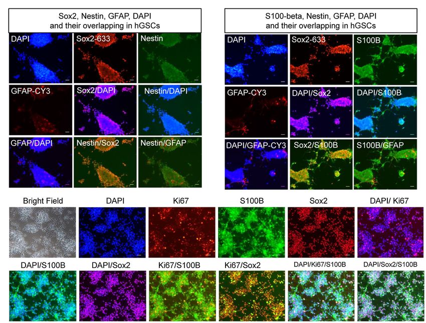
Han et al.                                                                                                      Rapidly Derived Glioma Stem Cells

INTRODUCTION                                                           Plates Coating With Poly-L-Ornithine
                                                                       and Laminin
Malignant glioma is a highly lethal brain cancer, and                  Culture plates were pre-covered with poly-L-ornithine (Sigma
glioblastoma (GBM) is the most aggressive glioma type. The             P3655) and laminin (Thermo Fisher 23017015) for hGSC culture
5 year overall survival for GBM patients is less than 10%              and passage as previously described (Han et al., 2017). In brief,
with the median survival of 14–16 months (Wankhede et al.,             each 100 mm dish was treated with 10 ml 0.5 μg/ml poly-L-
2012; Campos et al., 2016). It is difficult to characterize            ornithine (10 mg/ml stock concentration dissolved in water) and
GBM subtypes posing a challenge to choose an appropriate               maintained at room temperature for 16 h on a flat, clean tabletop.
therapeutic approach for patients due to a limited                     The following day, 1 × PBS (Hyclone SH30258.01) was used
understanding of the underlying molecular biology (Campos              applied to wash the dishes after the poly-L-ornithine supernatant
et al., 2016; Karim et al., 2016). Therefore, there is an urgent       was discarded. Then, 10 ml 1 × PBS containing 5 μg/ml laminin
need to understand the progression of GBM and to develop               was added to the dish and was left to keep infiltrating for at
effective therapies for patients with GBM.                             least 16 h at room temperature. Finally, the coated dishes were
   Glioblastoma is a highly heterogeneous tumor with various           stored at −20°C. Before cell culture, the coated dishes were
cellular subtypes (Aum et al., 2014; Soeda et al., 2015) and           incubated at 37°C, and the supernatant was discarded.
genetic properties (Sullivan et al., 2014; Li et al., 2015). The
tumor mass is complex and contains mature cells, GSCs, and
neural stem cells (NSCs). The concept of GSCs emerged in               Glioma Stem Cells Derivation and Culture
the early 2000s but remained to be fully characterized (Hemmati        After strictly following the ethical process, about 300 mg of tumor
et al., 2003; Singh et al., 2003; Galli et al., 2004). As a            surgical samples was quickly transferred to a sterile 50 ml centrifuge
subpopulation of tumor cells, GSCs are the driving force of            tube and brought to the biosafety cabinet of the laboratory. The
tumor recurrence and can self-renew and differentiate (Taga            surgical samples were carefully removed with sterile forceps and
and Tabu, 2020). Unlike GSCs, NSCs are tumor-tropic cells              were placed into the sterile 100 mm Corning cell culture dish.
that can be used as vehicles to selectively deliver various            Surgical specimens were washed with 1 × HBSS (Gibco 14170-
anticancer agents to tumor sites (Mooney et al., 2018).                112) at least six times. 1 × HBSS was pre-cold at 4°C. After
   Recently, personalized therapies have been gaining momentum         each washing, the samples were fished into a new sterile culture
and are thought to significantly change the outcome for patients       dish, and new Hanks’ balanced salt solution (HBSS) was added
with GBM (Sundar et al., 2014; Graham-Gurysh et al., 2020;             for a full cleaning. Then, the samples were evenly divided into
Samec et al., 2020; White et al., 2020). Ideally, therapy for          new 100-mm dishes. Open the lid, carefully remove the tumor
patients with GBM would only kill GSCs but not affect NSCs.            tissue with sterile forceps, and place it on the lid of the sterile
Therefore, it is important to separate GSCs from tissue samples        100 mm Corning cell culture dish. Cut into small pieces with
after surgery and to identify the differences between GSCs             a sterile blade, and then, cut into mud shape with ophthalmic
and normal NSCs. The development of personalized therapy               forceps. Muddy tissue samples were placed into centrifuge tubes
needs to occur quickly with high efficiency because of the             containing 3 ml 1 U/ml Dispase II (Roche 04942078001) in
high recurrence rate and short recurrence time. The key is             DMEM/F12 (Gibco 11330-033) or DMEM (Gibco 11995-065).
to establish efficient protocols to quickly isolate pure GSCs          1 ml blue tips were used to head to blow and mixed the digestive
from patient samples and do comparisons between GSCs and               enzyme and muddy tissue thoroughly and slowly, and then carefully
NSCs. This study was designed to establish a fast and efficient        transfer them into 15 ml sterile centrifuge tube. The tubes were
approach for obtaining high-purity glioma stem cells and to            incubated at 37°C in a water-bath for 30 min to allow digestion.
explore the morphological and genetic differences between              During the period, the centrifuge tube can be taken out to check
GSCs and NSCs. We describe the features of GSC origin and              the digestion condition, and it is usually shaken evenly every
compare differences in the properties and regulatory genes in          10 min. Be careful not to over digest. After digestion, tissues
GSCs and NSCs.                                                         were centrifuged at 1,000 g for 3 min, and the supernatant was
                                                                       discarded carefully with a 1 ml blue tip. Cells suspended in 3 ml
                                                                       DMEMF12 or DMEM. Re-suspended cells were centrifuged again
MATERIALS AND METHODS                                                  at 1,000 g for 3 min, and the supernatant was also discarded
                                                                       carefully with a 1 ml blue tip. The precipitate was blown no
Surgical Glioma Samples                                                more than eight times each time with only 1 ml tips. Finally,
Surgical samples and basal data from human patients with               the precipitate was suspended in DMEM/F12 (Gibco 11330-033)/
glioma were obtained strictly according to Ethics Committee            N2 (Gibco 17502-048)/B27 (Gibco 17504-044)/GlutaMAX (Gibco
permission. Patients with glioma provided informed consent             35050061)/bFGF (HumanZyme HZ-1272)/epidermal growth factor
and donated their surgical specimens to the study. All the             (EGF; R & D Systems 236-EG)/heparin (Sigma H3393)/penicillin/
tumor samples were obtained and taken to the laboratory in             streptomycin (Gibco C14-15070-063) medium or DMEM/10%
time for follow-up treatment. Glioma tumors were categorized           FBS (Gibco 10099)/GlutaMAX/penicillin/streptomycin medium.
as grades II–IV using the WHO guidelines (Wesseling and                Fibroblast growth factor (FGF), EGF, and heparin concertations
Capper, 2018). After anonymous processing, surgical samples            were 20 ng/ml. Cells were seeded onto coating with poly-L-
were coded by the research number.                                     ornithine and laminin or non-coated plates.

Frontiers in Genetics | www.frontiersin.org                        2                                         July 2021 | Volume 12 | Article 639858
Han et al.                                                                                                         Rapidly Derived Glioma Stem Cells

Immunofluorescence Staining                                                downregulated genes. Kyoto Encyclopedia of Genes and Genomes
Human glioma stem cells (hGSCs) were assessed using staining               (KEGG) and Gene ontology (GO) analyses were also performed
assays as described previously (Han et al., 2017). In brief, cells         using an online database (g: Profiler; Reimand et al., 2007). The
were grown for 3 or 7 days in optimized culture conditions and             datasets presented in this study can be found in online repositories
fixed with 4% paraformaldehyde for 12 min at room temperature.             at https://bigd.big.ac.cn/gsa-human/browse/HRA000521, accession
Then, cells were permeabilized with 2.5% Triton X-100 (Sigma               no: prjca004144.
V900502) in PBS and incubated for 15 min at room temperature.
Then, we discarded the supernatant and blocked non-specific
reactions with 5% bovine serum albumin (Solarbio, A8010) in                STATISTICAL ANALYSIS
1 × PBS for 1.5 h at room temperature. Cells were incubated
                                                                           All data were obtained from three or more replicates. Quantitative
with primary antibodies to Sox2 (Goat, Santa Cruz sc-17320),
                                                                           data are presented as mean ± standard deviation. Statistical
nestin (Mouse, Millipore MAB5326), glial fibrillary acidic protein
                                                                           analysis was performed using GraphPad Prism 7.0 (GraphPad
(GFAP; Rabbit, Abcam ab7260), S100-beta (Mouse, Sigma S2352),
                                                                           Software, United States). For multiple comparisons, Student’s t-test
or Ki67 (Rabbit, Thermo Fisher 14-5698-82) for 2 days at 4°C.
                                                                           was applied to check significance values. Value of p lower than
After three washes with 0.1% Tween-20 (Sigma 655206) in 1 × PBS,
                                                                           0.05 were considered statistically significant. Value of p were
cells were incubated with secondary antibodies (Alexa Fluor
                                                                           calculated from at least three independent experiments. Statistical
647-conjugated donkey anti-goat IgG 705-605-003, Alexa Fluor
                                                                           significance was denoted as: *p < 0.05, **p < 0.01, and ***p < 0.001.
488-conjugated donkey anti-mouse IgG 715-545-150, Alexa Fluor
                                                                           All error bars represent the standard deviation of the mean.
Cy3-conjugated donkey anti-rabbit IgG 711-165-152, Alexa
Fluor Cy3-conjugated donkey anti-goat IgG 705-165-003, and
Alexa Fluor 647-conjugated donkey anti-rabbit antibody 712-605-
153, Jackson ImmunoResearch). Antibodies were dissolved in PBS             RESULTS
containing 2.5% bovine serum albumin and incubated with cells
at room temperature for 2 h. Cells were washed with 0.1%
                                                                           Establishing an Efficient Protocol for
Tween-20 in PBS three times, and the nucleus stained with                  Human Glioma Stem Cells Isolation
4′,6-diamidino-2-phenylindole (Sigma D9542). Immunofluorescence            and Expansion
staining images were captured using an inverted fluorescence               Primary GBM tissues contain many different types of cells
microscope (Nikon TE2000).                                                 including endothelial cells, non-stem tumor cells, GSC-derived
                                                                           endothelial-like cells, blood vessels, and hGSCs (Figure 1A).
                                                                           To better understand the molecular mechanisms of hGSCs,
RNA Extraction, Sequencing, and Gene                                       we aimed to develop a protocol to isolate the hGSCs from
Expression Analysis                                                        GBM tissues by primary culture. Various kinds of culture media
Passage 10 hGSCs were grown in DMEM/F12 medium on non-coated               and additives were used to culture GSCs in different studies
plates for 7 days. Passage 4 human neural stem cells (hNSCs)               (Zhang et al., 2020), and serum-free medium has been generally
were cultured in the same medium on coated plates for 7 days.              preferred (Lee et al., 2006). For comparison and for considering
The medium was sucked away, and cells were washed once with                the heterogeneous nature of tumors, we decided to include
PBS. Then, we added 2 ml of TRIzol (Invitrogen 15596026) and               both serum-supplemented medium and serum-free medium
extracted the total RNA from the cells. For each cell type, two            for our optimization. For the serum-free condition, we preferred
biological replicate RNA samples were collected for RNA sequencing.        DMEM/F12 medium over neural basal medium with N2 and
RNA integrity was determined using an Agilent 2100 Bioanalyzer             B27 supplements. For serum-free medium, FGF, EGF, and
(Agilent; Palo Alto, CA, United States). RNA quantity was determined       heparin were added (Figure 1B). Primary cell cultures were
using a NanoDrop (Thermo Fisher Scientific, Wilmington, DE).               also tested and maintained under both coated and non-coated
Poly-A-containing mRNA molecules were purified using ploy-T                conditions (Figure 1B). In total, we have shown optimization
oligo-attached magnetic beads, fragmented, and primed for cDNA             resulted from four different culture conditions (Figure 1B).
synthesis using the Illumina TruSeqTM RNA sample preparation                  To derive GSC culture, patient surgical samples were washed
kit (Illumina Inc., San Diego, CA) according to the manufacturer’s         in (HBSS)-PS six times and then chopped into small chunks.
protocol. cDNA was converted into double-stranded DNA using                The chunks were digested with 30-min incubation in 1 U/ml
the kit reagents. dsDNA was purified using AMPure XP beads                 Dispase II at 37°C. The disassociated cells were then spun
(Invitrogen, Carlsbad, CA) and was end-repaired and A-tailed               down, washed, and re-suspended for primary culture at indicated
following Illumina’s protocol. After adapter ligation, PCR was used        culture conditions (Figure 1B) with medium changed every
to enrich DNA fragments with adapter molecules on both ends                3 days (Figure 1B).
and to amplify the amount of DNA in the library. The resulting
molecular libraries were pooled and sequenced on a HiSeq 2500              The Choice of the Optimal Method to
sequencer (Illumina Inc., San Diego, CA). Then, the FPKM values            Separate Human Glioma Stem Cells
were analyzed as the gene expression base. Differentially expressed        We monitored the culture closely through frequent observations
gene (DEG) analysis was performed using online software (Morpheus,         after seeding. At 16 h, images of hGSCs were acquired at different
https://software.broadinstitute.org/morpheus) to identify up- and          magnifications (4×, 10×, and 20×) and small but visible cell clusters

Frontiers in Genetics | www.frontiersin.org                            3                                        July 2021 | Volume 12 | Article 639858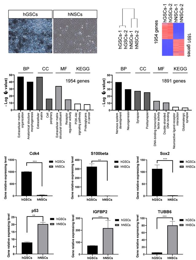
Han et al.                                                                                                                        Rapidly Derived Glioma Stem Cells

                      A

                      B

  FIGURE 1 | Schematic and flowchart describing how human glioma stem cell (hGSC) culture was derived. (A) hGSC isolation from glioma tissue and removal of
  other cells, including endothelial cells, glioma stem cells (GSCs)-derived endothelial-like cells, and non-stem tumor cells. (B) hGSC derivation procedures.

started to appear (Figure 2A). At 40 h, significant large cell                      spheres (Weiswald et al., 2015). In contrast, although they were
spheres were shown in coated plates (Figure 2B). However, closer                    significantly smaller, the small colonies in the non-coated plates
observations revealed that there were more small sphere colonies                    supplemented with three factors were round with clear boundaries,
in the non-coated plates supplemented with 3F (Figure 2B). There                    reminiscent of healthy neurospheres of high proliferative potentials.
are around 15–20 spheroids under each 4 × field in non-c N2B273f
condition (Figure 2C). Further examination found that the                           Highly Efficient Derivations of Human
morphology of cell spheres became irregular in the coated plates                    Glioma Stem Cells From Glioblastoma
and in the non-coated plate with FBS. The larger spheres were                       Patients
likely resulted from the adherence and spreading of the spheres,                    Then, we sought to determine whether this culture system is suitable
an indication of fast proliferation and possible differentiation of                 for surgical samples from different patients. Primary tumor cell

Frontiers in Genetics | www.frontiersin.org                                     4                                              July 2021 | Volume 12 | Article 639858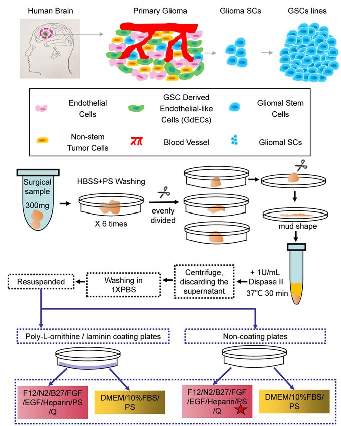
Han et al.                                                                                                                            Rapidly Derived Glioma Stem Cells

                            A

                            B                                                                      C

  FIGURE 2 | Separating hGSCs using different methods to identify an efficient and high-quality approach. (A) Representative photos of glioma tissue cell germination
  and morphology following culture in a different medium for 16 h. Yellow dotted line on the right shows the clonal morphology after 16 h germination under different
  culture conditions. Non-c FBS: culture cells on non-coated plates with DMEM + 10%FBS; Non-c N2B273f: culture cells on the non-coated plates with DMEM/F12 +
  N2B27 + 3 factors [Fibroblast growth factor (FGF), epidermal growth factor (EGF), and heparin]; coated FBS: culture cells on coated plates with DMEM + 10% FBS;
  coated N2B273f: culture cells on coated plates with DMEM/F12 + N2B27 + 3 factors (FGF, EGF, and heparin). (B) Primary glioma cells from glioma tissue cultured in a
  different medium for 40 h. (C) Single sphere area and spheres number of hGSCs in per 100x field of vision (non-c FBS with n = 3; Non-c N2B273f with n = 5; coated
  FBS with n = 3; and coated N2B273d with n = 3). Scale bar, 50 μm. Data are represented as mean ± SD. Student’s t-test; *p < 0.05; **p < 0.01.

cultures were successfully obtained for all the ten samples from                          Considering the short median survival time for patients
non-selected patients. Among them, four hGSCs cell lines were                          with GBM, personalized medicine requires a robust culture
successfully established (No. 2, No. 3, No. 4, and No. 6; Figure 3A)                   system that can be used to passage multiple generations of cells
with the presence of abundant healthy spheres.                                         to produce sufficient numbers of hGSCs for drug selection

Frontiers in Genetics | www.frontiersin.org                                        5                                               July 2021 | Volume 12 | Article 639858
Han et al. Rapidly Derived Glioma Stem Cells
tests. As shown in Figure 3B, we could routinely culture these
A
cells continuously for at least over 9–10 passages. We did notice
that the number of spheres decreased slightly and some of
them became adherent around passage 10 cells. Nonetheless,
we could continue the culture for all of them for many more
passages. In conclusion, our culture system, using a non-coated
plate with a medium supplemented with three factors, is a
stable culture system that can maintain hGSCs for many
generations and is suitable for various surgical samples.
Molecular Confirmation and
Characterization of Human Glioma Stem
Cells
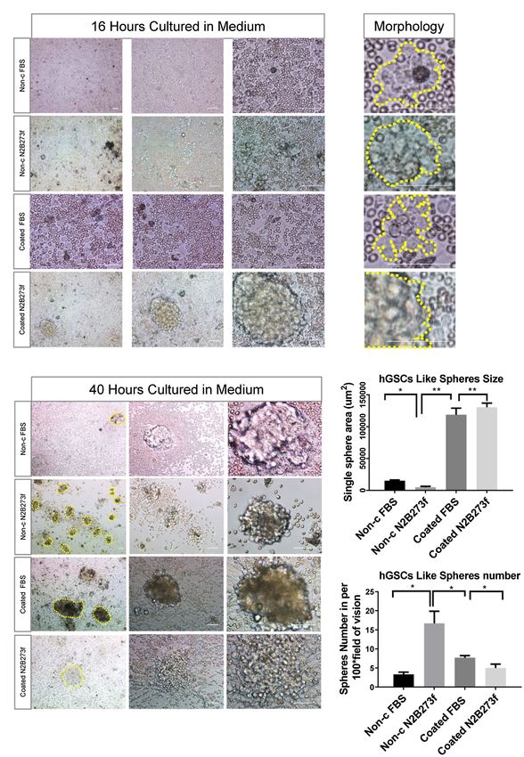
To confirm the identities of the derived hGSCs, we stained
them for SRY-box containing gene 2 (Sox2), nestin, glial fibrillary
acidic protein (GFAP), and S100-beta (Figures 4A,B). As
expected, all of them were highly expressed in the derived
cells except GFAP which manifested a low expression in some
of the cells. Furthermore, these cells also had strong proliferation
potentials as demonstrated by their continuous passaging ability
and Ki67 staining (Figure 4C). Together, these cellular analyses B
supported their identities as GSCs.
To further confirm and characterize the identities of these
GSCs, we carried out transcriptomic analysis by RNA sequencing.
For comparison, we decided to include hNSCs. hGSCs and
hNSCs were morphologically different. hGSCs formed spheres,
while hNSCs cells exhibited strong adherent abilities (Figure 5A).
Consistent with that, their molecular signatures were rather
different too (Figure 5B). Different expression gene analysis
showed 1954 genes had higher expression and 1891 genes had
lower expression in hGSCs, compared to hNSCs (Figure 5B).
GO and KEGG analyses revealed many pathways were
differentially expressed in hGSCs or hNSCs including those
related to the extracellular matrix and neurogenesis processes
(Figure 5C). In this current study, for their relevance to GBM,
we have chosen to focus on a number of genes related to cell
cycle and stemness. As shown in Figure 5D, the expression
level of cyclin-dependent kinase 4 (Cdk4), S100-beta, and Sox2
was all higher in hGSCs than that in hNSCs. In contrast, the
expression of the p53 tumor suppressor gene was lower in
hGSCs than that in hNSCs (Figure 5E). IGFB2 and tubulin
beta 6 class V (TUBB6), two potential therapeutic targets in
GBM treatment (Jiang et al., 2020), both displayed significantly
lower expression in hGSCs than in hNSCs (Figure 5F).
DISCUSSION
FIGURE 3 | hGSCs isolated from different glioma tissues in P0 generation
Intensive efforts are under ways to predict potential therapeutic
and different generations from P0 after passage. (A) Representative images
targets and to expand GBM treatment options. For example, of hGSCs isolated from different individual glioma tissues in P0 generation.
DEG analysis has been used to identify differences in gene hGSCs were cultured in optimized culture conditions for 7 days. (B) hGSCs
expression patterns between samples from patients with GBM can be passaged for over 10 generations. Scale bar, 50 μm.
and normal brain samples. Very recently, CMPK2, CRLS1,
PGS1, SLC22A5, and SOAT1 were identified as essential microarray data K-M curve analyses also identified SLC12A5,
for GBM growth but non-toxic to remove from healthy CCL2, IGFBP2, and PDPN as independent predictors of survival
brain tissue (Larsson et al., 2020). DEG, multivariate Cox, and in patients with GBM (Yang et al., 2020). Differentially expressed
Frontiers in Genetics | www.frontiersin.org 6 July 2021 | Volume 12 | Article 639858Han et al. Rapidly Derived Glioma Stem Cells
A B
C
FIGURE 4 | Confirmation and characterization of hGSCs. (A–C) hGSCs were fixed and immunostained with GFAP, Sox2, and nestin (A); S100-beta, Sox2, and
GFAP (B); and Ki67, S100-beta, and Sox2 (C). Scale bar, 50 μm.
mitochondrial-focused gene (DEMFG) analysis also suggested accompanied by a rapid and safety test in the same system (da
microtubular TUBB6 as a potential therapeutic target in GBM Hora et al., 2019). Recently, people can develop neuronal organoid
(Jiang et al., 2020). Besides, Immunocore, an immune-infiltration- model mimicking GBM features from induced pluripotent stem
based signature, was also being considered as a potentially cells (iPSC) by mutated c-met gene (Hwang et al., 2020). However,
reliable prognostic and predictive tool for GBM (Tang et al., due to the multiple procedures of organoid cultures, it takes
2020). However, all these potential therapeutic strategies need around 21–45 days to obtain a mature drug screening organoid
to be further confirmed by experimentations before they could (Hubert et al., 2016; Ogawa et al., 2018; Jacob et al., 2020).
move forward to clinics. This is where a quick and robust For the high-grade GBM patients, it is an emergency to make
drug screening system is required. A primary cell culture system treatment decision within a short time course. Our GSC culture
like ours may fulfill both the speed and quality requirements. system could be established within only 24 h to 3 days immediately
First, hGSC primary culture could be established timely within after surgery, which may save some times as compared to the
1–3 days after surgery. Secondly, hGSCs can be maintained complex organoid culture system.
for more than 10 passages, and hence, our approach can help Personalized medicine therapy is a promising approach
provide a large amount of cell material for drug screening for GBM. However, it requires timely selection of appropriate
and potential therapeutic target selection. therapeutic targets or anticancer agents. A desired strategy
Brain organoids are a new approach for GBM modeling (Qian for patients with GBM is to only kill hGSCs but not hNSCs.
et al., 2016). Comparing to the two-dimensional culture of GSC Therefore, it is necessary to understand the morphological
cells, the organoid culture system acted as a 3D system may and molecular characteristics of hGSCs. Sox2 is a stem cell
reproduce a better niche for GBM study (Gomez-Oliva et al., marker of adult neurogenesis (Steiner et al., 2006), and nestin
2020). Organoid models are very useful for studies of essential is a marker used to examine neurogenesis within the
tumor biology and also suitable for preclinical investigations, adult brain (Doyle et al., 2001). The high levels of Sox2 and
such as drug screening and analysis of antitumor effects nestin expression observed in hGSCs are indicative of their
Frontiers in Genetics | www.frontiersin.org 7 July 2021 | Volume 12 | Article 639858Han et al. Rapidly Derived Glioma Stem Cells
A B
C
D
E F
FIGURE 5 | Molecular characterization and comparison of hGSCs and human neural stem cells (hNSCs). (A) hGSCs and hNSCs were morphologically different.
(B) Gene clustering analysis of hGSCs and hNSCs and differential gene expression analysis of hGSCs and hNSCs. (C) The Gene ontology analysis (biological
process, cell component and molecular functions) and KEGG analysis of upregulated genes (left) and downregulated genes (right). (D) The expression of Cdk4,
S100-beta, and Sox2 was higher in hGSCs than that in hNSCs. (E) The expression of tp53 was lower in hGSCs than that in hNSCs. (F) Decreased IGFBP2 and
TUBB6 expression was observed in hGSCs, compared to hNSCs. Data are represented as mean ± SD with n = 3 biological replicates.
Frontiers in Genetics | www.frontiersin.org 8 July 2021 | Volume 12 | Article 639858Han et al. Rapidly Derived Glioma Stem Cells
stemness. GFAP and S100-beta are mature astrocyte markers ETHICS STATEMENT
(Garcia et al., 2004). GFAP expression was low in hGSCs,
but surprisingly, the S100-beta expression was high in GSCs. The studies involving human participants were reviewed and
S100 beta was first identified as an astrocyte marker (Castets approved by Ethics Committee of Shanghai 10th people’s Hospital.
et al., 1997). Recently, some malignant diseases showed highly The patients/participants provided their written informed consent
expressed S100-beta as well. The importance of understanding to participate in this study.
the differences between hGSCs and hNSCs was previously
noted in clinical treatments as well (Stoyanov et al., 2018).
In the present study, we focused to compare several key
genes. As expected, we observed higher expression of cell AUTHOR CONTRIBUTIONS
cycle genes and lower expression of p53 tumor suppressor
gene expression in hGSCs than that in hNSCs. We have ZG, X-XH, and HH conceived and designed the research.
analyzed the 10 GBM-related genes reported recently (Jiang X-XH and CC performed sample collection, experiments,
et al., 2020; Larsson et al., 2020; Yang et al., 2020) and six and data analysis. L-MY, MW, D-YH, and JR helped perform
of them showed expression differences between hGSCs and the cell experiments. M-HZ, L-YZ, W-HZ, and WH performed
hNSCs, further validating our hGSC derivation approach. data analysis. X-XH and CC wrote the manuscript. All authors
In summary, we have established a fast and efficient protocol contributed to the article and approved the submitted
to obtain high-purity GBM stem cell culture for both basic version.
research and translational research. The approach detailed here
has the very potential to facilitate drug screening directly with
patient-relevant cells and enable personalized medicine practice.
FUNDING
We hope that similar approaches like ours may 1 day prove
to be beneficial to GBM patients. This work was supported by funds from the National Key
R&D Program of China (2019YFA0110300), National Natural
DATA AVAILABILITY STATEMENT Science Foundation of China (81901031 to X-XH, 31600819
to CC, and 32070862 and 31571058 to ZG), Natural Science
The datasets presented in this study can be found in online Foundation of Shanghai (19ZR1445400) to X-XH, and Shanghai
repositories. The names of the repository/repositories and Municipal Population and Family Planning Commission
accession no: prjca004144. (20174Y0216) to CC.
for a precision medicine approach to interstitial glioblastoma therapy. J. Control.
REFERENCES Release 323, 282–292. doi: 10.1016/j.jconrel.2020.04.028
Han, X., Yu, L., Ren, J., Wang, M., Liu, Z., Hu, X., et al. (2017). Efficient and
Aum, D. J., Kim, D. H., Beaumont, T. L., Leuthardt, E. C., Dunn, G. P., and
fast differentiation of human neural stem cells from human embryonic stem
Kim, A. H. (2014). Molecular and cellular heterogeneity: the hallmark of
cells for cell therapy. Stem Cells Int. 2017:9405204. doi: 10.1155/2017/9405204
glioblastoma. Neurosurg. Focus. 37:E11. doi: 10.3171/2014.9.FOCUS14521
Hemmati, H. D., Nakano, I., Lazareff, J. A., Masterman-Smith, M.,
Campos, B., Olsen, L. R., Urup, T., and Poulsen, H. S. (2016). A comprehensive
Geschwind, D. H., Bronner-Fraser, M., et al. (2003). Cancerous stem cells
profile of recurrent glioblastoma. Oncogene 35, 5819–5825. doi: 10.1038/onc.
can arise from pediatric brain tumors. Proc. Natl. Acad. Sci. USA 100,
2016.85
15178–15183. doi: 10.1073/pnas.2036535100
Castets, F., Griffin, W. S., Marks, A., and Van Eldik, L. J. (1997). Transcriptional Hubert, C. G., Rivera, M., Spangler, L. C., Wu, Q., Mack, S. C., Prager, B. C.,
regulation of the human S100 beta gene. Mol. Brain Res. 46, 208–216. doi: et al. (2016). A three-dimensional organoid culture system derived from
10.1016/s0169-328x(96)00298-7 human glioblastomas recapitulates the hypoxic gradients and cancer stem
da Hora, C. C., Schweiger, M. W., Wurdinger, T., and Tannous, B. A. (2019). cell heterogeneity of tumors found in vivo. Cancer Res. 76, 2465–2477. doi:
Patient-derived glioma models: from patients to dish to animals. Cell 8:1177. 10.1158/0008-5472.CAN-15-2402
doi: 10.3390/cells8101177 Hwang, J. W., Loisel-Duwattez, J., Desterke, C., Latsis, T., Pagliaro, S., Griscelli, F.,
Doyle, K. L., Khan, M., and Cunningham, A. M. (2001). Expression of the et al. (2020). A novel neuronal organoid model mimicking glioblastoma
intermediate filament protein nestin by sustentacular cells in mature olfactory (GBM) features from induced pluripotent stem cells (iPSC). Biochim. Biophys.
neuroepithelium. J. Comp. Neurol. 437, 186–195. doi: 10.1002/cne.1278 Acta Gen. Subj. 1864:129540. doi: 10.1016/j.bbagen.2020.129540
Galli, R., Binda, E., Orfanelli, U., Cipelletti, B., Gritti, A., De Vitis, S., et al. Jacob, F., Salinas, R. D., Zhang, D. Y., Nguyen, P. T. T., Schnoll, J. G., Wong, S. Z.
(2004). Isolation and characterization of tumorigenic, stem-like neural H., et al. (2020). A patient-derived glioblastoma organoid model and biobank
precursors from human glioblastoma. Cancer Res. 64, 7011–7021. doi: recapitulates inter- and intra-tumoral heterogeneity. Cell 180, 188–204. doi:
10.1158/0008-5472.CAN-04-1364 10.1016/j.cell.2019.11.036
Garcia, A. D., Doan, N. B., Imura, T., Bush, T. G., and Sofroniew, M. V. (2004). Jiang, L., Zhu, X., Yang, H., Chen, T., and Lv, K. (2020). Bioinformatics analysis
GFAP-expressing progenitors are the principal source of constitutive neurogenesis discovers microtubular tubulin beta 6 class V (TUBB6) as a potential therapeutic
in adult mouse forebrain. Nat. Neurosci. 7, 1233–1241. doi: 10.1038/nn1340 target in glioblastoma. Front. Genet. 11:566579. doi: 10.3389/fgene.2020.566579
Gomez-Oliva, R., Dominguez-Garcia, S., Carrascal, L., Abalos-Martinez, J., Karim, R., Palazzo, C., Evrard, B., and Piel, G. (2016). Nanocarriers for the
Pardillo-Diaz, R., Verastegui, C., et al. (2020). Evolution of experimental treatment of glioblastoma multiforme: current state-of-the-art. J. Control.
models in the study of glioblastoma: toward finding efficient treatments. Release 227, 23–37. doi: 10.1016/j.jconrel.2016.02.026
Front. Oncol. 10:614295. doi: 10.3389/fonc.2020.614295 Larsson, I., Uhlen, M., Zhang, C., and Mardinoglu, A. (2020). Genome-scale
Graham-Gurysh, E. G., Murthy, A. B., Moore, K. M., Hingtgen, S. D., metabolic Modeling of glioblastoma reveals promising targets for drug
Bachelder, E. M., and Ainslie, K. M. (2020). Synergistic drug combinations development. Front. Genet. 11:381. doi: 10.3389/fgene.2020.00381
Frontiers in Genetics | www.frontiersin.org 9 July 2021 | Volume 12 | Article 639858Han et al. Rapidly Derived Glioma Stem Cells
Lee, J., Kotliarova, S., Kotliarov, Y., Li, A., Su, Q., Donin, N. M., et al. (2006). Sundar, S. J., Hsieh, J. K., Manjila, S., Lathia, J. D., and Sloan, A. (2014). The
Tumor stem cells derived from glioblastomas cultured in bFGF and EGF role of cancer stem cells in glioblastoma. Neurosurg. Focus. 37:E6. doi:
more closely mirror the phenotype and genotype of primary tumors than 10.3171/2014.9.FOCUS14494
do serum-cultured cell lines. Cancer Cell 9, 391–403. doi: 10.1016/j.ccr.2006.03.030 Taga, T., and Tabu, K. (2020). Glioma progression and recurrence involving
Li, R., Li, H., Yan, W., Yang, P., Bao, Z., Zhang, C., et al. (2015). Genetic and maintenance and expansion strategies of glioma stem cells by organizing
clinical characteristics of primary and secondary glioblastoma is associated self-advantageous niche microenvironments. Inflamm. Regener. 40, 1–5. doi:
with differential molecular subtype distribution. Oncotarget 6, 7318–7324. 10.1186/s41232-020-00142-7
doi: 10.18632/oncotarget.3440 Tang, X., Xu, P., Chen, A., Deng, G., Zhang, S., Gao, L., et al. (2020). Prognostic
Mooney, R., Hammad, M., Batalla-Covello, J., Abdul Majid, A., and Aboody, K. S. and predictive value of an immunoscore signature in glioblastoma multiform.
(2018). Concise review: neural stem cell-mediated targeted cancer therapies. Front. Genet. 11:514363. doi: 10.3389/fgene.2020.514363
Stem Cells Transl. Med. 7, 740–747. doi: 10.1002/sctm.18-0003 Wankhede, M., Bouras, A., Kaluzova, M., and Hadjipanayis, C. G. (2012).
Ogawa, J., Pao, G. M., Shokhirev, M. N., and Verma, I. M. (2018). Glioblastoma Magnetic nanoparticles: an emerging technology for malignant brain tumor
model using human cerebral organoids. Cell Rep. 23, 1220–1229. doi: 10.1016/j. imaging and therapy. Expert. Rev. Clin. Pharmacol. 5, 173–186. doi: 10.1586/
celrep.2018.03.105 ecp.12.1
Qian, X., Nguyen, H. N., Song, M. M., Hadiono, C., Ogden, S. C., Hammack, C., Weiswald, L. B., Bellet, D., and Dangles-Marie, V. (2015). Spherical cancer
et al. (2016). Brain-region-specific organoids using mini-bioreactors for models in tumor biology. Neoplasia 17, 1–15. doi: 10.1016/j.neo.2014.12.004
modeling ZIKV exposure. Cell 165, 1238–1254. doi: 10.1016/j.cell.2016.04.032 Wesseling, P., and Capper, D. (2018). World Health Organization 2016 classification
Reimand, J., Kull, M., Peterson, H., Hansen, J., and Vilo, J. (2007). g:Profiler–a of gliomas. Neuropathol. Appl. Neurobiol. 44, 139–150. doi: 10.1111/nan.12432
web-based toolset for functional profiling of gene lists from large-scale White, K., Connor, K., Clerkin, J., Murphy, B. M., Salvucci, M., O'Farrell, A. C.,
experiments. Nucleic Acids Res. 35, W193–W200. doi: 10.1093/nar/gkm226 et al. (2020). New hints towards a precision medicine strategy for IDH
Samec, N., Zottel, A., Videtic Paska, A., and Jovcevska, I. (2020). Nanomedicine wild-type glioblastoma. Ann. Oncol. 31, 1679–1692. doi: 10.1016/j.
and immunotherapy: a step further towards precision medicine for glioblastoma. annonc.2020.08.2336
Molecules 25:490. doi: 10.3390/molecules25030490 Yang, J., Wang, L., Xu, Z., Wu, L., Liu, B., Wang, J., et al. (2020). Integrated
Singh, S. K., Clarke, I. D., Terasaki, M., Bonn, V. E., Hawkins, C., Squire, J., analysis to evaluate the prognostic value of signature mRNAs in glioblastoma
et al. (2003). Identification of a cancer stem cell in human brain tumors. multiforme. Front. Genet. 11:253. doi: 10.3389/fgene.2020.00253
Cancer Res. 63, 5821–5828. Zhang, L., Yu, H., Yuan, Y., Yu, J. S., Lou, Z., Xue, Y., et al. (2020). The
Soeda, A., Hara, A., Kunisada, T., Yoshimura, S., Iwama, T., and Park, D. M. necessity for standardization of glioma stem cell culture: a systematic review.
(2015). The evidence of glioblastoma heterogeneity. Sci. Rep. 5:7979. doi: Stem Cell Res. Ther. 11, 1–7. doi: 10.1186/s13287-020-01589-8
10.1038/srep07979
Steiner, B., Klempin, F., Wang, L., Kott, M., Kettenmann, H., and Kempermann, G. Conflict of Interest: The authors declare that the research was conducted in
(2006). Type-2 cells as link between glial and neuronal lineage in adult the absence of any commercial or financial relationships that could be construed
hippocampal neurogenesis. Glia 54, 805–814. doi: 10.1002/glia.20407 as a potential conflict of interest.
Stoyanov, G. S., Dzhenkov, D., Ghenev, P., Iliev, B., Enchev, Y., and Tonchev, A. B.
(2018). Cell biology of glioblastoma multiforme: from basic science to Copyright © 2021 Han, Cai, Yu, Wang, Hu, Ren, Zhang, Zhu, Zhang, Huang,
diagnosis and treatment. Med. Oncol. 35, 1–10. doi: 10.1007/ He and Gao. This is an open-access article distributed under the terms of the
s12032-018-1083-x Creative Commons Attribution License (CC BY). The use, distribution or reproduction
Sullivan, J. P., Nahed, B. V., Madden, M. W., Oliveira, S. M., Springer, S., in other forums is permitted, provided the original author(s) and the copyright
Bhere, D., et al. (2014). Brain tumor cells in circulation are enriched for owner(s) are credited and that the original publication in this journal is cited, in
mesenchymal gene expression. Cancer Discov. 4, 1299–1309. doi: accordance with accepted academic practice. No use, distribution or reproduction
10.1158/2159-8290.CD-14-0471 is permitted which does not comply with these terms.
Frontiers in Genetics | www.frontiersin.org 10 July 2021 | Volume 12 | Article 639858You can also read