A time to heal: microRNA and circadian dynamics in cutaneous wound repair
←
→
Page content transcription
If your browser does not render page correctly, please read the page content below

Clinical Science (2022) 136 579–597
https://doi.org/10.1042/CS20220011

Review Article

A time to heal: microRNA and circadian dynamics in
cutaneous wound repair
Sandra Fawcett1,2 , Raida Al Kassas1,2 , Iain M Dykes1,2 , Alun TL Hughes2,3 , Fawaz Ghali2,4 and
  Kehinde Ross1,2
1 Schoolof Pharmacy and Biomolecular Science, Liverpool John Moores University, Liverpool, United Kingdom; 2 Instiute for Health Research, Liverpool John Moores University,
Liverpool, United Kingdom; 3 School of Biological and Environmental Sciences, Liverpool John Moores University, Liverpool, United Kingdom; 4 School of Computer Science and
Mathematics, Liverpool John Moores University, Liverpool, United Kingdom
Correspondence: Kehinde Ross (o.k.ross@ljmu.ac.uk)

                                                                                                                                                                                             Downloaded from http://portlandpress.com/clinsci/article-pdf/136/8/579/932145/cs-2022-0011.pdf by guest on 05 July 2022
                                             Many biological systems have evolved circadian rhythms based on the daily cycles of day-
                                             light and darkness on Earth. Such rhythms are synchronised or entrained to 24-h cycles,
                                             predominantly by light, and disruption of the normal circadian rhythms has been linked to
                                             elevation of multiple health risks. The skin serves as a protective barrier to prevent microbial
                                             infection and maintain homoeostasis of the underlying tissue and the whole organism. How-
                                             ever, in chronic non-healing wounds such as diabetic foot ulcers (DFUs), pressure sores,
                                             venous and arterial ulcers, a variety of factors conspire to prevent wound repair. On the
                                             other hand, keloids and hypertrophic scars arise from overactive repair mechanisms that fail
                                             to cease in a timely fashion, leading to excessive production of extracellular matrix (ECM)
                                             components such as such as collagen. Recent years have seen huge increases in our un-
                                             derstanding of the functions of microRNAs (miRNAs) in wound repair. Concomitantly, there
                                             has been growing recognition of miRNA roles in circadian processes, either as regulators or
                                             targets of clock activity or direct responders to external circadian stimuli. In addition, miR-
                                             NAs are now known to function as intercellular signalling mediators through extracellular
                                             vesicles (EVs). In this review, we explore the intersection of mechanisms by which circa-
                                             dian and miRNA responses interact with each other in relation to wound repair in the skin,
                                             using keratinocytes, macrophages and fibroblasts as exemplars. We highlight areas for fur-
                                             ther investigation to support the development of translational insights to support circadian
                                             medicine in the context of these cells.

                                          Introduction
                                          Chronic, non-healing wounds have emerged as a major public health crisis associated with enormous
                                          negative impact on quality of life and healthcare budgets [1–3]. Factors driving the elevated incidence
                                          of chronic wounds include the ageing population, the diabetes epidemic, and the intersection of these
                                          phenomena, that is, diabetic patients living longer [1–4]. Wound management costs are dominated by
                                          diabetes-related amputations [2] and have been estimated at £5.3 billion annually [5] in the U.K. and a
                                          staggering $28.1–96.8 billion per year in the U.S.A. [6]. Comparative cost analyses are less readily avail-
                                          able for the Global South, but it is noteworthy that every three in four (79%) people with diabetes live in
                                          low- and middle-income countries [7]. Further, the prevalence of diabetic foot ulcers (DFUs) reported
                                          for Africa (7.2%) was higher than that of Europe (5.1%) or Asia (5.5%) but lower than the 13.0% preva-
Received: 07 January 2022                 lence in North America [4]. However, the management of DFUs in Africa is burdened by a high rate of
Revised: 01 April 2022                    mortality during hospitalisation [8], perhaps reflecting a paucity of healthcare resources compared with
Accepted: 04 April 2022                   high-income countries.
Version of Record published:                 Healthy skin provides wrap-around protection for the body against the external environment, prevent-
21 April 2022                             ing mechanical and chemical damage to the underlying tissues and resisting microbial invasion [9]. The

© 2022 The Author(s). This is an open access article published by Portland Press Limited on behalf of the Biochemical Society and distributed under the Creative Commons Attribution   579
License 4.0 (CC BY).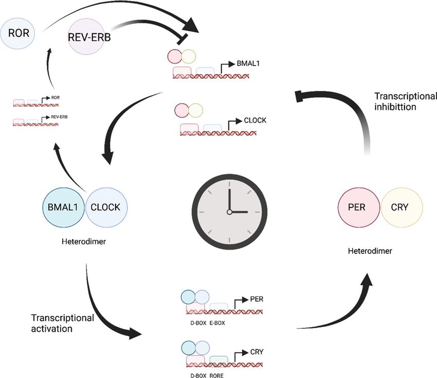
Clinical Science (2022) 136 579–597
                                                                                                                                             https://doi.org/10.1042/CS20220011

                                                                                                                                                                                             Downloaded from http://portlandpress.com/clinsci/article-pdf/136/8/579/932145/cs-2022-0011.pdf by guest on 05 July 2022
        Figure 1. The normal physiology of the skin
        Multilayered organ containing stratum corneum or non-viable epidermis, the viable interfollicular epidermis consisting of ker-
        atinocytes, the vascular- and appendage-rich dermis and the subcutaneous adipose layer containing adipocytes. Biological clocks
        are known to regulate the behaviour of keratinocytes, fibroblasts and macrophages and may impact intercellular transfer of miRNAs
        via extracellular vesicles (EVs). Created in BioRender.

        top layer of the skin is the epidermis, followed by the dermis and hypodermis (Figure 1). The epidermis consists
        primarily of keratinocytes that undergo terminal differentiation and, in combination with a lipid-rich matrix, form
        the epidermal barrier [10,11]. The dermis lies beneath the epidermis and contains fibroblasts that secrete extracellular
        matrix (ECM) proteins such as collagen, fibronectin and laminin and the hydrated proteoglycan gel in which these
        proteins are embedded [12]. The dermis also contains the microvasculature that supplies blood to the skin cells, as
        does the underlying hypodermis [13]. The hypodermis also contains subcutaneous adipose tissue (SAT) that supplies
        growth factors to the dermis and serves as an energy reservoir [14,15].
           Wound healing is orchestrated by complex interactions among skin cells, immune cells and endothelial cells that
        underpin the various overlapping phases of wound repair, spanning haemostasis, inflammation, angiogenesis, prolif-
        eration and remodelling [15,16]. Reciprocal signalling between keratinocytes and fibroblasts [17], keratinocytes and
        immune cells such as neutrophils and macrophages [18] and between cells and the ECM [19,20] that provide robust
        systems to support wound healing.
           While tissue repair is deficient in chronic non-healing wounds, it goes into overdrive in hypertrophic scars and
        keloids, which are fibroproliferative skin disorders associated with a pathological elevation of ECM components such
        as collagen, hyaluronan and fibronectin due to a massive increase in fibroblast numbers in the dermis [21–23]. Both
        keloids and hypertrophic scars are thought to arise from prolonged inflammation of the reticular dermis, with the
        magnitude of inflammation driving the growth of excessive scar tissue beyond the original wound borders in the case
        of keloids [21].
           Over the last decade, it has become clear that small non-coding RNA molecules of the microRNA (miRNA) fam-
        ily play pivotal roles in the regulation of cells and associated processes during wound repair [24–26]. In addition,
        keratinocytes, fibroblasts, neutrophils and macrophages all exhibit circadian rhythms, ubiquitous daily changes in

580   © 2022 The Author(s). This is an open access article published by Portland Press Limited on behalf of the Biochemical Society and distributed under the Creative Commons Attribution
      License 4.0 (CC BY).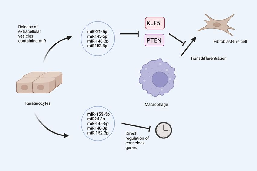
Clinical Science (2022) 136 579–597
https://doi.org/10.1042/CS20220011

behaviour and physiology that are driven by endogenous biological clocks [27–30]. However, the effects of miRNA
on the circadian properties of keratinocytes, fibroblasts and macrophages have received little attention in relation to
wound repair. Likewise, the impact of core clock gene activity on miRNA expression in these cells during wound
healing has not been established.
   In this review, we explore mechanisms by which circadian and miRNA responses interact with each other in relation
to wound healing in the skin. We begin with brief outlines of circadian biology and miRNA, respectively, and also
introduce small extracellular vesicles (EVs) (exosomes), which have gained prominence as mediators of intercellular
miRNA function in wound repair. We then examine a number of scenarios in which miRNAs may contribute to
circadian responses associated with wound healing, with a focus on keratinocytes and macrophages.

Brief primer on circadian rhythms
Circadian rhythms are fundamental processes that pervade biology and are thought to offer a selective advantage by
optimising physiological and behavioural activities to appropriate temporal niches [31]. The endogenous pacemakers

                                                                                                                                                                                             Downloaded from http://portlandpress.com/clinsci/article-pdf/136/8/579/932145/cs-2022-0011.pdf by guest on 05 July 2022
that generate these rhythms are found in most cells throughout the body and are underpinned by a set of so-called
‘core clock genes’, the transcriptional/translational activities of which form positive and negative feedback loops that
oscillate with a period of approximately 24 h [32].
   Central to this core molecular clockwork are the genes Clock and Bmal1, as well as the Cryptochrome 1/2 (Cry1/2)
and Period 1/2 (Per1/2) genes. The positive drive for this mechanism is provided by a heterodimeric transcription
factor consisting of BMAL1 (encoded by aryl hydrocarbon receptor nuclear translocator like, ARNTL and CLOCK
(or its analogue neuronal PAS domain protein 2, NPAS2)) that acts on E-box elements in the promoters of the Per
and Cry genes, activating their transcription (Figure 2). After translation, PER and CRY proteins form heterodimers
that mediate a negative feedback limb by inhibiting BMAL1/CLOCK-dependent transcriptional activation of the
Cry1/2 and Per1/2 promotors. BMAL1/CLOCK also activates the expression of Rev-erb α/β (also known as nuclear
receptor subfamily 1, group D member 1, NR1D1 and NR1D2) and retinoic acid-related orphan receptors (RORs
α/β). The REV-ERB α/β and RORs α/β proteins competitively inhibit and activate Bmal1 expression, respectively.
Various other genes and their proteins, including Dbp and E4bp4, and Dec1/2, form additional auxiliary loops.
These proteins, together with dynamic phosphorylation and ubiquitination involving casein kinase 1δ/ε (CK1δ/ε)
and F-box/LRR-repeat protein 3 (Fbxl3, a component of a ubiquitin ligase complex), provide further control over
the system, fine-tuning the approximate 24-h rhythms that it generates [33]. In addition, post-transcriptional mech-
anisms spanning poly(A) tail length, RNA methylation, alternative splicing and miRNAs confer further regulatory
control of the circadian clock, as reviewed very recently [34].
   Rhythms driven by these cell-autonomous circadian oscillators must be synchronised with both the external and in-
ternal environments in order to generate biologically relevant timing signals, and this is achieved through the actions
of a variety of factors, including environmental light, food, exercise and temperature [35]. In vertebrates, including
humans, the extended circadian system consists of a ‘master’ circadian pacemaker located within the suprachiasmatic
nucleus (SCN) of the hypothalamus and peripheral oscillators found throughout the rest of the body. A principal role
of the SCN is to integrate both external time signals and feedback from the periphery, and transmit this information
to peripheral oscillators, which use it to appropriately control the timing of local, tissue-specific, aspects of physiology
[36].

Circadian function in skin
Peripheral oscillators are found in almost all cells throughout the body and the skin is no exception. Both rhythmic
activities and functional implications of circadian rhythms for the biology of skin have been widely described [37].
Daily variation in gene expression have been reported in the epidermis and dermis, as well as in cutaneous and SATs
and dermal hair follicles [38–40]. At the cellular level epidermal keratinocytes, hair follicle cells, dermal fibroblasts
and dermal macrophages all generate rhythms on a daily timescale, as do subcutaneous adipocytes [41].
   Several mouse models have helped to establish key roles for core clock genes in the skin. For example, in
Bmal1-knockout mice, SAT was depleted in aged mice compared with their younger counterparts [42]. Loss of SAT
and other features of early ageing in the Bmal1-deficient mice were linked to circadian effects on reactive oxygen
species (ROS) homoeostasis in selected tissues [42]. Indeed, we now know that deletion of Bmal1 leads to the accu-
mulation of ROS in both the epidermis and the dermis [43]. Further, adipose-derived stem cells and miRNA-loaded
small extracellular vesicles (sEVs) released by these cells play key roles in wound healing, as reviewed elsewhere [44],
though the impact of Bmal deletion on sEV density and composition in normal and wounded skin remains to be
established. Defective wound healing is, however, seen in multiple mouse strains bearing core circadian clock gene

© 2022 The Author(s). This is an open access article published by Portland Press Limited on behalf of the Biochemical Society and distributed under the Creative Commons Attribution   581
License 4.0 (CC BY).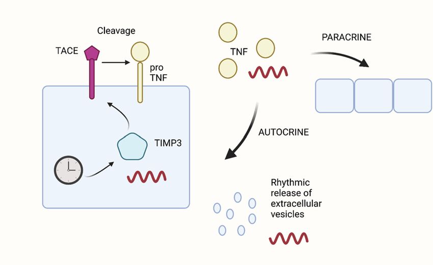
Clinical Science (2022) 136 579–597
                                                                                                                                             https://doi.org/10.1042/CS20220011

                                                                                                                                                                                             Downloaded from http://portlandpress.com/clinsci/article-pdf/136/8/579/932145/cs-2022-0011.pdf by guest on 05 July 2022
        Figure 2. The core molecular clock
        The heterodimeric transcription factor BMAL1:CLOCK1 promotes the expression of PER and CRY proteins. The PER:CRY protein
        complex in turn represses BMAL1:CLOCK-dependent transcriptional activation of PER and CRY promotors. BMAL1/CLOCK also
        activates the expression of ROR and REV-ERB (NR1D1 and NR1D2) proteins. These confer additional control as ROR competitively
        activates BMAL1 expression and REV-ERB proteins competitively inhibit BMAL1 expression. Created in BioRender.

        mutations [45] and time-of-day of wounding differentially impacts on the rate of healing in diurnal and nocturnal
        species [46,47]. On the other hand, wound healing was accelerated in mice lacking NPAS2, and this was associated
        with enhanced collagen synthesis, migration, and proliferation of dermal fibroblasts [48].
           Further studies have provided evidence for autonomous oscillations of molecular clock components in mouse skin,
        particularly in the epidermis and hair follicles, and rhythmic expression of PER2 was completely lost in Cry1/Cry2
        double knockout mice lacking molecular clocks [49]. Crucially, Bmal1 controls the expression of stem cell regulatory
        genes and studies on mice lacking Bmal1 in basal epidermal keratinocytes, or on Per1/Per2 double knockout mice,
        revealed the molecular clock modulates stemness within the hair follicle stem cell niche [50]. Specifically, the hair
        follicle bulges of mice lacking Bmal1 had fewer proliferative cells and more dormant stem cells as the mice aged. On
        the other hand, a later independent study found that keratinocyte-specific deletion of Bmal1 results in constitutively
        elevated cell proliferation and abolishes time-of-day-dependent cell proliferation rhythms in the interfollicular epi-
        dermis and upper hair follicles [51]. Interestingly, this later study also found that accumulation of ROS in mouse skin
        appears to be temporally de-coupled from DNA replication, at least when the hair follicles of the skin were in the
        telogen (resting) phase [51].

582   © 2022 The Author(s). This is an open access article published by Portland Press Limited on behalf of the Biochemical Society and distributed under the Creative Commons Attribution
      License 4.0 (CC BY).
Clinical Science (2022) 136 579–597
https://doi.org/10.1042/CS20220011
Furthermore, the temporal segregation of biological processes has also been observed in human epidermal stem
cells (EPSCs), with keratinocyte differentiation associated with late-night and early-morning hours, while DNA repli-
cation, UV protection and cell division proceed in the afternoon and evening hours [52]. Notably, studies on CLOCK
(Clk/Clk) mouse mutants showed that even the hydration of the topmost layer of the epidermis, the stratum corneum,
was regulated by clock genes [53]. This was associated with a rhythmic expression of aquaporin 3 that was observed
in both mouse skin and in synchronised HaCaT keratinocytes [53].
Given that inflammation is a key early phase of wound repair, it is noteworthy that inflammatory responses, espe-
cially those driven by the Toll-like-receptor 7 agonist imiquimod (IMQ), have been linked to circadian dynamics in
the epidermis [54,55]. Nakao and colleagues found psoriasis-like disease observed upon topical application of IMQ
was attenuated or exacerbated in Clock and Per2 mutant mice, respectively, compared with wildtype mice [54]. More
recently, Andersen and colleagues showed that diurnal variation in both EPSC proliferation and epidermal thick-
ness appeared to be lost upon treatment of mouse skin with IMQ [55]. Gene expression studies found that IMQ
mobilised greater interferon (IFN) responses after daytime application to mouse skin and this was associated with
greater daytime expression of approximately a quarter (53 out of 202) interferon-sensitive genes (ISGs) examined
Downloaded from http://portlandpress.com/clinsci/article-pdf/136/8/579/932145/cs-2022-0011.pdf by guest on 05 July 2022
[55]. Importantly, IMQ dampened rhythmic Bmal1 expression and down-regulated Rev-erbα (Nr1d1) and Dbp. On
the other hand, the inflammatory response to IMQ was potentiated in the epidermis of Bmal1 knockout mice, with
elevated expression of interferon-regulatory factor 7 (IRF7), a master regulator of ISG transcription, as well as in-
creased serum levels of IFN-β [55]. Thus, Bmal1 appears to support repression of inflammatory axes associated with
ISG expression in the skin and type 1 IFN levels in the blood.
The above observations together point to the importance of core clock genes such as Bmal1 in skin processes as-
sociated with wound healing. Very recently, however, Reddy and colleagues presented evidence indicating that 24-h
oscillations of the transcriptome, proteome, and phosphoproteome can persist in skin fibroblasts and liver slices from
Bmal1 knockout mice in the absence of exogenous stimuli [56]. Therefore, circadian gene expression in peripheral
tissues might have mechanisms beyond Bmal1 to provide oscillatory control of biological processes; a combination
of E26 transformation-specific (ETS) family transcription factors and redox oscillations were uncovered as candi-
date drivers of such Bmal1-independent rhythmicity. However, these findings raise many questions with regard to
BMAL1-independent ‘noncanonical’ circadian rhythmicity and have been challenged by independent investigators
[57–60].
Intriguingly, in both nocturnal rodents and diurnal humans, wounds suffered during the daily active phase heal
more rapidly than those suffered during the inactive phase [46], whilst exposure to dim light during the dark phase
of a daily 24-h cycle, a stimulus known to be disruptive to normal circadian function, delays and impairs wound
healing in mice [61]. At the gross tissue level, circadian control of skin physiology is taken to elevate mechanisms that
minimise damage to the skin during the day, whilst promoting growth and repair mechanisms at night [37]. Impor-
tantly, an elegant recent study on mice found that while the majority of circadian gene expression in the epidermis
may be associated with organismal factors linked to photic stimulation and the SCN, the core clock machinery of
the epidermis appears to oscillate directly in response to cyclic changes in light, even though individual epidermal
keratinocytes may have functioning cell autonomous clocks [62].
MiRNAs at the interface of skin repair and rhythms
Since their discovery over two decades ago, miRNAs have been established as key regulators of normal and pathophys-
iological cell behaviour that repress gene expression in a post-transcriptional manner [63]. These small non-coding
RNA molecules are generated from diverse genomic loci that yield primary miRNA transcripts which are processed
to give rise to precursor then mature miRNAs that are typically ∼22 nucleotides long. The mature miRNA is loaded
into Argonaute proteins to form the RNA-induced silencing complex (RISC) that executes the repression of messen-
ger RNA transcripts through mechanisms involving mRNA degradation and, to a lesser extent, translation inhibition
[64].
Recent years have seen a plethora of miRNAs implicated in wound repair processes spanning proliferation, resolu-
tion of inflammation, formation of granulation tissue, angiogenesis and migration [65–76]. Moreover, miRNA activity
has been linked to circadian mechanisms in a wide variety of tissues, including the brain [77,78], heart [79], liver [80]
and pineal gland [81]. Indeed, both rhythmic expression of miRNAs and miRNA alteration of circadian function
have been described, as well as direct interactions between miRNAs and elements of the core circadian molecular
machinery [77,82–84]. In skin, however, the relationships between miRNAs and circadian processes, including in
cutaneous wound repair, remain poorly understood. This is especially the case in relation to autocrine and paracrine
mechanisms of miRNA transmission between cells via EVs.
© 2022 The Author(s). This is an open access article published by Portland Press Limited on behalf of the Biochemical Society and distributed under the Creative Commons Attribution 583
License 4.0 (CC BY).Clinical Science (2022) 136 579–597
https://doi.org/10.1042/CS20220011
EVs for intercellular miRNA transfer
One of the more recently discovered aspects of miRNA function is their role as mediators of intercellular commu-
nication, transferred from one cell to another via EVs. These EVs, which include sEVs known as exosomes (EXOs),
microvesicles (MVs) and apoptotic bodies (ABs), convey diverse molecular cargoes to their target cells, including
miRNA, RNA, DNA, proteins and metabolites. The miRNA cargo of EXOs appears to be distinct from that of ABs
and MVs, which are more similar [85]. More importantly, EV-derived miRNAs from various mesenchymal and ep-
ithelial cells have been implicated in wound repair [86–96].
The three classes of EVs differ in size, but with some overlap: EXOs have a size range of 40–150 nm [97–99],
while MVs range approximately from 100 to 350 nm [100], but can also reach approximately 1 μm [101]. ABs re-
sult from cellular breakdown during apoptosis and are the largest class, being 1–5 μm [101]. These overlapping size
ranges present a challenge to the acquisition of pure populations of each class, as does overlapping epitope expression.
Therefore, the International Society for Extracellular Vesicles (ISEV) [102] recommends providing quantitative de-
tails on isolation methods used and referring to all particles as ‘extracellular vesicles’, describing their physical features
including size and epitope expression [102].
Downloaded from http://portlandpress.com/clinsci/article-pdf/136/8/579/932145/cs-2022-0011.pdf by guest on 05 July 2022
EXOs are derived from the endosomal pathway by invagination of the endosome membrane to form intraluminal
vesicles within the multivesicular body (MVB). The formation of the MVB is regulated by the endosomal sorting
complex required for transport (ESCRT), which recognises ubiquitinated membrane proteins and directs them into
the intraluminal vesicles [103]. Subsequently, the MVB can either be subjected to the lysosomal degradation of vesicle
contents or can fuse with the plasma membrane to release EXOs.
In contrast, MVs (also known as shedding vesicles, microparticles and ectosomes), which were first reported in
platelets in 1967, are released by various immune cell types that circulate in the blood, including platelets and neu-
trophils [104–106], as well as tumour cells [107]. MVs are produced by budding directly from the plasma membrane
rather than an intracellular precursor. Interestingly, studies are beginning show that the levels of specific MVs can
vary in a circadian fashion [108,109].
Keratinocytes, fibroblasts and adipose tissue are rich sources of miRNA-loaded EVs [110–113]. Although the cir-
cadian behaviour of these cell is well-established, we do not know whether their ability to generate and release EVs
follows a circadian pattern. Likewise, the impact of time of day on the loading of miRNA and other cargoes into
EVs has not been defined. miRNA-loaded EVs from the above cell types target macrophages during wound repair
[94,95], and macrophages also follow circadian rhythms [114]. With these perspectives in mind, we thus appraise ker-
atinocytes and macrophages in relation to the interplay of miRNA and circadian processes with relevance to wound
healing.
Circadian miRNA possibilities in keratinocyte–macrophage
interactions
Epidermal keratinocytes are critical for both normal skin physiology and successful wound closure and healing. In-
deed, following physical damage to the skin, basal keratinocytes migrate to the wound site and proliferate, with daugh-
ter cells terminally differentiating to seal the wound. Keratinocyte migration and proliferation are controlled by an ar-
ray of cytokines, growth factors and miRNAs, involving cross-talk among keratinocytes, dermal fibroblasts, activated
immune cells and endothelial cells. The circadian clock has been shown to drive rhythmic expression of genes associ-
ated with epidermal keratinocyte physiology [115]. However, the relationships between such circadian behaviour and
miRNAs have received limited attention, especially in relation to wound healing. In this regard, miR-21-5p is of par-
ticular interest because of its known ability to modulate wound repair, as we have reviewed elsewhere [25]. Further, di-
urnal expression of miR-21-5p has been observed in murine heart and lung, and the induction of miR-21-5p appeared
to occur in a Per2-dependent manner [116]. In addition, exposure of mice or humans to intense light up-regulated
miR-21-5p in murine hearts and human plasma, respectively [116]. Notably, the opsin family of light-sensitive G
protein-coupled receptors associated with phototransduction, have recently been shown to be expressed in human
epidermal keratinocytes and melanocytes [117–121]. Importantly, photic entrainment has been observed in mouse
outer ear and macrovibrissal pad dermal tissues, with opsin 5 serving as a photopigment that drove phase-setting
effects ex vivo (particularly over 370–415 nm) and photoentrainment of the local circadian clock in vivo [121]. Fur-
ther, using a reporter assay for per1 transcription, blue light (410 nm) has been shown to reduce PER1 expression
in normal human keratinocytes [122]. Together, these observations call for studies on relevant mouse models and ex
vivo human skin to establish whether environmental light directs clock gene-dependent miRNA expression in skin
cells, which may explain some of the observed effects of blue light on keratinocyte proliferation, differentiation and
migration [118,123].
584 © 2022 The Author(s). This is an open access article published by Portland Press Limited on behalf of the Biochemical Society and distributed under the Creative Commons Attribution
License 4.0 (CC BY).Clinical Science (2022) 136 579–597
https://doi.org/10.1042/CS20220011
Downloaded from http://portlandpress.com/clinsci/article-pdf/136/8/579/932145/cs-2022-0011.pdf by guest on 05 July 2022
Figure 3. Hypothetical model for rhythmic release of keratinocyte EVs
The core clock transcription/translation pathway regulates rhythmic expression of TIMP3, an enzyme required for synthesis of the
disintegrin metalloprotease ADAM17 or TACE. TACE mediates extracellular cleavage of membrane-bound TNF to release the free
growth factor. TNF can act in either an autocrine or paracrine fashion to regulate rhythmic release of EVs. Created in BioRender.
Very recent work has demonstrated circadian expression of miR-21-5p and miR-21-3p in atherosclerotic plaque
macrophages, leading to diurnal changes in macrophage apoptosis [124]. Work is needed to determine whether
wound macrophages can exhibit similar behaviour, especially given recent studies showing miR-21-5p from ker-
atinocyte EXOs promoted the transdifferentiation of wound-resident macrophages into fibroblast-like cells [94,95].
This appears to be an important step in resolution of the inflammatory phase of wound healing and adds miR-21-5p
to the growing number of miRNAs implicated in anti-inflammatory polarisation of macrophages [125,126]. However,
whether miR-21 expression occurs in a circadian manner in cutaneous wound macrophages remains to be established.
Interestingly, polarisation of the macrophages towards a fibroblast phenotype associated with resolution of inflam-
mation was associated with miR-21-5p-dependent down-regulation of the transcription factor, Krüppel-like factor 5
(KLF5) and the phosphatase and tensin homologue (PTEN) [94]. KLF5 is a member of the Krüppel-like factor family
of transcriptional regulators and regulates diverse cellular processes, including proliferation, migration and differ-
entiation [127]. PTEN dephosphorylates phosphatidylinositol (3,4,5)-trisphosphate to inactivate PI3K/AKT/mTOR
signalling and is a well-established target of miR-21-5p [128]. At present, however, it is not clear whether secretion of
keratinocyte EV or their contents occurs in a circadian fashion, nor do we know whether such putative circadian dy-
namics apply equally to EXO, MVs and ABs. However, we can hypothesise that EV release from keratinocytes occurs
in a circadian fashion based on two observations. First, tumour necrosis factor α (TNFα), a major pro-inflammatory
cytokine highly expressed at the wound site induces EV release from human keratinocytes [94]. Secondly, the secre-
tion of TNFα is regulated by TIMP3, a CLOCK-controlled diurnal gene that inhibits the TNFα-converting enzyme
(TACE; also known as ADAM17) responsible for release of membrane-bound TNFα into the surrounding milieu
as active TNFα [129,130]. Ultraviolet radiation (UVB) down-regulated CLOCK, BMAL1 and TIMP3 mRNA and
protein in human keratinocytes [129]. The expression on TIMP3 was periodic and the levels of TNFα and other in-
flammatory cytokines secreted under UVB conditions depended on TIMP3 expression. These observations suggest
light-dependent diurnal variations in epidermal TNFα levels may generate rhythmic patterns of EV release from ker-
atinocytes (Figure 3). Notably, TNFα and a plethora of other inflammatory markers were recently detected in suction
© 2022 The Author(s). This is an open access article published by Portland Press Limited on behalf of the Biochemical Society and distributed under the Creative Commons Attribution 585
License 4.0 (CC BY).Clinical Science (2022) 136 579–597
https://doi.org/10.1042/CS20220011
Table 1 MiRNAs predicted to regulate KLF5 and present in keratinocyte EVs
Mean read counts in
miRNA families broadly Cumulative weighted context GSE106453 primary Predicted to regulate
conserved among vertebrates score in TargetScan 7.2 keratinocyte EVs PTEN
miR-148-3p; 152-3p −0.48 1299; 26 Yes
miR-145-5p −0.41 46 Yes
miR-21-5p; 590-5p −0.39 12067 Yes
miR-140-3p.2 −0.26 144 Yes
miR-143-3p −0.18 4832 No
miR-23-3p −0.18 381 Yes
miR-375 −0.16 38 No
miR-141-3p; 200a-3p −0.15 1889; 112 Yes
miR-101-3p.1 −0.15 84 Yes
miR-182-5p −0.15 3597 Yes
Downloaded from http://portlandpress.com/clinsci/article-pdf/136/8/579/932145/cs-2022-0011.pdf by guest on 05 July 2022
miR-25-3p; 32-5p −0.12 333; 3 Yes
miR-96-5p; 1271-5p −0.12 36; 3 Yes
miR-142-5p −0.06 7 Yes
blister fluid from skin [131], which may prove to be a useful biofluid for minimally invasive delineation of circadian
variation in TNFα, EV and miRNA in human skin.
Full characterisation of keratinocyte EV-associated miRNA is important because apart from miR-21-5p, there may
be additional exosomal miRNAs that target KLF5 and PTEN in wound macrophages. A search of TargetScan 7.2
(http://www.targetscan.org/vert 72/) revealed 20 conserved miRNA families that may regulate the KLF5 transcript,
and 13 of these were among 381 keratinocyte miRNAs detected by Parker and colleagues in EVs from both primary
human keratinocytes and the HaCaT keratinocyte cell line [85]; see Table 1. These studies relied on cultured cells and
therefore need data from native or reconstituted human epidermal samples to support the key findings. Nonetheless,
three miRNAs – miR-148-3p, miR-152-3p and miR-145-5p – had higher context scores than miR-21-5p, perhaps
suggesting even more likelihood of KLF5 repression. This led us to examine the levels of miR-148-3p, miR-152-3p and
miR-145-5p in EVs in the RNAseq data (GSE106453) reported by Parker and colleagues [85]. While miR-21-5p was
relatively abundant (>12000 counts), amounts of miR-148-3p, miR-152-3p and miR-145-5p were low, with mean read
counts of 1299, 26 and 46, respectively (Table 1). Hence whether these miRNAs can be transferred from keratinocytes
to macrophages in functionally significant amounts remains to be seen. Nonetheless, it is noteworthy that miR-145-5p
represses KLF5 in cancer cell lines [132–134] and bronchial epithelial cells [135], while miR-152-3p has been reported
to suppress KLF5 in RAW264.7 macrophages, B cells and cervical cancer cells [136–138].
Notably, miR-145-5p and miR-148-3p were identified among 120 miRNAs reported very recently to modulate cir-
cadian periodicity in a genome-wide miRNA screen using an osteosarcoma cell line [139]. Hence, a picture emerges
in which transfer of keratinocyte EV-derived miRNAs may concomitantly repress KLF5 and modulate the circadian
properties of recipient macrophages (Figure 4). This raises questions as to whether the efficiency of miRNA-mediated
macrophage polarisation depends on parallel changes in circadian parameters or, more broadly, the circadian pro-
teome [140].
In addition, 11 of the 13 miRNAs predicted to target KLF5 were also predicted to target PTEN, including
miR-21-5p, miR-148-3p, miR-152-3p and miR-145-5p (Table 1). There is evidence that miR-152-3p represses PTEN
in human dermal fibroblasts (HDFs) and human umbilical vein endothelial cells [141,142]. Notably, conditional de-
pletion of PTEN from mouse skin leads to nuclear accumulation of BMAL1 in the hair follicles and interfollicular
epidermis [143,144]. This presumably exerts pro-healing effects as Squarize and colleagues very recently showed that
depletion of BMAL1 slows epidermal regeneration in mouse skin by impairing keratinocyte proliferation [43]. Ques-
tions that arise include whether the effects of keratinocyte EV-derived miR-21-5p on macrophages are supported by
other miRNAs such as miR-152-3p and whether these effects modulate the macrophage circadian behaviour through
a PTEN:BMAL1 axis. It will thus be important, in the context of chronic diabetic wounds, to determine how di-
abetic factors (elevated inflammation, ROS, advanced glycation end products) affect the circadian features of the
PTEN:BMAL1 pathway in keratinocytes, macrophages and other cell types such as fibroblasts, endothelial cells and
neutrophils.
586 © 2022 The Author(s). This is an open access article published by Portland Press Limited on behalf of the Biochemical Society and distributed under the Creative Commons Attribution
License 4.0 (CC BY).Clinical Science (2022) 136 579–597
https://doi.org/10.1042/CS20220011
Downloaded from http://portlandpress.com/clinsci/article-pdf/136/8/579/932145/cs-2022-0011.pdf by guest on 05 July 2022
Figure 4. A model for regulation of macrophages by keratinocyte miRNA
Epidermal keratinocytes release EVs replete with miRNAs [85]. Some of these miRNAs (miR-21-5p) promote polarisation (trans-
differentiation to fibroblast-like cells) by repression of KLF5 and PTEN [94]. Others (miR-155-5p, miR-24-3p) modulate circadian
rhythm [78,114]. A subset of miRNAs (miR-145-5p, miR148-3p, miR-152-3p) may target KLF5 and PTEN and also modulate circa-
dian rhythm (see main text for details). Created in BioRender.
Macrophages and miRNA: exosomal miR-24-3p as a putative
regulator of PER2
Cross-talk between macrophages and keratinocytes is a crucial determinant of macrophage fate during tissue repair
[94,95,145]. Epidermal keratinocytes release EXOs that are rich in miRNAs [146], approximately 400 of which are
confidently annotated miRNAs [85]. Of the well-established miRNAs detected in keratinocyte EXOs, miR-24-3p is
particularly interesting because using the PER::LUCIFERASE mouse, Takahashi and colleagues found that miR-24-3p
repressed the PER2 transcript in mouse embryonic fibroblasts as well as various central and peripheral tissues, result-
ing in lower levels of PER2 protein [78]. This raises the possibility that elevation of miR-24-3p could directly repress
PER2 in other cell types, such as macrophages recruited to the site of injury during wound repair, thereby regulating
the circadian behaviour of these immune cells. Such elevation of miR-24-3p could be mediated via EXOs or other EVs
engulfed by macrophages infiltrating the wound as there is evidence that macrophages recruited to the site of tissue
injury selectively internalise keratinocyte-derived EXOs [94,95] and levels of miR-24-3p were very recently shown to
be moderately elevated in serum and serum-derived EXOs of DFU patients [147].
Although the ability of miR-24-3p to target PER2 and circadian dynamics in macrophages has not been established,
it is easy to envisage a scenario where miR-24-3p supports the suppression of PER2 amplitudes in response to TNFα,
interferon γ (IFNγ) and Toll-like receptor (TLR) ligands Pam3CSK4 and lipopolysaccharide (LPS) [148]. Such re-
pression of PER2 may drive changes in macrophage behaviour in relation to autonomous circadian metabolism and
phagocytosis as reported very recently [140].
On the other hand, overexpression of miR-24-3p in macrophages has been shown to dampen secretion of
pro-inflammatory cytokines, TNFα and IL-6 [149]. Given that the amplitudes of PER2 oscillations in macrophages
were suppressed by TNFα [148], could miR-24-3p modulate circadian dynamics indirectly by depleting TNFα from
© 2022 The Author(s). This is an open access article published by Portland Press Limited on behalf of the Biochemical Society and distributed under the Creative Commons Attribution 587
License 4.0 (CC BY).Clinical Science (2022) 136 579–597
https://doi.org/10.1042/CS20220011
the macrophage microenvironment during wound repair? In this scenario, exosomal miR-24-3p uptake would help
decrease secreted TNFα levels, and this in turn would dampen PER2 rhythms. This calls for studies in which secreted
TNFα levels in conditioned media of macrophages are compared directly with the amplitude of PER2 expression in
the presence and absence of exogenous miR-24-3p. In addition, since cytokine (IL-4 and IL-13)-dependent polari-
sation of macrophages towards the anti-inflammatory M2 phenotype was dramatically enhanced in miR-24-loaded
macrophages [149], it will be interesting to determine whether miR-24 controls PER2 rhythmicity under these con-
ditions and how such putative modulation of PER2 affects macrophage polarisation.
Given that levels of miR-24-3p were very recently shown to be moderately elevated in serum and serum-derived
EXOs of DFU patients [147], it is also worth investigating how serum-derived miR-24-3p may contribute to differ-
ential macrophage polarisation into distinct subsets spanning the M1 pro-inflammatory:M2 anti-inflammatory axis
during wound healing observed in diabetic mice [150]. Experiments will need to be designed very carefully to de-
termine the putative impact of serum-derived exosomal miR-24-3p on circadian dynamics in macrophages during
wound repair and how this in turn modulates macrophage polarisation. However, the miRNA profiles of macrophages
in normal versus diabetic skin wounds have not been defined to our knowledge.
Downloaded from http://portlandpress.com/clinsci/article-pdf/136/8/579/932145/cs-2022-0011.pdf by guest on 05 July 2022
Macrophages and miR-155-5p-dependent regulation of BMAL1
The thrust of the above argument is that keratinocyte-derived miRNAs may impact the circadian biology
of macrophages. This raises questions as to which other keratinocyte-derived miRNAs may be transferred to
macrophages in a functionally relevant manner and how many of these may modulate circadian rhythms in the recip-
ient macrophages. One strong candidate in this regard is miR-155-5p, which was also detected in keratinocyte EVs
reported by Parker and colleagues [85]. In macrophages, miR-155-5p was one of the earliest miRNAs shown to be
elevated in response to TLR stimulation [151]. Recent work from O’Neill and colleagues linked uncovered circadian
variation in miR-155-5p expression, and linked this to the regulation of BMAL1 [114]. Induction of miR-155-5p and
its host gene (miR-155HG) by LPS was two-fold higher in mouse peritoneal cells isolated at zeitgeber time 12 (ZT12)
compared with ZT0. The elevated miR-155-5p expression was associated with increased susceptibility to LPS-induced
sepsis and lethality and approximately three-fold increase in TNFα secretion. Analysis with TargetScan identified two
miR-155-5p-binding sites in the 3 UTR of mouse Bmal1 transcript and one miR-155-5p site in the human BMAL1
mRNA, and the activity of the former was confirmed by luciferase reporter assays. Importantly, treatment of human
macrophages with LPS reduced BMAL1 expression. This was associated with miR-155-5p as LPS-dependent deple-
tion of BMAL1 was abrogated in human peripheral blood mononuclear cells (PBMCs) loaded with an miR-155-5p
inhibitor [114]. Given that BMAL1 was known to attenuate NF-κB activation by sequestering CLOCK [152], the au-
thors linked inhibition of miR-155-5p to reduced activation of NF-κB by LPS in PBMCs. What was less clear was the
effects of the miR-155-5p:BMAL1 axis on the expression of canonical circadian genes with E-box responsive promot-
ers. In any case, the picture that emerges is one where fluctuations in endogenous miR-155-5p in macrophages may
be augmented by keratinocyte-derived miR-155-5p given that very recent work from Ghatak and colleagues showed
that keratinocyte-derived EXO miRNA drives conversion of wound macrophages from proinflammatory into prores-
olution state [95]. Although they focused on miR-21-5p, other miRNAs from the 381 keratinocyte EXO miRNAs
identified by Parker and colleagues may also be transferred from keratinocytes to macrophages via EVs, including
miR-155-5p [85]. The overall dynamics of BMAL1 expression in macrophages would thus depend on the integration
of endogenous and paracrine miRNAs (Figure 4). These ideas call for co-culture experiments of keratinocytes and
macrophages, using keratinocytes in which MIR155HG has been deleted using gene-editing techniques.
Do keratinocyte-derived EVs modulate the circadian clock in
fibroblasts?
A recent study found high levels of miR-142-3p in EXOs derived from EPSC cell lines [96]. The EXOs enhanced
wound healing in rat skin and appeared to dampen fibroblast differentiation to myofibroblasts by silencing trans-
forming growth factor β (TGF-β1) signalling. Studies with a Transwell co-culture model with EPSC in the lower
chamber and HDFs, in the insert allowed the authors to suggest EXO transfer from EPSCs to HDFs mediated si-
lencing of TGF-β1 expression [96]. However, the roles of EXOs in this context need confirmation, for instance with
experiments that incorporate inhibitors of EXO formation and release [153].
Nonetheless, it was interesting that the authors observed elevated EXO levels of two miRNAs that may target
TGF-β1 signalling: miR-142-3p and miR-425-5p were approximately 30- and 40-fold higher, respectively, than those
of the miR-16 control. This is interesting from a circadian perspective because miR-142-3p is a clock-controlled
miRNA, expression of which is activated by BMAL1 [154]. In addition, miR-142-3p directly targets the 3 UTR
588 © 2022 The Author(s). This is an open access article published by Portland Press Limited on behalf of the Biochemical Society and distributed under the Creative Commons Attribution
License 4.0 (CC BY).Clinical Science (2022) 136 579–597
https://doi.org/10.1042/CS20220011
of BMAL1, inhibiting BMAL1 expression [154]. The miR-142 gene was shown to contain an E-box to which
CLOCK:BMAL1 bound. Functional studies confirmed CLOCK:BMAL1 co-expression increased activity of an
miR-142-luciferase reporter and expression of miR-142-3p [154]. There is also evidence for miR-142-3p-dependent
regulation of BMAL in HaCaT keratinocytes [155] and in the SCN of mice [156]. We thus have the intriguing possi-
bility that the putative ability of exomosal miR-142-3p to inhibit fibroblast differentiation into myofibroblasts stems
not only from suppression of TGF-β1 signalling but also from the miR-142-3p-dependent repression of BMAL1.
Consistent with this, silencing of BMAL1 in normal human lung fibroblasts inhibited their differentiation into my-
ofibroblasts [157]. More importantly, there is a clear need to establish a mouse model with epidermis-specific deletion
of miR-142-3p to fully delineate the physiological relationships among miR-142-3p, BMAL1 and wound healing.
Some miRNAs control the circadian clock and collagen expression
Very recent work from Kay and colleagues screened a library of 989 miRNA mimics against Bmal1 and Per2 luciferase
reporters in a model cell line and identified 120 miRNAs that altered the circadian oscillations [139]. Surprisingly, nei-
Downloaded from http://portlandpress.com/clinsci/article-pdf/136/8/579/932145/cs-2022-0011.pdf by guest on 05 July 2022
ther miR-24 nor miR-155 (both discussed above) were reported among the miRNAs they identified, perhaps raising
questions about the sensitivity of their assay. Notably, 54 of these miRNAs mapped to 35 miRNA clusters, including
the miR-29b-1/miR-29a, miR-29c/miR29-b-2 cluster, the let-7a-1/let-7f-1/let-7d cluster and the miR-183; miR-96;
miR-182 cluster. The miR-183/96/182 cluster was selected for further study given previous reports suggesting roles
for this cluster in circadian processes [158,159]. The dominant mature miRNAs from the pre-miR-183/96/182 clus-
ter are miR-182-5p, miR-96-5p and miR-183-5p. These mature miRNAs have seed regions that are similar but not
identical and are predicted to target CLOCK and PER2. The circadian period lengths were altered in cells in which
pre-miR-96, pre-miR-182 and pre-miR-183 genomic regions were deleted using clustered regularly interspaced short
palindromic repeats (CRISPR)/CRISPR-associated protein 9 (Cas9) gene-editing approaches. Further, in mice lack-
ing miR-183/96/182 expression, circadian patterns were altered at the behavioural level. Circadian parameters were
also altered in the ex vivo cultures of the SCN, retina and lungs, those the precise changes differed. Reporter gene,
miRNA and protein expression indicated that PER2 was a target of miR-96-5p as predicted, but the effects were all
rather modest: for example, overexpression miR-96-5p dropped relative PER2 expression at the first time point mea-
sured from 0.7 to 0.5, and the putative effect of miR-96 deletion on PER2 expression in the knockout cells was not
shown.
Notably, miR-29a-3p, miR-29b-2-5p and miR-29c-3p were also detected in the screen by Kay and colleagues for
miRNAs that modulate circadian output [139]. One study has reported miR-29a/b/c-dependent regulation of human
PER1 in human lung epithelial (A549) cells [160], providing a mechanistic basis for the effects of the miR-29 family
on circadian rhythms. It is worth noting that depletion of miR-29 has been implicated in excessive collagen expression
associated with fibrosis in diverse pathophysiological contexts, including keloids and hypertrophic scars [161–163].
Circadian rhythmicity in collagen biosynthesis
Kadler and colleagues demonstrated very recently that mouse tendon collagen is controlled by rhythmic expression
of four proteins, namely SEC61, TANGO1, PDE4D and VPS33B [164]. Rhythmic expression of these genes appears to
separate production of procollagen at night from assembly of fibrils during the day. Cathepsin K (CTSK) is responsible
for the rhythmic breakdown/degradation of collagen which overall leads to a maintenance of a homoeostatic level
of functioning collagen within the tendon. Given the similarities between tendon and dermal fibroblasts [165], the
physiological strains and biomechanical tensions that are in place on skin tissue when in the active period could
position rhythmic collagen production as an additional factor in the circadian dynamics of fibroblasts during wound
healing, alongside the rhythmic actin dynamics [46]. Interestingly, as PDE4D is a cAMP-specific phosphodiesterease,
there may also be a role for circadian patterning of cAMP levels coupled to PDE4D expression. Indeed, Hastings and
colleagues defined cAMP as an integral component of the circadian pacemaker in the SCN over a decade ago, and
alterations in cAMP dynamics changed circadian parameters of the SCN as well as peripheral tissues and cultured
fibroblasts [166].
From miRNA perspective, it is noteworthy that the expression of PDE4D may be controlled by several unre-
lated miRNAs, including miR-101-3p and miR-144-3p, miR-139-5p, miR-208a-3p, miR-203a-3p, miR-130a-3p and
miR-219a-5p [167–172]. In contrast, there is limited evidence for miRNA regulation of MIA3 (the gene that encodes
TANGO1) apart from miR-30a-5p and miR-222-3p in colorectal cancer cells [173,174], even though the roles of
TANGO1 in collagen secretion have received much attention recently [175–181]. Likewise, VPS33B and SEC61A1
have not been established as miRNA targets, except for miR-218-5p in the case of SEC61A1 [182].
© 2022 The Author(s). This is an open access article published by Portland Press Limited on behalf of the Biochemical Society and distributed under the Creative Commons Attribution 589
License 4.0 (CC BY).Clinical Science (2022) 136 579–597
https://doi.org/10.1042/CS20220011
Conclusion
Chronic non-healing wounds such as DFUs, pressure sores, venous and arterial ulcers present clinical problems which
require safe and effective treatment. The use of miRNA is an attractive and exciting approach for developing gene
therapy for non-healing wounds. Recently, there has been increasing interest in the role of interaction of miRNA with
circadian processes on wound repair. Moreover, miRNAs are known to function as intercellular signalling mediators
through EVs. These are unique properties for miRNAs and indicate their great clinical potential in wound healing
and repair. However, there is some controversy over the general ability of EVs to accumulate and deliver function-
ally relevant amounts of miRNAs to target cells [183]. Nonetheless, within a given tissue context (i.e. skin) paracrine
EV-mediated miRNA transfer may be physiologically significant. Indeed, a very recent study found that miRNA sort-
ing into EVs occurred in a cell-specific manner and sequence motifs, typically towards the 3 end of mature miRNA
(away from the seed region), regulate EV-loading versus cellular retention [184]. In any case, it is clear that miRNA
expression through the delivery of mimics or inhibitors can enhance wound healing processes [25,26]. Further, with
recent single-cell RNA sequencing uncovering four fibroblast subpopulations in normal scars and keloids, it will be
Downloaded from http://portlandpress.com/clinsci/article-pdf/136/8/579/932145/cs-2022-0011.pdf by guest on 05 July 2022
interesting to determine miRNA associations with CRY1 and other core clock gene transcripts within each fibroblast
subpopulation [185].
The literature reveals a wide range of miRNA that have been involved in wound healing processes. But the delivery
of miRNA to wound tissues and its cellular uptake by wound cells are associated with many limitations related to the
biological barriers and physicochemical nature of the molecules. A delivery system must be designed to overcome
these challenges and ensure successful targeting of miRNA to the site of action without casing immune reactions.
Advanced delivery strategies based on nanotechnology can provide the answers and solutions for the delivery issues.
The future trend of innovative nano-miRNA therapy opens new avenues for advanced treatments that may lead to
more effective, safer, faster wound healing and scarless skin.
Competing Interests
The authors declare that there are no competing interests associated with the manuscript.
Open Access
Open access for this article was enabled by the participation of Liverpool John Moores University in an all-inclusive Read & Pub-
lish agreement with Portland Press and the Biochemical Society under a transformative agreement with JISC.
Abbreviations
AB, apoptotic body; CRISPR, clustered regularly interspaced short palindromic repeats; Cry1/2, cryptochrome 1/2; DFU, di-
abetic foot ulcer; ECM, extracellular matrix; EPSC, epidermal stem cell; EV, extracellular vesicle; EXO, exosome; HDF, hu-
man dermal fibroblast; IFN, interferon; IMQ, imiquimod; ISG, interferon-sensitive gene; KLF5, Krüppel-like factor 5; LPS,
lipopolysaccharide; miRNA, microRNA; MV, microvesicle; MVB, multivesicular body; NPAS2, neuronal PAS domain protein
2; PBMC, peripheral blood mononuclear cell; Per1/2, period 1/2; PTEN, phosphatase and tensin homologue; ROR, retinoic
acid-related orphan receptor; ROS, reactive oxygen species; SAT, subcutaneous adipose tissue; SCN, suprachiasmatic nucleus;
sEV, small extracellular vesicle; TGF-β1, transforming growth factor β; TLR, Toll-like receptor; TNFα, tumour necrosis factor α.
References
1 Sen, C.K. (2019) Human wounds and its burden: an updated compendium of estimates. Adv. Wound Care (New Rochelle) 8, 39–48,
https://doi.org/10.1089/wound.2019.0946
2 Olsson, M., Jarbrink, K., Divakar, U., Bajpai, R., Upton, Z., Schmidtchen, A. et al. (2019) The humanistic and economic burden of chronic wounds: a
systematic review. Wound Repair Regen. 27, 114–125, https://doi.org/10.1111/wrr.12683
3 Pierpont, Y.N., Dinh, T.P., Salas, R.E., Johnson, E.L., Wright, T.G., Robson, M.C. et al. (2014) Obesity and surgical wound healing: a current review.
ISRN Obes. 2014, 638936, https://doi.org/10.1155/2014/638936
4 Zhang, P., Lu, J., Jing, Y., Tang, S., Zhu, D. and Bi, Y. (2017) Global epidemiology of diabetic foot ulceration: a systematic review and meta-analysis
(dagger). Ann. Med. 49, 106–116, https://doi.org/10.1080/07853890.2016.1231932
5 Guest, J.F., Ayoub, N., McIlwraith, T., Uchegbu, I., Gerrish, A., Weidlich, D. et al. (2015) Health economic burden that wounds impose on the National
Health Service in the U.K. BMJ Open 5, e009283, https://doi.org/10.1136/bmjopen-2015-009283
6 Nussbaum, S.R., Carter, M.J., Fife, C.E., DaVanzo, J., Haught, R., Nusgart, M. et al. (2018) An economic evaluation of the impact, cost, and medicare
policy implications of chronic nonhealing wounds. Value Health 21, 27–32, https://doi.org/10.1016/j.jval.2017.07.007
7 International Diabetes Federation (2019) IDF Diabetes Atlas., 9th edn, Brussels, Belgium, Available at: https://www.diabetesatlas.org
8 Rigato, M., Pizzol, D., Tiago, A., Putoto, G., Avogaro, A. and Fadini, G.P. (2018) Characteristics, prevalence, and outcomes of diabetic foot ulcers in
Africa. A systemic review and meta-analysis. Diabetes Res. Clin. Pract. 142, 63–73, https://doi.org/10.1016/j.diabres.2018.05.016
590 © 2022 The Author(s). This is an open access article published by Portland Press Limited on behalf of the Biochemical Society and distributed under the Creative Commons
Attribution License 4.0 (CC BY).Clinical Science (2022) 136 579–597
https://doi.org/10.1042/CS20220011
9 Proksch, E., Brandner, J.M. and Jensen, J.M. (2008) The skin: an indispensable barrier. Exp. Dermatol. 17, 1063–1072,
https://doi.org/10.1111/j.1600-0625.2008.00786.x
10 Wikramanayake, T.C., Stojadinovic, O. and Tomic-Canic, M. (2014) Epidermal differentiation in barrier maintenance and wound healing. Adv. Wound
Care (New Rochelle) 3, 272–280, https://doi.org/10.1089/wound.2013.0503
11 van Smeden, J., Janssens, M., Gooris, G.S. and Bouwstra, J.A. (2014) The important role of stratum corneum lipids for the cutaneous barrier function.
Biochim. Biophys. Acta 1841, 295–313, https://doi.org/10.1016/j.bbalip.2013.11.006
12 Kendall, R.T. and Feghali-Bostwick, C.A. (2014) Fibroblasts in fibrosis: novel roles and mediators. Front. Pharmacol. 5, 123,
https://doi.org/10.3389/fphar.2014.00123
13 Deegan, A.J. and Wang, R.K. (2019) Microvascular imaging of the skin. Phys. Med. Biol. 64, 07TR01, https://doi.org/10.1088/1361-6560/ab03f1
14 Man, A.W.C., Xia, N. and Li, H. (2020) Circadian rhythm in adipose tissue: novel antioxidant target for metabolic and cardiovascular diseases.
Antioxidants (Basel) 9, 968, https://doi.org/10.3390/antiox9100968
15 Rodrigues, M., Kosaric, N., Bonham, C.A. and Gurtner, G.C. (2019) Wound healing: a cellular perspective. Physiol. Rev. 99, 665–706,
https://doi.org/10.1152/physrev.00067.2017
16 Gonzalez, A.C., Costa, T.F., Andrade, Z.A. and Medrado, A.R. (2016) Wound healing - a literature review. An. Bras. Dermatol. 91, 614–620,
https://doi.org/10.1590/abd1806-4841.20164741
Downloaded from http://portlandpress.com/clinsci/article-pdf/136/8/579/932145/cs-2022-0011.pdf by guest on 05 July 2022
17 Russo, B., Brembilla, N.C. and Chizzolini, C. (2020) Interplay between keratinocytes and fibroblasts: a systematic review providing a new angle for
understanding skin fibrotic disorders. Front. Immunol. 11, 648, https://doi.org/10.3389/fimmu.2020.00648
18 Brazil, J.C., Quiros, M., Nusrat, A. and Parkos, C.A. (2019) Innate immune cell-epithelial crosstalk during wound repair. J. Clin. Invest. 129,
2983–2993, https://doi.org/10.1172/JCI124618
19 Schultz, G.S., Davidson, J.M., Kirsner, R.S., Bornstein, P. and Herman, I.M. (2011) Dynamic reciprocity in the wound microenvironment. Wound Repair
Regen. 19, 134–148, https://doi.org/10.1111/j.1524-475X.2011.00673.x
20 Rousselle, P., Montmasson, M. and Garnier, C. (2019) Extracellular matrix contribution to skin wound re-epithelialization. Matrix Biol. 75-76, 12–26,
https://doi.org/10.1016/j.matbio.2018.01.002
21 Ogawa, R. (2017) Keloid and hypertrophic scars are the result of chronic inflammation in the reticular dermis. Int. J. Mol. Sci. 18,
https://doi.org/10.3390/ijms18030606
22 Huang, C. and Ogawa, R. (2021) Keloidal pathophysiology: current notions. Scars Burn. Heal. 7, 2059513120980320,
https://doi.org/10.1177/2059513120980320
23 Limandjaja, G.C., Niessen, F.B., Scheper, R.J. and Gibbs, S. (2020) The Keloid disorder: heterogeneity, histopathology, mechanisms and models. Front.
Cell. Dev. Biol. 8, 360, https://doi.org/10.3389/fcell.2020.00360
24 Petkovic, M., Sorensen, A.E., Leal, E.C., Carvalho, E. and Dalgaard, L.T. (2020) Mechanistic actions of microRNAs in diabetic wound healing. Cells 9,
https://doi.org/10.3390/cells9102228
25 Ross, K. (2021) MiR equal than others: microRNA enhancement for cutaneous wound healing. J. Cell. Physiol. 236, 8050–8059,
https://doi.org/10.1002/jcp.30485
26 Bibby, G., Krasniqi, B., Reddy, I., Sekar, D. and Ross, K. (2021) Capturing the RNA castle: exploiting microRNA inhibition for wound healing. FEBS J.,
https://doi.org/10.1111/febs.16160
27 Sandu, C., Dumas, M., Malan, A., Sambakhe, D., Marteau, C., Nizard, C. et al. (2012) Human skin keratinocytes, melanocytes, and fibroblasts contain
distinct circadian clock machineries. Cell. Mol. Life Sci. 69, 3329–3339, https://doi.org/10.1007/s00018-012-1026-1
28 Ella, K., Csepanyi-Komi, R. and Kaldi, K. (2016) Circadian regulation of human peripheral neutrophils. Brain Behav. Immun. 57, 209–221,
https://doi.org/10.1016/j.bbi.2016.04.016
29 Aroca-Crevillen, A., Adrover, J.M. and Hidalgo, A. (2020) Circadian features of neutrophil biology. Front. Immunol. 11, 576,
https://doi.org/10.3389/fimmu.2020.00576
30 Keller, M., Mazuch, J., Abraham, U., Eom, G.D., Herzog, E.D., Volk, H.D. et al. (2009) A circadian clock in macrophages controls inflammatory immune
responses. Proc. Natl. Acad. Sci. U.S.A. 106, 21407–21412, https://doi.org/10.1073/pnas.0906361106
31 Finger, A.M., Dibner, C. and Kramer, A. (2020) Coupled network of the circadian clocks: a driving force of rhythmic physiology. FEBS Lett. 594,
2734–2769, https://doi.org/10.1002/1873-3468.13898
32 Rijo-Ferreira, F. and Takahashi, J.S. (2019) Genomics of circadian rhythms in health and disease. Genome Med. 11, 82,
https://doi.org/10.1186/s13073-019-0704-0
33 Yi, J.S., Diaz, N.M., D’Souza, S. and Buhr, E.D. (2021) The molecular clockwork of mammalian cells. Semin. Cell Dev. Biol.,
https://doi.org/10.1016/j.semcdb.2021.03.012
34 Anna, G. and Kannan, N.N. (2021) Post-transcriptional modulators and mediators of the circadian clock. Chronobiol. Int. 38, 1244–1261,
https://doi.org/10.1080/07420528.2021.1928159
35 Xie, Y., Tang, Q., Chen, G., Xie, M., Yu, S., Zhao, J. et al. (2019) New insights into the circadian rhythm and its related diseases. Front. Physiol. 10,
682, https://doi.org/10.3389/fphys.2019.00682
36 Koronowski, K.B. and Sassone-Corsi, P. (2021) Communicating clocks shape circadian homeostasis. Science 371,
https://doi.org/10.1126/science.abd0951
37 Matsui, M.S., Pelle, E., Dong, K. and Pernodet, N. (2016) Biological rhythms in the skin. Int. J. Mol. Sci. 17, https://doi.org/10.3390/ijms17060801
38 Wu, G., Ruben, M.D., Schmidt, R.E., Francey, L.J., Smith, D.F., Anafi, R.C. et al. (2018) Population-level rhythms in human skin with implications for
circadian medicine. Proc. Natl. Acad. Sci. U.S.A. 115, 12313–12318, https://doi.org/10.1073/pnas.1809442115
39 Sherratt, M.J., Hopkinson, L., Naven, M., Hibbert, S.A., Ozols, M., Eckersley, A. et al. (2019) Circadian rhythms in skin and other elastic tissues. Matrix
Biol. 84, 97–110, https://doi.org/10.1016/j.matbio.2019.08.004
© 2022 The Author(s). This is an open access article published by Portland Press Limited on behalf of the Biochemical Society and distributed under the Creative Commons 591
Attribution License 4.0 (CC BY).You can also read