Acylated Flavone O-Glucuronides from the Aerial Parts of Nepeta curviflora - MDPI
←
→
Page content transcription
If your browser does not render page correctly, please read the page content below
molecules
Article
Acylated Flavone O-Glucuronides from the Aerial
Parts of Nepeta curviflora
Maysaa Rabee 1 , Øyvind Moksheim Andersen 2 , Torgils Fossen 2 , Kjersti Hasle Enerstvedt 2 ,
Hijazi Abu Ali 1 and Saleh Rayyan 1, *
 1   Department of Chemistry, Birzeit University, Birzeit 627, Palestine; maysa.rabee@hotmail.com (M.R.);
     habuali@birzeit.edu (H.A.A.)
 2   Department of Chemistry and Centre for Pharmacy, University of Bergen, Allégt. 41,
     N-5007 Bergen, Norway; oyvind.andersen@uib.no (Ø.M.A.); torgils.fossen@uib.no (T.F.);
     kjersti.enerstvedt@uib.no (K.H.E.)
 *   Correspondence: sarayyan@birzeit.edu; Tel.: +970597586124; Fax: +97022982084

 Academic Editor: Natalizia Miceli

 Received: 3 July 2020; Accepted: 11 August 2020; Published: 20 August 2020

 Abstract: Nepeta curviflora Boiss. (Syrian catnip) is native to the Middle East. This medicinal
 plant is commonly used against nervous disorders, rheumatic pains, and high blood pressure.
 Herbal infusions prepared from various Nepeta spp. are extensively consumed as functional
 food. However, limited information has been known about the phenolic constituents of
 Syrian catnip. In this study, two acylated flavone 7-O-glucuronides, apigenin 7-O-(2”-O-
 (2000 -(E-caffeoyl)-β-glucuronopyranosyl)-β-glucuronopyranoside) (1) and luteolin 7-O-(2”-O-(2000 -
 (E-caffeoyl)-β-glucuronopyranosyl)-β-glucuronopyranoside) (2), along with the known phenolic
 compounds rosmarinic acid, caffeic acid, apigenin, and apigenin 7-O-β-glucopyranoside were
 isolated from the aerial parts of N. curviflora. The characterizations of these compounds were based
 on high-resolution mass spectrometry, UV, and extensive use of multidimensional NMR spectroscopy.
 The new compounds (1 and 2) were identified in the unmodified state and as dimethylesters.

 Keywords: syrian catnip; Nepeta curviflora; lamiaceae; acylated flavone 7-O-glucuronosides

1. Introduction
     The genus Nepeta (Lamiaceae) is widely distributed in Europe, North Africa, North America, India,
and Asia, including the Mediterranean countries. It contains around 300 species, some of which are
used in traditional medicine [1]. Intake of some Nepeta spp. has been associated with positive health
effects, including antispasmodic, antiasthmatic, and anti-inflammatory activities, as well as efficiently
maintaining and balancing serum lipids [2–5]. Some species (for instance Nepeta menthoides) have also
been used as traditional herbal medicine against nervous disorders, rheumatic pains, and high blood
pressure [6], while the aqueous extracts of N. menthoides have possible benefits in controlling the mood
of patients suffering from major depression [7]. As a consequence, herbal infusions prepared from
Nepeta spp. are nowadays considered as functional food [8].
     The potential beneficial pro-health effects associated with the intake of Nepeta species have been
suggested to be partly attributable to their content of essential oils and phenolic compounds [1,9–16],
which have been reported as the major secondary metabolites of this genus [16–18]. Among these,
rosmarinic acid appears to be the most abundant phenolic compound [1,17,19–22].
     Syrian catnip, N. curviflora Boiss., (syn. Glechoma curviflora (Boiss.) Kuntze), a medicinal plant native
to the Middle East, has been reported to exhibit antioxidant [23], phytotoxic [24] as well as nematicidal
activities [25]. While the screened antimicrobial activity of dimethylsulfoxide extract of N. curviflora
apparently is low [26], however, methanolic extracts of leaves and stem of this plant have shown

Molecules 2020, 25, 3782; doi:10.3390/molecules25173782                        www.mdpi.com/journal/molecules
Molecules 2020, 25, 3782 2 of 9
efficacy against more than 88.8% of the tested microorganisms [26]. Only volatile chemical constituents
have previously been reported from N. curviflora [24–27]. Although phenolic compounds of some
Nepeta spp. have been suggested to be responsible for a wide range of biological activities [20,28,29],
the specific characterization of this group of phenolic compounds appears underinvestigated in this
genus. The aim of this study was thus to isolate and elucidate individual phenolic constituents of the
aerial parts of Syrian catnip.
2. Results and Discussion
The plant was identified by Dr. Munir Naser at Birzeit University, and a voucher specimen of
N. curviflora has been deposited at Al-Quds University Herbarium (accession number Nc2019Lam11)
and at the seeds bank of the Union of Agricultural Work Committees (UAWC) (accession number
UB-435-19/s).
The HPLC chromatogram of the methanolic extract of the aerial parts (stems, leaves, and flower)
of N. curviflora recorded at 360 nm showed two major and several minor compounds. This extract
was purified by partition against hexane, followed by Amberlite XAD-7 absorption chromatography.
The flavonoids in the purified extract were further fractionated by Sephadex LH-20 chromatography,
and pure compounds (1, 2, and 4) were thereafter isolated by preparative HPLC of selected Sephadex
LH-20 fractions.
Compounds 3 and 4 were identified as the methylesters of the known phenolic compounds
rosmarinic acid and caffeic acid, respectively (Tables S1 and S2, supplementary materials). The esters
were most probably made by the solvent (acidified methanol) during isolation. Compounds 5 and 6 were
isolated from the flowers of the plant and identified as apigenin (5) and apigenin 7-O-β-glucopyranoside
(6) by UV (Table S1) and NMR (Table S3) spectroscopy.
The downfield region of the 1 H-NMR spectrum of 1 (Figure S1) showed an AA’XX’ system
at δ 7.94 (H-20 /60 ) and δ 6.95 (H-30 /50 ), a one proton singlet at δ 6.85 (H-3) and an AX system at
δ 6.76 (d, J = 2.2 Hz; H8) and δ 6.39 (d, J = 2.2 Hz; H6), in accordance with a 7-O-substituted
apigenin derivative (Tables S1 and S3). Based on HSQC (Figure S3), HMBC (Figure S4), and H2BC
(heteronuclear 2-bond correlation) (Figure S6) NMR spectra of 1, 15 carbon resonances belonging to
the aglycone and 9 resonances corresponding to an acyl moiety, were assigned (Table 1). The presence
of two glycopyranosyl units was further suggested from both the 1 H and 13 C-NMR spectra (Table 1).
The relationships between the 1 H sugar resonances of each sugar unit were assigned by the 1 H-1 H
COSY experiment (Figure S7), and the corresponding 13 C resonances were then assigned by the
HSQC experiment. The coupling constants (7.5 Hz and 8.2 Hz) for the two anomeric protons and the
twelve 13 C resonances were consistent with two O-β-glucuronopyranosyl units [30–33]. The additional
presence of the two singlets at δ 3.63 and δ 3.52 ppm (methoxy groups), which showed cross peaks
with the carbonyl groups at δ 169.10 and δ 169.17, respectively, in the HMBC spectrum, suggested the
presence of a methyl ester group attached to each of the two glucuronopyranosyl units. The downfield
shift of H-2000 (δ 4.62) of the terminal glucuronopyranosyl unit indicated acyl substitution. The presence
of an AMX system at δ 7.03 (J = 2.1; H-2””), δ 6.97 (J = 2.1, 8.5; H-6””), and δ 6.76 (J = 8.5; H-5””), and
trans-oriented olefinic protons at δ 7.43 (J = 15.8 Hz, H-β) and δ 6.24 (J = 15.8 Hz, H-α) established the
identity of the acyl-group to be (E)-caffeoyl. The cross peaks at δ 5.44/162.19 (H-1”/C-7), δ 3.52/101.42
(H-2”/C-1000 ), δ 4.88/81.01 (H-1000 /C-2”), and at δ 4.62/165.87 (H-2000 /C=O) in the HMBC spectrum
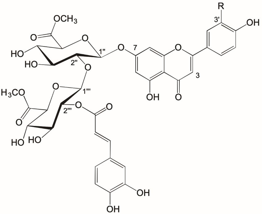
of 1 confirmed the linkages between the aglycone, sugar, and (E)-caffeoyl moieties (Figure 1). The
high-resolution ESI+ -MS spectrum of 1 (Figure S9) showed a [M + H]+ ion at m/z 813.1881 corresponding
to the empirical formula C38 H37 O20 + (calc. 813.1878 Da) in agreement with the dimethyl ester of
apigenin 7-O-(2”-O-(2000 -(E-caffeoyl)-β-glucuronopyranosyl)-β-glucuronopyranoside) (1) (Figure 1).Molecules 2020, 25, 3782 3 of 9
Table 1. 1 H and 13 C spectral data (δ in ppm) for 1 and 2 dissolved in DMSO-d6 at 25 ◦ C.
1 (1 H) 1 (13 C) 2 (1 H) 2 (13 C)
Aglycone
2 164.39 164.66
3 6.85 s 103.25 6.73 s 103.31
4 182.10 182.03
5 161.28 161.34
6 6.39 d 2.2 99.25 6.39 d 2.1 99.29
7 162.19 162.22
8 6.76 d 2.2 94.44 6.73 d 2.1 94.34
9 157.01 157.06
10 105.52 105.57
10 121.12 121.50
20 7.94 ‘d‘ 8.8 128.66 7.43 br 119.25
30 6.95 ‘d‘ 8.8 116.15 145.99
40 161.50 150.14
50 6.95 ‘d‘ 8.8 116.15 6.92 d 8.5 116.18
60 7.94 ‘d‘ 8.8 128.66 7.43 dd 2.2, 8.5 121.36
7-O-glucuronopyranosyl
1” 5.44 d 7.5 97.35 5.46 d 7.5 97.37
2” 3.52 m 81.01 3.53 m 81.07
3” 3.37 m 74.73 3.37 m 74.77
4” 3.37 m 71.54 3.37 m 71.58
5” 4.19 d 7.5 74.70 4.19 d 9.4 74.75
6” 169.10 169.15
OCH3 3.63 s 52.09 3.63 s 52.14
2”-O-β-glucuronopyranosyl
1000 4.88 d 8.2 101.42 4.89 d 8.3 101.46
2000 4.62 d 8.2 73.52 4.63 dd 8.3, 9.6 73.58
3000 3.47 m 73.56 3.48 t 9.6 73.61
4000 3.36 m 72.02 3.36 t 9.6 72.08
5000 3.85 d 8.2 75.37 3.85 d 9.6 75.42
6000 169.17 169.22
OCH3 3.52 s 51.71 3.52 s 51.75
2000 -O-caffeoyl
C=O 165.87 165.94
α 6.24 d 15.8 114.73 6.23 d 15.8 114.75
β 7.43 d 15.8 144.72 7.44 d 15.8 144.79
1”” 125.86 125.89
2”” 7.03 d 2.1 114.83 7.04 d 2.2 114.86
3”” 145.65 145.72
4”” 148.29 148.36
5”” 6.76 m 115.82 6.77 d 8.1 115.87
6”” 6.97 dd 2.1, 8.5 121.30 6.97 dd 2.2, 8.1 121.36
s = singlet, d = doublet, t-triplet, dd = double doublet, m = multiplet, br = broad. See Figure 1 for structures.Molecules 2020, 25, 3782 4 of 9
Molecules 2020, 24, x FOR PEER REVIEW 3 of 9
Figure 1. Structures
Figure Structuresofoftwo
twonew flavonoids
new 1 (R=H)
flavonoids andand
1 (R=H) 2 (R=OH) isolated
2 (R=OH) from aerial
isolated parts of
from aerial Nepeta
parts of
curviflora.
Nepeta curviflora.
The NMR Table resonances
1. 1H and 13Cofspectral
2 weredata
very(δsimilar
in ppm)toforthose
1 and of compound
2 dissolved 1 (Table
in DMSO-d 1).
6 at 25 However,
°C. the
1
main differences were shown in the aromatic region where the H NMR spectrum of 2 (Figure
1 (1H) 1 (13C) 2 (1H) 2 (13C)
S11) revealed a 3H AA’X system at δ 7.43 (H-20 , br), δ 7.43 (H-60 , br), and δ 6.92 (J = 8.5 Hz;
H-50 ), a 1H singlet at δ 6.73 (H-3) and a 2H Aglycone
AX system at δ 6.73 (d, J = 2.2 Hz; H8) and δ
6.39 (d, J = 2.2 Hz; H6), in accordance with a 7-O-glycosylated luteolin derivative.
2 164.39 164.66 The cross
peaks at δ 35.46/162.22 (H-1”/C-7),
6.85 s 103.25
δ 3.53/101.46 (H-2”/C-16.73000 ), sδ 4.89/81.07 (H-1 000 /C-2”), and at
103.31
4 (H-2000 /C=O) in the HMBC
δ 4.63/165.94 182.10
spectrum of 2 (Figure S13) confirmed 182.03the linkages
5 161.28
between the aglycone, sugar, and (E)-caffeoyl moieties (Figure 1). The high-resolution 161.34 ESI+ –MS
spectrum of6 2 (Figure 6.39 S17) dshowed
2.2 a [M + 99.25 +
H] ion at m/z 6.39 d 2.1 corresponding99.29
829.1830 to the empirical
7 + 162.19
formula C38 H37 O21 (calc. 829.1827 Da) in agreement with the dimethylester form of luteolin 162.22
7-O-(2”-O-(28 000 -(E-caffeoyl)-β-glucuronopyranosyl)-β-glucuronopyranoside)
6.76 d 2.2 94.44 6.73 d 2.1 (Figure94.34
1).
9
Previously, we have reported that the 157.01
free carboxyl group of glucuronyl moieties 157.06 of flavonoids
10 105.52
readily will be esterified with methanol during extraction and isolation processes involving acidified105.57
methanol as1′ solvent [30]. This is in accord 121.12
with the identification of both 1 and 2 121.50 as dimethylesters
2′ 7.94 `d` 8.8
caused by methylesterfication of the two 128.66
glucuronyl moieties 7.43 br
of these flavonoids. 119.25
Small amounts of
3′ 6.95 `d` 8.8 116.15
parental 1 and 2 without their methylesters were indeed detected by LC-MS analysis of Sephadex 145.99
4′
LH-20 fractions of the purified extract of N. 161.50
curviflora (FigureS S10 and S18). 150.14
5′ 6.95 `d` 8.8 116.15 6.92 d 8.5
Antibacterial activity was measured using the agar diffusion method. Sephadex LH-20 fractions 116.18
containing 6′
both 1 and 7.94 `d` 8.8 in DMSO
2 dissolved 128.66
did not reveal 7.43 dd 2.2, 8.5 activity. However,
antibacterial 121.36 the cruder
XAD-7 purified material showed some 7-O-glucuronopyranosyl
antibacterial activity, suggesting other compounds in the extract
1′′
to be considered 5.44antibacterial
in future d 7.5 97.35 studies.
activity 5.46 d 7.5 97.37
2′′ 3.52 m 81.01 3.53 m 81.07
3. Materials
3′′and Methods 3.37 m 74.73 3.37 m 74.77
4′′ 3.37 m 71.54 3.37 m 71.58
3.1. General
5′′ 4.19 d 7.5 74.70 4.19 d 9.4 74.75
UV-Vis6′′absorption spectra were recorded 169.10 on-line during HPLC analysis using 169.15 a photodiode
OCH3 (HP 1050)3.63
array detector s
(Agilent 52.09 Santa Clara,3.63
Technologies, CA, 1
s USA). H (600.13 52.14MHz) and 13 C
2′′-O-β-glucuronopyranosylMolecules 2020, 25, 3782 5 of 9
(150.90 MHz) NMR spectra were obtained on a Bruker Biospin AV-600 MHz instrument equipped
with a TCI 1 H-13 C/15 N CryoProbe (Bruker BioSpin, Zürich, Switzerland), and on a Bruker Biospin
AV-850 MHz equipped with a CryoProbe (Bruker BioSpin, Zürich, Switzerland). All experiments were
recorded at 298K and the chemical shift values were set relative to the deutero-methyl 13 C signal and
the residual 1 H signal of the solvent ((CD3 )2 SO) at δ 39.6 and δ 2.49, respectively. Low-resolution mass
spectra were recorded on a LC-MS system (Agilent Technologies, Santa Clara, CA, USA) consisting
of an Agilent 1200 series LC module (binary pump, column compartment/oven, and auto sampler),
equipped with an Agilent ZORBAX SB-C18 (RRHT 2.1 × 50 mm, 1.8 µm), with an Agilent 6420A mass
spectrometer equipped with a triple quadrupole (QqQ configuration) mass analyzer using electrospray
ionization (ESI) as detector.
High-resolution mass spectrometry (ESI+ /TOF), spectra were recorded using a JEOL AccuTOF
JMS-T100LC instrument (JEOL, Peabody, USA) in combination with an Agilent Technologies 1200 Series
HPLC system. A Zorbax Eclipse-C18 (Agilent Technologies, Santa Clara, CA, USA) (50 × 2.1 mm,
length × i.d., 1.8 µm) column was used for separation. Two solvents, A (H2 O + 0.2 % HCOOH, v/v)
and B (acetonitrile + 0.2 % HCOOH, v/v), were used for elution. The following solvent compositions
were used: 0 min (0 % B), 0–2 min (0 to 10% B, linear gradient), 2–15 min (10 to 80% B, linear gradient).
The flow rate was 0.4 mL/min and the temperature was kept at 60 ◦ C.
3.2. Isolation of Flavones
The aerial parts of Syrian catnip (stems, leaves, and flowers) were collected in May 2016 from
the close surrounding area of Birzeit University, Palestine (Coordinates: Latitude: 31◦ 570 18.76” N;
Longitude: 35◦ 100 30.32” E). The collected plant material was dried and thereafter stored for
approximately one month. The dried aerial part (755 g) was extracted three times over night at
4 ◦ C, with 5L 10% H2 O in MeOH (v/v; containing 0.5% trifluoroacetic acid, TFA). The combined extract
was concentrated under reduced pressure in order to remove methanol. The nonpolar compounds
were removed by partition against hexane. Approximately 1/3 of the resulting aqueous phase was
subjected to Amberlite XAD-7 column chromatography. Part of the XAD-7 purified extract (2.34 g) was
further purified and separated on a 100 × 5 cm Sephadex LH-20 column using acetonitrile—H2 O:TFA
(10:90:0.2; v/v) and MeOH—H2 O:TFA (80:20:0.2; v/v) as mobile phase with a flow rate of 4 mL/min
(Supporting Information, Table S4).
A mixture of compounds 1, 2, and 4 was obtained in the combined fractions 25 and 26 achieved
by Sephadex LH-20 chromatography (Table S4). Pure compounds 1, 2, and 4 were then isolated from
fractions 25 and 26 by preparative HPLC. Pure compound 3 was eluted in Sephadex LH-20 fraction 33
(Table S4). The isolation of compounds 5 and 6 was based on the extraction of 195 g dried flowers of N.
curviflora followed by the same purification and separation steps as indicated above. The dried XAD-7
purified extract (2.31 g) was fractionated by Sephadex LH-20 chromatography, and pure 5 and 6 were
obtained in fractions 27 and 38, respectively (Supporting Information, Table S5).
3.3. Preparative HPLC
Preparative HPLC was performed using a Gilson 321 preparative HPLC equipped with a UV
detection (Dionex UltiMate 3000 Variable Wavelength Detector) (Dionex Corporation, Sunnyvale, CA,
US). The system was equipped with an Econosil C18 column (250 mm × 22 mm; length × I.D., 5.0 µm;
Fortis Technologies Ltd., Neston, UK). The elution protocol consisted of solvents A, H2 O containing
0.5% TFA (v/v) and B, acetonitrile containing 0.5% TFA (v/v). The following gradient was used: 100% A
in 0–5 min, 10% B (isocratic elution) for the next 46 min (6–52 min), 20% B (isocratic elution) for the next
12 min (53–65 min), 50% B (isocratic elution) for the next four minutes (66–70 min), and then back to the
starting conditions (100% A, isocratic elution) in 4 min (71–75 min). The flow rate was 12.0 mL min−1 .Molecules 2020, 25, 3782 6 of 9
3.4. Analytical HPLC
Analytical HPLC was performed using Agilent 1100 HPLC system (Agilent Technologies,
Santa Clara, CA, USA) equipped with a HP 1050 diode array detector and with an ODS-Hypersil
column (20 × 0.5 cm, length × i.d., 5 µm; Supelco, Bellefonte, PA, USA), using the solvents A, H2 O
containing 0.5% TFA (v/v) and B, acetonitrile containing 0.5% TFA (v/v). The following gradient (B in A)
was used: 10 to 14% B in 0–10 min, 14% B (isocratic elution) for the next 4 min, 14 to 40% B (linear
gradient) from 14–32 min, 40% B (isocratic elution) from 32–43 min, followed by a linear gradient 40%
B-10% B for 3 min to re-establish the starting conditions. The flow rate was 1.0 mL min−1 .
3.5. Biological Activity
The antibacterial activities of Sephadex LH-20 fractions containing both 1 and 2, and XAD-7
purified material, were investigated against five gram-positive (Staphylococcus aureus, Micrococcus luteus,
Bacillus subtilis, Enterococcus faecalis, and Staphylococcus epidermidis) and four gram-negative
(Escherichia coli, Klebsiella pneumonia, Proteus mirabilis, and Proteus aeruginosa) bacteria. The antibacterial
test was carried out by using the agar diffusion method. The sterile saline was prepared by dissolving
0.5 g of NaCl in 500 mL of water (0.1% of NaCl) before this solution was autoclaved. The single bacterial
colonies were dissolved in the sterile saline until the turbidity of the suspended cells reached the
McFarland 0.5 standard. The bacterial inocula were spread on the surface of Mueller–Hinton nutrient
agar using a sterile cotton swab. Then, the wells (6 mm in diameter) in the agar plate were made by
using Sterile glassy borer [34,35]. The samples were dissolved in DMSO in concentrations of 6 mg/mL,
and 25 µL of each were added into their respective wells before the plates were incubated at 37 ◦ C for
12–24 h. Gentamycin (G) and Erythromycin (E) were used as positive controls, while DMSO was used
as negative control. The activities of the samples were determined by measuring the inhibition zone
diameter in millimeter. The results were determined by calculating the average of three trials.
4. Conclusions
In this investigation, individual phenolic constituents of the aerial parts of Syrian catnip have
been characterized on the basis of extensive spectroscopic analyses. Two new flavonoids (1 and 2)
along with two known flavonoids (5 and 6) and the methylesters of rosmarinic acid (3) and caffeic acid
(4) have been identified. Flavonoids glycosylated with glucuronic acid have previously been reported
from several Nepeta spp. [36–38]. However, the findings of 1 and 2 in N. curviflora are the first report of
acylated flavone glucuronides in the genus Nepeta, which might have chemotaxonomic significance
within the genus.
Supplementary Materials: The following are available online. Figure S1: 1D 1 H NMR spectrum of apigenin
7-O-(2”-O-(2000 -(E-caffeoyl)-β-glucuronopyranosyl)-β-glucuronopyranoside) (1). Figure S2: 1D 13 C CAPT NMR
spectrum of apigenin 7-O-(2”-O-(2000 -(E-caffeoyl)-β-glucuronopyranosyl)-β-glucuronopyranoside) (1). Figure
S3: 2D 1 H-13 C edited HSQC NMR spectrum of apigenin 7-O-(2”-O-(2000 -(E-caffeoyl)-β-glucuronopyranosyl)-β-
glucuronopyranoside) (1). Figure S4: 2D 1 H-13 C HMBC NMR spectrum of apigenin 7-O-(2”-O-(2000 -(E-caffeoyl)-β-
glucuronopyranosyl)-β-glucuronopyranoside) (1). Figure S5: 2D 1 H-13 C HSQC-TOCSY NMR spectrum of
apigenin 7-O-(2”-O-(2000 -(E-caffeoyl)-β- glucuronopyranosyl)-β-glucuronopyranoside) (1). Figure S6: 2D 1 H-13 C
H2BC NMR spectrum of apigenin 7-O-(2”-O-(2000 -(E-caffeoyl)-β-glucuronopyranosyl)-β- glucuronopyranoside)
(1). Figure S7: 2D 1 H-1 H COSY NMR spectrum of apigenin 7-O-(2”-O-(2000 -(E-caffeoyl)-β-glucuronopyranosyl)-
β-glucuronopyranoside) (1). Figure S8: 2D 1 H-1 H ROESY NMR spectrum of apigenin 7-O-(2”-O-(2000 -(E-caffeoyl)-β-
glucuronopyranosyl)-β-glucuronopyranoside) (1). Figure S9: HR Mass spectrum of the dimethylester form
of apigenin 7-O-(2”-O-(2000 -(E-caffeoyl)-β-glucuronopyranosyl)- β-glucuronopyranoside) (1). Figure S10: Mass
spectrum of apigenin 7-O-(2”-O-(2000 -(E-caffeoyl)-β-glucuronopyranosyl)- β-glucuronopyranoside) (1). Figure S11:
1D 1 H NMR spectrum of luteolin 7-O-(2”-O-(2000 -(E-caffeoyl)-β- glucuronopyranosyl)-β-glucuronopyranoside)
(2). Figure S12: 1D 13 C CAPT NMR spectrum of luteolin 7-O-(2”-O-(2000 -(E-caffeoyl)-β-glucuronopyranosyl)-
β-glucuronopyranoside) (2). Figure S13: 2D 1 H-13 C HMBC NMR spectrum of luteolin 7-O-(2”-O-(2000 -(E-caffeoyl)-
β-glucuronopyranosyl)-β-glucuronopyranoside) (2). Figure S14: 2D 1 H-13 C edited HSQC NMR spectrum of
luteolin 7-O-(2”-O-(2000 -(E-caffeoyl)-β-glucuronopyranosyl)-β-glucuronopyranoside) (2). Figure S15: 2D 1 H-13 C
H2BC NMR spectrum of luteolin 7-O-(2”-O-(2000 -(E-caffeoyl)-β-glucuronopyranosyl)-β-glucuronopyranoside) (2).Molecules 2020, 25, 3782 7 of 9
Figure S16: 2D 1 H-1 H COSY NMR spectrum of luteolin 7-O-(2”-O-(2000 -(E-caffeoyl)-β-glucuronopyranosyl)-
β-glucuronopyranoside) (2). Figure S17: HR mass spectrum of the methyl ester form of luteolin
7-O-(2”-O-(2000 -(E-caffeoyl)-β-glucuronopyranosyl)-β-glucuronopyranoside) (2). Figure S18: Mass spectrum
of luteolin 7-O-(2”-O-(2000 -(E-caffeoyl)-β-glucuronopyranosyl)-β-glucuronopyranoside) (2). Table S1:
Chromatographic (HPLC) and spectral (UV and MS) data recorded for 1–6 from Nepeta curviflora. Table
S2: 1 H and 13 C spectral data (ppm) and coupling constants (Hz) for compounds 3 and 4 dissolved in DMSO-D6 at
25 ◦ C. Table S3: 1 H and 13 C-NMR chemical shift values of apigenin (5) and apigenin 7-O-β-glucuronopyranoside
(6) in DMSO-D6 at 298 K. Table S4: Solvent composition and elution volumes used for separation of XAD-7
purified extract of aerial parts of Nepeta curviflora using a 100 × 5 cm Sephadex LH-20 column. The flow rate was
4 mL/min. Table S5: Composition and elution volumes used for separation of XAD-7 purified extract of flowers of
Nepeta curviflora using a 100 × 5 cm Sephadex LH-20 column. The flow rate was 4 mL/min. Table S6: In-vitro
anti-bacterial activity data for compound 1 and 2 and for the XAD purified materials against gram-negative
bacteria. Inhibition zone diameter is in millimetre. Table S7: In-vitro anti-bacterial activity data for compounds 1
and 2 and for the XAD purified materials against gram positive bacteria. Inhibition zone diameter is in millimeter.
Author Contributions: S.R., H.A.A., Ø.M.A., and T.F. planned, and designed the project. S.R. and M.R. isolated
the pure natural products. K.H.E. performed LC-MS analysis. T.F. recorded NMR spectra of isolated fractions.
S.R. and T.F. determined the structures of the isolated compounds. S.R., T.F., and Ø.M.A. wrote the manuscript.
All authors have read and agreed to the published version of the manuscript.
Funding: This research received no external funding.
Acknowledgments: We acknowledge Bjarte Holmelid (Dept. of Chemistry, University of Bergen) for recording of
high-resolution mass spectra. Azmi Doudin and Asem Mubarak (both Department of Chemistry, Birzeit University)
are acknowledged for technical assistance, Ibrahim Shalash (Department of Chemistry, Birzeit University) for
the help with the analytical HPLC and the different chemicals used in this work. Khaled Sawalha (Botanist and
Director of AQU Herbarium) and the Union of Agricultural Work Committees (UAWC) are acknowledged for
storing voucher specimen of N. curviflora. This work was partly supported by the Research Council of Norway
through the Norwegian NMR Platform, NNP (226244/F50). The authors thank the office of Vice-President for
Academic Affairs at Birzeit University for financial support.
Conflicts of Interest: The authors declare no conflict of interest.
References
1. Jamzad, Z.; Grayer, R.J.; Kite, G.C.; Simmonds, M.S.J.; Ingrouille, M.; Jalili, A. Leaf surface flavonoids
in Iranian species of Nepeta (Lamiaceae) and some related genera. Biochem. Syst. Ecol. 2003, 31, 587–600.
[CrossRef]
2. Ali, L.; Ali, S.; Rizvi, T.S.; Khan, A.; Hassan, Z.; Al-Harrasi, A.; Hussain, J. Antioxidant Flavonoids from
Nepeta floccosa Benth. Rec. Nat. Prod. 2015, 9, 567–571.
3. Baser, K.H.C.; Kirimer, N.; Kurkcuoglu, M.; Demirci, B. Essential oils of Nepeta species growing in Turkey.
Chem. Nat. Compd. 2000, 36, 356–359. [CrossRef]
4. Dabiri, M.; Sefidkon, F. Composition of essential oil of Nepeta crassifolia Boiss and Buhse. Flav. Fragr. J. 2003,
18, 225–227. [CrossRef]
5. Rapisarda, A.; Galati, E.M.; Tzakou, O.; Flores, M.; Miceli, N. Nepeta sibthorpii Bentham (Lamiaceae):
Micromorphological analysis of leaves and flowers. Farmaco 2001, 56, 413–415. [CrossRef]
6. Naghibi, F.; Mosaddegh, M.; Motamed, S.M.; Ghorbani, A. Labiatae family in folk medicine in Iran: From
Ethnobotany to Pharmacology. Iran. J. Pharm. Res. 2005, 2, 63–79.
7. Firoozabadi, A.; Kolouri, S.; Zarshenas, M.; Salehi, A.; Mosavat, S.H.; Dastgheib, S.A. Efficacy Nepeta menthoides
Boiss and Buhse freeze-dried aqueous extract on anxiety of patients with depression: A double-blind
randomized controlled clinical trial. Iran J. Med. Sci. 2016, 41, 164–170. [CrossRef]
8. Dienaitėa, L.; Pukalskienėa, M.; Matiasb, A.A.; Pereirab, C.V.; Pukalskasa, A.; Venskutonisa, P.R. Valorization
of six Nepeta species by assessing the antioxidant potential, phytochemical composition and bioactivity of
their extracts in cell cultures. J. Funct. Foods 2018, 45, 512–522. [CrossRef]
9. Barhoumi, L.M.; Al-Jaber, H.I.; Abu Zarga, M.H. Volatile Organic Compounds and Essential Oil Composition
of Selected Organs of Nepeta curviflora Collected from two Region in Jordan. Jord. J. Chem. 2017, 12, 101–112.
10. Bernardi, M.M.; Kirsten, T.B.; Salzgeber, S.A.; Ricci, E.L.; Romoff, P.; Guilardi Lago, J.H.; Lourenc, L.M.
Antidepressant-like effects of an apolar extract and chow enriched with Nepeta cataria (catnip) in mice.
Psychol. Neurosci. 2010, 3, 251–258. [CrossRef]Molecules 2020, 25, 3782 8 of 9
11. Hadi, N.; Sefidkon, F.; Shojaeiyan, A.; Šiler, B.; Jafari, A.A.; Mišić, D. Phenolic’s composition in four endemic
Nepeta species from Iran cultivated under experimental field conditions: The possibility of the exploitation
of Nepeta germplasm. Ind. Crop. Prod. 2017, 95, 475–484. [CrossRef]
12. Joshi, N.; Sah, G.C. GC–MS analysis and antimicrobial activity of essential oil of Nepeta coerulescens. Int. J.
Res. Pharm. Pharm. 2014, 3, 68–71.
13. Kaviarasan, S.; Sundarapandiyan, R.; Anuradha, C.V. Protective action of fenugreek (Trigonella foenum
graecum) seed polyphenols against alcohol-induced protein and lipid damage in rat liver. Cell. Biol. Toxicol.
2008, 24, 391–400. [CrossRef]
14. Mahboubi, M.; Kazempour, N.; Ghazian, F.; Taghizadeh, M. Chemical composition: Antioxidant and
antimicrobial activity of Nepeta persica Boiss essential oil. Herba Pol. 2011, 57, 62–71.
15. Nestorović, J.; Mišić, D.; Šiler, B.; Soković, M.; Glamočlija, J.; ’Cirić, A.; Maksimović, V.; Grubišić, D. Nepeta
lactone content in shoot cultures of three endemic Nepeta species and the evaluation of their antimicrobial
activity. Fitoterapia 2010, 81, 621–626. [CrossRef]
16. Süntar, I.; Nabavi, S.M.; Barreca, D.; Fischer, N.; Efferth, T. Pharmacological and chemical features of Nepeta
L. genus: Its importance as a therapeutic agent. Phytother. Res. 2018, 32, 185–198.
17. Mišić, D.; Šiler, B.; Gašić, U.; Avramov, S.; Živković, S.; Nestorović Živković, J.; Milutinović, M.; Tešičć, Ž.
Simultaneous UHPLC/DAD/(+/-) HESI-MS/MS analysis of phenolic acids and Nepeta lactones in methanol
extracts of Nepeta species: A possible application in chemotaxonomic studies. Phytochem. Anal. 2015, 26,
72–85.
18. Formisano, C.; Rigano, D.; Senatore, F. Chemical constituents and biological activities of Nepeta species.
Chem. Biodivers. 2011, 8, 1783–1818. [CrossRef]
19. Lee, S.Y.; Lee, C.Y.; Eom, S.H.; Kim, Y.K.; Park, N.I.; Park, S.U. Rosmarinic acid production from transformed
root cultures of Nepeta Cataria L. Sci. Res. Essays 2010, 5, 1122–1126.
20. Mihaylova, D.; Georgieva, L.; Pavlov, A. In vitro antioxidant activity and phenolic composition of
Nepeta cataria L. extracts. Int. J. Agric. Sci. Tech. 2013, 1, 74–79.
21. Modnicki, D.; Tokar, M.; Klimek, B. Flavonoids and phenolic acids of Nepeta cataria L: Var citriodora (Becker)
balb (Lamiaceae). Acta. Pol. Pharm. 2007, 64, 247–252.
22. Trivellini, A.; Lucchesini, M.; Maggini, R.; Mosadegh, H.; Villamarin, T.S.S.; Vernieri, P.; Mensuali-Sodi, A.;
Pardossi, A. Lamiaceae phenols as multifaceted compounds: Bioactivity, industrial prospects and role of
positive-stress. Ind. Crop. Prod. 2016, 83, 241–254. [CrossRef]
23. Al-Qudah, M.A. Antioxidant activity and chemical composition of essential oils of fresh and dried Jordanian
Nepeta curviflora Boiss. J. Biol. Act. Prod. Nat. 2016, 6, 101–111.
24. Mancini, E.; Arnold, N.A.; Feo, V.D.; Formizano, C.; Rigano, D.; Piozzi, F.; Senatore, F. Phytotoxic effects
of essential oils of Nepeta curviflora Boiss and Nepeta nuda L subsp albiflora growing wild in Lebanon.
J. Plant. Inter. 2009, 4, 253–259.
25. Musso, L.; Scaglia, B.; Al Haj, G.; Apostolides, N.A.; Adani, F.; Scarì, G.; Dallavalle, S.; Iriti, M. Chemical
Characterization and Nematicidal Activity of the Essential Oil of Nepeta nuda L. ssp pubescens and Nepeta
curviflora Boiss from Lebanon. J. Essent. Oil Bear. Pl. 2017, 20, 1424–1433. [CrossRef]
26. Al-Bakri, A.G.; Afifi, F.U. Evaluation of antimicrobial activity of selected plant extracts by rapid XTT
colorimetry and bacterial enumeration. J. Microbiol. Meth. 2007, 68, 19–25. [CrossRef]
27. Barbour, E.K.; Al Sharif, M.; Sagherian, V.K.; Habre, A.N.; Talhouk, R.S.; Talhouk, S.N.J. Screening of selected
indigenous plants of Lebanon for antimicrobial activity. J. Ethnopharmacol. 2004, 93, 1–7. [CrossRef]
28. Cigremis, Y.; Ulukanli, Z.; Ilcim, A.; Akgoz, M. In vitro antioxidant and antimicrobial assays of acetone
extracts from Nepeta meyeri Bentham. Eur. Rev. Med. Pharm. Sci. 2010, 14, 661–668.
29. Proestos, C.; Lytoudi, K.; Mavromelanidou, O.; Zoumpoulakis, P.; Sinanoglou, V. Antioxidant Capacity of
Selected Plant Extracts and Their Essential Oils. Antioxidants 2013, 2, 11–22. [CrossRef]
30. Fossen, T.; Slimestad, R.; Øvstedal, D.O.; Andersen, Ø.M. Covalent anthocyanin-flavonols complexes from
flowers of chive, Allium schoenoprasum. Phytochemistry 2000, 54, 317–323. [CrossRef]
31. Fossen, T.; Andersen, Ø.M. Spectroscopic techniques applied to flavonoids. In Flavonoids: Chemistry,
Biochemistry and Applications; Andersen, Ø.M., Markham, K.R., Eds.; CRC Press: Boca Raton, FL, USA, 2006;
pp. 37–142.
32. Kowalska, I.; Stochmal, A.; Kapusta, I.; Janda, B.; Pizza, C.; Piacente, S.; Oleszek, W. Flavonoids from barrel
medic (Medicago truncatula) aerial parts. J. Agric. Food Chem. 2007, 55, 2645–2652. [CrossRef]Molecules 2020, 25, 3782 9 of 9
33. Stochmal, A.; Simonet, A.M.; Macias, F.A.; Oleszek, W. Alfalfa (Medicago sativa L.) flavonoids. 2. Tricin and
chrysoeriol glycosides from aerial parts. J. Agric. Food Chem. 2001, 49, 5310–5314. [CrossRef]
34. Parada, J.; Atria, A.N.; Wiese, G.; Rivas, E.; Corsini, G. Synthesis, characterization and antibacterial activity
of cobalt (III) complex with phenanthroline and Maltose. J. Chil. Chem. Soc. 2014, 59, 2636–2639. [CrossRef]
35. Rahman, M.M.; Sheikh, M.M.I.; Sharmin, S.A.; Islam, M.S.; Rahman, M.A.; Rahman, M.M.; Alam, M.F.
Antibacterial activity of leaf juice and extracts of Moringa oleifera Lam. Against some human patho-genic
bacteria. Chiang Mai University. J. Nat. Sci. 2009, 8, 219–228.
36. Güvenalp, Z.; Özbek, H.; Kuruüzüm-Uz, A.; Kazaz, C.; Demirezer, L.Ö. Secondary metabolites from Nepeta
heliotropifolia. Turk. J. Chem. 2009, 33, 667–675.
37. Olennikov, D.N.; Akobirshoeva, A.A. Flavonoids and phenylpropanoids of Nepeta glutinosa and Ziziphora
Pamiroalaica. Chem. Nat. Compd. 2016, 52, 909–912. [CrossRef]
38. Tomas-Barberan, F.A.; Gil, M.I.; Ivanchev, S.; Tomas-Lorente, F. External and vacuolar flavonoids from Nepeta
transcaucasica. Biochem. Syst. Ecol. 1992, 20, 589–590. [CrossRef]
Sample Availability: Samples of the compounds can be obtained from the authors.
© 2020 by the authors. Licensee MDPI, Basel, Switzerland. This article is an open access
article distributed under the terms and conditions of the Creative Commons Attribution
(CC BY) license (http://creativecommons.org/licenses/by/4.0/).You can also read