Biodegradable and Peroxidase-Mimetic Boron Oxynitride Nanozyme for Breast Cancer Therapy
←
→
Page content transcription
If your browser does not render page correctly, please read the page content below
RESEARCH ARTICLE
 www.advancedscience.com

Biodegradable and Peroxidase-Mimetic Boron Oxynitride
Nanozyme for Breast Cancer Therapy
Lula Zeng, Yuxin Han, Zhiwei Chen, Kang Jiang, Dmitri Golberg, and Qunhong Weng*

 nanotechnology are breeding innovative
Nanomaterials having enzyme-like activities are recognized as potentially diagnosis and therapy methods for tack-
important self-therapeutic nanomedicines. Herein, a peroxidase-like artificial ling this cancer.[12–14] To this end, many
enzyme is developed based on novel biodegradable boron oxynitride (BON) nanomaterials have been designed and
nanostructures for highly efficient and multi-mode breast cancer therapies. implemented based on the principles of
 photothermal therapy,[15,16] photodynamic
The BON nanozyme catalytically generates cytotoxic hydroxyl radicals, which
 therapy,[17,18] magnetotherapy,[19,20] etc.
induce apoptosis of 4T1 cancer cells and significantly reduce the cell viability However, these therapies rely on external
by 82% in 48 h. In vivo experiment reveals a high potency of the BON stimuli, like light or field, which signif-
nanozyme for breast tumor growth inhibitions by 97% after 14-day treatment icantly limits the clinical translations
compared with the control, which are 10 times or 1.3 times more effective and applications of these nanomateri-
 als. Therefore, much attention starts to
than the inert or B-releasing boron nitride (BN) nanospheres, respectively.
 be paid on developing non-invasive and
This work highlights the BON nanozyme and its functional integrations within self-therapeutic nanomaterials that work
the BN nanomedicine platform for high-potency breast cancer therapies. without involvements of drugs and ex-
 ternal stimuli such like nanozymes, that
 is, enzyme-mimetic inorganic or organic
 nanomaterials.[21–23]
1. Introduction These artificial enzymes, particularly the peroxidase-like
 nanozymes, have aroused great interest and become a new
Breast cancer is ranked as the most commonly diagnosed can- anti-cancer methodology. Reactive oxygen species (ROS) are
cer worldwide based on the recent statistics of the World Health endogenic byproducts of cell metabolism, including singlet oxy-
Organization, affecting health and lives of millions of women.[1] gen, superoxide anion free radicals, and hydroxyl free radicals
In recent decades, great efforts have been made in the breast (·OH), which play a crucial role in the process of intracellular
cancer diagnosis and therapy,[2] and many strategies have been signal transduction.[24] Among the ROS species, the hydroxyl
developed to prevent and treat this disease, including surgery, radical has a high reactivity and can cause significant damage
chemotherapy,[3] radiotherapy,[4,5] hormone therapy,[6,7] and tar- to protein, lipid, and DNA of cells.[25] The rapid proliferation of
geted therapy.[8,9] However, breast cancer has the characteristics tumor cells usually leads to a large amount of hydrogen peroxide
of rapid growth, easy metastasis and recurrence, and generation accumulated in the cells.[26] Peroxidase-mimetic nanozymes
of drug resistance after therapies,[10,11] making the eradication take advantage of this feature and convert hydrogen peroxide
of the tumor cells and complete rehabilitation very challenging, into harmful ROS, which could be utilized for novel cancer
particularly for the late-stage patients. Besides the standard ther- treatments. Peroxidase-like nanomaterials, such as, ferromag-
apy strategies mentioned above, the emerging nanoscience and netic nanoparticles,[27] porous carbon nanospheres,[28] and
 gold nanoparticle-loaded mesoporous silica-coated graphene
 (GSF@AuNPs), can catalyze H2 O2 to generate ROS like
L. Zeng, Y. Han, Z. Chen, K. Jiang, Prof. Q. Weng horseradish peroxidase.[29] It was found that the nitrogen-doped
College of Materials Science and Engineering
 porous carbon nanospheres had shown multiple enzyme-like ac-
Hunan University
Changsha 410082, P. R. China tivities and were utilized for in vivo ROS species regulations. The
E-mail: wengqh@hnu.edu.cn higher N content in the nanospheres, the higher enzyme-like
Prof. D. Golberg effect on inhibiting tumors was observed.[28] The peroxidase-like
Centre for Materials Science and School of Chemistry and Physics activity of nanoparticles may cause enhanced cleavage of nucleic
Queensland University of Technology (QUT) acids, proteins, and polysaccharides.[27] Compared with natural
Brisbane 4000, Australia
 enzymes that would be easily denatured and degraded when en-
 The ORCID identification number(s) for the author(s) of this article ter circulation system, the artificial nanozymes have numerous
 can be found under https://doi.org/10.1002/advs.202101184 advantages in higher chemical stability, much easier synthesis,
© 2021 The Authors. Advanced Science published by Wiley-VCH GmbH. and adjustable catalytic performance, making them competent
This is an open access article under the terms of the Creative Commons for in vivo catalysis.[30]
Attribution License, which permits use, distribution and reproduction in Hexagonal boron nitride (h-BN), a structural analog of
any medium, provided the original work is properly cited. graphite, has excellent oxidation resistance, thermal and chem-
DOI: 10.1002/advs.202101184 ical stability.[31] As a non-toxic skin lightener, it has been adopted

Adv. Sci. 2021, 2101184 2101184 (1 of 12) © 2021 The Authors. Advanced Science published by Wiley-VCH GmbH
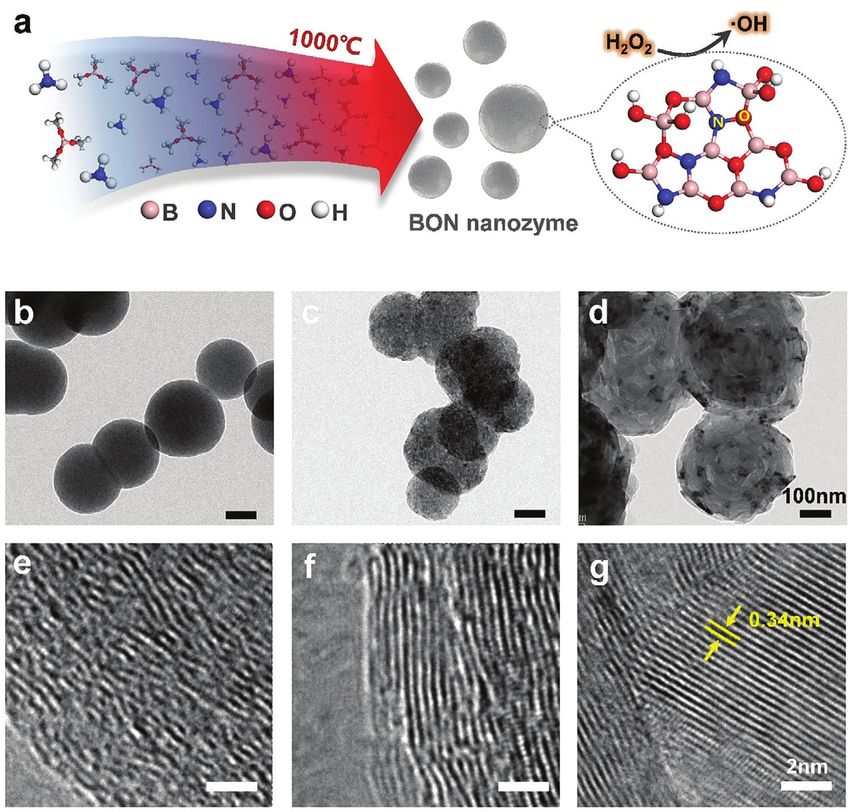
www.advancedsciencenews.com www.advancedscience.com

in many cosmetic products,[32] and also shows tremendous po- persed BON nanospheres (Figure S3, Supporting Information),
tentials for biomedical applications.[33] Purified h-BN and BN indicating an excellent stability of the formed BON colloid solu-
nanomaterials have excellent biocompatibility.[34] After function- tion in water. Further annealing treatment of the BON samples
alization, the hydrophobic BN structures could be changed into results in structural evolutions of the nanospheres. As indicated
highly water-soluble materials, which were employed for anti- in Figure 1c,d, the BON1000 annealed at 1000 °C for 4 h generally
cancer drug delivery to achieve impressive cancer cell inhibition maintains the sphere morphology with the formation of porous
efficiency.[35] Besides, the BN materials themselves were also ex- structures, while the BON1400 sample reorganizes into a crys-
plored for direct cancer therapies. For example, BN nanotubes talline and hollow structure. In this study, these BN nanomateri-
(BNNTs) and BN nanoparticles were proven as boron neutron als are employed as the B-releasing and inert BN nanospheres,
therapeutic agents that can effectively treat breast cancer.[36] Li et respectively.[37] Accordingly, the particle size of the BON sam-
al. found that hollow BN nanospheres could enduringly release ples determined by DLS increases after the annealing treatments
boric acid for prostate cancer treatment and achieved high cancer along with the decrease of Zeta potential (Figure S3, Supporting
inhibitions.[37] Because these developed cancer treatments based Information).
on BN agents must rely on external stimuli or the release of ther- Electron energy loss spectroscopy (EELS), X-ray absorption
apeutic agents, it is difficult to realize the approaches in cancer near edge structure spectroscopy (XANES), X-ray photoelectron
therapeutic practices. However, these progresses highlight the spectroscopy (XPS) and Fourier transform infrared (FTIR)
great potential of BN materials, as an integrable nanomedicine spectroscopy were employed for chemical composition and
platform, toward future multifunctional cancer diagnosis and chemical state analysis of the BON samples. EELS maps shown
therapy. in Figure 2a reveal uniform distributions of the B, N, and O el-
 In this work, we have developed a brand-new peroxidase- ements in BON nanospheres. The corresponding EEL spectrum
mimetic nanozyme based on biodegradable boron oxynitride confirms that the main compositions are B, N, and O with a
(BON) structure, which can in vitro and in vivo catalyze hydro- minor C component. The molar ratio of B:C:N:O in the BON
gen peroxide to efficiently generate hydroxyl radicals. The BON is 45:6:29:20 (Figure 2b). In N K-edge XANES (Figure 2c), the
nanospheres were synthesized via a high-temperature pyrolysis sharp peaks seen at ≈401 eV in BON and h-BN are caused by
of organic borates in ammonia. Their structures and properties the X-ray photoexcitation of N1s electrons to * (N-B), while the
were systematically analyzed employing various microscopic photoexcitation band at a higher energy position of 403.0 eV for
and spectroscopic methods. The catalytic performance of the the BON sample is originated from the N1s → * (N-O).[38,39]
nanozyme was proved directly by electron spin resonance (ESR) Compared with h-BN, the N1s → * transition in BON also
and further evaluated by a fluorescent trapping agent. The shifts to higher photo energy.
mechanism of breast cancer cell death induced by BON was Importantly, both the B1s and N1s XPS spectra reveal the dis-
evaluated by a flow cytometry. These results showed that the tinct binding energy features of the BON sample compared with
BON nanozyme could be degraded in 7 days in aqueous solu- BON1000 and BON1400 (Figure 2d). In either B1s or N1s spec-
tion; they enter 4T1 cancer cell lysosomes through phagocytosis, tra, both the BON1000 and BON1400 show the main B-N com-
which reduced the viability of the cancer cells by 82% in 48 h ponents with a minor B-O peak at 192.2 eV in the B1s spectra
by triggering the cell apoptosis process. In vivo experiment or a minor N-H peak at 399.5 eV in the N1s spectra. However,
confirmed the high potency of the BON nanozyme for breast there is a prominent N-O peak at the binding energy of 401.7 eV
tumor inhibitions by 97% after 14 days’ treatment compared that appears in the BON N1s spectrum, which is seldom observed
with the control, a much higher number compared with those and reported for BN materials.[40] From FT-IR spectra (Figure 2e),
of the inert and B-releasing BN nanospheres. This contribution it is concluded that all samples exhibit two prominent peaks at
provides a new platform for powerful nanozyme designs toward 780 and 1380 cm–1 assigned to the out-of-plane B-N-B bending
broad biomedical applications. vibration and in-plane B-N stretching vibration mode, respec-
 tively. The BON possesses broad peaks at 3200 and 3400 cm–1 ,
2. Results and Discussions corresponding to the N-H and O-H vibrations, in accordance
 with the XPS results. The band at 920 cm–1 belongs to B-N-O
2.1. Microscopic and Spectroscopic Characterizations of Boron vibration,[41] which diminishes in BON1000 and BON1400 sam-
Oxynitride ples after further annealing treatments. As shown in Figure 2f,
 the X-ray diffraction (XRD) pattern of BON has two broad and
As shown by the scanning electron microscopy (SEM) images weak diffraction peaks at ≈26° and ≈42° belonging to the BN(002)
(Figure S2, Supporting Information) and the transmission elec- and BN(100) planes,[42] these become shaper with the raise of an-
tron microscopy (TEM) images (Figure 1a,b), the pyrolytically nealing temperature. This indicates the formation of the h-BN
synthesized BON has a spherical morphology with the particle phase with higher crystallinity after the annealing treatments, in
size ranging from 100 to 500 nm, in agreement with the dynamic accordance with the TEM, XPS, and FTIR results.
light scattering (DLS) measurement (Figure S3, Supporting In- The synthesized BON nanospheres without further high-
formation). It exhibits excellent dispersibility in water even at temperature annealing treatments usually have very low crys-
a high concentration of 5 mg mL−1 . High-resolution TEM im- tallinity. It is noted that h-BN has an intrinsic planar layered struc-
ages reveal the amorphous structure of the BON, whose crys- ture like graphite. Deviations from this structure would result
tallinity increases along with the raise of annealing temperature, in metastable phases. The existence of high oxygen content in
as shown in Figure 1b–g. Zeta potential measurement reveals the BN nanospheres synthesized by the CVD reaction between
a high and negative surface potential of −47.4 mV for the dis- B(OMe)3 and NH3 was reported.[43,44] Oxygen was considered

Adv. Sci. 2021, 2101184 2101184 (2 of 12) © 2021 The Authors. Advanced Science published by Wiley-VCH GmbH

www.advancedsciencenews.com www.advancedscience.com

Figure 1. Morphological and structural analysis of BON nanospheres. a) Schematic illustration of the pyrolytic synthesis of BON nanospheres. b–d)
TEM images of BON, BON1000, and BON1400. The scale bars are 100 nm. e–g) High-magnification TEM images of BON, BON1000, and BON1400.
The scale bars are 2 nm.

as a crucial factor to form the nanosphere morphology. Further nanozyme has excellent biodegradability, and would not cause
annealing treatment of the BN nanospheres in Ar atmosphere long-term toxicity when used as nanomedicines.
would evaporate B-O species and lead to the formation of hol-
low BN spheres, in which the void size increases along with the
increase of the treatment temperature.[44] 2.3. Peroxidase-Like Catalytic Performances

 To evaluate catalytic activity of BON and the efficiency of ·OH
2.2. Degradability of Boron Oxynitride generations, a series of qualitative and semiquantitative analy-
 ses were performed. As shown in Figure 3b, the generation of
The biodegradability of BON nanozymes was investigated ·OH radicals was first determined using the coumarin as a trap-
through observations of their structural evolutions in an aque- ping agent to form the highly fluorescent 7-hydroxycoumarin.[45]
ous solution. As shown in Figure 3a, the morphology of the BON After mixing the BON and coumarin solution with H2 O2 , the
nanospheres changes continuously when incubated at a concen- fluorescence peak at 457 nm arisen from 7-hydroxycoumarin
tration of 0.1 mg mL−1 at 37 °C with shaking. First, the size of the gradually increases. As shown in Figure 3c, the catalytic potency
nanospheres obviously shrinks in 0.5 day and then the structure of BON is strong and endurable, which makes it an excellent
of nanosphere changes into hollow spheres in 3 days. With con- nanozyme to continuously produce harmful ·OH. As a compari-
tinuing incubation, the original nanosphere-like BON becomes son, the typical Fe2+ solution shows strong but transitory catalytic
a flat structure (in 5 days) and decomposes almost completely activity for H2 O2 decompositions (Figure S5, Supporting Infor-
after 7 days’ degradation. Figure S4, Supporting Information, mation). However, there are no noticeable fluorescent signals of
shows the XRD patterns of the decomposed products of BON, 7-hydroxycoumarin detected at the same testing conditions for
suggesting the formations of boric acid and NH4 B5 O8 ·4H2 O af- BON1000 and BON1400 samples, as shown in Figure S6, Sup-
ter the degradation. These results suggest that the prepared BON porting Information.

Adv. Sci. 2021, 2101184 2101184 (3 of 12) © 2021 The Authors. Advanced Science published by Wiley-VCH GmbH

www.advancedsciencenews.com www.advancedscience.com

Figure 2. Microscopic and spectroscopic characterizations of BON, BON1000, and BON1400. a) EELS maps of B-K, N-K, C-K, and O-K of BON sample.
b) EEL spectrum of BON. c) N K-edge XANES spectrum of BON. The h-BN data were reproduced from ref. [39] for comparison. d) XPS B1s and N1s spectra
of BON, BON1000, and BON1400 samples. e) FTIR spectra of BON, BON1000, and BON1400. f) XRD patterns of BON, BON1000, and BON1400.

 The generation of ·OH radicals was also more intuitively DA agent, suggesting the efficient ROS accumulations in live
demonstrated by MB degeneration experiments and ESR detec- 4T1 cells. These results verify the extracellular and intracellular
tions. It was observed that after 1 h reaction of BON, H2 O2 , and peroxidase-like behaviors of the developed BON nanospheres.
MB, the absorbance of MB solutions decreased to 50% of the orig- The ·OH radicals react with the DNA, enzymes and other
inal value (Figure S7, Supporting Information). The MB was al- biomolecules in the cell, causing damages and cell apoptosis.
most completely degraded after 12 h reaction (Figure S7, Sup- On one hand, the tumor shows increased ROS levels compared
porting Information). We also carried out ESR measurement to with normal tissues.[47] The accumulation of ROS in cancer cells
detect the generated ·OH radicals using DMPO as a spin-trapping makes the cells under a high oxidative stress state, and may cause
agent. After catalytic reaction for 20 min, the ESR spectrum con- the destructions of cell components and lead to cell apoptosis or
firms the spin signals of DMPO-trapped ·OH radicals with the necrosis.[48] On the other hand, tumor tissues are prone to higher
ratio of 1:2:2:1 (Figure 3d).[46] Furthermore, the intracellular ROS sensitivity to ROS rather than the normal ones because of the
generation was confirmed by a DCF-DA assay. As shown in Fig- lower level of antioxidant enzymes in tumors.[49] Therefore, in-
ure 3e, prominent green fluorescence appears in 4T1 cells that tracellular stimulation of forming high levels of ROS becomes
are incubated with the BON nanozyme and treated with the DCF- a promising anti-cancer strategy.[50] ROS induces cell apoptosis

Adv. Sci. 2021, 2101184 2101184 (4 of 12) © 2021 The Authors. Advanced Science published by Wiley-VCH GmbH

www.advancedsciencenews.com www.advancedscience.com

Figure 3. Enzymatic performance determinations for BON. a) SEM images of BON (0.1 mg mL−1 ) after degradations over different time at 37 °C.
b) Fluorescence spectrum evolution for detection of ·OH radicals catalyzed by BON (200 µg mL−1 ) using coumarin as the trapping agent. Ex = 332 nm.
c) Plots of fluorescence intensity (Em = 457 nm) and differential intensity versus reaction time. d) ESR spectra DMPO-·OH spin adduct generated from
BON-catalytic decomposition of H2 O2 . e) DCFH-DA fluorescence microscopic images of the 4T1 cells incubated without (left) and with (right) BON.
The scale bars are 40 µm.

through the death receptor-dependent extrinsic pathway includ- 2.4. Biocompatibility and Cellular Uptake
ing tumor necrosis factor-alpha and Fas ligand and the mitochon-
drial intrinsic pathway.[51] High concentration of ROS can induce Since the demonstrations of high ·OH production efficiency for
opening of mitochondrial permeability transition pores through the BON nanozyme, it is of great interest to know the inhibi-
regulating the conformation of adenine nucleotide translocase tion effect for cancer cells. Figure 4a illustrates the viability of
in mitochondrial inner membrane, and further reduce mito- 4T1 cells cocultured with BON, BON1000, and BON1400 at dif-
chondrial transmembrane potential, release cytochrome-c, and ferent concentrations for 24 h. At the same concentration, the
apoptosis-inducing factor, and then activate caspase signaling cytotoxicity of BON is much higher than those of BON1000 and
cascade and induce apoptosis.[52] BON1400, suggesting a remarkable inhibition effect of the BON

Adv. Sci. 2021, 2101184 2101184 (5 of 12) © 2021 The Authors. Advanced Science published by Wiley-VCH GmbH
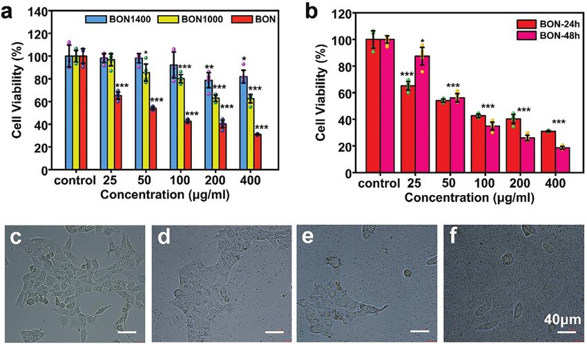
www.advancedsciencenews.com www.advancedscience.com

Figure 4. In vitro viability assays of BON nanozyme. a) Viability of 4T1 cells incubated with BON, BON1000, and BON1400 at different concentrations
for 24 h. b) Cell viability incubated with BON for 24 and 48 h. c–f) Optical microphotographs of 4T1 cells cocultured with BON at different concentrations
(c: control, d: 25 µg mL−1 , e: 100 µg mL−1 , f: 400 µg mL−1 ) for 24 h. The scale bars in (c–f) are 40 µm. Data in (a,b) are shown as mean ± SD; n = 5 per
group. Student’s t-test was used to calculate p-values by SPSS software. *p < 0.05, **p < 0.01, ***p < 0.001.

for 4T1 cells. The regarded cell viability is 50% at the BON con- The diameters of the prepared BON nanospheres are mainly lo-
centration of 50 µg mL−1 and further reduces to 31% when the cated within a range of 100–500 nm, and thus are assumed to en-
concentration increases to 400 µg mL−1 . In contrast, when co- ter cells through an endocytic pathway mediated by caveolin or
cultured with BON1000 and BON1400, even at a high concen- clathrin.[54] BON can be accumulated in lysosomes and behave
tration of 400 µg mL−1 , the resultant cell viabilities still reach to like an artificial peroxidase, thereby increasing the level of ROS.
63% and 82%. The cytotoxicity of BON1400 is not significantly Since both the generated ROS and released B compounds can
different from that of pure h-BN (Figure S9, Supporting Infor- reduce the 4T1 viability,[37,55] it is necessary to distinguish the
mation), which has been verified to have good biocompatibility. impacts between these effects. We performed dialysis for the
After coculturing with BON for 48 h, the cell viability is further re- BON solutions and collected the eluates for cytotoxicity assay.
duced to 18% (Figure 4b), indicating the high potency for in vitro As shown in Figure S14, Supporting Information, the released B
4T1 cell inhibitions. From the microscopic images (Figure 4c–f), compound from BON has mild cytotoxicity for 4T1 cells after co-
it can also be seen that the cell density tends to reduce and the incubation at the same condition. It exhibits no obvious cytotoxic-
cell morphology utterly deteriorates along with the increase of the ity for the eluates from the BONs with concentrations lower than
BON concentration. 100 µg mL−1 ; when the BON concentrations are 200 and 400 µg
 Considering the accumulation effects of the BON nanospheres mL−1 , the measured cell viabilities are 85% and 70%, respec-
in the reticuloendothelial system, practically in liver,[37] we fur- tively, which are much higher than those directly treated by the
ther carried out in vivo experiments and performed serum bio- BON samples. These results suggest that the cytotoxicity of BON
chemical analysis and H&E staining experiments of the Balb/c for 4T1 cells should mainly originate from the generated ROS
mice administered with BON samples after 7 days. As shown catalyzed by peroxidase-mimetic nanozyme. This is very differ-
in Figure S10, Supporting Information, there are no significant ent from the BON1000 sample, whose dialysis eluate shows very
effects on mouse liver functions compared with control group, close cytotoxicity results with those directly treated by BON1000,
suggesting good biocompatibility of BON nanozyme for normal indicating a B release-dominated cytotoxicity origin of the sam-
tissues. ple. This is in accordance with the previous report.[37] The present
 The BON nanospheres are blue fluorescent under UV light ex- studies indicate that, besides the cytotoxicity arising from the re-
citation (Figure S12, Supporting Information), which can be em- leased soluble B compounds in the BON1000, the cytotoxicity for
ployed for material tracking. As revealed by colocalization assay the BON nanozyme mainly roots in the peroxidase-mimetic for-
(Figure 5a–d), the BON nanozymes are prone to accumulate at mation of ·OH radicals, a mechanism seldom reported for the
the 4T1 cell lysosomes. It is also of interest to understand cell up- known BN materials.
take mechanisms for nanomedicines. Previous studies have re- In recent years, there have been numerous cutting-edge
vealed an important role of particle size for a nanomaterial to en- catalysts developed for industrial reactions based on oxygen
ter cells. Nanomaterials with diameters larger than 500 nm tend modified h-BN structures. For example, the edge O-terminated
to enter cells through a caveolae-mediated internalization mech- BN nanosheets were found to have highly selective catalytic
anism, while the ones with a diameter less than 200 nm tend dehydrogenation of propane to propene.[56] BN nanosheet-
to enter cells through clathrin-mediated endocytic pathways.[53] constituted microspheres were developed for catalytic oxidative

Adv. Sci. 2021, 2101184 2101184 (6 of 12) © 2021 The Authors. Advanced Science published by Wiley-VCH GmbH
www.advancedsciencenews.com www.advancedscience.com

Figure 5. Cellular uptake and cell death mechanism assays. a) Bright-field microscopic image of 4T1 cells that incubated with BON and Lyso-Tracker
Red. Fluorescence microscopic images of 4T1 cells at the b) Ex = 540 nm and c) Ex = 350 nm. d) Merged image of (b) and (c). e) Flow cytometry with
Annexin V-FITC/PI assay of 4T1 cells after 24 h co-incubation with BON at different concentrations. The scale bars in (a–d) are 10 µm.

dehydrogenation of propane to produce propylene and 4T1 cells increases significantly. When the concentration of BON
ethylene.[57] And indeed, the property of catalytic genera- reaches 200 and 400 µg mL−1 , 73.1% or 80.0% of total cells are
tions of ·OH radicals had been reported for BN quantum dots, in early and late apoptosis, respectively, accompanied with only
which were prepared by sonication and hydrothermal treatment 10.1% and 1.8% belonging to cellular necrosis (similar to the con-
of edge hydroxylated BN nanosheets. It was proposed that the trol group, 2.1%); the proportions of early and late apoptotic cells
free B radicals at the edges and defective sites of the BN quantum in the BON samples with 200 and 400 µg mL−1 are about 3.3 and
dots were responsible for the resultant catalytic performances 3.6 times as high as that for the concentration of 50 µg mL−1 ;
because of the detected quenching effect of DPPH.[58] This there are no early or late apoptosis cells detected in the control
result highlights the potentials of BN nanomaterials for artificial group. The dialysate of BON (400 µg mL−1 ) was also collected to
nanozyme applications. As the structural analog of h-BN, carbon coculture with 4T1 cells; the flow cytometry in Figure S15, Sup-
nanomaterials doped with nitrogen atoms have been reported porting Information, reveals 6.1% of the cells in early apoptosis
to show enzyme-like activities.[28] The regarded peroxidase-like and 4.0% in late apoptosis, the numbers which are significantly
behaviors therein were ascribed to the involvement of N-O lower than those for the samples incubated with BON directly
structures.[28] In the present study, rich N-O bonds were found at the same concentration. This also supports our claim that the
in the BON sample, which completely disappeared in the sam- peroxidase-like catalytic generation of ROS of BON nanozyme
ples after annealing at 1000 and 1400 °C, and thus were also plays the key role in regulating the apoptosis of 4T1 cells.
tentatively attributed as the catalytic sites. Successful clarifying
the catalytic mechanisms will unambiguously guide future
rational designs of high-performance artificial nanozymes. 2.6. In Vivo Anticancer Effect

 To further test and understand the anticancer effect of BON
2.5. Cell Death Mechanism Induced by Boron Oxynitride nanozyme in vivo, we explored the therapeutic effect of BON
Nanozyme on mice bearing 4T1 tumors (Figure 6a). When the subcuta-
 neously formed tumors grew up to ≈100 mm3 , the mice were
Flow cytometry was adopted to explore the anticancer mecha- subjected to the injections of samples as well as PBS as a control.
nism of BON nanozyme with Annexin V-FITC/PI assay. Annexin Among all experimental groups, BON significantly suppresses
V-FITC+/PI− stained cells indicate early apoptotic, while the the tumor growth with the average tumor volume of 29 mm3 af-
cells stained Annexin V-FITC+/PI+ are considered as late stage ter 14-day treatment (Figure 6b). For the BON1000 group, it was
apoptotic. The Annexin V-FITC−/PI+ staining result accounts found that the growth rate of tumors was remarkably reduced
for cell necrotic. As shown in Figure 5e, the BON nanozyme ex- compared with that of the control group. And the tumor growth
hibits dose-dependent cytotoxicity. With the increase of BON con- for the inert BON1400-treated mice shows no significant differ-
centration, the proportion of early apoptosis and late apoptosis of ence with that of the control. After 14-day treatment, the average

Adv. Sci. 2021, 2101184 2101184 (7 of 12) © 2021 The Authors. Advanced Science published by Wiley-VCH GmbH
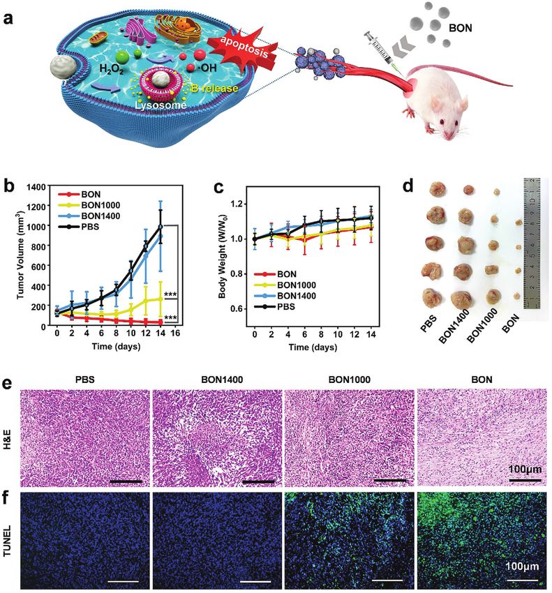
www.advancedsciencenews.com www.advancedscience.com

Figure 6. In vivo evaluations of BON nanozyme for breast cancer therapy. a) Schematic illustration of therapeutic mechanism of BON. b) Tumor growth
curves of 4T1 tumors in BALB/c mice treated at different conditions. c) Body weights of different groups of mice. d) Photographs of exfoliated tumors
of different groups. e) Optical photographs of tumor sections stained with H&E. f) Corresponding fluorescent images of tumor sections subjected to
TUNEL staining. The scale bars in (e,f) are 100 µm. Data in (b,c) are shown as mean ± SD; n = 5 per group. Student’s t-test was used to calculate
p-values by SPSS software. *p < 0.05, **p < 0.01, ***p < 0.001.

Adv. Sci. 2021, 2101184 2101184 (8 of 12) © 2021 The Authors. Advanced Science published by Wiley-VCH GmbH
www.advancedsciencenews.com www.advancedscience.com

tumor volumes in the control group, BON1400, and BON1000 ing a new powerful tool based on BN structures toward potent
are 985, 891, and 261 mm3 , respectively. These results also con- cancer therapies.
firm that the BON1000 can suppress tumor growth by about 74% One critical issue in cancer treatment is the difficulty eliminat-
compared with the control because of releasing B compounds.[37] ing cancer cells completely, which will cause failures of the ther-
While to the BON nanozyme, besides the release of B species, the apies. To eliminate cancer cells as much as possible, an emerg-
catalytic generations of ROS lead to excellent anticancer potency ing strategy is to combine different diagnostic and therapeutic
in vivo, which achieves the inhibition of tumor growth by 97%. functions within one material platform. For example, by inte-
And amazingly, the tumor tissue in one mouse of BON group grating multiple functions of photothermal, photodynamic, mag-
was found to be completely eliminated through visual inspection netotherapy, chemotherapy together in carbon materials,[66] or
(Figure 6d). During the treatment, there was no significant dif- up-conversion nanoparticles,[67] the reported cancer therapeutic
ference in the mice body weight of the experimental groups com- efficacies were enormously improved compared with the corre-
pared with the control, indicating a high biocompatibility of BON sponding monofunctional materials. Herein, for the BN nano-
samples for biomedical applications (Figure 6c). material, it is possible to in parallel integrate the therapeutic
 Thereafter, the tumor tissues were stained by H&E and functions of neutron capture therapy, boron-releasing therapy,
triphosphate nick end labeling (TUNEL) to evaluate the thera- chemotherapy, as well as, the nanozyme therapy that discovered
peutic effects in different groups. From the results of H&E stain- in the present work. In addition, BON nanozyme exhibits strong
ing (Figure 6e), the apoptosis/necrosis of the tumor tissue in the blue fluorescence, which can be exploited for real-time tracking
BON group is the most serious, which is consistent with the re- of the treated materials. This is also readily integrable into the BN
sults of the tumor growth curve. In addition, from the results nanomedicine platform. It is known that the bandgap of h-BN
of H&E staining of the main organ tissues (heart, liver, spleen, could be effectively tuned through heteroatom-doping or func-
lung, and kidney) of each group of mice (Figure S16, Support- tionalization. Through oxygen doping and hydroxyl functional-
ing Information), there are no obvious damages for these or- ization, the bandgap of BN was successfully reduced to 2.1 eV and
gan tissues, which further proves the excellent biosafety of the the resultant BN materials showed strong cyan fluorescence.[39]
BON nanozyme. The apoptotic cells on the TUNEL stained tis- All these progresses highlight the great potentials to design and
sue sections show green color fluorescence, and the nuclei ex- develop BN nanomedicine platforms integrated with multiple
hibit blue fluorescence. From Figure 6f, it is clear that the BON- different diagnostic and therapeutic functions.
treated tumors have the strongest green fluorescence among all
the groups, indicating that the BON can effectively induce apop-
tosis for the 4T1 cells and function as a potent anticancer drug. 3. Conclusions
 The development of nanomedicine provides tremendous op-
 In summary, we have designed the biodegradable peroxidase-
portunity for cancer diagnosis and therapy, which can maximize
 mimetic BON nanospheres containing rich N-O bonds for effi-
the clinical efficacy and reduce side effects compared with tradi-
 cient breast cancer therapy. In vitro experiments have shown that
tional medicine forms.[59] In recent years, the potentials of BN
 BON can effectively and continuously catalyze hydrogen peroxide
nanomaterials for anticancer and biomedicine applications start
 to generate highly reactive hydroxyl radicals. Colocalization ex-
to be recognized. First, as a type of high B-rich agent, BNNTs were
 periment has revealed that the blue-fluorescence BON nanozyme
proposed for neutron capture cancer therapy.[60] Boron neutron
 can accumulate specifically in lysosomes and stimulate 4T1 cells
capture therapy requests a high B content and targeted delivery
 to generate hydroxyl radicals, thereby inducing cell apoptosis.
capability of the neutron capture agent to increase the therapeutic
 The BON nanozyme has been found to reduce the viability of
efficacy relative to conventional radiotherapeutic drugs. Numer-
 the 4T1 cancer cells in vitro by 82% in 48 h. It also exhibits high
ous B-rich nanoparticles have been developed with remarkable
 potency for breast tumor inhibitions by 97% after 14-day treat-
B species enrichment in tumors compared with blood and other
 ment, much more effective than the inert and B-releasing BN
organs, such as, BNNTs,[61] boron carbide,[62] and carborane cage-
 nanospheres. The present study integrates multiple functions of
attached CNTs.[63] Through morphological controls and surface
 peroxidase-like nanozyme, boron-release therapy, and drug track-
modifications, BN nanomaterials could be used as an effective
 ing into the biodegradable BON nanomaterial, opening the way
carrier of doxorubicin and realized much enhanced intracellu-
 toward rational designs of the multi-mode BN nanomedicine
lar drug delivery to LNCap prostate cancer cells,[35] or load Au-
 platform for potent cancer diagnosis and therapies.
ristatin PE to trigger the mitochondrial-mediated apoptosis of
Hep G2 cells.[64] Surface chemical modifications of BN nano-
materials could further enhance the specific targeting perfor- 4. Experimental Section
mances by ligand-receptor recognitions and promote the thera- Synthesis of Boron Oxynitride: The BON sample was synthesized
peutic efficacy.[43,65] Recent advances suggested that hollow BN through a pyrolysis method.[44] As shown in Figure S1, Supporting In-
nanospheres could slowly and endurably release water-soluble formation, trimethyl borate vapor was carried by N2 and NH3 gas (N2 :
B compounds, which were found to efficiently inhibit LNCap 800 mL min−1 , NH3 : 300 mL min−1 ) and introduced into a horizontal
prostate cancer cell growth. This result represents a remark- tube furnace with a temperature of 1000 °C. White solid product formed
able progress of BN nanomedicine toward non-invasive therapy and was collected at the outlet tube wall regions of the quartz tube. For
 comparison, the BON product was proceeded for further heat treatments
of cancers.[37] However, compared with other nanomedicines,
 at 1000 and 1400 °C for 4 h to generate BON1000 and BON1400 samples,
designs and explorations of BN nanomaterials on anticancer respectively.
applications are still in their infancy. This study uncovers the Material Characterizations: The morphology and composition of sam-
peroxidase-like nanozyme activity of BON nanospheres, provid- ples were characterized by a SEM (TESCAN MIRA3 LMH) and a

Adv. Sci. 2021, 2101184 2101184 (9 of 12) © 2021 The Authors. Advanced Science published by Wiley-VCH GmbH
www.advancedsciencenews.com www.advancedscience.com

Cs-correction TEM miscroscope equipped with EELS (Titan Themics G2 and incubated at room temperature in dark for 15 min for flow cytometry
60–300). Surface zeta-potential and particle size were determined using a analysis (BD FACSCalibur).
Zetasizer Nano ZSP (Malvern Instruments) based on DLS. XRD were mea- In Vivo Anti-Tumor Experiments: All animal experiments were carried
sured on a BrukerAXS D8 Advance using Cu K target. XANES data was out according to Hunan University’s guidelines on animal use and care.
collected on the XAFCA beamline at Singapore synchrotron light source The tumor models were established by injecting 100 µL 4T1 cell suspen-
using a transmission mode. The electron energy was 700 MeV and the sion (around 7 × 10⁶ cells) subcutaneously into BALB/c mice (≈16 g,
beam current was below 200 mA. FTIR were taken on a Bruker Vertex 70 female). After raising for 7 days, the tumor volumes in mice grew to
FTIR spectrophotometer in the range of 4000–400 cm−1 . XPS (Thermo ≈100 mm3 . Then, 20 BALB/c mice were divided into four groups randomly
Scientific K-Alpha) was employed to study the chemical states of samples. (n = 5). They were injected with BON, BON1000, and BON1400 solutions
All the XPS spectra were calibrated by principal C1s binding energy peak intratumorally. Every mouse was subjected to an injected dose of 0.4 mg
at 284.6 eV. ESR spectra were collected on a EMXmicro-6/1/P/L system. on day zero and day seven. While in the control group, PBS was injected
Photoluminescence (PL) spectra were acquired on a JASCO FP-8500 spec- into tumors instead. In order to evaluate the effect of anti-tumor treat-
trofluorometer, and UV–vis spectra were collected on a SHIMADZU UV- ment, the body weights and tumor volumes of mice were recorded every
2600 spectrophotometer. Fluorescence images were taken by an inverted other day. After 14 days treatment, mice were euthanized to make their
fluorescence microscope (Motic, MXH-100). neck dislocated painlessly. Then, tissues from heart, liver, spleen, lung,
 Evaluation of ·OH Generation: First, to qualitatively determine gener- kidney, and mouse tumors were collected and stained with hematoxylin
ated hydroxyl radicals (·OH), 500 µL 2 mg mL−1 BON solution was mixed and eosin (H&E) and transferase-mediated deoxyuridine TUNEL staining
with 20 µL H2 O2 and a spin-trapping agent (DMPO, 0.1 M) in a dark en- to evaluate the biological safety and antitumor effects.
vironment. After 20 min, the solution was transferred to a quartz tube Statistical Analysis: All the data were presented as mean ± SD. Stu-
for ESR measurement. Besides, the catalytic generation of ·OH was also dent’s t-test and one-way ANOVA method were adopted to assess the
evaluated by the degradation of methylene blue (MB). Specifically, 100 µL difference between groups by using SPSS software. Differences were
12.5 mg L−1 MB solution was added into a 96-well plate. Then, 100 µL considered statistically significant at p < 0.05. (*p < 0.05, **p < 0.01,
200 µg mL−1 BON and different amounts of H2 O2 were added to make ***p < 0.001)
the final H2 O2 concentrations of 10, 1, and 0.1 mM, respectively. The ab-
sorbance at 620 nm was measured by a microplate reader (Thermo Sci-
entific, Multiscan FC). For semiquantitative evaluations, coumarin was Supporting Information
used as the trapping agent of ·OH to form the highly fluorescent 7-
 Supporting Information is available from the Wiley Online Library or from
hydroxycoumarin to assess the produced ·OH.[45] Specifically, a 2 mL solu-
 the author.
tion with 200 µg mL−1 BON and 5 mM coumarin was prepared. Then, 5 µL
H2 O2 was added to the solution to initiate the reaction. The fluorescence
intensity of 7-hydroxycoumarin (Ex: 332 nm Em: 467 nm) was measured Acknowledgements
in every 5 min.
 Intracellular Reactive Oxygen Species Detection: 4T1 cells were sowed The authors would like to thank Singapore Synchrotron Light Source for
on a 96-well plate with the density of ≈1 × 10⁴ cells per well, which were collecting the XANES data. The technical helps in animal experiments pro-
cultured in 90 µL Dulbecco’s modified Eagle’s medium (DMEM, Gibco) vided by Dr. Mei Chen and Dr. Xingyi Ge in Hunan University are also
containing 10% fetal bovine serum (Gibco), 1% antibiotic, and antimy- acknowledged. This work was supported by the National Nature Science
cotic solution (Gibco) in a humidified atmosphere with 5% CO2 at 37 °C. Foundation of China (NSFC; no. 21903021), the Innovation Platform and
After the complete attachment, 10 µL BON (4 mg mL−1 ) was added and Talent Plan of Hunan Province (2019RS1027), the Fundamental Research
incubated for 6 h and then the culture medium was replaced by the fresh Funds for the Central Universities (Hunan University: 531119200114), and
one containing 5 µM DCF-DA. After 10 min, the cells were washed with the Australian Research Council (ARC) Laureate Grant No. FL160100089.
DMEM three times to remove the adsorbed DCF-DA on the cell surfaces
and observed by an inverted fluorescence microscope under the excitation
wavelength of 480 nm. Conflict of Interest
 In Vitro Cell Viability Assay: 4T1 cells were seeded and cultured in a 96-
 The authors declare no conflict of interest.
well plate at the same conditions as described above. Then, 10 µL BON,
BON1000, and BON1400 solutions were added to the culture medium
to make the final sample concentrations of 25, 50, 100, 200 and 400 µg Author Contributions
mL−1 , respectively. After coculturing for 24 h, cell viabilities were mea-
sured by cell-counting kit-8. The optical density (OD) was recorded on the Q.W. conceived the research and supervised the project. L.Z. performed
microplate reader with a filter of 450 nm. By comparing the absorbance the material synthesis, characterization, and anticancer assays. Y.H., Z.C.,
between the control group and the experimental groups, the cell viability and K.J. participated in material preparations and characterizations. All
was calculated. the authors were involved in data analysis and result discussions. L.Z. and
 Colocalization Assay: BON sample was cocultured with cells for 4 h. Q.W. wrote the manuscript with the inputs from all authors.
Then, the cells were washed with PBS to remove the attached samples on
cell surfaces. Then Lyso-Tracker Red (Lyso-Tracker Red, Ex = 500–577 nm,
 Em = 590 nm, Beyotime) was added for co-incubation with cells for addi- Data Availability Statement
tional 10 min. Finally, the fluorescence images of the cells were recorded by The data that support the findings of this study are available from the cor-
an inverted fluorescence microscope under the required excitation wave- responding author upon reasonable request.
lengths.
 Apoptosis Assay: The Annexin V-FITC apoptosis detection kit was
used to analyze the 4T1 cell apoptosis exposed to BON, BON1000, and Keywords
BON1400 by flow cytometry. In brief, 3 × 10⁶ 4T1 cells were sowed in a
six-well plate. After 24 h incubation, the nanozyme samples were added to biodegradable, boron nitride, breast cancer, nanozyme, peroxidase
make the final concentrations of 50, 200, and 400 µg mL−1 , respectively.
The cells were cocultured for another 24 h and washed with PBS. Then, Received: March 23, 2021
the Annexin V-FITC binding buffer was added to prepare the cell suspen- Revised: May 6, 2021
sion. Finally, 5 µL of Annexin V-FITC and 10 µL of PI solutions were added Published online:

Adv. Sci. 2021, 2101184 2101184 (10 of 12) © 2021 The Authors. Advanced Science published by Wiley-VCH GmbH
www.advancedsciencenews.com www.advancedscience.com

 [29] S. K. Maji, A. K. Mandal, K. T. Nguyen, P. Borah, Y. L. Zhao, ACS Appl.
 Mater. Interfaces 2015, 7, 9807.
 [1] International Agency for Research on Cancer, https://gco.iarc.
 [30] H. Ding, B. Hu, B. Zhang, H. Zhang, X. Yan, G. Nie, M. Liang, Nano
 fr/today/data/factsheets/cancers/39-All-cancers-fact-sheet.pdf (ac-
 Res. 2021, 14, 570.
 cessed: January 2021).
 [31] Q. H. Weng, X. B. Wang, X. Wang, Y. Bando, D. Golberg, Chem. Soc.
 [2] J. Y. He, C. C. Li, L. Ding, Y. N. Huang, X. L. Yin, J. F. Zhang, J. Zhang,
 Rev. 2016, 45, 3989.
 C. J. Yao, M. R. N. Liang, R. P. Pirraco, J. Chen, Q. Lu, R. Baldridge,
 [32] C. Y. Su, H. Z. Tang, K. Chu, C. K. Lin, Ceram. Int. 2014, 40, 6903.
 Y. Zhang, M. H. Wu, R. L. Reis, Y. L. Wang, Adv. Mater. 2019, 31,
 [33] G. Ciofani, S. Danti, G. G. Genchi, B. Mazzolai, V. Mattoli, Small 2013,
 1902419.
 9, 1672.
 [3] S. Rodrigues-Ferreira, A. Nehlig, H. Moindjie, C. Monchecourt, C.
 [34] X. Chen, P. Wu, M. Rousseas, D. Okawa, Z. Gartner, A. Zettl, C. R.
 Seiler, E. Marangoni, S. Chateau-Joubert, M.-E. Dujaric, N. Servant,
 Bertozzi, J. Am. Chem. Soc. 2009, 131, 890.
 B. Asselain, P. de Cremoux, M. Lacroix-Triki, M. Arnedos, J.-Y. Pierga,
 [35] Q. H. Weng, B. J. Wang, X. B. Wang, N. Hanagata, X. Li, D. Q. Liu, X.
 F. Andre, C. Nahmias, Proc. Natl. Acad. Sci. U. S. A. 2019, 116, 23691.
 Wang, X. F. Jiang, Y. Bando, D. Golberg, ACS Nano 2014, 8, 6123.
 [4] F. Castro, M. L. Pinto, C. L. Pereira, K. Serre, M. A. Barbosa, K. Ver-
 [36] L. P. Li, J. Y. Li, Y. X. Shi, P. Du, Z. Z. Zhang, T. Liu, R. P. Zhang, Z. B.
 maelen, F. Gartner, R. M. Goncalves, O. De Wever, M. J. Oliveira, Bio-
 Liu, ACS Nano 2019, 13, 13843.
 materials 2020, 257, 120218.
 [37] X. Li, X. P. Wang, J. Zhang, N. Hanagata, X. B. Wang, Q. H. Weng, A.
 [5] Y. Chao, L. G. Xu, C. Liang, L. Z. Feng, J. Xu, Z. L. Dong, L. L. Tian, X.
 Yi, K. Yang, Z. Liu, Nat. Biomed. Eng. 2018, 2, 611. Ito, Y. Bando, D. Golberg, Nat. Commun. 2017, 8, 13936.
 [6] J. Xu, J. Lv, Q. Zhuang, Z. Yang, Z. Cao, L. Xu, P. Pei, C. Wang, H. Wu, [38] Q. W. Liu, C. Chen, M. Du, Y. W. Wu, C. J. Ren, K. N. Ding, M. X. Song,
 Z. Dong, Y. Chao, C. Wang, K. Yang, R. Peng, Y. Cheng, Z. Liu, Nat. C. J. Huang, ACS Appl. Nano Mater. 2018, 1, 4566.
 Nanotechnol. 2020, 15, 1043. [39] W. T. Zheng, J. H. Guo, Y. Sakamoto, M. Takaya, X. T. Li, P. J. Chao,
 [7] L. J. Ochyl, J. D. Bazzill, C. Park, Y. Xu, R. Kuai, J. J. Moon, Biomaterials Z. S. Jin, K. Z. Xing, J. E. Sundgren, Diamond Relat. Mater. 2010, 10,
 1897.
 2018, 182, 157.
 [40] Q. H. Weng, D. G. Kvashnin, X. Wang, O. Cretu, Y. Yang, M. Zhou, C.
 [8] Z. Y. Xiao, E. Levy-Nissenbaum, F. Alexis, A. Luptak, B. A. Teply, J. M.
 Zhang, D. M. Tang, P. B. Sorokin, Y. Bando, D. Golberg, Adv. Mater.
 Chan, J. J. Shi, E. Digga, J. Cheng, R. Langer, O. C. Farokhzad, ACS
 2017, 29, 1700695.
 Nano 2012, 6, 696.
 [41] P. M. Sudeep, S. Vinod, S. Ozden, R. Sruthi, A. Kukovecz, Z. Konya,
 [9] H. Wang, D. J. Mooney, Nat. Mater. 2018, 17, 761.
 R. Vajtai, M. R. Anantharaman, P. M. Ajayan, T. N. Narayanan, RSC
[10] O. A. Martin, R. L. Anderson, K. Narayan, M. P. MacManus, Nat. Rev.
 Adv. 2015, 5, 93964.
 Clin. Oncol. 2017, 14, 32.
 [42] V. Guerra, C. Y. Wan, V. Degirmenci, J. Sloan, D. Presvytis, T. McNally,
[11] S. W. Brady, J. A. McQuerry, Y. Qiao, S. R. Piccolo, G. Shrestha, D.
 Nanoscale 2018, 10, 19469.
 F. Jenkins, R. M. Layer, B. S. Pedersen, R. H. Miller, A. Esch, S. R.
 [43] S. N. Feng, H. J. Zhang, S. Xu, C. Y. Zhi, H. Nakanishi, X. D. Gao,
 Selitsky, J. S. Parker, L. A. Anderson, B. K. Dalley, R. E. Factor, C. B.
 Mater. Sci. Eng., C 2019, 96, 552.
 Reddy, J. P. Boltax, D. Y. Li, P. J. Moos, J. W. Gray, L. M. Heiser, S.
 [44] C. C. Tang, Y. Bando, Y. Huang, C. Y. Zhi, D. Golberg, Adv. Funct. Mater.
 S. Buys, A. L. Cohen, W. E. Johnson, A. R. Quinlan, G. Marth, T. L.
 2008, 18, 3653.
 Werner, A. H. Bild, Nat. Commun. 2018, 9, 1231.
 [45] C. Zhang, W. L. Liu, X. F. Bai, S. X. Cheng, Z. L. Zhong, X. Z. Zhang,
[12] J. J. Shi, P. W. Kantoff, R. Wooster, O. C. Farokhzad, Nat. Rev. Cancer
 Biomaterials 2019, 199, 1.
 2017, 17, 20.
 [46] Q. H. Weng, Y. Ide, X. B. Wang, X. Wang, C. Zhang, X. F. Jiang, Y.
[13] R. van der Meel, E. Sulheim, Y. Shi, F. Kiessling, W. J. M. Mulder, T.
 Lammers, Nat. Nanotechnol. 2019, 14, 1007. M. Xue, P. C. Dai, K. Komaguchi, Y. Bando, D. Golberg, Nano Energy
[14] P. Grodzinski, M. Kircher, M. Goldberg, A. Gabizon, ACS Nano 2019, 2015, 16, 19.
 13, 7370. [47] H. Pelicano, D. Carney, P. Huang, Drug Resist. Updates 2004, 7, 97.
[15] Y. Y. Jiang, X. H. Zhao, J. G. Huang, J. C. Li, P. K. Upputuri, H. Sun, [48] J. L. Martindale, N. J. Holbrook, J. Cell. Physiol. 2002, 192, 1.
 X. Han, M. Pramanik, Y. S. Miao, H. W. Duan, K. Y. Pu, R. P. Zhang, [49] T. D. Oberley, L. W. Oberley, Histol. Histopathol. 1997, 12, 525.
 Nat. Commun. 2020, 11, 1857. [50] C. Gorrini, I. S. Harris, T. W. Mak, Nat. Rev. Drug Discovery 2013, 12,
[16] J. C. Li, K. Y. Pu, Chem. Soc. Rev. 2019, 48, 38. 931.
[17] Y. Zhang, F. M. Wang, C. Q. Liu, Z. Z. Wang, L. H. Kang, Y. Y. Huang, [51] S. Galadari, A. Rahman, S. Pallichankandy, F. Thayyullathil, Free Rad-
 K. Dong, J. S. Ren, X. G. Qu, ACS Nano 2018, 12, 651. icals Biol. Med. 2017, 104, 144.
 [52] S. Orrenius, A. Gogvadze, B. Zhivotovsky, Annu. Rev. Pharmacol. Tox-
[18] X. S. Li, J. F. Lovell, J. Yoon, X. Y. Chen, Nat. Rev. Clin. Oncol. 2020, 17,
 icol. 2007, 47, 143.
 657.
 [53] J. Rejman, V. Oberle, I. S. Zuhorn, D. Hoekstra, Biochem. J. 2004, 377,
[19] X. L. Liu, B. Yan, Y. Li, X. W. Ma, W. B. Jiao, K. J. Shi, T. B. Zhang, S. Z.
 159.
 Chen, Y. He, X. J. Liang, H. M. Fan, ACS Nano 2020, 14, 1936.
 [54] S. Behzadi, V. Serpooshan, W. Tao, M. A. Hamaly, M. Y. Alkawareek,
[20] Y. Du, X. L. Liu, Q. Liang, X. J. Liang, J. Tian, Nano Lett. 2019, 19, 3618.
 E. C. Dreaden, D. Brown, A. M. Alkilany, O. C. Farokhzad, M. Mah-
[21] Y. Y. Huang, J. S. Ren, X. G. Qu, Chem. Rev. 2019, 119, 4357.
 moudi, Chem. Soc. Rev. 2017, 46, 4218.
[22] L. Z. Gao, J. Zhuang, L. Nie, J. B. Zhang, Y. Zhang, N. Gu, T. H. Wang,
 [55] R. I. Jenie, A. Hermawan, E. Meiyanto, R. Y. Utomo, P. Su, R. A. Susi-
 J. Feng, D. L. Yang, S. Perrett, X. Yan, Nat. Nanotechnol. 2017, 2, 577.
 darti, M. Ikawati, ID201906703-A, 2020.
[23] J. Y. Zhang, S. H. Wu, X. M. Lu, P. Wu, J. W. Liu, Nano Lett. 2019, 19,
 [56] J. T. Grant, C. A. Carrero, F. Goeltl, J. Venegas, P. Mueller, S. P. Burt, S.
 3214.
 E. Specht, W. P. McDermott, A. Chieregato, I. Hermans, Science 2016,
[24] A. Manke, L. Y. Wang, Y. Rojanasakul, Biomed Res. Int. 2013, 2013,
 354, 1570.
 942916.
 [57] L. Cao, P. C. Dai, J. Tang, D. Li, R. H. Chen, D. D. Liu, X. Gu, L. J. Li,
[25] M. L. Circu, T. Y. Aw, Free Radicals Biol. Med. 2010, 48, 749.
 Y. Bando, Y. S. Ok, X. B. Zhao, Y. Yamauchi, J. Am. Chem. Soc. 2020,
[26] T. P. Szatrowski, C. F. Nathan, Cancer Res. 1991, 51, 794.
 142, 8755.
[27] L. Z. Gao, K. M. Giglio, J. L. Nelson, H. Sondermann, A. J. Travis,
 [58] Z. Liu, J. Q. Liu, S. Mateti, C. M. Zhang, Y. X. Zhang, L. F. Chen, J. M.
 Nanoscale 2014, 6, 2588.
 Wang, H. B. Wang, E. H. Doeven, P. S. Francis, C. J. Barrow, A. J. Du,
[28] K. L. Fan, J. Q. Xi, L. Fan, P. X. Wang, C. H. Zhu, Y. Tang, X. D. Xu, M.
 Y. Chen, W. R. Yang, ACS Nano 2019, 13, 1394.
 M. Liang, B. Jiang, X. Y. Yan, L. Z. Gao, Nat. Commun. 2018, 9, 1440.

Adv. Sci. 2021, 2101184 2101184 (11 of 12) © 2021 The Authors. Advanced Science published by Wiley-VCH GmbH
www.advancedsciencenews.com www.advancedscience.com

[59] W. P. Fan, B. Yung, P. Huang, X. Y. Chen, Chem. Rev. 2017, 117, [63] Z. Yinghuai, A. T. Peng, K. Carpenter, J. A. Maguire, N. S. Hosmane,
 13566. M. Takagaki, J. Am. Chem. Soc. 2005, 127, 9875.
[60] D. A. Buzatu, J. G. Wilkes, D. Miller, J. A. Darsey, T. Heinze, A. Birls, [64] W. Li, X. Xie, T. T. Wu, H. Lin, L. J. Luo, H. Yang, J. B. Li, Y. Xin, X. D.
 R. Beger, US Patent 7608240 B2, 2009. Lin, Y. J. Chen, Colloids Surf., B 2019, 181, 305.
[61] J. Capala, B. H. Stenstam, K. S. Skold, P. M. Rosenschold, V. Giusti, [65] E. S. Permyakova, L. Y. Antipina, P. V. Kiryukhantsev-Korneev, A. M.
 C. Persson, E. Wallin, A. Brun, L. Franzen, J. O. Carlsson, L. Salford, Kovalskii, J. Polcak, A. Manakhov, K. Y. Gudz, P. B. Sorokin, D. V.
 C. Ceberg, B. Persson, L. Pellettieri, R. Henriksson, J. Neuro-Oncol. Shtansky, Nanomaterials 2019, 9, 1658.
 2003, 62, 135. [66] Z. Liu, J. T. Robinson, S. M. Tabakman, K. Yang, H. J. Dai, Mater. Today
[62] M. W. Mortensen, P. G. Sorensen, O. Bjorkdahl, M. R. Jensen, H. J. 2011, 14, 316.
 G. Gundersen, T. Bjornholm, Appl. Radiat. Isot. 2006, 64, 315. [67] C. Wang, L. A. Cheng, Z. A. Liu, Biomaterials 2011, 32, 1110.

Adv. Sci. 2021, 2101184 2101184 (12 of 12) © 2021 The Authors. Advanced Science published by Wiley-VCH GmbH
You can also read