BIOLOGY Notes Fbise Class 9 th
←
→
Page content transcription
If your browser does not render page correctly, please read the page content below

Federal board Fbise Class 9th Notes Biology Chapter 4 Cell and Tissue
                               Visit Now: perfect24u.com

                                                                                  th
 Fbise Class 9
     Notes
  BIOLOGY

1|P age                Visit Now: perfect24u.com
Federal board Fbise Class 9th Notes Biology Chapter 4 Cell and Tissue
                                  Visit Now: perfect24u.com

  Cell and Tissue Chapter No 04
   Q1: Define cell, unicellular and multicellular organisms?
   Ans: Cell
   The basic structural and functional unit of all living organisms is called cell
   Unicellular Organisms: -
   Those organisms which are made from single cell are called unicellular organisms.
   Example:
   Amoeba, Paramecium etc.
   Multicellular organisms: -
   Those organisms which are made from more than one cells are called multicellular organisms.
   Example:
   All plants and animals

   Q2: Define tissue? Also discuss the types of tissue. (additional Question)
   TISSUE: -
   Definition: -
   Group of cells which perform particular function is called tissue.
   Types of Tissue: -
   There are two types of tissue.
             Simple tissues
            Compound Tissues.
   Simple Tissues: -
   Those tissues which are made from the same kind of cell are called simple tissues. They are found both in
   plants and animals.
   Compound Tissues: -
   Those tissues which are made from different kind of cells are called compound tissues. They are present
   in plant but absent in animals.

   Q3: Define microscopy? Explain Microscope and their types?
   Ans: Microscopy:
   The use of microscope to observe very minute living organisms is known as microscopy.
   Microscope: -
   It is an instrument which is used for the observation of those things which cannot be seen with naked eyes.
   Discovery: -
   Zacharias Janssen and his son Hans Janssen were two eye glass makers in Holland. They discovered first
   microscope in 1595. It was simply a tube with lenses at each end and its magnification from 3x to 9x.
   Anton van leeuwenhoek (1632-1723):
   Anton van leeuwenhoek was Dutch scientist made much better microscope and observed small organism
   under it. The magnification power of leeuwenhoek’s microscope was more than 250x. He is considered to
   be the first microscopist.

   Q4: Define magnification and resolution of microscope?
   Ans: Magnification: -
   The capacity of microscope to enlarge the apparent size of a small object is called magnification.
   The magnification power of electron a light microscope is 1500x.
   Resolution: -
   The capacity of microscope to differentiate between two close objects is called Resolution.
   The human eye can differentiate between two points, which are at least o.1 mm apart. This is
   known as resolution of human eye. The resolution power of light microscope is o.2μm.

   Q5: Write the characteristic features of light and electron microscope?
   Ans: Light microscope:

2|P age                       Visit Now: perfect24u.com
Federal board Fbise Class 9th Notes Biology Chapter 4 Cell and Tissue
                                 Visit Now: perfect24u.com

   Definition:
   Those microscopes which uses light to make the image of an object is called light microscope.
   Light pathway:
   Light passes through the simple and then through two glass lenses.
   Image formation:
   Lens produces an enlarge image of the sample and the second lens magnifies the image more. After
   passing through the object and lenses, the light is projected into the viewers eye when an enlarge
   and clear image is formed.
   Magnification:
   The magnification of a light microscope is 1500x. It can magnify objects only about 1500 times.
   Resolution:
   The resolution of light microscope is 0.2μm.
   Electron microscope:
   Definition:
   It is the most advance form of microscope which use beam of electron to make the image of an
   object.
   Image formation:
   In electron microscope, the object and the lenses are placed in a vacuum chamber and a beam of
   electron is passed through object. Electrons pass through or are reflected from the object
   and make the image.
   Magnetic lenses;
   Magnetic lenses focus the electron beam on a screen and make much enlarge image.
   Resolution:
   Resolution of electron microscope is 0.2mm.
   Types of Electron Microscope: -
   There are two types of electron microscope.
   Scanning electron microscope (SEM): -
            It uses an electron beam to scan the surface that has been coated with metal.
            The SEM does not have great magnifying power.
   Transmission Electron Microscope (TEM): -
            It is used for the study of internal structure of cell or any other object.
            It can magnify objects about 250,000 times.

   Q6: Differentiate between Light and Electron microscope?
   Ans: Comparison between light and electron microscope:

                                    Light Microscope                  Electron Microscope

   Radiation source                 Light                             Beams of Electron

   Lenses                           Optical                            Magnetic
   Magnification                    10,000 times greater than the     100 times greater than
                                    naked eye                         light microscope

   Resolution                       500 times of the naked eye        400 times of the light
                                                                      microscope

   Images                           2 D images                        TEM show 2D While
                                                                      SEM Shows 3D Images

3|P age                    Visit Now: perfect24u.com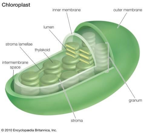
Federal board Fbise Class 9th Notes Biology Chapter 4 Cell and Tissue
                                 Visit Now: perfect24u.com

   Wave length                      400 – 700`                        0.005nm

   Q7: Write the brief history of cell theory?
   Ans: History of Cell Theory: -
   Robert Hook:-
   Robert Hook was an English scientist who discovered cell in 1665. He observed piece of cork under
   his self-made microscope. He observed small chambers like honey comb structures and he named
   it cell.
   Anton van Leeuwen Hook:-
   Leeuwen Hook was a Dutch scientist. He studied a drop or pond water under his own
   made microscope. The magnification power of his microscope was 300x. He noticed tiny
   creature swimming in the drop of pond water. Leeuwen Hook was the first man to observe single
   celled organisms called unicellular organisms.
   Jean Baptist de-Lamarck:
   In 1809, jean Baptist de-Lamarck proposed that “Nobody can have life if its constituent parts are
   not formed by cellular tissues.”
   Dolland:-
   Dolland in 1827 improved the quality of lenses. After that all the scientist were interested
   in microscopy.
   Robert Brown:-
   In 1831, a British botanist Robert Brown discovered the nucleus in the plant cell.
   Mathias Schleiden:-
   In 1839 a German Botanist Matthias schleiden studied plant tissues and made the first statement
   of the cell theory. He stated that “all plants are made up of cells”
   Theodor Schwann: -
   In 1839 a German Zoologist Schwann found that all the animals are made from cells.
   Thus, schleiden and Schwann proposed cell theory in its Italian from i.e., “all living things are
   composed of living Cells”.
   EvenglodaPurkyne: -
   Purkyne was an English scientist. In 1840 he proposed that all the cellular contents are
   living materials and gave them the name of “Protoplasm”.
   Rudolf Vivchow;-
   In 1855, Rudolf Virchow, German physician gave his hypothesis that every cell comes from a
   pre-existing cell. (“Omnis Cellula a celula”).
   Louis Pasteur:-
   In 1862, a French scientist Louis Pasteur experimentally proved the hypothesis of Rudolf
   Virchow. He experimented on bacteria and found that bacteria are produced from pre-existing
   bacteria.

   Q8: Write the main points of cell theory?
   Ans: Cell Theory:-
   Cell theory was first proposed by two German scientists, Botanist Matthias Schleiden and
   Zoologist Theodor Schwan.
   Main Points:-
          All living organisms are made from one or more cells.
          Cell is the basic structural and functional unit of all living organism.
          New cells arise from pre-existing cells by cells division.

4|P age                    Visit Now: perfect24u.com
Federal board Fbise Class 9th Notes Biology Chapter 4 Cell and Tissue
                                  Visit Now: perfect24u.com

   Q9: What do you meant by acellular or sub-cellular particles?
   Ans: According to first principle of the cell theory all organisms are composed of one or more cells.
   Discovery of virus prions and viroid claims that the statement is not universal. They are not
   composed of cells rather they are sub-cellular or acellular particles. As they show some
   characteristics of living organisms i.e., they can increase in number and can transmit their
   characteristics to the next generation.

   Q10: Describe the structure of Cell Wall?
   Ans: Structure of Eukaryotic Cell:-
   Cell Wall:-
   It is nonliving structure present in bacteria, plants, fungi and some protists.
   Location:-
   Cell Wall is located outside the cell membrane.
   Chemical Composition:-
   The cell wall of plant cell is made from cellulose while the cell wall of Fungi and prokaryotes are
   made from chitin and murein respectively.
   Structural of Cell Wall:-
   Cell Wall is mainly composed of three main layers.
   i). Primary Wall:-
   It is the outer layer of cell wall which is composed of cellulose. Cellulose molecules are arranged
   in crisscross manner.
   ii. Secondary wall:-
   It is the second layer of cell wall which lies inner to the primary wall. It is comparatively thick
   and rigid than the primary wall.
   iii. Middle Lamella:-
   It is the inner layer between primary walls of two adjacent cells.
   Function of Cell Wall:-
            Protection: It protects the cellular contents from the outer environment.
            Support: It gives support to the plant cell.
            Shape: It gives proper shape to the cell.
            Rigidity: It provides rigidity to the cell.

   Q 11: Explain the structure and function of cell membrane?
   Ans: Cell Membrane:-
   All prokaryotic and eukaryotic cells have a thin and elastic cell membrane covering the
   cytoplasm. It is the outermost layer of the animal cell. In the cells of bacteria plants fungi and some
   protists, cell membrane lies beneath cell wall.
   Chemical Composition:-
   Chemically cell walls are composed of 20-40% lipids and 60-80% proteins and also contain some
   carbohydrates.
   Structure of Cell Membrane:-
   Many scientists presented different models for the structure of cell membrane. Among them the
   most acceptable model is Fluid mosaic model.
   Fluid Mosaic Model:-
   In 1972 singer and Nicolson presented a model about the structure of cell membrane which is
   known as fluid mosaic model. According to this model, lipids bilayer is a sea and the protein are
   floating over it while some stay embedded in the bilayer. Carbohydrates molecules are
   joined with proteins or with lipids.

5|P age                      Visit Now: perfect24u.com
Federal board Fbise Class 9th Notes Biology Chapter 4 Cell and Tissue
                                  Visit Now: perfect24u.com

   Cell membrane as semi permeable membrane:-
   Cell membrane is a semi permeable membrane because it controls the inflow and outflow
   of material of cell. It is thin delicate and elastic. It controls the movement of the molecule passing
   through it. It allow only the passage of water and other small molecules such as gases while other
   substances such as glucose, amino acids etc can slowly diffuse through it.
   Function:-
           Protection: It protects the inner parts of cell.
           Shape: It gives proper shape to the cell.
           Regulation: It regulates the inflow and outflow of substances.
           Binding site: It provides binding sites for ATP and other biological molecule.

   Q 12: Define cytoplasm? Describe the structure of cytoplasm.
   Ans: Cytoplasm:-
   The portion of the cell which lies between the nuclear membrane and cell membrane is called
   cytoplasm. It contains a variety of cell organelles and other substances.
   Characteristics:
   It is translucent, living and viscous substances.
   Main Parts:-
   It consists of two main parts.
   Soluble Part:-
   Soluble Parts contain about 70% Water and 30% organic and inorganic substances.
   Insoluble Part:-
   Cell organelles are the insoluble part of cytoplasm.
   Portion:
   Cytoplasm is divided into two portions.
   i) Ectoplasm: The outer clear portion is called ectoplasm.
   ii) Endoplasm: The inner granular portion is called endoplasm.
   Function:
            The cytoplasm of the cell provides space for the proper functioning of the organelles.
            It also act as the site for various metabolic reactions for example Glycolysis (breakdown of
             glucose during cellular respiration) Cytoplasm store useful substance like protein,
             lipid, vitamin, carbohydrates.
   Q13: Describe the structure and function of Endoplasmic Reticulum?
   Ans: Endoplasmic Reticulum:-
   Terminology:-
             Endo means internal
             Plasm means cytoplasm
             Reticulum mean network.
   Structure:
   It is a network of interconnected channels present throughout cytoplasm.
   These membranes enclosed flattened sacs called Cisternae.

6|P age                     Visit Now: perfect24u.com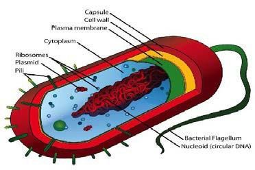
Federal board Fbise Class 9th Notes Biology Chapter 4 Cell and Tissue
                                 Visit Now: perfect24u.com

   Types of Endoplasmic Reticulum:-
   There are two type of endoplasmic reticulum.

   Read more: Federal G9 Biology Notes Chapter 3 (Biodiversity) PDF

   i. Rough endoplasmic reticulum:-
   The endoplasmic reticulum which has small granules called Ribosome present on its surface is
   called rough endoplasmic reticulum. It is attached with nuclear membrane.
   Function:-
          Rough endoplasmic reticulum is involved in protein synthesis.
          Transport of materials from nuclear membrane to cytoplasm.
          They give support to the cell.
   ii. Smooth Endoplasmic Reticulum:-
   The endoplasmic reticulum which has no ribosome’s present on its surface is called
   smooth endoplasmic reticulum. It is attached with cell membrane.
   Function:-
          It plays an important role in the formation of lipids.
          They transfer materials from one part of cytoplasm to another.
          Detoxification of toxic materials.
          They give support to the cell.

   Q14: Describe the structure and function of mitochondria?
   Ans: Mitochondria:-
   Mitochondria are the important organelle of a eukaryotic cell but absent in prokaryotes.
   Shape:-
   Mitochondria are oval, rod shaped or filamentous shape bodies.
   Discovery:-
   They were discovered by Granules in 1850 in muscle cell by electron microscope.
   Structure:-
   Mitochondrion is bounded by double membrane. The outer membrane is smooth and the inner
   membrane is inwardly folded these folds are called cristae. Cristae increase the surface area of
   respiratory process.
   Power House:-
   Mitochondria are energy producing organelles therefore they are called powerhouse of the cell.
   Replication:-
   Mitochondria are the self-replicating organelle.
   Function:-
          Mitochondria play a role in cellular respiration. They produce energy rich
       ATP
           molecules.
7|P age                    Visit Now: perfect24u.com
Federal board Fbise Class 9th Notes Biology Chapter 4 Cell and Tissue
                                 Visit Now: perfect24u.com

          Most of the enzymatic activities of the cell are carried out by mitochondria.

8|P age                     Visit Now: perfect24u.com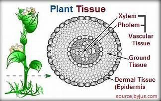
Federal board Fbise Class 9th Notes Biology Chapter 4 Cell and Tissue
                                 Visit Now: perfect24u.com

   Q15: Explain the structure and function of Golgi Bodies?
   Ans: Golgi Bodies:-
   Golgi Bodies were first discovered by an Italian Scientist Camillo Golgi in 1898.
   Shape:-
   Golgi bodies are in the form of granules, rods, threads or canals.
   Structure:-
   Golgi bodies consist of stacks of flattened sacs made of membrane which are arranged parallel to
   each other called cisternae.
   Other Name:-
   They are also known as Golgi apparatus. In plants it is generally known by the name of
   Dictyosomes.
   Function:-
          Golgi bodies store the secretory product.
          Golgi bodies modify and pack the secretory products at their margins into small rounded
          sacs called Golgi vesicles or lysosomes.
          They also synthesize complex carbohydrates from simple sugar.

   Q16: Describe the structure and function of Ribosome and Plastid?
   Ans: i) Ribosomes:-
   Ribosomes are the only organelles found in all prokaryotic and eukaryotic cells.
   Position:-
   They are either freely dispersed in cytoplasm or attached with ER(endoplasmic reticulum).
   Discovery:-
   Ribosomes were discovered by Palade in 1955.
   Structure:-
   Ribosomes have two subunits.
       Large subunit.
       Small subunit.
   Small and large subunit combines to form ribosome.
   Formation:-
   They are produced in nucleolus.
   Composition:
   A ribosomes is made of almost equal amount of protein and ribosomal RNA (rRNA).
   Size:-
   Eukaryotic Ribosomes:
          Ribosome present in eukaryotes is of 80S.
   Prokaryotic Ribosome:

9|P age                    Visit Now: perfect24u.com
Federal board Fbise Class 9th Notes Biology Chapter 4 Cell and Tissue
                                    Visit Now: perfect24u.com

              Ribosome of prokaryotic cell is of 70s,
     Group of Ribosome:
     Group of ribosome is called polysomes.
     Function:
     Ribosomes are the site of protein synthesis.
     ii.Plastid:
     Definition;
     Plastids are also membrane bounded organelles that only occur in plant and photosynthetic
     protest (algae)
     Types of Plastids:-
     There are three types of Plastids.
     i). Chloroplast:-
     It is the most important and abundant type of plastids. It is bounded by double membrane. The outer
     membrane is smooth while inner one give rise to membranous sac called thylakoids. The stack of
     thylakoids is known as granum (pl. grana).
     Position:-
     These are present in green parts of plants particularly in leaves.
     Colour:-
     Chloroplast are green in colour due to green pigment called chlorophyll.

     Read more: Federal G9 Biology Notes Chapter 2 Pdf Download

     Structure of chloroplast:-
     Chloroplast is bounded by double membranes. A smooth outer membrane and an inner folded
     membrane which is modified into stack / pile of coins.
     Granum:-
     Each coin of the granum is called thylakoid. They combine to form granum.
     Intergrana:-
     Granum is attached with each other by intergrana.
     Stroma;-
     The semi fluid and gelatinous matrix present inside the chloroplast is called stroma.
     Function;-
     The process of photosynthesis occurs in chloroplast.
     Photosynthesis complete in two steps, light reaction and dark reaction.
     a. Light Reaction;-
     Light reaction of photosynthesis occurs in grana.
     b. Dark Reaction:-
     Dark Reaction of photosynthesis occurs in stroma.

     ii. Chromoplast:-
     Location:-
     It is present in the petal of flowers and in the skin of ripened fruits.
10 | P a g e                   Visit Now: perfect24u.com
Federal board Fbise Class 9th Notes Biology Chapter 4 Cell and Tissue
Visit Now: perfect24u.com
Colour:-
In plants colours other than green are due to Chromoplasts.
11 | P a g e Visit Now: perfect24u.comFederal board Fbise Class 9th Notes Biology Chapter 4 Cell and Tissue
Visit Now: perfect24u.com
Function:-
Its bright colour attracts insects for pollination.
iii. Leucoplast:
Leucoplast is colorless plastids. They are present in underground parts of the plants particularly in
roots.
Position:-
They are present in underground part of the plant called roots.
Colour: -
Leucoplast is colourless.
Function:-
They store food materials such as starch, protein and lipid.
Q17: Write short note on cytoskeleton?
Ans: Cytoskeleton:
Meaning:
Skeleton of cytoplasm.
Cytoskeleton is an important complex and dynamic cell component. It is invisible under
light microscope.
Structure:
Two important types of filaments make up the cytoskeletons are,
i. Microtubules
ii. Microfilaments
i. Microtubules: -
Microtubules are made up of a protein called actin. These microfilaments are approximately one-
third of the diameter of a microtubule.
Function: -
Microtubules are also the major component of cilia and flagella.
Microfilaments are often used by cells to change their shapes and to hold the structure.
It maintains the cell’s shape, anchors organelles in place and moves parts of the cell in
processes of growth and motility.
Q18: Write short note on centrioles and Vacuole?
i. Centriole: -
These are the cell organelles that are present in animals’ cell and in unicellular organism. It is also
present in young plants and animals. They are two in number and are collectively called
centrosomes.
Location: -
They are small rounded bodies present near the nucleus.
Structure: -
These are hollow cylindrical structures called microtubule. Each centriole contains nine triplets of
microtubules (27) microtubules). Each microtubule is composed of tubulin proteins.
Diameter: -
Each microtubule is 0.2 micrometre in diameter.
Triplet: -
A group of three microtubules is called triplet. There are nine triplets in a centriole. Each
microtubule is composed of tubulin proteins. Tubulin protein has three-dimensional shape.
Function: -
Animal Cell: -
In animal cell they help in the formation of spindle fibers which help in the separation
and movement of chromosomes during cell division.
12 | P a g e Visit Now: perfect24u.comFederal board Fbise Class 9th Notes Biology Chapter 4 Cell and Tissue
Visit Now: perfect24u.com
Unicellular organisms: -
In some unicellular organisms it helps in the formation of cilia and flagella which are the
locomotory organs of some unicellular organisms.
ii. Vacuole: -
It is a single membrane bounded organelle present in the cytoplasm of both plants and animal
cells.
Tonoplast: -
Vacuole is bounded by single membrane called tonoplast. In plant cell there is a single
large vacuole present in the center while in animal cell there are many small vacuoles. In unicellular
organisms there are two vacuoles.
Food vacuole: Digestion of food occurs in food vacuole.
Contractile vacuole: Excretion (removal of metabolic waste and toxic materials).
Function: -
A plant vacuole store important material like water, amino acids, sugar and some
minerals.
Many cells take in materials from outside in the form of food and then digest the
materials with the help of lysosomes.
Some unicellular organisms use contractile vacuole for the elimination of wastes
from their body.
Q19: Describe the formation and function of lysosomes?
Ans: Lysosomes:
In the mid of twenty century, the Belgian scientists Christian Rene de Duve discovered
lysosomes. De Duve in 1947 won the Nobel prize for physiology and medicine.
Structure: -
These are single membrane bound organelles. Lysosomes contain strong digestive enzymes and
work for the breakdown of food and waste materials within the cell.
Function:
A lysosomes fuses with the vacuole that contains the targeted materials and its enzymes break down
the materials. It is also termed as suicide bags of cell because it helps during cell death (apoptosis).
Q20: Explain the structure of nucleus?
Ans: Nucleus: -
It is the most important part of cell. It is also called brain of the cell because it controls all the
cellular activities.
Discovery: -
Shape: -
Nucleus is spherical in shape.
Location; -
In animals cell nucleus is present in the center.
In plant cells, it is pushed to the side due to the presence of large central vacuole.
Structure of Nucleus: -
Following are the main parts of the nucleus.
i. Nuclear Membrane: -
Nucleus is bounded by a double membrane called nuclear envelope. It is present
in eukaryotes but absent in prokaryotes.
Porous: -
Small pores are present on the surface of the nucleus. The pores allow the exchange of materials
between the nucleus and cytoplasm.
13 | P a g e Visit Now: perfect24u.comFederal board Fbise Class 9th Notes Biology Chapter 4 Cell and Tissue
Visit Now: perfect24u.com
ii. Nucleoplasm: -
Inside the nucleus there is a granular matrix called nucleoplasm. It contains chromosomes and
round shape structures called nucleolus. (Plural-nucleoli).
iii. Nucleoli: -
These are one or two rounded structures present in the nucleoplasm.
Function of Nucleoli: -
It is responsible for the formation of ribosomal RNA. Ribosomal RNA plays an important role in
the formation protein synthesis.
iv. Chromosomes: -
Chromosomes are in the form of a network of fine threads present in Nucleoplasm.
Terminology: -
The word chromosomes have derived from two words.
Chroma mean colour.
Soma mean bodies.
Chemical Composition: -
Chromosomes are made of DNA and proteins.
Structure: -
Each chromosome is composed of two main parts.
Two chromatids.
One centromere.
Function of chromosomes; -
It is responsible for the transmission of hereditary characteristics from parents to off springs.
Number of Chromosomes; -
Members of same species having the same numbers of chromosomes. Chromosomes in different
species are given below.
Specie Chromosomes Pairs
Human 46 23
Radish 18 9
Onion 16 8
Q21: Difference between prokaryotic and eukaryotic cell?
S.No Prokaryotic Cell Eukaryotic Cell
1 They have no true nucleus. They have true nucleus.
2 Due to the absence of nuclear Due to the presence of nuclear
membrane chromosomes are membrane chromosomes are present in the
dispersed in cytoplasm nucleus.
3 Ribosome is small in size (70s) Ribosome is large in size (80s)
4 Cells are small in size 0.5nm Cells are large in size from 10nm to 100nm in
in diameter. diameter.
14 | P a g e Visit Now: perfect24u.comFederal board Fbise Class 9th Notes Biology Chapter 4 Cell and Tissue
Visit Now: perfect24u.com
5 The cell wall is made of murein. The cell wall of plants is made of cellulose
while in fungi is made of chitin.
6 Example: Example:
Bacteria and cyanobacteria Plants, Animal and Fungi.
(Blue green algae)
Q 22: Why cells are specific in their function?
Ans: Cell specificity: -
The basic structure and functional unit of all living organisms is called cell. Every living
organism is composed of different types of cell. Each type of the cell performs a specific
function.
Examples: -
Plants: -
Xylem cells are responsible for the transport of water and dissolved minerals from roots to
leaves.
Phloem cells are responsible for the transport of food from leaves to all parts of plant body.
Root hair cells are responsible for the absorption of water and dissolved minerals.
Cells involve in photosynthesis have chloroplast.
Animals: -
In animals nerve cells are responsible for the transmission of impulses.
In muscular cells are responsible for movement.
Red blood cells carry oxygen and white blood cells kill foreign agents.
Relation between cell function and cell structure:
Cell of one type may differ from those of other types in following respects.
Size and Shape • Nerve Cells are long for the transmission of nerve impulse.
• Xylem cells are tube like and have thick walls for conduction
of water and support.
• Red blood cells are round to accommodate globular
haemoglobin.
Surface area to •Root hair cells have large surface area for the maximum
volume ration absorption of water and salts.
Presence or absence of • Cell involved in making secretion have more complex ER
organelles and Golgi apparatus.
• Cells involved in photosynthesis have chloroplasts.
15 | P a g e Visit Now: perfect24u.comFederal board Fbise Class 9th Notes Biology Chapter 4 Cell and Tissue
Visit Now: perfect24u.com
Q 23: Cell as an open system? Justify the statement.
16 | P a g e Visit Now: perfect24u.comFederal board Fbise Class 9th Notes Biology Chapter 4 Cell and Tissue
Visit Now: perfect24u.com
Ans: A cell works as an open system. That is
It takes in substances needed for its metabolic activities through its cell membrane.
Then it performs the metabolic processes assigned to it.
Products and by products are formed in metabolism.
Cell either utilizes the products or transports them to other cells.
The by products are either stored or are excreted out of the cell.
Q24: Explain how surface area to volume ratio limits cell size.
Ans: Cell size and surface are to volume ratio:
Cells are varying greatly in size. The smallest cells are bacteria, with diameter between 0.1 m to
1.0 m. The bulkiest cells are bird eggs, and the longest cells are muscle and nerve cells. Most cells
are small in size.
Large cells have less surface area in relation to their volume while small cells of the
same shape have more surface area.
In the figure below show 1 large cell and 27 small cells. In both cases the total volume is the same:
Volume: 30μm x 30μm x 30μm = 27,000μm3
In contrast to total volume, the total surface areas are very different. Because
the cubical shape has 6 side, its surface area is 6 times the area of 1 side.
The total surface areas of the cubes are as follows.
Surface area of 1 large cube = 6 x (30μm x 30μm) = 5400μm3
Surface area of 1 small cube = 6 x (10μm x 10μm) = 600μm3
Surface area of 27 small cubes = 27 x 600μm2 = 16,200μm2
This relationship between cell size and surface area to volume ratio works to limits cell size. As the
size of a cell increases cell volume increases more rapidly than its surface area.
The need of nutrients and rate of waste production are directly proportional to cell volume. The cell
takes up nutrients and excretes wastes through its surface cells membrane. So, a large
volume demands large surface area.
Hence it concluded that the membranes of small cells can serve their small volumes more easily
than the membrane of large cell.
Q25: Explain the phenomenon involved in the passage of materials across the cell
membrane?
Ans: Cell membrane is called differentially or semi permeable membrane as it controls the
inflow and outflow of materials to cells. Cell membrane maintains equilibrium inside as well as
outside the cell. The control of the passage of molecules into and out of cells is made possible
through following phenomena.
Types: -
There are two types of movement.
Active Transport: -
Definition: -
The movement of molecules across cell membrane from lower concentration region to higher
concentration with the expenditure of metabolic energy is called active transport.
For the active transport of substances carrier proteins present in cell membrane use energy and
move them against concentration gradients. Na+ /K+ are actively transported across the
requirement of the cell/body.
Example: -
Nerve impulse is carried when Na+ is actively transported across the membrane from nerve cell to
outside.
Passive Transport: -
Definition: -
17 | P a g e Visit Now: perfect24u.comFederal board Fbise Class 9th Notes Biology Chapter 4 Cell and Tissue
Visit Now: perfect24u.com
The movement of molecules from higher concentration region to lower concentration
region without the expenditure of energy is called passive transport.
Example: -
i. Diffusion
ii. Facilitate diffusion
iii. Osmosis.
1. Diffusion: -
Definition: -
The movement of molecules from higher concentration region to lower concentration
region without the expenditure of energy is called diffusion.
It is a type of passive transport.
Explanation: -
Only small molecules can diffuse through cell membrane e.g. water, carbon dioxide, oxygen and
some other simple molecules. Diffusion is slow process yet it is efficient and rapid enough to
fulfil the requirement of the cells.
Important: -
• Substances such as glucose, O2 and CO2 can easily diffuse through the membrane.
Glucose is present in higher concentration after the food is digested in small intestine.
Therefore, glucose is transported to villi from the inner space of small intestine through
diffusion to be stored in the form of glycogen.
• Carbon dioxide and oxygen are among the few simple molecules that can cross the cell
membrane by diffusion. Gases exchange in gills and lungs operates by this process.
2. Facilitated Diffusion: -
Definition:
The movement of molecules from high to low concentration with the help of transport proteins
present in cell membrane.
Many molecules do not diffuse freely across cell membrane because of their size or charge. Such
molecules are taken into or out of the cells with facilitated diffusion is higher than simple
diffusion.
Facilitated diffusion is also a type of passive transport because there is no expenditure of energy
in this process.
18 | P a g e Visit Now: perfect24u.comFederal board Fbise Class 9th Notes Biology Chapter 4 Cell and Tissue
Visit Now: perfect24u.com
3. Osmosis: -
Definition: -
The movement of solvent molecules from higher concentration to lower concentration
region through semi permeable membrane is called osmosis.
Example of Osmosis: -
Plant cell absorb water and store it in vacuole by osmosis.
Q26: Define turgidity and plasmolysis?
Ans: 1. Turgidity:
Definition:
When a cell absorbs water and become swell is called turgid cell and this phenomenon is called
turgidity.
Mechanism:
When we place the plant cell in pure water or in dilute solution. The plant absorbs water
by osmosis and stores it in vacuole. Due to this storing of water the plant cell will swell.
Turgor Pressure: -
The internal pressure exerted on the cell wall is called turgor pressure.
Importance: -
i. It keeps the herbaceous plants erect.
i.e. helps in opening and closing of stomata.
iii. Some flower open during day time and close at night time this is due to change in turgor.
iv. It gives proper shape to the cell.
v. The upward movement of water and dissolved minerals is due to turgor.
2. Plasmolysis: -
Definition: -
The condition in which plant cell loose water and become shrink is called plasmolysis. And the
cell is also called plasmolysis cell.
Mechanism: -
When a plant cell is placed in a solution having lower water potential than the cell contents. The
water leaves the cell by osmosis. In this way the cell become shrink. This phenomenon is called
plasmolysis and the cell is also called plasmolysis cell.
19 | P a g e Visit Now: perfect24u.comFederal board Fbise Class 9th Notes Biology Chapter 4 Cell and Tissue
Visit Now: perfect24u.com
Q27: Discuss endocytosis and exocytosis?
Ans: 1. Endocytosis: -
Definition: -
The movement of materials from outside environment to inside the cell is called endocytosis.
Endocytosis occurs in following steps.
i. A portion of cell membrane invaginates (depressed inward)
ii. The material from outside is taken inside the invagination.
iii. The open end of the invagination seal and form a small vesicle.
iv. The vesicle detaches from the cell membrane and moves into the cytoplasm.
Forms: -
There are two forms of endocytosis.
Phagocytosis: -
The endocytosis of solid objects is called phagocytosis. It is also called eating of cell.
Pinocytosis: -
The endocytosis of liquid substances is called Pinocytosis. It is called dinking of a cell.
2. Exocytosis: -
The movement of waste materials from inside of the cell to the outside environment is
called
Exocytosis. Exocytosis occurs in following steps.
i. The bulky materials are packed inside a membrane and a vesicle is formed.
ii. The vesicle moves to the cell membrane.
iv. The vesicle fuses with the membrane and releases its contents into the extracellular
environment.
This process adds new membrane which replaces the part of cell membrane lost during
endocytosis.
Q28: What is Filtration? Justify the statement.
Ans: Filtration: -
Filtration is a process by which small molecules are forced to move across semi-
permeable membrane with the aid of hydrostatic (water) pressure of blood pressure.
In filtration the pressure cannot force large molecules, such as proteins to pass through
membranes pores.
Example: -
In our body filtration occurs in the kidney and helps us filter out harmful substances.
20 | P a g e Visit Now: perfect24u.comFederal board Fbise Class 9th Notes Biology Chapter 4 Cell and Tissue
Visit Now: perfect24u.com
Q29: Define tissue? Discuss various types of plant tissues?
Ans: Tissue: -
A group of cells which perform a particular function is called tissue.
Types of Plant Tissues: -
There are two types of plant tissue.
i. Simple Tissue.
ii. Compound Tissue.
i. Simple Tissues: -
Definition: -
The tissue which is made from single type of cells called simple tissue.
Types of simple tissue: -
There are two types of simple tissue.
A. Meristematic tissues.
B. Permanent tissues.
A. Meristematic tissue (Embryonic tissues): -
Definition: -
These tissues are made up of cells which have the ability to divide.
Location: -
They are present on the apical point and lateral side of roots, stem and branches of a plant.
Properties: -
i. The cells of meristematic tissue are closely packed i.e. they have no intercellular spaces.
ii. They have no small vacuoles.
iii. Nucleus is present in the center of the cell.
iv. They have a delicate and thin cell wall.
v. They have power of cell division.
There are two main types of meristematic tissues recognized in plants.
1. Apical meristem:
These are located at the apices or tips of roots and shoots. When they divide, they cause increase
in the length of plant. Such a grown is called primary growth.
2. lateral meristems: -
These are located on the lateral sides of roots and shoot. By divide they result in the increase of
thickness of root and shoot such a growth is called secondary growth.
Lateral meristems are further of two types.
a. Vascular cambium:
Vascular cambium is present between the xylem and phloem tissues. Its cells divide and form
new xylem tissues toward the center and new phloem tissue towards the outside.
b. Cork cambium:
Cork cambium is present in the outer lateral sides and its cells are responsible for making the
characteristic corky layer.
B. Permanent tissue: -
21 | P a g e Visit Now: perfect24u.comFederal board Fbise Class 9th Notes Biology Chapter 4 Cell and Tissue
Visit Now: perfect24u.com
Permanent tissue originates from the meristematic tissue. These tissues are composed of
cells which do not have the ability to divide.
Types of permanent tissue: -
There are three types of permanent tissue.
1. Epidermal tissue.
2. Ground tissue.
3. Supporting tissue.
1. Epidermal Tissue: -
Terminology: -
The word epidermal is derived from two Greek words.
• Epi means above
• Dermis means skin.
Location: -
These tissues are present as outermost protective layer of roots, stems, branches and leaves.
Characteristics: -
i. Cell of the epidermal tissue are flattened and irregular in shape.
ii. They are thick walled and closely packed with no intercellular spaces.
iii. In stem, the walls of these are covered with waxy materials which prevent loss of water.
iv. In leaves, the epidermal tissue has small ground opening called stomata for gaseous exchange.
Function: -
Leaves: -
In leaves the epidermal tissues have small pores called stomata for gaseous exchange and
for transpiration.
Stem: -
In stem the epidermal tissues have root hairs which are responsible for absorption of water.
Root: -
In roots the epidermal tissues have root hairs which are responsible for absorption of water.
2. Ground tissues: -
Location: -
They are present in all parts of plant except epidermal and vascular tissues.
Composition: -
Ground tissues are composed of thin walled cells called parenchyma cells.
Properties of Parenchyma cell: -
i. These are thin walled living cells.
ii. They are oval or polygonal in shape.
iii. They have a large vacuole. Nucleus is present in peripheral position due presence of
large vacuole.
v.In leaves the ground tissue contains chlorophyll and is called mesophyll tissues. It prepares
food.
Function: -
Parenchyma cells store food.
3. Supporting tissues: -
They give support and flexibility to the plant body. They are of two types.
a. Collenchyma tissues
b. Sclerenchyma tissues
a. Collenchyma tissues (Kolla-glue)
They are flexible, living cells, elongated and polygonal with tapering end.
Location: -
They are found in young stem, midrib of leaves, petals of flower and in herbaceous plants stem.
22 | P a g e Visit Now: perfect24u.comFederal board Fbise Class 9th Notes Biology Chapter 4 Cell and Tissue
Visit Now: perfect24u.com
Function:
They provide strength to different parts of the plant.
b. Sclerenchyma tissues (Greek, Scleros –hard): -
They are thick walled dead cells. Their cell wall has lignin which is the main chemical
component of wood.
Function: -
They give support to woody plants.
2. Compound Tissue: -
The tissue made from different type of cells performing a common function called compound
tissues.
Compound tissues in plants are xylem and phloem.
i. Xylem tissue: -
Xylem tissue is composed of:
Vessels.
Tracheid.
a.Vessels:-
i. Vessel cells are short, wide and have thick secondary walls.
ii. These cells are dead, hollow and joins together to form long tubes.
b. Tracheid: -
They are spindle shape and closed at both ends. They overlap with each other with pair of pits
present. The pits allow water to pass from cell to cell.
Function of Xylem: -
Xylem transport water and dissolved mineral salts from roots to upper parts of plants.
These tissues also give support to plant body.
Phloem: -
Phloem is consisting of:
a. Sieve tube cells
b. Companion cells
a. Sieve tube cells: -
They are tube like structures opened at both ends. They have small pores at each end called sieve
plates which join one sieve tube cell with another.
Function: -
They transport prepared food.
b.Companion cells:-
The sieve tube cells are accompanied by nucleated cells called companion cells.
Function:-
They control the movement of food materials in sieve tube cells.
Function of Phloem:-
Phloem transport organic food from leaves to all other parts of the plant body.
23 | P a g e Visit Now: perfect24u.comFederal board Fbise Class 9th Notes Biology Chapter 4 Cell and Tissue
Visit Now: perfect24u.com
Q30: Discuss different types of animal tissues?
Ans: Animal Tissue:-
Animals tissues are classified according to their structure and function into major four types.
1.Epithelial tissue:-
Epithelial tissue are made from epithelium cells they form the outermost layer of skin.
Characteristics:
The cells in this type of tissue are very closely packed together and joined with little space with
them.
Function:-
They help to protect organisms from microorganism, injury and fluid loss.
These tissues are commonly classified on the basis of the shape of the cells as well as the number
of cell layers. Some types include.
i. Simple squamous epithelium:-
A single layer of tightly packed, flattened cells. e.g. in lining of air sacs of lungs, heart and blood
vessels etc.
ii. Simple cuboidal epithelium:-
It consists of single layer of tightly packed, cube-shaped cells. e.g., found in kidney tubules and
small glands.
iii. Simple columnar epithelium
Consist of single layer of elongated cells. e.g. in lining of digestive tract and gallbladder etc.
iv Ciliated columnar epithelium
A tuft of cilia is present at the top of each columnar cell. e.g. in lining of trachea and bronchi.
v. Stratified squamous epithelium
Consist of many layers of flattened cells. e.g. inner lining of oesophagus and at the surface of
skin.
2. Connective tissue:
As the name implies, connective tissue serves “connecting” function. It supports and binds other
tissue. Its cells are scattered throughout an extracellular matrix.
Types of connective tissues:
i. Loose connective tissue:
Most common type, matrix contains loosely arranged collagen
(a protein) fiber.
Widely distributed under epithelial tissues
It holds organs at their specific place.
ii. Fibrous connective tissue:
Matrix contains tightly packed collagen fibers.
Found in tendons, which attach muscles and bones.
In ligaments, which join two bones
24 | P a g e Visit Now: perfect24u.comFederal board Fbise Class 9th Notes Biology Chapter 4 Cell and Tissue
Visit Now: perfect24u.com
iii. Adipose tissue:
Swollen cells due to the presence of larger number of fat droplets.
Found around kidneys, under skin, in abdomen etc.
Provides energy when fat is oxidized insulator against heat loss protects and supports organ.
iv. Cartilage:
Matrix contains bundles of collagen fibers embedded in a rubbery substance.
Provide support while allowing flexibility.
Found around the ends of bones in external ear, in nose, trachea in discs between vertebras, as
skeleton in many fishes.
v. Bone:
Matrix contains collagen fibers embedded in calcium salts.
Supports, protect, provide lever system for movement for movement, store calcium and forms
blood cells.
Found in skeleton.
vi. Blood:
Matrix is not solid but in the form of fluid, red and white blood cells are suspended in plasma.
Transport substance from one part of the body to the other and responsible for immunity.
Found in blood vessel.
3. Muscular tissue: -
Muscle tissue consists of bundles of long cells called muscle fibers. It is the most abundant tissue
in a typical animal. The cells of this tissue have ability to contract and relax.
Types:
There are three types of muscle in vertebrates.
i. Skeleton muscle.
ii. Smooth muscle.
iii. Cardiac muscle.
i. Skeleton muscle:
The characteristics of skeletal muscles are
Skeletal muscles are attached to our bones and cause movement in them. i.e. movement in
them i.e. Movement of arms and legs etc.
These are striated or stripped muscles because they have alternate light and dark bands.
They are also called voluntary muscle because their movement is under our control.
Their working is fast but they fatigue easily.
The cells of skeletal muscles contain many nuclei.
Example, muscle of arms and legs.
ii. Smooth Muscle:
Smooth muscles are working smoothly and slowly.
They are involuntary muscle mean that they do not work under our will.
They cannot fatigue easily.
These are non-striated or strapped muscle because they have no alternate light and
dark bands.
Fibers of smooth muscles are multinucleated.
Examples are muscles of digestive, respiratory, circulatory and urinary tract etc.
iii. Cardiac muscle:
Cardiac muscles make our heart.
Composed of striated cells that are branched and each contain a single nucleus.
They are also involuntary in action.
They contract and relax rhythmically without getting any fatigue.
Examples are the muscle of our heart.
25 | P a g e Visit Now: perfect24u.comFederal board Fbise Class 9th Notes Biology Chapter 4 Cell and Tissue
Visit Now: perfect24u.com
4. Nerve tissues: -
Nerve tissue is composed of nerve cells called neuron. So, neuron is the basic structural
and functional unit of nervous system. They transmit impulses to the brain. These are two types of
nervous system.
Central nervous system
Peripheral nervous system
Central nervous system: -
Central nervous system is composed of brain and spinal card.
Peripheral nervous system: -
Peripheral nervous system is composed of the nervous that arise from brain and spinal card.
Q31: Difference between plant cell and animal cell?
S.No Plant Cell Animal Cell
1 Cell wall is present in plant cell. Cell wall is absent in animal cell.
2 Plastids are present in plant cell. Plastid are absent in animal cell.
3 Plant cell have one large vacuole. Animal cell have many small vacuoles.
4 Centrosomes are absent in plant cell. Centrosomes are present in animal cell.
5 Plant nucleus is not present in center Animal nucleus is present in the center
of a cell. of a cell.
26 | P a g e Visit Now: perfect24u.comFederal board Fbise Class 9th Notes Biology Chapter 4 Cell and Tissue
Visit Now: perfect24u.com
SHORT QUESTIONS
B. Write short Answer of the following Questions.
Q1: Who proposed the cell theory and what are the main points of the cell theory?
Ans: See Q.No. 8
Q2: Differentiate between simple and compound tissue?
Ans: Simple and compound tissues;
S.No Simple tissues` Compound tissues
1 Simple tissues are made of one Compound tissues are composed of
type of cells. more than one type of cells.
2 Meristematic tissue is responsible Xylem tissue transport water
for primary growth of root and and dissolved salt from roots to
stem. all part
of the plants.
3 Epidermal tissue forms a single Epithelial tissues form the outside
outer layer of root, stem and cover or the organs.
leaves.
4 Ground tissue store food. Muscle tissue produce movement.
5 Collenchyma tissue gives Nerve tissue transmit nerve impulse
flexibility.
Q3: How cell membrane helps in maintaining equilibrium while exchange materials with
environment?
Ans: Each cell is surrounded by cell membrane which is selectively permeable membrane.
It helps in maintaining equilibrium by controlling outflow and inflow of materials. Selective
molecules can move from the cell to outside or from outside to the cell. It freely allows
the passage of water and other small molecules such as gases. Other substances like glucose, fatty
acid and ions etc can slowly diffuse through it.
Q4: Differentiate between endocytosis and exocytosis?
Ans: See Q No 27
Q5: How does turgor pressure develops in a plant cell?
Ans: Turgor pressure:
In the force within the cell that pushes the plasma membrane against the cell wall.
Most plant cell live in hypotonic environment because there is low concentration of solute
in extracellular fluids than in their cells. As a result, the water moves into cytoplasm, then
to vacuole by osmosis. As the water enters the vacuole, it increases in size and push the
cell contents against cell wall so turgor pressure is developed.
Turgor pressure of the cells is responsible for,
i. Keeping herbaceous plants erect.
ii. It helps in opening and closing of stomata.
iii. Some flower open during day time and close at night time, this is due change in turgor
pressure.
27 | P a g e Visit Now: perfect24u.comFederal board Fbise Class 9th Notes Biology Chapter 4 Cell and Tissue
Visit Now: perfect24u.com
iv. it gives proper shape to the cell.
28 | P a g e Visit Now: perfect24u.comFederal board Fbise Class 9th Notes Biology Chapter 4 Cell and Tissue
Visit Now: perfect24u.com
Long Question
C. Give detailed Answers to the following Question.
Q.1: Root hairs are adapted to absorption and xylem to support. Relate their functions to
their structure.
Ans: Absorption of Root:
Root hairs are adopted for absorption because it provides large surface area for absorption. They
grow out into the space between the soil particles where they are in direct contact with water. The
cytoplasm of the root hairs has higher concentration of salts than the soil water, so water move by
osmosis into the root hairs. After their entry into the root hairs. Water and salt must move
through the epidermis and cortex of the root, and then into the xylem tissue in the center of the root.
Xylem role in support:
Xylem also helps in the support of plant body because xylem contains fibers. Fibers are
elongated cells which give support to the plant. Due to the presence of lignin, the secondary walls
of its cells are thick and rigid that is why xylem tissue provides support to the plant body.
Q2: Discuss different types of tissue found in plants. Elaborate your answer with relevant
diagrams?
Ans: See Q No. 29
Q3: Describe the nervous, muscular and epithelial tissues?
Ans: See Q No. 30
Q4: Write a note on the structure of cell wall, cell membrane, mitochondria and chloroplast
of a plant cell?
Ans: See Q.No. 10 (Cell Wall).
Q.No. 11 (Cell membrane).
Q.No 14 (Mitochondria).
Q.No 16 (Chloroplast)
Sindh textbook board Jamshoro books pdf download 2021
KPK Text Book Board New 2021 PDF Download
Top 9th Class Math Notes PDF Download Cha 1 to 9
Unit 1 Simplicity and Humility of Hazrat Muhammad (PBUH) Calss 10 Notes
Adamjee Mathematics 9th Model Papers 2021
English notes for class 11 kpk board Chapter 1 Responsibilities of the Youth
Unit 02 The Champions Class 10 English Notes
Class 12 English notes Lesson 1 to 20 for KPK
Class 10th English book Notes For KPK Board 2021-22 Pdf Download
29 | P a g e Visit Now: perfect24u.comYou can also read