CELL DEATH AND THE P53 ENIGMA DURING MAMMALIAN EMBRYONIC DEVELOPMENT - SONAM RAJ, SUSHIL K JAISWAL, MELVIN L DEPAMPHILIS
←
→
Page content transcription
If your browser does not render page correctly, please read the page content below
Downloaded from https://academic.oup.com/stmcls/article/40/3/227/6516995 by guest on 12 August 2022
Cell Death and the p53 Enigma During Mammalian
Embryonic Development
Sonam Raj, Sushil K Jaiswal, Melvin L DePamphilis
Stem Cells, 2022, 40, 227–238
https://doi.org/10.1093/stmcls/sxac003
Advance access publication 28 January 2022
Concise Review

Cell Death and the p53 Enigma During Mammalian
Embryonic Development
Sonam Raj1,‡, Sushil K. Jaiswal2,3,‡, Melvin L. DePamphilis2,*,
1
 National Cancer Institute, Bethesda, MD 20892, USA
2
 National Institute of Child Health and Human Development, Bethesda, MD 20892, USA
3
 Present address: National Human Genome Research Institute, Bethesda, MD 20892, USA

                                                                                                                                                       Downloaded from https://academic.oup.com/stmcls/article/40/3/227/6516995 by guest on 12 August 2022
*Corresponding author: Melvin L. DePamphilis, National Institute of Child Health and Human Development, Bldg. 6A, Rm 3A15, 6 Center Dr, Bethesda, MD
20892, USA. Email: depamphm@nih.gov
‡
 S.R. and S.K.J. contributed equally to this review.

Abstract
Twelve forms of programmed cell death (PCD) have been described in mammalian cells, but which of them occurs during embryonic devel-
opment and the role played by the p53 transcription factor and tumor suppressor remains enigmatic. Although p53 is not required for mouse
embryonic development, some studies conclude that PCD in pluripotent embryonic stem cells from mice (mESCs) or humans (hESCs) is
p53-dependent whereas others conclude that it is not. Given the importance of pluripotent stem cells as models of embryonic development
and their applications in regenerative medicine, resolving this enigma is essential. This review reconciles contradictory results based on the
facts that p53 cannot induce lethality in mice until gastrulation and that experimental conditions could account for differences in results with
ESCs. Consequently, activation of the G2-checkpoint in mouse ESCs is p53-independent and generally, if not always, results in noncanonical
apoptosis. Once initiated, PCD occurs at equivalent rates and to equivalent extents regardless of the presence or absence of p53. However,
depending on experimental conditions, p53 can accelerate initiation of PCD in ESCs and late-stage blastocysts. In contrast, DNA damage fol-
lowing differentiation of ESCs in vitro or formation of embryonic fibroblasts in vivo induces p53-dependent cell cycle arrest and senescence.
Key words: apoptosis; cell cycle; differentiation; embryo; p53; pluripotent; programmed cell death; stem cells.

Graphical Abstract

Neither cell cycle arrest nor programmed cell death requires p53 prior to gastrulation, at which stage DNA damage induces p53-dependent cell
cycle arrest and senescence.

    Significance Statement
    Programmed cell death (PCD) and survival are inherent components of mammalian development. In addition to its role as “guardian of the
    genome”, the p53 transcription factor and tumor suppressor has been reported to regulate at least six different forms of PCD. Given the
    importance of pluripotent stem cells as models of embryonic development and their applications in regenerative medicine, identifying which
    of the 12 forms of PCD respond to stress imposed at the beginning of embryogenesis and the role of p53 in regulating them is essential.

Received: 31 October 2021; Accepted: 20 December 2021.
Published by Oxford University Press 2022. This work is written by (a) US Government employee(s) and is in the public domain in the US.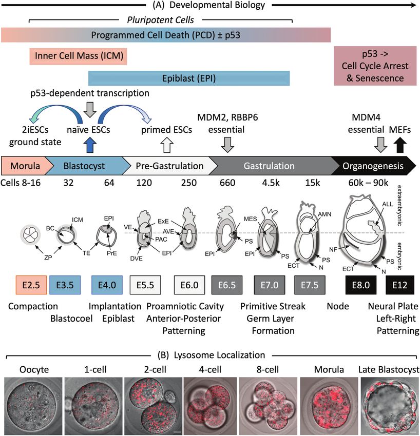
228                                                                                                Stem Cells, 2022, Vol. 40, No. 3

Introduction                                                      can be induced by DNA damage, unfolded proteins, reactive
                                                                  oxygen species (ROS), or disruption of cell division using
Cell death is a normal event in mammalian development,
                                                                  either an intrinsic or an extrinsic pathway. The intrinsic
as well as a cellular response to stressful conditions. The
                                                                  pathway regulates mitochondrial permeability via the Bcl-2
mechanisms that cause cell death are categorized as either
                                                                  family of cytokines. The extrinsic pathway is triggered by
necrosis or programmed cell death (PCD). Necrosis results
                                                                  ligand binding to tumor necrosis factor-family receptors in
from the progressive degradative action of enzymes and is
                                                                  the plasma membrane that activate caspases. Both pathways
typically followed by inflammation. Necrosis requires nei-
                                                                  activate initiator caspase (CASP) 2, 8, 9, or 10, which then
ther energy nor effector proteases1; it is simply a response
                                                                  activate effector CASP3, 6, and 7, which then degrade cel-
to physical damage or pathology that does not occur during
                                                                  lular proteins indiscriminately. Expression of proapoptotic
normal animal development.2 In contrast, PCD is a se-
                                                                  genes Bax, Puma, Bid, and Bcl-2, as well as Casp6 and
quence of genetically programmed events by which a cell
                                                                  Apaf1, a coactivator of Casp9, are upregulated by the p53
provokes its own demise in response to a stimulus. PCD
                                                                  transcription factor.
occurs in mammals as early as the blastocyst stage during
                                                                     Apoptosis is recognized by DNA fragmentation, accumula-
preimplantation development and as late as tissue homeo-

                                                                                                                                      Downloaded from https://academic.oup.com/stmcls/article/40/3/227/6516995 by guest on 12 August 2022
                                                                  tion of cells containing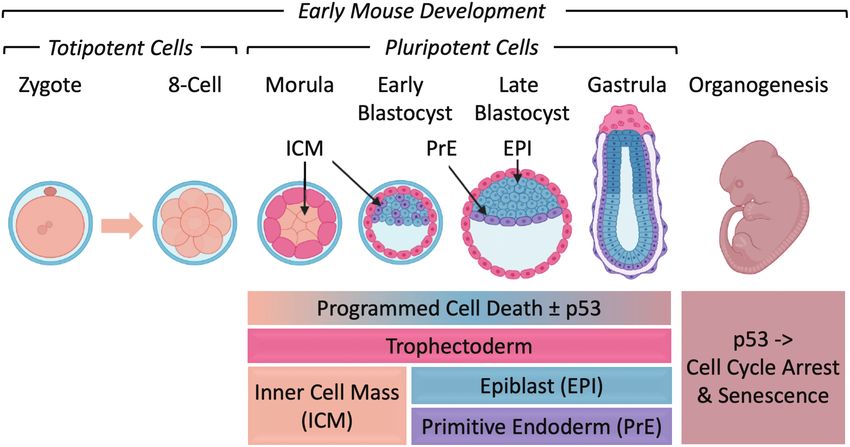
Stem Cells, 2022, Vol. 40, No. 3                                                                                                                      229

                                                                                                                                                             Downloaded from https://academic.oup.com/stmcls/article/40/3/227/6516995 by guest on 12 August 2022
Figure 1. Early mouse embryonic development. (A) The number of cells, days post-coitum (E2.5-E12), and morphogenetic events are indicated.
ALL, allantois; AMN, amnion; AVE, anterior visceral endoderm; BC, blastocyst cavity; DVE, distal visceral endoderm; ECT, ectoderm; EPI, epiblast;
ExE, extraembryonic ectoderm; ICM, inner cell mass; MES, mesoderm; N, node; NF, neural fold; PAC, proamniotic cavity; PrE, primitive endoderm;
PS, primitive streak; TE, trophectoderm; VE, visceral endoderm; ZP, zona pellucida. Adapted from Ref. 116. Preimplantation development begins with
totipotent blastomeres (1-8 cell stage) encapsulated by the zona pellucida. Totipotent cells can give rise to both placental and embryonic cells. When the
blastomeres develop cell-to-cell adhesion (compaction), the outer blastomeres differentiate into the trophectoderm while the remaining blastomeres
form the inner cell mass. The epithelial trophoblast cells (trophectoderm) are multipotent; they differentiate only into cells required for implantation
and placentation. The inner cell mass (recognized upon formation of a blastocoel cavity) differentiates into the epiblast and the primitive endoderm.
Postimplantation development begins when the primitive endoderm differentiates into multipotent visceral and parietal endoderm. Mesoderm
and ectoderm are derived from the epiblast during gastrulation. Gastrulation begins at the primitive streak, from which mesoderm and endoderm
progenitor’s ingress and begin to differentiate.117 Mouse embryonic fibroblasts (MEFs) are derived from E12-E14 embryos. Ablation of the Mdm2,
Rbbp6, or Mdm4 gene is lethal in embryos at the indicated times. Mouse embryonic stem cells (mESCs) are derived from the epiblast in blastocysts.49
mESCs cultured in the presence of serum and LIF interleukin-6 are considered “naïve” pluripotent cells, because they can give rise to all the cells of the
embryo, but not to the trophectoderm. Naïve mESCs cultured in defined medium (no serum) containing 2 metabolic inhibitors are considered totipotent
“ground-state” ESCs (2iESCs), because they give rise to both extraembryonic and embryonic cells. Naïve mESCs cultured in the presence of activin
and fibroblast growth factor generate pluripotent “primed” ESCs, because they give rise to the same cells as “naïve mESCs,” but they cannot generate
chimeric animals.118 Human embryonic stem cells (hESCs) and mouse epiblast stem cells (EpiSCs) are derived from the epiblast of post-implantation
blastocysts.48 (B) Images of LysoTracker Red stained oocytes and preimplantation embryos revealed that the number of lysosomes increased after
fertilization.36 Scale bar is 10 µm.

  γH2AX is easily detected throughout preimplantation de-                      is recruited to sites of double-strand DNA breaks43 was not
velopment, thereby revealing the presence of double-strand                     detected. Thus, p53-dependent PCD does not appear to be
DNA breaks even in the absence of any induced DNA                              induced by low levels of double-strand DNA breaks. In fact,
damage.42 In contrast, the p53-binding protein “53BP1” that                    regardless of the presence or absence of p53, induction of
230 Stem Cells, 2022, Vol. 40, No. 3
Table 1. Hallmarks of programmed cell death in mammals.
Form p53 Morphology Biochemistry
Associated with development
Apoptosis18,19 p53 Cell rounding, nuclear condensation, mem- Activates CASP3 and PARP1, DNA fragmentation and
brane blebbing, apoptotic bodies loss, ΔΨm dissipation, phosphatidylserine exposure
Anoikis20 p53 Anchorage-dependent cells detach from the Cleaved EMC proteins (laminin, fibronectin,
extracellular matrix vitronectin) → apoptosis
Lysosome Plasma membrane repair, lysosome mem- Release of lysosomal hydrolytic enzymes (cathepsins),
dependent21 brane permeabilization lysosomal iron-induced oxidative injury
Autophagy p53 Autophagic vacuolization LC3-I to LC3-II conversion, increases autophagic flux
dependent22,23 and lysosomal activity
Associated with disease
Necroptosis24 p53 Cell swells, PMR, moderate chromatin con- Activates RIPK1, RIPK3, and MLKL, cytosolic
Downloaded from https://academic.oup.com/stmcls/article/40/3/227/6516995 by guest on 12 August 2022
densation necrosome formation
Oxeiptosis25 Apoptosis-like ROS-dependent, activates KEAP1 and NFE2L2.
caspase-independent, no AIFM1 translocation
Ferroptosis26 p53 Small mitochondria (mt), reduced mt-crista, Iron accumulates, lipid peroxidation, ΔΨm dissipation,
elevated mt-membrane densities, mt- LC3-I to LC3-II conversion, glutaminolysis, caspase-
membrane rupture independent
Parthanatos27 p53 Chromatin condensation, large DNA frag- Oxidative stress (ROS)-induced, PARP1 activation,
ments, no cell swelling, apoptotic bodies or ΔΨm dissipation, caspase-independent, NAD+ and ATP
small DNA fragments, PMR depletion, accumulates PARP polymers, AIFM1 trans-
location
Alkaliptosis28 Necrosis-like Intracellular alkalinization, activates NF-κB, caspase-
independent
Pyroptosis29 No cell swelling, PMR, bubbling, moderate Activates CASP1, CASP3, and GSDMD, GSDMD-N-
chromatin condensation induced pore formation, IL1B released
Entotic30 One cell invades another Activates adhesion proteins and actomyosin, LC3-
associated phagocytosis
Netotic31 PMR, nuclear membrane collapse, chroma- Forms NETs, release and translocation of granular en-
tin fiber release zymes, histone citrullination
Note: ΔΨm is mitochondrial membrane potential.37 Reactive oxygen species is reactive oxygen species. Neutrophil Extracellular Traps (NETs) are
neutrophil extracellular traps. LC3 is MAP1LC3B. Dying cells release small vesicular apoptotic bodies.38 Plasma membrane rupture releases intracellular
molecules that propagate inflammatory response.39 Apoptosis-inducing factor 1 (AIFM1) translocates from mitochondria to nucleus.40 EMC is extracellular
matrix. Adapted from Ref. 17.
NMRs, neutrophil extracellular traps; PARP, poly(ADP-ribose) polymerase; PMR, plasma membrane rupture; ROS, reactive oxygen species.
double-strand DNA breaks by X-irradiation of two-cell em- populations in pre- or peri-implantation embryonic epiblast
bryos retarded their development to the late blastocyst stage whereas primed mESCs (termed EpiSCs) isolated from post-
but did not prevent it.44 Wild-type blastocysts exhibited 2 implantation blastocysts model the postimplantation epi-
to 3-times more cells with DNA damage than p53−/− blasto- blast.47 hESCs are similar to primed mESCs.48 Ground state
cysts, thereby revealing that p53 accelerates PCD, but is not mESCs are produced by culturing naïve mESCs with inhibi-
required for PCD. tors of MAP2K1/MEK1 and FRAT2/GSK-3, and therefore
Autophagy-dependent PCD is unlikely because termed 2iESCs.49 An intermediate state between naïve and
autophagy is essential for preimplantation development.45 primed ESCs has recently been described.50
Autophagy-defective oocytes derived from oocyte-specific Doxorubicin/Adriamycin is an anticancer drug commonly
Atg5 (autophagy-related 5) knockout mice failed to de- used to induce PCD in mammalian cells by causing double-
velop beyond the 4- to 8-cell stages if fertilized by Atg5−/− strand DNA breaks. Doxorubicin induces PCD equally well
sperm, but they did develop if fertilized by wild-type sperm. in either p53+/+ or p53−/− naïve ESCs, as evident from visual
However, lysosomes rapidly accumulate after fertilization inspection of cultured cells, DNA loss, annexin-V binding,
(Fig. 1B) and lysosome accumulation is required for de- propidium iodide, and trypan blue staining, cleaved PARP,
velopment,36 suggesting that induced stress might activate and CASP3.51 Robust PCD did not require p53 or its primary
lysosome-dependent PCD. In fact, PIKfyve, a phospho- targets, the CDK2 inhibitor p21 and pro-apoptotic protein
inositide kinase essential for maintaining lysosome homeo- PUMA, to cleave PARP and CASP3 (Fig. 2A), arrest cell pro-
stasis and autophagic flux,35 is essential for preimplantation liferation (cells accumulate with 4N DNA content, Fig. 2C)
mouse development.46 and complete apoptosis (cells accumulate withStem Cells, 2022, Vol. 40, No. 3 231 Figure 2. Cell cycle arrest and apoptosis in naïve ESCs are not dependent on p53. (A) Doxorubicin/Adriamycin (Dox) induced DNA damage (γH2AX Downloaded from https://academic.oup.com/stmcls/article/40/3/227/6516995 by guest on 12 August 2022 expression), DNA damage response (PARP to c-PARP cleavage) and apoptosis (CASP3 to c-CASP3 cleavage) in ESCs derived from p53+/+ and p53−/− mouse blastocysts (BD-ESCs, “chronic phenotype”). ESCs were cultured with or without 500 nM Dox. At the times indicated, attached and unattached cells were combined, and total cellular proteins analyzed by immunoblotting. (B) PCD was detected by translocation of AIFM (red) from cytoplasm to nuclei (blue) in BD-ESCs cultured with 500 nM Dox for 16 h. Scale bar is 15 µm. (C) A transient accumulation of cells with 4N DNA content is characteristic of a DNA damage-induced G2-arrest. The G2-checkpoint was activated within 24 h and apoptosis within 72 h by 50 nM Dox in both conditional knockout p53−/− ESCs and their p53+/+ parent (cKO-ESCs, “acute phenotype”). Attached and unattached cells were combined, and their DNA content quantified by fluorescence-activated cell sorting. Cells with
232 Stem Cells, 2022, Vol. 40, No. 3
PIKfyve inhibitors induce non-canonical apoptosis (no p53-dependent transcriptional activity exists as early as
caspase-3 cleavage) in mouse and human ESCs,51,55 as well as late-stage blastocysts and is confined to the epiblast in post-
in autophagy-dependent human cells,35,56 thereby confirming implantation embryos75 (Figure 3A). Double-strand DNA
the dependence of pluripotent stem cells on either lysosome breaks introduced by X-irradiation of embryos at either E3.5
homeostasis or autophagic flux. Because PIKfyve inhibitors (blastocysts) or E9.5 (organogenesis) revealed that p53+/+
alter lysosome permeability and cathepsin maturation,35,57 embryos die more frequently than p53−/− embryos, whereas
these results suggest lysosome-dependent PCD. p53−/− embryos exhibit more developmental anomalies.76
Necroptosis initiates cell death in the absence of caspase X-irradiated p53+/+ embryos undergoing organogenesis con-
cleavage by activating death receptors in the plasma mem- tain a greater number of apoptotic cells than p53−/− embryos.
brane that trigger assembly of a “necrosome complex” fol- p53 facilitates apoptosis in X-irradiated embryos only after
lowed by permeabilization of the plasma membrane and preimplantation embryos developed into late-stage blasto-
an inflammatory response. High levels of autophagosome- cysts (E5).44 No significant change in cell proliferation was
associated proteins ATG5, ATG8/LC3, or SQSTM1/p62 pro- observed following X-irradiation, but late-stage p53+/+ blasto-
mote necrosome assembly and activation in human cancer cysts exhibited 2 to 3-times more apoptotic cells than p53−/−
Downloaded from https://academic.oup.com/stmcls/article/40/3/227/6516995 by guest on 12 August 2022
cells,58-62 suggesting that disruption of autophagy by inhib- blastocysts. Thus, p53-dependent transcriptional activity and
ition of PIKfyve, which causes accumulation of LC3 and p62, apoptosis are first evident in late-stage blastocysts and in-
might trigger necroptosis in ESCs. Thus, different cellular crease during organogenesis.
stresses appear to trigger different forms of PCD.
p53 Regulation
PCD in Postimplantation Mouse Embryos p53 activity is tightly regulated posttranslationally. Under
Leukemia inhibitory factor (LIF) deprivation of naïve ESCs in- normal conditions, p53 expression is very low; it is a short-
duces differentiation, and comparison of LIF deprived p53+/+ lived protein whose stability and activity are regulated by
with p53−/− ESCs revealed that the roles of p53 in cell cycle phosphorylation, methylation, and acetylation events, and
regulation, apoptosis, and senescence are acquired during pluri- by association with specific p53 regulatory proteins such
potent stem cell differentiation.51 Senescence prevents cell pro- as MDM2 and MDM4/MDMX,77-79 RBBP6/PACT,80-82 and
liferation permanently, while retaining cell function. Wild-type PRKRA/RAX/PACT.83,84 MDM2, MDM4, and RBBP6 are
mouse embryonic fibroblasts (MEFs, Fig. 1) treated with doxo- essential for cell viability and embryonic development. Mice
rubicin undergo G1-arrest and senescence, but p53−/− MEFs fail lacking p53 and mice lacking both p53 and MDM2 dis-
to do so and ultimately undergo noncanonical apoptosis.51,63-66 play the same incidence and spectrum of spontaneous tumor
As little as 50 nM doxorubicin induces p53-independent apop- formation,65 thereby revealing that, in the absence of p53,
tosis in ESCs (Fig. 2D), but MEFs require excessive concen- MDM2 has no effect on cell proliferation, cell cycle regula-
trations of doxorubicin (Fig. 3B).51,67 Only MEFs expressing tion, or tumorigenesis. Thus, ablation of the Mdm2, Mdm4,
an oncogene exhibit p53-dependent apoptosis.68-70 Thus, sen- or Rbbp6 gene in mouse embryos is lethal, but only in the
escence rather than apoptosis is the normal response to DNA presence of p53 protein. Thus, unregulated p53 expression
damage in differentiated cells. during embryonic development is lethal.
In the absence of pro-apoptotic genes Bax and Bak, MEFs
appear to undergo autophagy-dependent PCD in response to
either etoposide or Staurosporine.71,72 p53−/− MEFs were not
p53-Dependent Lethality
characterized. Alternatively, DNA damage in MEFs lacking Unregulated p53 activity does not induce embryonic lethality
both Bax and Bak induced “parthanatos” (programable ne- until the onset of gastrulation (Fig. 1). For Mdm2−/− embryos,
crosis), a mechanism largely controlled by p53-mediated demise occurs after implantation of the embryo in the wall
transcription of cathepsin Q in cooperation with DNA of the uterus but before day 7.5 of gestation (≈E5.5).85,86
damage-induced ROS.73,74 Parthanatos is a PARP1-dependent Deletion of the Mdm2 gene has no additional effect on cell
form of PCD that relies on the AIFM1-macrophage migration proliferation, cell cycle control, or tumorigenesis when p53
inhibitory factor (MIF) pathway. MIF is an AIFM1-binding gene is absent.65,87 Therefore, lethality in the absence of
protein with nuclease activity that produces large DNA frag- MDM2 is due solely to p53 activity. For RBBP6/PACT−/− em-
ments. Thus, alternative forms of PCD can be activated in dif- bryos, lethality occurs after implantation but before E7.5.81
ferent types of cells. For Mdm4/Mdmx−/− embryos, lethality occurs between E7.5
and E12 from the p53-dependent arrest of cell proliferation
(presumably senescence).88-90 All 3 phenotypes could be res-
p53 Regulation of Cell Proliferation, PCD, and cued by transferring the mutated p53 negative regulator gene
Senescence (Mdm2, RBBP6, or Mdm4) to a p53-nullizygous background,
p53-dependent transcription is first detected during mouse de- in which case mice develop normally. Thus, embryonic death
velopment at the late blastocyst stage, but p53 levels are not in the absence of a p53 regulator resulted from activation of
great enough to induce embryonic lethality until gastrula- p53 protein.
tion. Therefore, a role for p53 in arresting cell proliferation or
p53-Dependent Senescence
activating PCD or senescence begins with cell differentiation.
Given that p53 activity is first detectable during the late blasto-
cyst stage and confined to the epiblast in early gastrula, p53
p53 Activity expression is too low to induce either cell cycle arrest or cell
Expression of an ectopic EGFP reporter gene driven by death upon release from post-translational regulation until
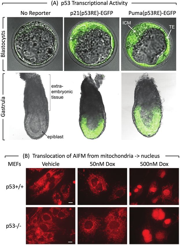
a p53-dependent response element demonstrated that after the blastocyst has implanted (E4.5) and gastrulation hasStem Cells, 2022, Vol. 40, No. 3 233
Downloaded from https://academic.oup.com/stmcls/article/40/3/227/6516995 by guest on 12 August 2022
Figure 3. p53 activity and MEF PCD response at the beginning of mouse development. (A) p53 activity assayed in embryos isolated from mice
homozygous for reporter genes expressing enhanced green fluorescence protein driven by either the Cdkn1a/p21 or the Bbc3/Puma gene’s p53
response element.75 At embryonic day E3.5, fluorescence was detected in the inner cell mass (ICM), and trophectoderm (TE) of blastocysts. The
large blastocoel cavity identifies these examples as late-stage blastocysts containing early epiblast (Fig. 1). At embryonic day E6.5, fluorescence was
detected in the epiblast but not in the extraembryonic tissue of gastrula. (B) MEFs cultured for 24 h with doxorubicin and then stained for “apoptosis-
inducing factor” AIFM.51 Scale bar is 15 μm. Translocation of AIFM from mitochondria to nucleus occurred in both p53+/+ and p53−/− cells, thereby
confirming non-canonical apoptosis in MEFs treated with 500 nM doxorubicin, but not in MEFs with 50 nM doxorubicin.
begun (E6.25-E7.5). However, DNA damage begins to accu- of cells with 4N DNA content in response to DNA damage
mulate in Mdm2−/− blastocysts, but less so if embryos also lack prior to induction of apoptosis.98 Double-strand DNA breaks
the p53-dependent proapoptotic gene Bax, suggesting that un- induced by culturing cells with doxorubicin activated the G2
regulated p53 initiates apoptosis in blastocysts.91 Nevertheless, checkpoint in naïve mESCs regardless of the presence or ab-
embryonic lethality still occurs at E6.5-E7.5 due to the arrest sence of p53,51,95,97,99 p21, or PUMA51 (Fig. 2A). In contrast,
of cell proliferation (cell senescence) rather than PCD. both hESCs and mouse EpiSCs derived from the epiblast in
postimplantation blastocysts exhibit a G1 checkpoint.48
PCD is not dependent on p53. Of the 9 studies that inves-
p53 and Naïve ESCs
tigated the role of p53 in PCD, 3 concluded that p53 is not
Cell cycle checkpoints are p53-independent. The G1 check- required51,93,97 and 6 concluded that p53 is required.95,96,100-102
point is a response to cell stress that retards entrance into To resolve this paradox, Jaiswal et al51 quantified the effects
the S phase.92 Naïve mESCs lack a G1 DNA damage check- of doxorubicin on p53+/+ and p53−/− ESCs derived by 2 dif-
point.51,93-97 The G2 checkpoint is a transient accumulation ferent methods. To eliminate the possibility that conclusions234 Stem Cells, 2022, Vol. 40, No. 3
depended on either the source or derivation of p53−/− ESCs, Experimental Conditions Could Account for
both wild-type and p53−/− ESCs derived directly from blasto- Contradictory Conclusions
cysts were characterized in parallel with ESCs in which the
Cell Culture
p53 genes were ablated in vitro. ESCs isolated from p53−/−
blastocysts exhibit the effects of p53 loss through multiple Culture conditions are critical to maintaining the pluripotent
generations in vivo (chronic phenotype), whereas p53−/− ESCs state.49 Suboptimal conditions promote DNA damage108 and
engineered in vitro from p53+/+ ESCs exhibit the effects of im- ESC differentiation, thereby selecting for p53 dependence.109
mediate p53 loss (acute phenotype). ESCs under stress characteristically undergo either differen-
To eliminate methodology-dependent biases, the rate and tiation or apoptosis.110,111 In fact, the culture conditions used
extent of cell cycle arrest and cell death were quantified by to convert naïve ESCs into 2iESCs enforce self-renewal and
time-dependent changes in DNA content (Fig. 2C and D), a dramatic loss of spontaneously differentiating cells; neither
by staining with annexin-V and propidium iodide to distin- primed ESCs nor differentiated somatic cells survive these
guish apoptosis from necrosis, by exclusion of trypan blue conditions.112 Remarkably, 2 studies used blastocyst derived-
to distinguish live cells from dead cells, by Western immuno- ESCs from the same source (Rudolf Jaenisch, MIT, Cambridge,
MA) but reported contradictory results. Culturing ESCs to
Downloaded from https://academic.oup.com/stmcls/article/40/3/227/6516995 by guest on 12 August 2022
blotting of p53, p21, PUMA, γH2AX, PARP, and CASP3 to
confirm genotypes, DNA damage, and caspase cleavage (Fig. “sub-confluence” before adding doxorubicin101 might have
2A), and by cellular localization of AIFM to confirm caspase- created conditions in which excessively high concentrations
independent apoptosis (Fig. 2B). of doxorubicin-induced apoptosis in p53+/+ cells more rapidly
The results revealed that, regardless of their derivation, than in p53−/− cells.51,93
naïve mouse ESCs do not require p53, p21, or PUMA either
Time
to activate the G2-checkpoint (Fig. 2C) or to undergo ro-
bust apoptosis (Fig. 2D). Depending on conditions such as The only effect of p53 on apoptosis in naïve ESCs was to
seeding density and doxorubicin concentration, p53 can accelerate its initiation. Once initiated, apoptosis continued
accelerate initiation of apoptosis in ESCs in response to at the same rate and to the same extent as in the absence of
DNA damage by 8.4 ± 0.5 h, but the rate and extent of p53. Experiments with a single time point and a single-drug
apoptosis in ESCs are equivalent and complete PCD within concentration cannot reveal the relationship between DNA
72 h, regardless of the presence or absence of p53. The in- damage and the significance of p53.
hibitory effect of only 50 nM doxorubicin is evident from
DNA Damage
visual inspection of cultured cells, and the lethal effect is
evident from the accumulation of cells withStem Cells, 2022, Vol. 40, No. 3 235
p53 Null Mutation lethality is first evident during gastrulation. Depending on ex-
Comparing p53+/+ with p53−/− ESCs is essential to establish a perimental conditions, p53 can accelerate initiation of PCD
role for p53. Two studies with contradictory conclusions re- in mESCs and late-stage blastocysts, but once initiated, PCD
lied on inadequately characterized ESCs.97,100 Another study occurs at equivalent rates and to equivalent extents regardless
on hESCs based its conclusion solely on changes in p53 ex- of the presence or absence of p53. Following either mESC dif-
pression in response to apoptotic stimuli.114 Studies that rely ferentiation in vitro or the formation of MEFs in vivo, DNA
upon changes in p53 protein in response to stress and p53 damage induces p53-dependent cell cycle arrest and senes-
inhibitors ignore the fact that the p53 transcription factor cence. Given the sensitivity of MEFs to p53-dependent sen-
regulates at least 343 target genes involved in maintaining escence, failure of embryonic development likely results from
genomic stability, cell differentiation, cell senescence, cell cycle cell senescence rather than PCD, although excessive DNA
regulation, and PCD.7 The fact that ectopic over-expression of damage induces PCD.
certain p53 mutations also suppressed doxorubicin-induced
apoptosis101 simply reflects the fact that p53 affects expres-
Funding
sion of hundreds of different genes, some of which affect
Downloaded from https://academic.oup.com/stmcls/article/40/3/227/6516995 by guest on 12 August 2022
apoptosis. Many naturally occurring p53 mutations have the The work was funded by the National Institute of Child
opposite effect; they gain additional oncogenic functions that Health and Human Development (NICHD) Intramural
endow cells with growth and survival advantages.115 Research Program, Grant/Award Numbers: ZIA HD000506,
ZIA HD000507.
Reproducibility
Two studies using the same source of ESCs (Yang Xu, Univ. Conflict of Interest
California, San Diego) concluded that p53 is not required for
cell cycle arrest51,96 and their results with p53+/+ ESCs are in- The authors declare no potential conflicts of interest.
distinguishable.51,96 However, one study concluded that p53
is essential for doxorubicin-induced apoptosis96 whereas the Author Contributions
other study concluded that it is not.51 The first study relied
on caspase-3 cleavage to confirm apoptosis, which they de- All the authors wrote and revised the manuscript. All the au-
tected with a monoclonal antibody specific for the cleaved thors have read and agreed to the published version of the
form. Thus, the fact that the extent of CASP3 cleavage was manuscript.
insignificant was not recognized. Moreover, the time delay
for initiation of apoptosis exhibited by p53−/− ESCs cultured Data Availability
with excess doxorubicin delayed the appearance of cleaved
caspase-3, thereby allowing cleaved-caspase-3 to be detected No new data were generated or analyzed in support of this
in p53+/+ cells under conditions where it appeared to be absent research.
in p53−/− cells. Apoptosis is also delayed in p53−/− ESCs cul-
tured under stress, such as the extremely high seeding density References
(260 000 cells/cm2) used in the first study.96
1. Galluzzi L, Bravo-San Pedro JM, Vitale I, et al. Essential versus ac-
cessory aspects of cell death: recommendations of the NCCD 2015.
Cell Death Differ. 2015;22(1):58-73. https://doi.org/10.1038/
Conclusions
cdd.2014.137
Of the 12 forms of PCD described in human cells, only 2. Wyllie AH, Kerr JF, Currie AR. Cell death: the significance of apop-
noncanonical apoptosis, autophagy-dependent, and lysosome- tosis. Int Rev Cytol. 1980;68:251-306. https://doi.org/10.1016/
dependent PCD have been reported in ESCs, preimplantation, s0074-7696(08)62312-8
or gastrulating embryos. However, autophagy-dependent 3. Hardy K, Handyside AH, Winston RM. The human blastocyst: cell
PCD might be confused with autophagy disruption which number, death and allocation during late preimplantation develop-
ment in vitro. Development. 1989;107(3):597-604.
could activate non-canonical apoptosis, lysosome-dependent
4. Fuchs Y, Steller H. Programmed cell death in animal development
PCD, or necroptosis. Another candidate is parthanatos.
and disease. Cell. 2011;147(4):742-758. https://doi.org/10.1016/j.
The importance of p53 in PCD has been characterized ex- cell.2011.10.033
tensively, but conclusions are often enigmatic. Based solely on 5. Munoz-Espin D, Canamero M, Maraver A, et al. Programmed
studies comparing wild-type with p53−/− ESCs, MEFs, or mice, cell senescence during mammalian embryonic development. Cell.
3 conclusions appear uncontested; p53 is not required for ac- 2013;155(5):1104-1118. https://doi.org/10.1016/j.cell.2013.10.019
tivation of the G2-checkpoint, for embryonic lethality prior 6. Coucouvanis E, Martin GR. Signals for death and survival:
to gastrulation, or for embryonic development. The form of a two-step mechanism for cavitation in the vertebrate em-
PCD and the role of p53 might change as preimplantation bryo. Cell. 1995;83(2):279-287. https://doi.org/10.1016/0092-
embryos develop from totipotent (2iESCs) to pluripotent 8674(95)90169-8
7 Jaiswal SK, Raj S, DePamphilis ML. Developmental acquisition of
(naïve ESCs) to primed pluripotent cells in post-implantation
p53 functions. Genes. 2021;12(11):1675.
embryos (hESCs, mEpiSCs). However, contradictory conclu-
8. Aubrey BJ, Kelly GL, Janic A, et al. How does p53 induce apoptosis
sions concerning the role of p53 during PCD in ESCs can and how does this relate to p53-mediated tumour suppression?
be reconciled by differences in experimental conditions, such Cell Death Differ. 2018;25(1):104-113. https://doi.org/10.1038/
as the amount of stress and the length of time stress was in- cdd.2017.169
duced, culture conditions, and assay conditions. 9. Levine AJ, Berger SL. The interplay between epigenetic changes and
In mice, p53 dependent transcription is first evident in late- the p53 protein in stem cells. Genes Dev. 2017;31(12):1195-1201.
stage blastocysts, and the ability of p53 to induce embryonic https://doi.org/10.1101/gad.298984.117236 Stem Cells, 2022, Vol. 40, No. 3
10. Bieging KT, Mello SS, Attardi LD. Unravelling mechanisms of p53- 32. Lee IH, Kawai Y, Fergusson MM, et al. Atg7 modulates p53 ac-
mediated tumour suppression. Nat Rev Cancer. 2014;14(5):359- tivity to regulate cell cycle and survival during metabolic stress.
370. https://doi.org/10.1038/nrc3711 Science. 2012;336(6078):225-228. https://doi.org/10.1126/sci-
11. Riley T, Sontag E, Chen P, et al. Transcriptional control of human ence.1218395
p53-regulated genes. Nat Rev Mol Cell Biol. 2008;9(5):402-412. 33. Yang Y, White E. Autophagy suppresses TRP53/p53 and oxidative
https://doi.org/10.1038/nrm2395 stress to enable mammalian survival. Autophagy. 2020;16(7):1355-
12. Shiloh Y, Y Z. The ATM protein kinase: regulating the cellular 1357. https://doi.org/10.1080/15548627.2020.1765522
response to genotoxic stress, and more. Nat Rev Mol Cell Biol. 34. Chen J. The cell-cycle arrest and apoptotic functions of p53 in
2013;14(4):197-210. tumor initiation and progression. Cold Spring Harb Perspect Med.
13. Sengupta S, Harris CC. p53: traffic cop at the crossroads of DNA 2016;6(3):a026104. https://doi.org/10.1101/cshperspect.a026104
repair and recombination. Nat Rev Mol Cell Biol. 2005;6(1):44- 35. Sharma G, Guardia CM, Roy A, et al. A family of PIKFYVE
55. https://doi.org/10.1038/nrm1546 inhibitors with therapeutic potential against autophagy-dependent
14. Tilgner K, Neganova I, Moreno-Gimeno I, et al. A human iPSC cancer cells disrupt multiple events in lysosome homeostasis.
model of Ligase IV deficiency reveals an important role for Autophagy. 2019;15(10):1694-1718. https://doi.org/10.1080/155
NHEJ-mediated-DSB repair in the survival and genomic stability 48627.2019.1586257
of induced pluripotent stem cells and emerging haematopoietic 36. Tsukamoto S, Hara T, Yamamoto A, et al. Functional analysis of
Downloaded from https://academic.oup.com/stmcls/article/40/3/227/6516995 by guest on 12 August 2022
progenitors. Cell Death Differ. 2013;20(8):1089-1100. https://doi. lysosomes during mouse preimplantation embryo development. J
org/10.1038/cdd.2013.44 Reprod Dev. 2013;59(1):33-39. https://doi.org/10.1262/jrd.2012-
15. Lozano G. Mouse models of p53 functions. Cold Spring Harb 096
Perspect Biol. 2010;2(4):a001115. https://doi.org/10.1101/ 37. Charlot JF, Pretet JL, Haughey C, et al. Mitochondrial translocation
cshperspect.a001115 of p53 and mitochondrial membrane potential (Delta Psi m) dissi-
16. Yu J, Zhang L. PUMA, a potent killer with or without p53. On- pation are early events in staurosporine-induced apoptosis of wild
cogene. 2008;27(suppl 1):S71-S83. https://doi.org/10.1038/ type and mutated p53 epithelial cells. Apoptosis. 2004;9(3):333-
onc.2009.45 343. https://doi.org/10.1023/b:appt.0000025810.58981.4c
17. Tang D, Kang R, Berghe TV, et al. The molecular machinery of 38. Battistelli M, Falcieri E. Apoptotic bodies: particular extracellular
regulated cell death. Cell Res. 2019;29(5):347-364. https://doi. vesicles involved in intercellular communication. Biology (Basel).
org/10.1038/s41422-019-0164-5 2020;9(1). https://doi.org/10.3390/biology9010021
18. Doherty J, Baehrecke EH. Life, death and autophagy. Nat Cell Biol. 39. Kayagaki N, Kornfeld OS, Lee BL, et al. NINJ1 mediates
2018;20(10):1110-1117. https://doi.org/10.1038/s41556-018- plasma membrane rupture during lytic cell death. Nature.
0201-5 2021;591(7848):131-136. https://doi.org/10.1038/s41586-021-
19. Singh R, Letai A, Sarosiek K. Regulation of apoptosis in health and 03218-7
disease: the balancing act of BCL-2 family proteins. Nat Rev Mol 40. Hevler JF, Zenezeni Chiozzi R, Cabrera-Orefice A, et al. Molec-
Cell Biol. 2019;20(3):175-193. https://doi.org/10.1038/s41580- ular characterization of a complex of apoptosis-inducing factor 1
018-0089-8 with cytochrome c oxidase of the mitochondrial respiratory chain.
20. Laszlo ZI, Lele Z, Zoldi M, et al. ABHD4-dependent develop- Proc Natl Acad Sci USA. 2021;118(39). https://doi.org/10.1073/
mental anoikis safeguards the embryonic brain. Nat Commun. pnas.2106950118
2020;11(1):4363. https://doi.org/10.1038/s41467-020-18175-4 41. Zakeri Z, Lockshin RA, Criado-Rodriguez LM, et al. A generalized
21. Stahl-Meyer J, Stahl-Meyer K, Jaattela M. Control of mitosis, caspase inhibitor disrupts early mammalian development. Int J Dev
inflammation, and cell motility by limited leakage of lysosomes. Biol. 2005;49(1):43-47. https://doi.org/10.1387/ijdb.041920zz
Curr Opin Cell Biol. 2021;71:29-37. https://doi.org/10.1016/j. 42. Ziegler-Birling C, Helmrich A, Tora L, et al. Distribution of p53
ceb.2021.02.003 binding protein 1 (53BP1) and phosphorylated H2A.X during
22. Denton D, Kumar S. Autophagy-dependent cell death. Cell Death mouse preimplantation development in the absence of DNA
Differ. 2019;26(4):605-616. https://doi.org/10.1038/s41418-018- damage. Int J Dev Biol. 2009;53(7):1003-1011. https://doi.
0252-y org/10.1387/ijdb.082707cz
23. White E. Autophagy and p53. Cold Spring Harb Perspect Med. 43. J L, Priest DG, Solano A, et al. Spatiotemporal dynamics of 53BP1
2016;6(4):a026120. https://doi.org/10.1101/cshperspect.a026120 dimer recruitment to a DNA double strand break. Nat Commun.
24. Weinlich R, Oberst A, Beere HM, et al. Necroptosis in development, 2020;11(1):5776.
inflammation and disease. Nat Rev Mol Cell Biol. 2017;18(2):127- 44. Wilson Y, Morris ID, Kimber SJ, et al. The role of Trp53 in the
136. https://doi.org/10.1038/nrm.2016.149 mouse embryonic response to DNA damage. Mol Hum Reprod.
25. Holze C, Michaudel C, Mackowiak C, et al. Oxeiptosis, a ROS- 2019;25(7):397-407. https://doi.org/10.1093/molehr/gaz029
induced caspase-independent apoptosis-like cell-death pathway. 45. Tsukamoto S, Kuma A, Murakami M, et al. Autophagy is es-
Nat Immunol. 2018;19(2):130-140. https://doi.org/10.1038/ sential for preimplantation development of mouse embryos.
s41590-017-0013-y Science. 2008;321(5885):117-120. https://doi.org/10.1126/sci-
26. Stockwell BR, Friedmann Angeli JP, Bayir H, et al. Ferroptosis: a ence.1154822
regulated cell death nexus linking metabolism, redox biology, and 46. Ikonomov OC, Sbrissa D, Delvecchio K, et al. The phosphoinositide
disease. Cell. 2017;171(2):273-285. kinase PIKfyve is vital in early embryonic development: preimplan-
27. Wang X, P G. Parthanatos in the pathogenesis of nervous system tation lethality of PIKfyve−/− embryos but normality of PIKfyve+/-
diseases. Neuroscience. 2020;449:241-250. mice. J Biol Chem. 2011;286(15):13404-13413. https://doi.
28. J L, Kuang F, Kang R, et al. Alkaliptosis: a new weapon for cancer org/10.1074/jbc.M111.222364
therapy. Cancer Gene Ther. 2020;27(5):267-269. 47. Smith A. Formative pluripotency: the executive phase in a devel-
29. Y H, Wang B, S L, et al. Pyroptosis, and its role in central nervous opmental continuum. Development. 2017;144(3):365-373. https://
system disease. J Mol Biol. 2021;167379. doi.org/10.1242/dev.142679
30. Hamann JC, Kim SE, Overholtzer M. Methods for the study of 48. Zaveri L, Dhawan J. Cycling to meet fate: connecting pluripotency
entotic cell death. Methods Mol Biol. 2019;1880:447-454. https:// to the cell cycle. Front Cell Dev Biol. 2018;6:57. https://doi.
doi.org/10.1007/978-1-4939-8873-0_28 org/10.3389/fcell.2018.00057
31. Inoue M, Enomoto M, Yoshimura M, et al. Pharmacological in- 49. Mulas C, Kalkan T, von Meyenn F, et al. Defined conditions for
hibition of sodium-calcium exchange activates NADPH oxidase propagation and manipulation of mouse embryonic stem cells.
and induces infection-independent NETotic cell death. Redox Biol. Development. 2019;146(6):dev178970. https://doi.org/10.1242/
2021;43:101983. https://doi.org/10.1016/j.redox.2021.101983 dev.178970Stem Cells, 2022, Vol. 40, No. 3 237
50. Kinoshita M, Barber M, Mansfield W, et al. Capture of mouse 68. Lowe SW, Bodis S, McClatchey A, et al. p53 status and the effi-
and human stem cells with features of formative pluripotency. cacy of cancer therapy in vivo. Science. 1994;266(5186):807-810.
Cell Stem Cell. 2021;28(3):453-471.e8. https://doi.org/10.1016/j. https://doi.org/10.1126/science.7973635
stem.2020.11.005 69. Lowe SW, Jacks T, Housman DE, et al. Abrogation of oncogene-
51. Jaiswal SK, Oh JJ, DePamphilis ML. Cell cycle arrest and apoptosis associated apoptosis allows transformation of p53-deficient cells.
are not dependent on p53 prior to p53-dependent embryonic stem Proc Natl Acad Sci USA. 1994;91(6):2026-2030. https://doi.
cell differentiation. Stem Cells. 2020;38(9):1091-1106. https://doi. org/10.1073/pnas.91.6.2026
org/10.1002/stem.3199 70. Vater CA, Bartle LM, Dionne CA, et al. Induction of apoptosis by
52. Tichy ED, Stephan ZA, Osterburg A, et al. Mouse embryonic stem tamoxifen-activation of a p53-estrogen receptor fusion protein
cells undergo charontosis, a novel programmed cell death pathway expressed in E1A and T24 H-ras transformed p53−/− mouse embryo
dependent upon cathepsins, p53, and EndoG, in response to fibroblasts. Oncogene. 1996;13(4):739-748.
etoposide treatment. Stem Cell Res. 2013;10(3):428-441. https:// 71. Shimizu S, Kanaseki T, Mizushima N, et al. Role of Bcl-2 family
doi.org/10.1016/j.scr.2013.01.010 proteins in a non-apoptotic programmed cell death dependent on
53. Abdelalim EM, Tooyama I. The p53 inhibitor, pifithrin-alpha, autophagy genes. Nat Cell Biol. 2004;6(12):1221-1228. https://
suppresses self-renewal of embryonic stem cells. Biochem Biophys doi.org/10.1038/ncb1192
Res Commun. 2012;420(3):605-610. https://doi.org/10.1016/j. 72. Arakawa S, Tsujioka M, Yoshida T, et al. Role of Atg5-dependent
Downloaded from https://academic.oup.com/stmcls/article/40/3/227/6516995 by guest on 12 August 2022
bbrc.2012.03.041 cell death in the embryonic development of Bax/Bak double-
54. J Z, Singh M, Selivanova G, et al. Pifithrin-alpha alters p53 post- knockout mice. Cell Death Differ. 2017;24(9):1598-1608. https://
translational modifications pattern and differentially inhibits p53 doi.org/10.1038/cdd.2017.84
target genes. Sci Rep. 2020;10(1):1049. 73. Tu HC, D R, Wang GX, et al. The p53-cathepsin axis cooperates
55. Chakraborty AR, Vassilev A, Jaiswal SK et al. Selective elimi- with ROS to activate programmed necrotic death upon DNA
nation of pluripotent stem cells by PIKfyve specific inhibitors. damage. Proc Natl Acad Sci USA. 2009;106(4):1093-1098.
Stem Cell Rep. 2021;17(2):397-412. https://doi.org/10.1016/j. 74. Vaseva AV, Marchenko ND, K J, et al. p53 opens the mito-
stemcr.2021.12.013 chondrial permeability transition pore to trigger necrosis. Cell.
56. Gayle S, Landrette S, Beeharry N, et al. Identification of apilimod as 2012;149(7):1536-1548.
a first-in-class PIKfyve kinase inhibitor for treatment of B-cell non- 75. Goh AM, Lim CY, Chiam PC, et al. Using targeted transgenic re-
Hodgkin lymphoma. Blood. 2017;129(13):1768-1778. https://doi. porter mice to study promoter-specific p53 transcriptional activity.
org/10.1182/blood-2016-09-736892 Proc Natl Acad Sci USA. 2012;109(5):1685-1690. https://doi.
57. O’Connell CE, Vassilev A. Combined inhibition of p38MAPK and org/10.1073/pnas.1114173109
PIKfyve synergistically disrupts autophagy to selectively target 76. Norimura T, Nomoto S, Katsuki M, et al. p53-dependent ap-
cancer cells. Cancer Res. 2021;81(11):2903-2917. https://doi. optosis suppresses radiation-induced teratogenesis. Nat Med.
org/10.1158/0008-5472.CAN-20-3371 1996;2(5):577-580. https://doi.org/10.1038/nm0596-577
58. Basit F, van Oppen LM, Schockel L, et al. Mitochondrial complex 77. Klein AM, de Queiroz R, M, Venkatesh D, et al. The roles and regu-
I inhibition triggers a mitophagy-dependent ROS increase leading lation of MDM2 and MDMX: it is not just about p53. Genes Dev.
to necroptosis and ferroptosis in melanoma cells. Cell Death Dis. 2021;35(9-10):575-601.
2017;8(3):e2716. https://doi.org/10.1038/cddis.2017.133 78. Ivanov GS, Ivanova T, Kurash J, et al. Methylation-acetylation in-
59. Arya BD, Mittal S, Joshi P, et al. Graphene oxide-chloroquine terplay activates p53 in response to DNA damage. Mol Cell Biol.
nanoconjugate induce necroptotic death in A549 cancer 2007;27(19):6756-6769. https://doi.org/10.1128/MCB.00460-07
cells through autophagy modulation. Nanomedicine (Lond). 79. Kruse JP, W G. Modes of p53 regulation. Cell. 2009;137(4):
2018;13(18):2261-2282. https://doi.org/10.2217/nnm-2018-0086 609-622.
60. Goodall ML, Fitzwalter BE, Zahedi S, et al. The autophagy 80. Hull R, Oosthuysen B, Cajee UF, et al. The Drosophila retino-
machinery controls cell death switching between apoptosis blastoma binding protein 6 family member has two isoforms and
and necroptosis. Dev Cell. 2016;37(4):337-349. https://doi. is potentially involved in embryonic patterning. Int J Mol Sci.
org/10.1016/j.devcel.2016.04.018 2015;16(5):10242-10266. https://doi.org/10.3390/ijms160510242
61. Kharaziha P, Chioureas D, Baltatzis G, et al. Sorafenib-induced de- 81. Li L, Deng B, Xing G, et al. PACT is a negative regulator of p53 and
fective autophagy promotes cell death by necroptosis. Oncotarget. essential for cell growth and embryonic development. Proc Natl
2015;6(35):37066-37082. https://doi.org/10.18632/oncotarget.5797 Acad Sci USA. 2007;104(19):7951-7956.
62. Sulkshane P, Teni T. BH3 mimetic Obatoclax (GX15-070) 82. Yang X, Zhang Q, Yang X, et al. PACT cessation overcomes
mediates mitochondrial stress predominantly via MCL-1 inhibi- ovarian cancer cell chemoresistance to cisplatin by enhancing p53-
tion and induces autophagy-dependent necroptosis in human oral mediated apoptotic pathway. Biochem Biophys Res Commun.
cancer cells. Oncotarget. 2017;8(36):60060-60079. https://doi. 2019;511(4):719-724. https://doi.org/10.1016/j.bbrc.2019.02.089
org/10.18632/oncotarget.11085 83. Bennett RL, Pan Y, Christian J, et al. The RAX/PACT-PKR stress
63. Micco R D, Cicalese A, Fumagalli M, et al. DNA damage response response pathway promotes p53 sumoylation and activation,
activation in mouse embryonic fibroblasts undergoing replicative leading to G(1) arrest. Cell Cycle. 2012;11(2):407-417. https://doi.
senescence and following spontaneous immortalization. Cell Cycle. org/10.4161/cc.11.2.18999
2008;7(22):3601-3606. 84. Fujitani K, Otomo A, Nagayama Y, et al. PACT/PRKRA and p53
64. Harvey M, Sands AT, Weiss RS, et al. In vitro growth characteristics regulate transcriptional activity of DMRT1. Genet Mol Biol.
of embryo fibroblasts isolated from p53-deficient mice. Oncogene. 2020;43(2):e20190017. https://doi.org/10.1590/1678-4685-
1993;8(9):2457-2467. GMB-2019-0017
65. Jones SN, Sands AT, Hancock AR, et al. The tumorigenic poten- 85. Montes de Oca Luna R, Wagner DS, Lozano G. Rescue of
tial and cell growth characteristics of p53-deficient cells are equiva- early embryonic lethality in Mdm2-deficient mice by dele-
lent in the presence or absence of Mdm2. Proc Natl Acad Sci USA. tion of p53. Nature. 1995;378(6553):203-206. https://doi.
1996;93(24):14106-14111. https://doi.org/10.1073/pnas.93.24.14106 org/10.1038/378203a0
66. Brown JM, Attardi LD. The role of apoptosis in cancer develop- 86. Jones SN, Roe AE, Donehower LA, et al. Rescue of embryonic
ment and treatment response. Nat Rev Cancer. 2005;5(3):231-237. lethality in Mdm2-deficient mice by absence of p53. Nature.
https://doi.org/10.1038/nrc1560 1995;378(6553):206-208. https://doi.org/10.1038/378206a0
67. Lowe SW, Ruley HE, Jacks T, et al. p53-dependent apoptosis 87. McMasters KM, Montes de Oca Luna R, Pena JR, et al. Mdm2 de-
modulates the cytotoxicity of anticancer agents. Cell. 1993;74(6):957- letion does not alter growth characteristics of p53-deficient embryo
967. https://doi.org/10.1016/0092-8674(93)90719-7 fibroblasts. Oncogene. 1996;13(8):1731-1736.238 Stem Cells, 2022, Vol. 40, No. 3
88. Migliorini D, Lazzerini Denchi E, Danovi D, et al. Mdm4 Stem Cell. 2017;21(4):449-455.e4. https://doi.org/10.1016/j.
(Mdmx) regulates p53-induced growth arrest and neuronal stem.2017.09.004
cell death during early embryonic mouse development. Mol 104. Ter Huurne M, Peng T, G Y, et al. Critical role for P53 in regulating
Cell Biol. 2002;22(15):5527-5538. https://doi.org/10.1128/ the cell cycle of ground state embryonic stem cells. Stem Cell Rep.
MCB.22.15.5527-5538.2002 2020;14(2):175-183.
89. Finch RA, Donoviel DB, Potter D, et al. mdmx is a negative regu- 105. Atashpaz S, Samadi Shams S, Gonzalez JM, et al. ATR expands
lator of p53 activity in vivo. Cancer Res. 2002;62(11):3221-3225. embryonic stem cell fate potential in response to replication stress.
90. Parant J, Chavez-Reyes A, Little NA, et al. Rescue of embry- Elife. 2020;9:e54756.
onic lethality in Mdm4-null mice by loss of Trp53 suggests a 106. Grow EJ, Weaver BD, Smith CM, et al. p53 convergently activates
nonoverlapping pathway with MDM2 to regulate p53. Nat Dux/DUX4 in embryonic stem cells and in facioscapulohumeral
Genet. 2001;29(1):92-95. https://doi.org/10.1038/ng714 muscular dystrophy cell models. Nat Genet. 2021;53:1207-1220.
91. Chavez-Reyes A, Parant JM, Amelse LL, et al. Switching mechanisms 107. Chen Z, Zhang Y. Loss of DUX causes minor defects in zygotic
of cell death in mdm2- and mdm4-null mice by deletion of p53 genome activation and is compatible with mouse development.
downstream targets. Cancer Res. 2003;63(24):8664-8669. Nat Genet. 2019;51(6):947-951. https://doi.org/10.1038/s41588-
92. Hume S, Dianov GL, Ramadan K. A unified model for the G1/S 019-0418-7
cell cycle transition. Nucleic Acids Res. 2020;48(22):12483- 108. Jacobs K, Zambelli F, Mertzanidou A, et al. Higher-density cul-
Downloaded from https://academic.oup.com/stmcls/article/40/3/227/6516995 by guest on 12 August 2022
12501. https://doi.org/10.1093/nar/gkaa1002 ture in human embryonic stem cells results in DNA damage and
93. Aladjem MI, Spike BT, Rodewald LW, et al. ES cells do not activate genome instability. Stem Cell Rep. 2016;6(3):330-341. https://doi.
p53-dependent stress responses and undergo p53-independent ap- org/10.1016/j.stemcr.2016.01.015
optosis in response to DNA damage. Curr Biol. 1998;8(3):145- 109. Mansouri A, Fukumitsu H, Schindehuette J, et al. Differentiation
155. https://doi.org/10.1016/s0960-9822(98)70061-2 of embryonic stem cells. Curr Protoc Neurosci. 2009;Chapter
94. Hong Y, Stambrook PJ. Restoration of an absent G1 arrest and 3:Unit3 6.
protection from apoptosis in embryonic stem cells after ionizing 110. Jain AK, Y X, McCarthy R, et al. LncPRESS1 is a p53-
radiation. Proc Natl Acad Sci USA. 2004;101(40):14443-14448. regulated LncRNA that safeguards pluripotency by disrupting
https://doi.org/10.1073/pnas.0401346101 SIRT6-mediated de-acetylation of histone H3K56. Mol Cell.
95. He H, Wang C, Dai Q, et al. p53 and p73 regulate apoptosis but 2016;64(5):967-981.
not cell-cycle progression in mouse embryonic stem cells upon 111. Maimets T, Neganova I, Armstrong L, et al. Activation of p53
DNA damage and differentiation Stem Cell Rep. 2016;7(6):1087- by nutlin leads to rapid differentiation of human embryonic
1098. https://doi.org/10.1016/j.stemcr.2016.10.008 stem cells. Oncogene. 2008;27(40):5277-5287. https://doi.
96. M L, H G, Tripathi BK, et al. An Apela RNA-containing negative org/10.1038/onc.2008.166
feedback loop regulates p53-mediated apoptosis in embryonic 112. Navarro P. 2i, or not 2i: the soliloquy of nanog-negative mouse
stem cells. Cell Stem Cell. 2015;16(6):669-683. embryonic stem cells. Stem Cell Rep. 2018;11(1):1-3. https://doi.
97. Prost S, Bellamy CO, Clarke AR, et al. p53-independent DNA org/10.1016/j.stemcr.2018.06.017
repair and cell cycle arrest in embryonic stem cells. FEBS Lett. 113. Tovy A, Spiro A, McCarthy R, et al. p53 is essential for DNA
1998;425(3):499-504. https://doi.org/10.1016/s0014-5793(98) methylation homeostasis in naive embryonic stem cells, and its
00296-8 loss promotes clonal heterogeneity. Genes Dev. 2017;31(10):959-
98. Stark GR, Taylor WR. Analyzing the G2/M checkpoint. Methods 972. https://doi.org/10.1101/gad.299198.117
Mol Biol. 2004;280:51-82. https://doi.org/10.1385/1-59259-788- 114. Setoguchi K, TeSlaa T, Koehler CM, et al. P53 regulates
2:051 rapid apoptosis in human pluripotent stem cells. J Mol
99. M L, Y H, Dubois W, et al. Distinct regulatory mechanisms and Biol. 2016;428(7):1465-1475. https://doi.org/10.1016/j.
functions for p53-activated and p53-repressed DNA damage re- jmb.2015.07.019
sponse genes in embryonic stem cells. Mol Cell. 2012;46(1):30-42. 115. Rivlin N, Brosh R, Oren M, et al. Mutations in the p53 tumor
100. Corbet SW, Clarke AR, Gledhill S, et al. P53-dependent and -in- suppressor gene: important milestones at the various steps of
dependent links between DNA-damage, apoptosis and mutation tumorigenesis. Genes Cancer. 2011;2(4):466-474. https://doi.
frequency in ES cells. Oncogene. 1999;18(8):1537-1544. org/10.1177/1947601911408889
101. de Vries A, Flores ER, Miranda B, et al. Targeted point mutations 116. Kojima Y, Tam OH, Tam PP. Timing of developmental events in
of p53 lead to dominant-negative inhibition of wild-type p53 func- the early mouse embryo. Semin Cell Dev Biol. 2014;34:65-75.
tion. Proc Natl Acad Sci USA. 2002;99(5):2948-2953. https://doi. https://doi.org/10.1016/j.semcdb.2014.06.010
org/10.1073/pnas.052713099 117. Bardot ES, Hadjantonakis AK. Mouse gastrulation: coordina-
102. Grandela C, Pera MF, Grimmond SM, et al. p53 is required for tion of tissue patterning, specification and diversification of cell
etoposide-induced apoptosis of human embryonic stem cells. fate. Mech Dev. 2020;163:103617. https://doi.org/10.1016/j.
Stem Cell Res. 2007;1(2):116-128. https://doi.org/10.1016/j. mod.2020.103617
scr.2007.10.003 118. Samanta M, Kalantry S. Generating primed pluripotent epi-
103. Ter Huurne M, Chappell J, Dalton S, et al. Distinct cell-cycle blast stem cells: a methodology chapter. Curr Top Dev Biol.
control in two different states of mouse pluripotency. Cell 2020;138:139-174. https://doi.org/10.1016/bs.ctdb.2020.01.005You can also read


