BMP9 reduces age-related bone loss in mice by inhibiting osteoblast senescence through Smad1-Stat1-P21 axis
←
→
Page content transcription
If your browser does not render page correctly, please read the page content below
www.nature.com/cddiscovery

                  ARTICLE                  OPEN

                  BMP9 reduces age-related bone loss in mice by inhibiting
                  osteoblast senescence through Smad1-Stat1-P21 axis
                  Jing-zun Xu1,2, Yan-man Zhou1,2,3, Lin-lin Zhang1,2, Xiao-jing Chen1,2, Yu-ying Yang                           1,2
                                                                                                                                       , Deng Zhang1,2, Ke-cheng Zhu1,2,
                                                                                  ✉                                            1,2 ✉
                  Xiao-ke Kong1,2, Li-hao Sun1,2, Bei Tao1,2, Hong-yan Zhao 1,2 and Jian-min Liu

                  © The Author(s) 2022

                      Age-related osteoporosis is characterized by the accumulation of senescent osteoblastic cells in bone microenvironment and
                      significantly reduced osteogenic differentiation. Clearing of the senescent cells is helpful to improve bone formation in aged mice.
                      Bone morphogenetic protein 9 (BMP9), a multifunctional protein produced and secreted by liver, was reported to improve
                      osteoporosis caused by estrogen withdrawal. However, the mechanism of BMP9 has not been fully elucidated, and its effect on
                      senile osteoporosis has not been reported. This study reveals that BMP9 significantly increases bone mass and improves bone
                      biomechanical properties in aged mice. Furthermore, BMP9 reduces expression of senescent genes in bone microenvironment,
                      accompanied by decreased senescence-associated secretory phenotypes (SASPs) such as Ccl5, Mmp9, Hmgb1, Nfkb1, and Vcam1.
                      In vitro, Bmp9 treatment inhibits osteoblast senescence through activating Smad1, which suppresses the transcriptional activity of
1234567890();,:

                      Stat1, thereby inhibits P21 expression and SASPs production. Furthermore, inhibiting the Smad1 signal in vivo can reverse the
                      inhibitory effect of BMP9 on Stat1 and downstream senescent genes, which eliminates the protection of BMP9 on age-related
                      osteoporosis. These findings highlight the critical role of BMP9 on reducing age-related bone loss by inhibiting osteoblast
                      senescence through Smad1-Stat1-P21 axis.

                      Cell Death Discovery (2022)8:254 ; https://doi.org/10.1038/s41420-022-01048-8

                  INTRODUCTION                                                                             [11] and deletion of senescent gene P16 rescues bone loss of OVX-
                  Aging is manifested as time-dependent degeneration of physiolo-                          mice [12], which highlights the pivotal role of senescence in
                  gical functions at cellular, tissue, and organismal levels, making the                   estrogen deficiency-induced osteoporosis. In addition, pioneer
                  individual more vulnerable to various diseases, such as osteoporosis                     work demonstrates that eliminating senescent cells or suppressing
                  [1]. Unlike postmenopausal osteoporosis due to estrogen defi-                             SASPs is effective in promoting bone formation in aged mice
                  ciency, which is characterized by hyperactivity of osteoclasts, the                      [6, 13]. Most of the previous studies focused on searching for
                  main pathological feature of age-related bone loss is decreased                          exogenous reagents to inhibit senescence, termed as senolytic or
                  osteoblast functions and reduced bone formation [2]. Growing                             senomorphic according to their effects on clearing senescent cells
                  evidence demonstrates that the proportion of senescent cells in                          or suppressing SASPs secretion, respectively [14–16]. Researches
                  bone microenvironment increases with age, which contributes to                           on identification of the intrinsic anti-aging proteins should not be
                  the deteriorated osteogenic capacity of osteoblasts [3–5]. Further-                      ignored.
                  more, senescent osteoblastic lineage cells produce a senescence-                            Bone morphogenetic protein 9 (BMP9), mainly produced by
                  associated secretory phenotype (SASP) signal that is communicated                        liver and circulates in bloodstream, is a pleiotropic cytokine that
                  to neighboring cells in local bone microenvironment, resulting in                        regulates proliferation and differentiation in various cells [17, 18].
                  excessive production and secretion of chemokines and pro-                                BMP9 has been demonstrated effective on promoting osteoblast
                  inflammatory factors, thereby creating a toxic microenvironment                           differentiation [19] and improving fracture healing [20] in estrogen
                  which contributes to age-related bone loss [6–8].                                        deficiency-induced osteoporosis. Nevertheless, the effect of BMP9
                     With the rising of aging population, the incidence of                                 on age-related bone loss has not been clarified and studies about
                  osteoporosis and fracture is increasing significantly [1], treatment                      the regulation of BMP9 on senescence in skeleton are quite
                  targeting cellular senescence in both postmenopausal and age-                            limited [21]. Since the role of cellular senescence in the
                  related osteoporosis has become a new strategy [9, 10]. In recent                        pathogenesis of osteoporosis cannot be underestimated, it is
                  studies, senescence of bone marrow-derived mesenchymal stem                              necessary to clarify the effect of BMP9 on this pathological
                  cells is involved in ovariectomized (OVX)-induced osteoporosis                           process.

                  1
                    Department of Endocrine and Metabolic Diseases, Shanghai Institute of Endocrine and Metabolic Diseases, Ruijin Hospital, Shanghai Jiao Tong University School of Medicine,
                  Shanghai, China. 2Shanghai National Clinical Research Center for Metabolic Diseases, Key Laboratory for Endocrine and Metabolic Diseases of the National Health Commission of
                  the PR China, Shanghai Key Laboratory for Endocrine Tumor, State Key Laboratory of Medical Genomics, Ruijin Hospital, Shanghai Jiao Tong University School of Medicine,
                  Shanghai, China. 3Department of Nephrology, Provincial Hospital affiliated to Shandong First Medical University, Jinan, Shan Dong Province, China. ✉email: hyanzhao@163.com;
                  ljm10586@rjh.com.cn

                  Received: 19 February 2022 Revised: 22 April 2022 Accepted: 27 April 2022

                  Official journal of CDDpress
J. Xu et al.
2
      In this study, we attempted to investigate the influence of BMP9        effect on biomechanical parameters in 6-month-old mice
    on bone quality and senescent bone microenvironment in aged              (Fig. 3A–D).
    mice. The precise mechanism was assessed on senescent                       To further verify the protective effect of BMP9 in old mice, bone
    osteoblast by RNA sequencing analysis and rescue experiments             biochemical markers and osteoblast/osteoclast activity were
    using specific activator and inhibitor. Findings from our research        analyzed. Overexpressing BMP9 in aged mice led to elevated
    may expand our understanding of the functionalities of BMP9 and          serum PINP level and increased osteogenic genes in bone,
    provide new perspective in the treatment of age-related                  including Osx, Alp, Ocn, and Col1a1 (Fig. 3E, G). However, there
    osteoporosis.                                                            was only a slight decrease in serum CTX-I level, which did not
                                                                             reach significant difference (Fig. 3F). Among the expression of
                                                                             osteoclast differentiation markers, only Nfatc1 and Dc-stamp were
    RESULTS                                                                  downregulated by BMP9 (Fig. 3H). These results indicate that the
    Aged mice exhibit reduced bone mass and aging bone                       bone-promoting effect of BMP9 is mainly through facilitating
    microenvironment                                                         osteogenesis.
    Micro-CT analysis revealed serious bone loss of 20-month-old mice           It was noteworthy that BMP9 only worked on aged mice, not
    when compared with the 6-month-old cohort (Fig. 1A). The                 the young (Fig. S2). It was known that the bone exhibits pro-
    volumetric bone mineral densities (vBMDs) of femur significantly          inflammatory and senescent microenvironment with age [24].
    decreased in aged mice (Fig. 1B, C). Furthermore, bone                   Whether the beneficial effects of BMP9 on bone quality were
    microstructure as visualized by hematoxylin–eosin staining               achieved by inhibiting senescence in bone? To address this
    (HE staining) of vertebrae slices revealed less and thinner              question, we detected the expression of key senescent genes in
    trabecular in old mice (Fig. 1D). To quantify the bone turnover          bone microenvironment and found that BMP9 significantly
    level, procollagen I N-terminal propeptide (PINP) and C-terminal         decreased P16 and P21 expression, while no effect on P53 was
    cross-linked telopeptide of type I collagen (CTX-I) in serum, which      observed (Fig. 3I, J). Additionally, most of the SASPs (such as Ccl5,
    represent bone formation and resorption [22], respectively, were         Mmp9, Hmgb1, Nfkb1, and Vcam1) expressed in bone micro-
    measured. The results showed that PINP and CTX-I in old mice             environment were also downregulated by BMP9 in aged mice
    were both lower than the young, indicating a state of decreased          (Fig. 3K). However, the anti-aging effect of BMP9 in young mice
    bone remodeling (Fig. 1E, F). We further investigated the                was not as obvious as the old (Fig. S3). Taken together,
    expression of key osteoblast and osteoclast differentiation              overexpression of BMP9 in vivo improves bone quality and
    markers in vertebrae and found that they were all decreased in           microenvironment only in aged mice and this effect may be
    20-month-old mice, which were consistent with the results of             related to inhibiting cellular senescence.
    serum biomarkers (Fig. 1G, H).
       To verify the aging bone microenvironment, we measured the            BMP9 attenuates senescence and promotes osteoblast
    expression of P16, P21, and P53, which were senescence-                  differentiation in vitro
    associated genes [8, 23], and found that they were elevated in           To further evaluate the effect of BMP9 on cellular senescence, we
    aged mice (Fig. 1I). In addition, a variety of SASPs (Ccl5, Mmp9,        used serial passaging and hydrogen peroxide solution (H2O2)
    Hmgb1, Cxcl5, Nfkb1, and Vcam1) were all highly expressed in             treatment, respectively, to induce MC3T3-E1 cells senescence
    aged bone microenvironment(Fig. 1J). These findings clearly               in vitro. Subsequently, recombinant mouse BMP9 protein was
    indicate that the skeleton exhibits bone loss and senescent              applied into the culture medium for 3 days. β-galactosidase staining
    features with age.                                                       was performed to examine the degree of senescence and the
                                                                             number of β-gal (+) cells was counted. The proportion of β-gal (+)
    BMP9 reduces age-related bone loss and improves bone                     cells reached 48.69% and 64.16% in the passage 17 of MC3T3-E1
    microenvironment                                                         cells and cells treated with H2O2, respectively. Treatment with
    To investigate the impact of BMP9 on bone loss in aged mice,             BMP9 significantly decreased this proportion to 19.45% in repeating
    BMP9 overexpressing adeno-associated virus (AAV-BMP9) was                passaged and 36.65% in H2O2-induced MC3T3-E1 cells (Fig. 4A, B, K,
    injected through tail vein (Fig. 2A). The elevated levels of BMP9 in     L). In addition, the markers of DNA damage response γ-H2AX were
    serum and liver were confirmed 12 weeks later (Fig. S1). Micro-CT         increased in MC3T3-E1 cells after senescence induction, and were
    analysis revealed that BMP9 overexpression improved the bone             significantly downregulated by BMP9 (Fig. 4C, M). Moreover, the
    mass of old mice, while no effect was observed in the young              expression of P21 was significantly increased in senescent cells,
    (Fig. 2B). In aged mice, trabecular vBMD and cortical vBMD were          while P53 unchanged (Fig. 4D, N), indicating that senescence of
    obviously increased after AAV-BMP9 treatment (Fig. 2C, H).               MC3T3-E1 cells might be regulated mainly by P21 rather than P53.
    Microarchitecture analysis of the trabecular bone in distal femur        BMP9 suppressed the expression of P21, accompanied with
    showed that the percentage of bone (Trabecular BV/TV) and                decreased SASPs (Fig. 4E, F, O, P). The above results indicate that
    trabecular thickness (Tb.Th) increased in 20-month-old mice with         BMP9 inhibits senescence of MC3T3-E1 cells in different
    BMP9 overexpression, while trabecular separation (Tb.Sp) and             senescence-inducing conditions.
    structure model index (SMI) decreased (Fig. 2D–G). BMP9 also                Furthermore, senescent MC3T3-E1 cells showed deteriorated
    increased cortical bone volume (Cortical BV/TV) and cortical             osteogenic potential as evidenced by impaired alkaline phospha-
    thickness (Ct.Th) in aged mice (Fig. 2I, J). HE staining visually        tase staining (Alp staining) and decreased expression of osteoblast
    showed the increase of trabecular bone in aged mice over-                differentiation markers Runx2, Osx, Alp, Ocn, and Col1a1 after
    expressing BMP9. However, no difference was observed in the 6-           7 days osteogenic induction. The impaired osteogenic ability of
    month-old groups (Fig. 2K). These results indicate that BMP9 only        senescent osteoblast could be rescued by BMP9 treatment
    reduces bone loss in aged mice, but dose not increase bone mass          (Fig. 4G–J, Q–T). These results suggest that BMP9 promotes
    of the young.                                                            osteogenic differentiation in senescent osteoblast at least partially
       Three-point bending test was performed to examine the                 through attenuating cellular senescence.
    mechanical properties of the femur. An overall deterioration of             As Alk1 is a specific receptor for BMP9 ligand [25], we next
    the biomechanical properties was observed in aged mice,                  knocked down Alk1 on MC3T3-E1 cells to examine the effect of
    characterized by markedly reduced elastic modulus, bending               Alk1-deficiency on osteoblast senescence induced by H2O2. Alk1
    stiffness, maximum bending load, and fracture energy. The                knockdown efficiency was confirmed by quantitative polymerase
    reduction in mechanical parameters could be improved by                  chain reaction (qPCR) and Western blot (Fig. S4A, B). After H2O2
    BMP9 overexpression in aged mice. Similarly, BMP9 showed no              treatment, Alk1-deficient cells showed significantly increased

                                                                                                    Cell Death Discovery (2022)8:254
J. Xu et al.
                                                                                                                                               3

Fig. 1 Aged mice exhibit reduced bone mass and aging bone microenvironment. A Representative images derived from micro-CT analysis
of 6-month-old and 20-month-old mice (n = 5). B, C Quantitative analysis of the volume BMD of cortical bone (B) and trabecular bone (C) by
micro-CT (n = 5). D Representative images of HE-stained decalcified vertebrae sections of 6-month-old and 20-month-old mice (n = 5) (Scale
bar, 500 μm). E, F The levels of serum bone turnover parameters PINP (E) and CTX-I (F) were detected by ELISA (n = 5). G, H qPCR analysis of
osteoblast (G) and osteoclast (H) differentiation markers in vertebrae (n = 5). I, J qPCR analysis of mRNA levels of senescent genes (I) and
SASPs (J) in vertebrae (n = 5). Data presented as mean ± SD. A t-test was used for comparison between two groups. vBMD = volumetric bone
mineral density. *P < 0.05; **P < 0.01; ***P < 0.001.

expression of P21 (Fig. S4C, D) and SASPs (Fig. S4E). The results of    (Fig. S4H, I). These results demonstrate that blocking the
β-galactosidase staining indicated that the proportion of senes-        BMP9 signaling pathway through knocking down its receptor
cent cells in Alk1-deficient osteoblasts was much higher than the        Alk1 accelerates osteoblast senescence in vitro. Furthermore, we
control cells (Fig. S4F, G). Moreover, after 7 days of osteoblastic     detected the expression of Alk1 in bone of 6-month and 20-
induction, the Alp staining and the expression of key osteoblast        month-old mice, respectively, and found a significantly reduction
differentiation genes suggested that Alk1 knockdown further             in aged mice (Fig. S5), which suggested a low active state of
impaired the differentiation capacity of senescent osteoblasts          BMP9 signaling in vivo.

Cell Death Discovery (2022)8:254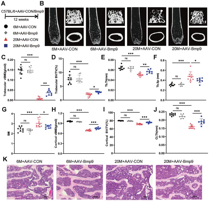
J. Xu et al.
4

    Fig. 2 BMP9 reduces age-related bone loss and improves bone microarchitecture. A Six-month-old and twenty-month-old mice were
    injected with AAV-BMP9 or AAV-CON, respectively. Twelve weeks after injection, mice were euthanized for subsequent experiments.
    B Representative images derived from micro-CT analysis, including 2D image construction of distal femur, 3D image reconstruction of
    trabecular bone of distal femur, and 3D image reconstruction of the femoral midshaft corticoid bone. C Quantitative analysis of the vBMD of
    trabecular by micro-CT. D–G Microarchitecture analysis of trabecular bone by micro-CT: Trabecular BV/TV (D), Tb.Th (E), Tb.Sp (F) and SMI (G).
    H Quantitative analysis of the vBMD of cortical by micro-CT. I, J Quantitative analysis of the BV/TV (I) and Ct.Th (J) of corticoid bone by micro-
    CT. K Representative images of HE-stained decalcified vertebrae sections (n = 6) (Scale bar, 500 μm). For micro-CT analysis: n = 9 for 6 M + AAV-
    CON group; n = 7 for 6 M + AAV-BMP9 group; n = 9 for 20 M + AAV-CON group; n = 8 for 20 M + AAV-BMP9 group. Data presented as mean ±
    SD. One-way ANOVA was used for comparison among multiple groups. AAV-CON = empty adeno-associated virus; AAV-BMP9 = BMP9
    overexpressing adeno-associated virus; BV/TV = percentage of bone volume; Tb.Th = trabecular thickness, Tb.Sp = trabecular separation,
    SMI = structure model index, Ct.Th = cortical thickness. *P < 0.05; **P < 0.01; ***P < 0.001. Ns, no significance.

    Stat1 is a key factor in regulating senescence of osteoblast               KEGG pathway analysis further displayed that the typical
    According to the above results, we demonstrated that BMP9 had              interferon-related signaling pathway Jak-Stat which included a
    the potential of suppressing senescence and promoting osteo-               series of pro-inflammatory factors was one of the most profound
    genesis. To further explore the specific molecular mechanism, RNA           pathways downregulated by BMP9 (Fig. 5B). Among these
    sequencing analysis of senescent MC3T3-E1 cells treated with or            downregulated genes, Stat1 is a crucial transcription factor
    without BMP9 was applied to uncover the differentially expressed           regulating the expression of P21 and multiple SASPs [27–29].
    genes.                                                                     We further analyzed the overlap of 479 highly expressed genes in
      Gene Ontology analysis revealed that genes regulated by BMP9             senescent MC3T3-E1 cells and 570 genes that were down-
    were associated with essential biological processes of osteoblast,         regulated by BMP9. Totally 116 genes were in this category,
    such as ossification, osteoblast differentiation, and extracellular         including Stat1 and its downstream target genes (Fig. 5C, D).
    matrix organization (Fig. 5A). Among the top 10 biological                    The expression of Stat1 was further examined in senescent
    processes, cellular responses to interferon-beta and interferon-           MC3T3-E1 cells with or without BMP9 treatment. Both serial
    gamma were associated with various SASPs production and                    passaging and H2O2-stimuli elevated the expression of Stat1,
    senescence [26], and were downregulated by BMP9 (Table S1).                which could be abolished by BMP9 (Fig. 5E, F). There was also a

                                                                                                       Cell Death Discovery (2022)8:254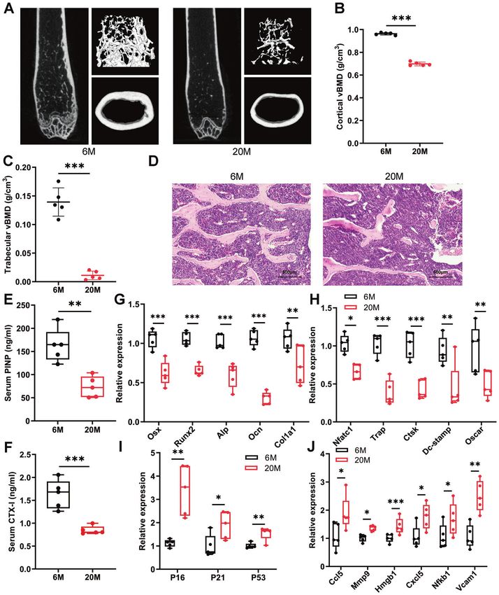
J. Xu et al.
                                                                                                                                                   5

Fig. 3 BMP9 improves bone biomechanical performance and bone microenvironment. A–D The right femur was isolated and subjected to
biomechanical properties analysis. The elastic modulus (A), bending stiffness (B), maximum bending load (C), and fracture energy (D) were
evaluated for each group. E, F The levels of serum bone turnover parameters PINP (E) and CTX-I (F) were detected by ELISA (n = 7). G, H qPCR
analysis of osteoblast (G) and osteoclast (H) differentiation markers in vertebrae. (n = 6). I qPCR analysis of senescent genes in vertebrae
(n = 6). J Western blot analysis of protein levels of senescent genes in vertebrae (n = 4). K qPCR analysis of SASPs in vertebrae (n = 6). For
biomechanical properties analysis: n = 9 for 6 M + AAV-CON group; n = 7 for 6 M + AAV-BMP9 group; n = 9 for 20 M + AAV-CON group; n = 8
for 20 M + AAV-BMP9 group. Data presented as mean ± SD. A t-test was used for comparison between two groups. One-way ANOVA was used
for comparison among multiple groups. *P < 0.05; **P < 0.01; ***P < 0.001. Ns, no significance.

remarkable increase of Stat1 expression in bone samples from                  The osteogenic potential of senescent MC3T3-E1 cells treated
aged mice, which was downregulated by AAV-BMP9 treatment                   with BMP9 and 2-NP was examined. Alp staining of senescent
(Fig. 5G). These findings suggest that BMP9 can suppress the                osteoblast in BMP9 + 2-NP group was weaker than the BMP9 group
expression of Stat1.                                                       (Figs. 6J, 7J). The elevated expression of differentiation markers in
                                                                           BMP9-treated cells was also reduced by 2-NP (Figs. 6K, L, 7K, L).
The suppression of Stat1 is crucial for the anti-senescence                These results indicate that the anti-senescence effect of BMP9 in
effect of BMP9 in osteoblast                                               osteoblast depends on its inhibition on Stat1.
To further verify the importance of Stat1 suppression in mediating
the anti-senescence effect of BMP9, a rescue experiment was                BMP9 inhibits osteoblast senescence through Smad1-Stat1-
performed by applying a specific transcriptional activator of Stat1         P21 axis
termed as 2-(1,8-Naphthyridin-2-ly)phenol (2-NP) [30]. Activation          According to the above findings, BMP9 attenuates cellular
of Stat1 was confirmed by elevated mRNA and protein expression,             senescence by downregulation of Stat1, leading to reduced P21
accompanied with increased P21 after 2-NP application (Fig. 6A–D,          expression. We then explored the regulatory mechanism of BMP9
Fig. 7A–D). The SASPs (Ccl5, Mmp9, Vcam1, and Hmgb1) reduced               on Stat1. Dual-luciferase reporter assay showed that treatment
by BMP9 were also increased with 2-NP treatment (Figs. 6E, F, 7E, F).      with BMP9 for 24 h significantly decreased the activity of Stat1
The proportion of β-gal (+) cells increased from 41.4% to 52.01% in        promotor (Fig. 8A). Since Smad1/5/9 is widely involved in the
serial passaged cells with 2-NP (Fig. 6G, H). In H2O2-treated cells, the   regulatory effect of BMP9 [31, 32], we detected their expression in
percentage was reduced to 40.9% by BMP9 and increased back to              bone of old mice, finding that the expression of Smad1 was
50.14% under 2-NP treatment (Fig. 7G, H). Consistently, nuclear            significantly elevated by BMP9 overexpression (Fig. 8B). We then
γ-H2AX expression was also elevated by 2-NP in both senescent              co-transfected Stat1 promoter reporter plasmid and Smad1-
groups in the presence of BMP9 (Figs. 6I, 7I).                             expressing vector in MC3T3-E1 cells for 36 h. The luciferase

Cell Death Discovery (2022)8:254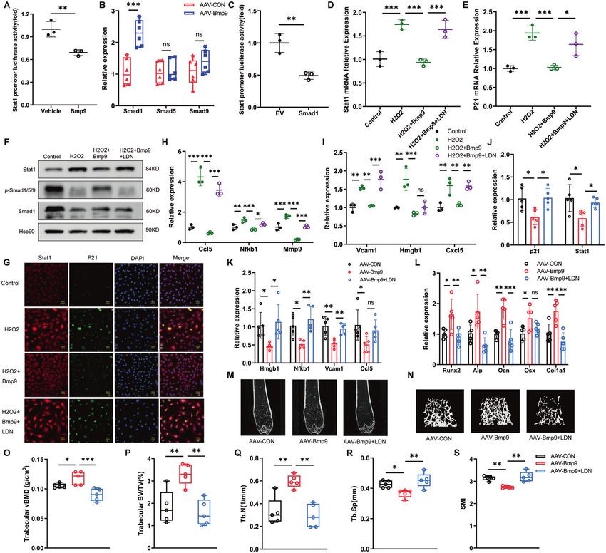
J. Xu et al.
6

    Fig. 4 BMP9 attenuates senescence and promotes osteoblast differentiation in MC3T3-E1 cells. Passage 7 of MC3T3-E1 cells were treated
    with vehicle as control group. Passage 17 of MC3T3-E1 cells were treated with vehicle or BMP9 for 3 days, respectively. A, B β-galactosidase
    staining was performed (A) and number of β-gal (+) cells was counted for each group (B) (Scale bar, 100 μm). C Immunofluorescence analysis
    of γ-H2AX expression (Scale bar, 50 μm). D qPCR analysis of mRNA levels of senescent genes P21 and P53. E, F qPCR analysis of mRNA levels of
    SASPs. G Alp staining of MC3T3-E1 cells suffered to osteogenic differentiation induction for 7 days with or without BMP9. H, I qPCR analysis of
    osteoblast differentiation markers in cells suffered to osteogenic differentiation induction for 7 days with or without BMP9. J Western blot
    analysis of protein levels of Runx2 and Osx in MC3T3-E1 cells suffered to osteogenic differentiation induction for 7 days with or without BMP9.
    Senescence of MC3T3-E1 cells was also induced by H2O2 treatment for 2 h, then fresh culture medium added with vehicle or BMP9 was
    replaced for another 3 days. K, L β-galactosidase staining was performed (K) and number of β-gal (+) cells was counted for each group (L).
    (Scale bar, 100 μm). M Immunofluorescence analysis of γ-H2AX expression (Scale bar, 50 μm). N qPCR analysis of mRNA levels of senescent
    genes P21 and P53. O, P qPCR analysis of mRNA levels of SASPs. Q ALP staining of control and H2O2-stimulating MC3T3-E1 cells suffered to
    osteogenic differentiation induction for 7 days with or without BMP9. R, S qPCR analysis of osteoblast differentiation markers in control and
    H2O2-stimulating MC3T3-E1 cells suffered to osteogenic differentiation induction for 7 days with or without BMP9. T Western blot analysis of
    protein levels of Runx2 and Osx in control and H2O2-stimulating MC3T3-E1 cells suffered to osteogenic differentiation induction for 7 days
    with or without BMP9. Data presented as mean ± SD. n = 3 biological replicates. One-way ANOVA was used for comparison among multiple
    groups. *P < 0.05; **P < 0.01; ***P < 0.001. Ns, no significance.

    activity of Stat1 was obviously downregulated, indicating the             H2O2-induced senescent cells (Fig. 8D–F). The number of P21 (+)
    inhibitory effect of Smad1 on Stat1 promoter activity (Fig. 8C).          Stat1 (+) cells was also increased with LDN193189 (Fig. 8G).
      With the use of Smad1/5/9 signaling pathway inhibitor                   Moreover, reduction of Ccl5, Nfkb1, Mmp9, Vcam1 and Cxcl5 in
    LDN193189 [33], activation of Smad1 induced by BMP9 was                   BMP9-treated senescent osteoblasts was partially neutralized by
    inhibited, resulting in increased expression of Stat1 and P21 in          the application of LDN193189 (Fig. 8H, I).

                                                                                                     Cell Death Discovery (2022)8:254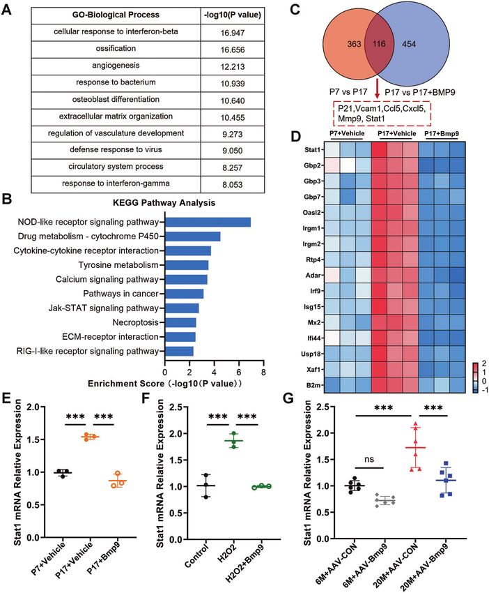
J. Xu et al.
                                                                                                                                                     7

Fig. 5 Stat1 is a key factor in regulating senescence of osteoblast. A GO analysis of differentially expressed genes between replicative
senescent MC3T3-E1 cells treated with vehicle or BMP9 for 3 days. B Top 10 downregulated pathways in KEGG analysis. C Overlap analysis of
upregulated genes between normal control with replicative senescent MC3T3-E1 cells and downregulated genes between senescent MC3T3-E1
cells treated with vehicle or BMP9. D Heatmap of Stat1 and its downstream target genes in passage 7 and passage 17 of MC3T3-E1 treated with
vehicle or BMP9 for 3 days. E qPCR analysis of mRNA level of Stat1 in cells of passage 7 and passage 17 with vehicle or BMP9 treatment for 3 days.
F qPCR analysis of mRNA level of Stat1 in control and H2O2-stimulating MC3T3-E1 cells treated with vehicle or BMP9 for 3 days. G qPCR analysis of
mRNA levels of Stat1 in 6-month-old and 20-month-old mice treated with AAV-CON or AAV-BMP9 (n = 6). Data presented as mean ± SD. n = 3
biological replicates for in vitro experiment. One-way ANOVA was used for comparison among multiple groups. *P < 0.05; **P < 0.01; ***P < 0.001.
Ns, no significance.

  To further investigate the effect of Smad1 inhibition in vivo,           manifested by increased expression of Hmgb1, Nfkb1, and Vcam1
aged mice with BMP9 overexpression were treated with                       (Fig. 8K). Furthermore, the expression of osteoblast-specific genes
LDN193189 injection. The decreased expression of P21 and Stat1             in vertebrae of aged mice was upregulated by BMP9 and
in the bone of BMP9-overexpressed mice was upregulated by                  downregulated by LDN193189 (Fig. 8L). Micro-CT showed that
LDN193189 intervention (Fig. 8J). In addition, LDN193189 reversed          LDN193189 eliminated the improvement on bone mass and
the inhibitory effect of BMP9 on SASPs in vivo, which was                  trabecular microarchitecture of aged mice caused by BMP9

Cell Death Discovery (2022)8:254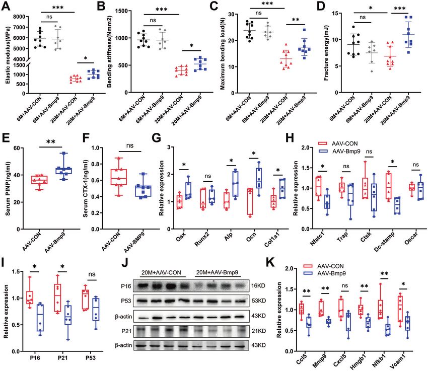
J. Xu et al.
8

    Fig. 6 The suppression of Stat1 is crucial for the anti-aging effect of BMP9 in replicative senescence of osteoblast. Passage 7 of MC3T3-E1
    cells were set as control group, passage 17 of MC3T3-E1 cells were treated with vehicle or BMP9 or BMP9 + 2-NP for 3 days, respectively.
    A, B qPCR analysis of Stat1 and P21 expression. C Western blot analysis of protein level of Stat1. D Immunofluorescence analysis of protein
    level of P21. (Scale bar, 50 μm). E, F qPCR analysis of mRNA levels of SASPs. G, H β-galactosidase staining was performed (G) and number of
    β-gal (+) cells was counted for each group (H) (Scale bar, 100 μm). I Immunofluorescence analysis of γ-H2AX expression (Scale bar, 50 μm).
    J Alp staining of MC3T3-E1 cells suffered to osteogenic differentiation induction for 7 days. K, L qPCR analysis of mRNA levels of osteoblastic
    differentiation markers. Data presented as mean ± SD. n = 3 biological replicates. One-way ANOVA was used for comparison among multiple
    groups. *P < 0.05; **P < 0.01; ***P < 0.001. Ns, no significance.

    overexpression (Fig. 8M–S). These results suggest that BMP9               attributed to senescence of osteoblastic lineage cells [9, 34].
    inhibits cellular senescence by activation of Smad1, which                Eliminating or reducing the burden of senescent cells represents a
    suppresses the promoter activity of Stat1, resulting in decreased         valuable therapeutic approach to age-related osteoporosis [3].
    P21 expression and SASPs production in osteoblast. Interfering            Based on this conception, our study aims to verify the anti-aging
    with the Smad1 signal can reverse the anti-aging effect of BMP9           effect of BMP9 and further explore the molecular mechanisms. In
    and undermine the protection of BMP9 on age-related bone loss.            this study, we find two features of BMP9’s regulation on age-
                                                                              related bone loss. First, the protective effect of BMP9 is observed
                                                                              only in aged mice, evidenced by elevated bone quality and
    DISCUSSION                                                                decreased expression of senescent genes and SASPs after BMP9
    In current study, we provide novel evidence that BMP9 reduces             overexpression in vivo. However, these beneficial effects are not
    bone loss and improves senescent bone microenvironment in                 observed in young mice receiving similar treatment. Consistently,
    aged mice. We further reveal that BMP9 inhibits expression of             recent study noted that mice treated with senolytic drugs
    senescent gene and SASPs in osteoblast through Smad1-Stat1-P21            (Dasatinib + Quercetin) from 18-month-old to 23-month-old
    axis, leading to improved osteoblastogenic differentiation. Our           showed improved vertebral bone quality parameters, whereas
    work uncovers a new mechanism for the protection effect of                same treatment during 6–23 months of age showed no obvious
    BMP9 on age-related bone loss.                                            effect [14]. It is suggested that an optimal treatment window is
       Age-related osteoporosis is characterized by significantly              necessary for BMP9 when senescent cells accumulating within
    decreased number of osteoblasts, which has been partially                 bone microenvironment and affecting bone homeostasis during

                                                                                                     Cell Death Discovery (2022)8:254
J. Xu et al.
9
Fig. 7 The suppression of Stat1 is crucial for the anti-aging effect of BMP9 in H2O2-induced senescence of osteoblast. Senescence of
MC3T3-E1 cells was induced by H2O2 treatment for 2 h, then fresh culture medium added with vehicle or BMP9 or BMP9 + 2-NP was replaced
for another 3 days. A, B qPCR analysis of Stat1 and P21 expression. C Western blot analysis of protein level of Stat1. D Immunofluorescence
analysis of protein level of P21. (Scale bar, 50 μm). E, F qPCR analysis of mRNA levels of SASPs. G, H β-galactosidase staining was performed (G)
and number of β-gal (+) cells was counted for each group (H) (Scale bar, 100 μm). I Immunofluorescence analysis of γ-H2AX expression (Scale
bar, 50 μm). J Alp staining of MC3T3-E1 cells suffered to osteogenic differentiation induction for 7 days. K, L qPCR analysis of mRNA levels of
osteoblastic differentiation markers. Data presented as mean ± SD. n = 3 biological replicates. One-way ANOVA was used for comparison
among multiple groups. *P < 0.05; **P < 0.01; ***P < 0.001. Ns, no significance.
aging. Second, the improvement on bone mass accomplished by suppression of senescent phenotype in both senescence-inducing
BMP9 is mainly through regulating bone formation by suppressing conditions, followed by promoted osteogenic differentiation. In
osteoblast senescence rather than inhibiting bone resorption, addition, we also found that osteoblasts lacking the BMP9-specific
which is inconsistent with the dual regulation of BMP9 in OVX- receptor Alk1 were more prone to senescence induced by H2O2
induced osteoporosis [35]. Since the cardinal feature of age- with a further degenerated differentiation capacity. Furthermore,
related osteoporosis is the accumulation of senescent osteoblastic we also detected the serum BMP9 level and the expression of Alk1
lineage cells in bone microenvironment, rather than a profound in vertebrae from both young and old mice. Consistently, the
increase in the activity of osteoclast, eliminating senescent content of BMP9 in serum and the expression of Alk1 in bone of
osteoclast progenitors cannot alleviate age-associated bone loss aged mice were significantly decreased, which represented a low
[36]. Therefore, targeting senescent osteoblastic cells is effective active state of BMP9 signal in vivo. Combined with the results of
for treatment of osteoporosis caused by aging. reduced bone mass in aged mice and the improvement after
In this study, BMP9 inhibited senescence of MC3T3-E1 cells after BMP9 overexpression, we cautiously make the point that BMP9
multiple passages, which was evidenced by reduced number of does have an inhibitory effect on senescent osteoblasts, which
β-gal (+) cells and decreased expression of senescence markers. contributes to its osteogenic promoting effect.
For serial passaging causing replicative senescence [37], it was After revealing the anti-senescence effect of BMP9, we further
used to study senescence of primary cells in previous studies explored the potential mechanism using RNA sequencing analysis
[38, 39]. To reconfirm our findings, another senescent cell model [42, 43]. The result showed markedly increased expression of Stat1
was established using H2O2, which was widely used to induce and its target genes in senescent osteoblast, which was consistent
senescence in different cell lines [40, 41]. BMP9 exhibited similar with prior studies showing the pro-aging effect of Stat1
Cell Death Discovery (2022)8:254J. Xu et al.
10
Fig. 8 BMP9 inhibits osteoblast senescence through Smad1-Stat1-P21 axis. A Relative Stat1 promotor luciferase activity after treated with
vehicle or BMP9 for 24 h (n = 3 biological replicates). B qPCR analysis of mRNA levels of Smad1, Smad5, and Smad9 in vertebrae of 20-month-
old mice treated with AAV-CON or AAV-BMP9 (n = 6). C MC3T3-E1 cells were co-transfected with Stat1 promoter reporter plasmid and empty
vector or Smad1-expressing vector for 36 h. The relative Stat1 promotor luciferase was examined (n = 3 biological replicates). D, E qPCR
analysis of mRNA levels of Stat1 (D) and P21 (E) of control and H2O2-induced MC3T3-E1 cells treated with BMP9 or BMP9 + LDN193189 for
3 days (n = 3 biological replicates). F Western blot analysis of protein levels of Stat1, p-Smad1/5/9, and total Smad1 in control and H2O2-
induced MC3T3-E1 cells treated with BMP9 or BMP9 + LDN193189 (n = 3 biological replicates). G Immunofluorescence analysis of P21(+) Stat1
(+) cells (Scale bar, 50 μm). H, I qPCR analysis of mRNA levels of SASPs in control and H2O2-induced MC3T3-E1 cells treated with BMP9 or
BMP9 + LDN193189 (n = 3 biological replicates). J qPCR analysis of mRNA levels of P21 and Stat1 in vertebrae (n = 5). K qPCR analysis of
mRNA levels of SASPs in vertebrae (n = 5). L qPCR analysis of mRNA levels of osteoblastic markers in vertebrae (n = 5). M, N Representative
images derived from micro-CT analysis, including 2D image construction of distal femur (M) and 3D image reconstruction of trabecular bone
of distal femur (N). O–S Quantitative analysis of the vBMD (O) and microarchitecture analysis of trabecular bone by micro-CT: Trabecular BV/TV
(P), Tb.N (Q), Tb.Sp (R) and SMI (S) (n = 5). Data presented as mean ± SD. One-way ANOVA was used for comparison among multiple groups.
*P < 0.05; **P < 0.01; ***P < 0.001. Ns, no significance.
[27, 28, 44]. It has been noted that Stat1 can induce cell cycle osteogenic differentiation when comparing with cells treated with
arrest by enhancing the transcriptional activity of P21 as a BMP9 alone. Therefore, we provide evidence that BMP9 inhibits
transcription factor [44, 45]. Our work showed that the expression senescence by downregulating the expression of Stat1, resulting
of Stat1 was significantly downregulated by BMP9 in senescent in decreased P21 expression.
osteoblast in vitro and aged bone microenvironment in vivo, Previous studies have shown that Smad1 is a key effector
indicating the novel role of BMP9 on regulating Stat1. To further downstream of BMP9, and is widely involved in various essential
evaluate the role of Stat1, we used a specific activator of Stat1 in biological processes [19, 31]. However, the effects of Smad1 on
the presence of BMP9 on senescent osteoblast, and found that the senescence and age-related osteoporosis are rarely investigated. It
senescent phenotype was recovered along with deteriorated was reported that Smad1 deletion led to increased P16 expression
Cell Death Discovery (2022)8:254J. Xu et al.
11
and shortened lifespan of osteoblast [46]. Consistently, we showed separation (Tb.Sp) and structure model index (SMI). The cortical thickness
the reduction of P16 by BMP9 overexpression in vertebrae of aged (Ct.Th) was assessed at the midshaft of femurs. All the analyses were
mice. In addition, we found that another essential senescent gene conducted in a blinded fashion.
P21 was also downregulated by BMP9 accompanied with elevated For biomechanical testing, the left femurs were cleaned of adhered
Smad1 expression. As P21 is a target gene of Stat1 [26, 47, 48], our tissue, wrapped by saline-soaked gauze, and tested immediately. Three-
point bending test was carried out at the midshaft of the femurs on a
work provides further evidence that there does exist an novel axis mechanical-testing machine (Instron 5569; Instron, Inc., Grove City, PA,
between Smad1 and Stat1/P21 signal in osteoblast. Previous USA). The elastic modulus, bending stiffness, maximum bending load, and
studies clarified the roles of Smad1 and Stat1 in proliferation, fracture energy were evaluated. All the analyses were conducted in a
differentiation, and functional regulation of different cells blinded fashion.
[27, 29, 32]; however, the interactions between them need to be
further investigated. This study uncovered a novel role for Smad1 Measurement of serum biomarkers
in repressing the transcriptional activity of Stat1, leading to Mice were fasted for at least 12 h before euthanized. Blood was collected
decreased P21 and SASPs production in osteoblasts. Moreover, we and allowed to clot at room temperature for 1 h, then centrifuged at
performed in vivo experiments to demonstrate that interfering 4000 rpm for 15 min at 4 °C. Serum samples were stored at −80 °C. Enzyme
with the Smad1 signal reversed the inhibitory effect of BMP9 on linked immunosorbent assay (ELISA) was employed to determine levels of
Stat1 and downstream senescent genes, which undermined the serum PINP, CTX-I (USCN Life Science, Wuhan, China), and BMP9
protection of BMP9 on age-related bone loss. Thus, in this study, (RayBiotech, Peachtree Corners, GA, USA) according to the manufacturer’s
from in vitro to in vivo, at cellular and molecular levels, we introductions. All the analyses were conducted in a blinded fashion.
provided series evidence from multiple aspects to demonstrate
the Smad1-Stat1-P21 signaling pathway regulated by BMP9, which HE staining
was helpful to expand the network of interactions between The fixed lumbar spines of mice were decalcified in 23% ethylenediami-
Smad1 and Stat1. Given the complexity and diversity of molecular netetraacetic acid at 4 °C for 5–7 days and embedded in paraffin. Bone
interactions in different cells, more in-depth mechanisms about sections (4 μm) were deparaffinized, and washed with water for 3 min.
Then hematoxylin solution (Beyotime, Shanghai, China) was applied for
mutual regulation between Smad family and Stat1 need further
10 min, washed with water, hydrochloric acid ethanol for 30 s, counter-
investigation. stained with eosin solution, and dehydrated. All the analyses were
In summary, our work highlights the critical role of BMP9 in conducted in a blinded fashion.
reducing age-related bone loss and improving bone quality by
inhibiting senescence in bone microenvironment. The anti-aging
effect of BMP9 is achieved by downstream Smad1, which Cell culture, osteogenic induction, and reagent treatment
Mycoplasma-free osteoblastic MC3T3-E1 cells (American Type Culture
suppresses transcriptional activity of Stat1, leading to decreased Collection, Manassas, VA, USA), a widely used clonal osteoblast-like cell line
expression of P21 and SASPs in senescent osteoblast. Our finding isolated from calvaria of embryonic mouse [52, 53], was cultured in α-
not only provides theoretical support for anti-aging treatment on modified minimal essential medium (Gibco, Thermo Fisher Scientific,
osteoporosis but also enriches the therapeutic value of BMP9 as Waltham, MA, USA) supplemented with 10% fetal bovine serum and 1%
translational medicine in the future. penicillin/streptomycin at 37 °C with 5% CO2. The culture medium was
changed every 2 days. For osteogenic induction, 10 mM β-glycerophosphate
disodium salt hydrate (Sigma-Aldrich, St. Louis, MO, USA) and 50 μM
MATERIALS AND METHODS ascorbic acid (Sigma-Aldrich, St. Louis, MO, USA) was added into the growth
Animals medium. Cells of passage 7 were classified as the normal control group,
Male C57BL/6 mice of 6 months (n = 16) and 20 months (n = 17) were while passage 17 as the replicative senescent group. 200 μM H2O2 and
arranged as young and old, respectively. The BMP9 overexpression model 100 ng/ml BMP9 (R&D Systems, Minneapolis, MN, USA) were used to induce
or inhibit cellular senescence. The same volume of phosphate buffer saline
was established as previously described [49, 50]. Briefly, 6-month and 20-
was given to control groups. To block the downstream pathways of BMP9,
month-old mice were both received a tail vein injection of empty adeno-
50 μM 2-NP (Abcam, Cambridge, UK) and 0.5 μM LDN193189 (Selleck
associated virus (AAV-CON) or AAV-BMP9, 5 × 10¹¹ vg/mouse, once. For the
aged mice received both AAV-BMP9 and LDN193189 treatment, 3 mg/kg Chemicals, Houston, TX, USA) were added to the cell culture medium at the
LDN193189 was used once every other day by intraperitoneal injection. same time with BMP9, respectively. Information of cell line used in this study
The same volume of normal saline was given to control groups. Animals is shown in Table S2. Information of biological modulators is listed in
were randomly assigned to experimental groups. Twelve weeks after AAV Table S3.
injection, mice were euthanized. Elevated level of BMP9 expression in liver
and serum was confirmed. Lumbar spines and femurs were collected for β-galactosidase staining assay
subsequent experiments. Mice were housed at 22 ± 2 °C and 55–60% Cells were stained following the manual of the Senescence β-galactosidase
humidity a specific pathogen-free environment, given water and a Staining Kit (Beyotime, Shanghai, China). In brief, cells with different
standard rodent chow diet with a 12-h light/dark cycle. All animal treatments were harvested and washed, then fixed and stained with the
experiments were conducted according to the Guide for the Care and Use β-galactosidase staining solution at 37 °C overnight. Level of senescence
of Laboratory Animals (National Institutes of Health publication 85–23, was quantified by visual examination of blue-stained cells and the number
revised 1996) and were approved by Shanghai Jiao Tong University School of β-gal (+) cells were counted with an inverted microscope. The
of Medicine Animal Study Committee. experiment was replicated three times.
Micro-CT analysis and three-point bending test Western blot and immunofluorescence assay
All procedures involving micro-CT were performed according to the Total protein lysates were obtained using RIPA buffer (Biocolors, Shanghai,
recommendation of the American Society for Bone and Mineral Research China) supplemented with Protease Inhibitor Cocktail (APExBIO, Suzhou,
[51]. Briefly, the right femurs of mice were isolated, fixed in 4% China). After denaturation, the protein lysates were subjected to SDS-PAGE
paraformaldehyde for 48 h, and then maintained in 75% ethanol. and transferred onto a polyvinylidene difluoride membrane (Millipore,
Quantitative analysis of distal femoral metaphysis and midshaft femoral Burlington, MA, USA). Afterward, membranes were blocked in 5% fat-free
diaphysis were performed using a high-resolution ex vivo micro-CT milk (Sangon Biotech, Shanghai, China) for 1 h at room temperature and
scanner (SkyScan 1176, Bruker, Kontich, Belgium). Using 2D data from then incubated with the primary antibodies against BMP9, Osx, P16, Alk1
scanned slices, 3D analysis was performed to calculate morphometric (1:1000 dilution, Abcam, UK), P21, P53, Runx2, Stat1, Smad1, p-Smad1/5/9,
parameters by a CT scan software (Ctan, Bruker, Kontich, Belgium). The β-actin (1:1000 dilution, CST, USA) and HSp90 (1:1000 dilution, Santa Cruz
following trabecular morphometric indexes were analyzed: volumetric Biotechnology, USA) overnight at 4 °C. HRP-conjugated secondary
bone mineral density (vBMD), percentage of bone volume (BV/TV), antibodies were incubated for 1 h at room temperature. The protein
trabecular number (Tb.N), trabecular thickness (Tb.Th), trabecular bands were detected with enhanced chemiluminescence reagents using
Cell Death Discovery (2022)8:254J. Xu et al.
12
the eBlot Touch Imager (eBlot, Shanghai, China). The experiment was Statistical analysis
replicated three times. All experimental data are presented as mean ± SD. Statistical significance
For immunofluorescence assay, cells were cultured on coverslips, was tested using a two-tailed Student’s t-test between two groups and
washed with phosphate buffer saline, and fixed with 4% paraformaldehyde one-way ANOVA followed by a Fisher’s least significant difference post hoc
for 15 min. After permeabilized in 0.5% Triton X-100 and blocked with 20% test was performed when comparing more than two groups. Statistical
goat serum, the slides were incubated with primary antibodies against analysis was performed using SPSS 25.0. P-value < 0.05 was considered
γ-H2AX (1:400 dilution, CST, USA), Stat1 (1:400 dilution, CST, USA) and P21 statistically significant.
(1:100 dilution, Santa Cruz Biotechnology, USA) overnight at 4 °C.
Afterward, the cells were incubated with Alexa Fluor 488-conjugated or
Alexa Fluor 594-conjugated secondary antibodies (1:500 dilution, CST, USA) DATA AVAILABILITY
for 1 h at room temperature and mounted with DAPI Fluoromount-G The datasets used during the current study are available from the corresponding
mounting media (SouthernBiotech, Birmingham, AL, USA). Images were author on reasonable request.
acquired using a Zeiss LSM880 confocal microscope (Zeiss, Inc., Thorn-
wood, NY, USA). The experiment was replicated three times. The
information of primary antibodies was shown in Table S4.
REFERENCES
1. Childs BG, Durik M, Baker DJ, van Deursen JM. Cellular senescence in aging and
Total RNA extraction and qPCR analysis age-related disease: from mechanisms to therapy. Nat Med. 2015;21:1424–35.
Total RNA was extracted from tissues or cells using the RNA isolator Total 2. Farr JN, Rowsey JL, Eckhardt BA, Thicke BS, Fraser DG, Tchkonia T, et al. Inde-
RNA Extraction Reagent (Vazyme, Shanghai, China). The RNA concentration pendent roles of estrogen deficiency and cellular senescence in the pathogenesis
and absorbance ratio at 260/280 nm of all samples were detected using a of osteoporosis: evidence in young adult mice and older humans. J Bone Min Res.
NanoDrop ND2000 spectrophotometer (Thermo Scientific, Waltham, MA, 2019;34:1407–18.
USA). Reverse transcription was performed using the PrimeScript™ Reverse 3. Farr JN, Fraser DG, Wang H, Jaehn K, Ogrodnik MB, Weivoda MM, et al. Identifi-
TranscriptMasterMix (TaKaRa Bio, Otsu, Japan). qPCR was conducted using cation of senescent cells in the bone microenvironment. J Bone Min Res.
the QuantStudio™Dx Real-Time PCR Instrument (Applied Biosystems, 2016;31:1920–9.
Foster City, CA, USA). The ΔΔCT method was used to evaluate the relative 4. Farr JN, Khosla S. Cellular senescence in bone. Bone 2019;121:121–33.
mRNA expression which was normalized to β-actin. The experiment was 5. Qadir A, Liang S, Wu Z, Chen Z, Hu L, Qian A, et al. Senile osteoporosis: the
replicated three times. Primer sequences are listed in Table S5. involvement of differentiation and senescence of bone marrow stromal cells. Int J
Mol Sci. 2020;21:349.
6. Farr JN, Xu M, Weivoda MM, Monroe DG, Fraser DG, Onken JL, et al. Targeting cellular
Alp staining assay
senescence prevents age-related bone loss in mice. Nat Med. 2017;23:1072–9.
Alp staining of differentiated MC3T3-E1 cells was performed using a 5-
7. Marie PJ. Bone cell senescence: mechanisms and perspectives. J Bone Min Res.
bromo-4-chloro-3-indolyl phosphate/tetranitro blue tetrazolium chloride
2014;29:1311–21.
(BCIP/NBT) alkaline phosphatase color development kit (Beyotime,
8. Piemontese M, Almeida M, Robling AG, Kim HN, Xiong J, Thostenson JD, et al. Old
Shanghai, China) according to the manufacturer’s instructions. In brief,
age causes de novo intracortical boneremodeling and porosity in mice. JCI
MC3T3-E1 cells were washed and fixed with 4% paraformaldehyde
Insight. 2017;2:e93771.
following the 7 days differentiation induction, and the staining solution
9. Khosla S, Farr JN, Kirkland JL. Inhibiting cellular senescence: a new therapeutic
was applied for 5-15 min at room temperature. Level of differentiation was
paradigm for age-related osteoporosis. J Clin Endocrinol Metab. 2018;103:1282–90.
quantified by visual examination of cells stained purple. The experiment
10. Geng Q, Gao H, Yang R, Guo K, Miao D. Pyrroloquinoline quinone prevents
was replicated three times.
estrogen deficiency-induced osteoporosis by inhibiting oxidative stress and
osteocyte senescence. Int J Biol Sci. 2019;15:58–68.
Lentivirus infection 11. Liu F, Yuan Y, Bai L, Yuan L, Li L, Liu J, et al. LRRc17 controls BMSC senescence via
Lentivirus-based Alk1 short hairpin RNA (shRNA) was purchased from mitophagy and inhibits the therapeutic effect of BMSCs on ovariectomy-induced
Shanghai Xitubio Biotechnology Co., Ltd (Shanghai, China). The shRNA bone loss. Redox Biol. 2021;43:101963.
targeting sequence was 5'-ACCTACATGTGGAGATCTTTG-3'. Lentivirus was 12. Li J, Karim MA, Che H, Geng Q, Miao D. Deletion of p16 prevents estrogen
infected at a multiplicity of infection (MOI) of 80 with 6 μg/ml polybrene for deficiency-induced osteoporosis by inhibiting oxidative stress and osteocyte
24 h. After 48 h of infection, culture medium containing puromycin (2 ug/ senescence. Am J Transl Res. 2020;12:672–83.
ml) was used to acquire stably transfected cells. Alk1 gene knockdown was 13. Chandra A, Lagnado AB, Farr JN, Monroe DG, Park S, Hachfeld C, et al. Targeted
verified by qPCR and Western blot analysis. reduction of senescent cell burden alleviates focal radiotherapy-related bone
loss. J Bone Min Res. 2020;35:1119–31.
14. Novais EJ, Tran VA, Johnston SN, Darris KR, Roupas AJ, Sessions GA, et al. Long-
RNA sequencing analysis term treatment with senolytic drugs dasatinib and quercetin ameliorates age-
One microgram total RNA from each sample was prepared and the quality dependent intervertebral disc degeneration in mice. Nat Commun. 2021;12:5213.
of isolated RNA was validated by Agilent 2100 bioanalyzer (Agilent, Santa
15. Shahbandi A, Rao SG, Anderson AY, Frey WD, Olayiwola JO, Ungerleider NA, et al. BH3
Clara, California, USA). cDNA libraries were constructed using NEBNext
mimetics selectively eliminate chemotherapy-induced senescent cells and improve
Ultra™ RNA Library Prep Kit (Illumina, San Diego, California, USA) and response in TP53 wild-type breast cancer. Cell Death Differ. 2020;27:3097–116.
sequencing was performed on IlluminaHiSeq 4000 (Illumina, San Diego, 16. Xu C, Shen WB, Reece EA, Hasuwa H, Harman C, Kaushal S, et al. Maternal
California, USA). After quality control, raw sequencing data was pretreated diabetes induces senescence and neural tubedefects sensitive to the seno-
into trimmed data and further mapped to mouse genome reference using morphic rapamycin. Sci Adv. 2021;7:eabf5089.
HISAT2. The differentially expressed genes and transcripts were identified 17. Bouvard C, Tu L, Rossi M, Desroches-Castan A, Berrebeh N, Helfer E, et al. Different
by setting a threshold at fold change ≥ 2.0, p-value < 0.05.
cardiovascular and pulmonary phenotypes for single- and double-knock-out
mice deficient in BMP9 and BMP10. Cardiovasc Res. 2021. https://doi.org/
Plasmids transfection and dual-luciferase reporter assay 10.1093/cvr/cvab187.
The mouse Stat1 promoter region (−2000 to +50 from transcriptional start 18. Zhao X, Huang B, Wang H, Ni N, He F, Liu Q, et al. A functional autophagy
site) was cloned into PGL3-basic-Luc vector. Plasmids encoding Smad1 pathway is essential for BMP9-induced osteogenic differentiation of mesenchy-
were constructed using PSC2 + vector. All constructs were verified by DNA mal stem cells (MSCs). Am J Transl Res. 2021;13:4233–50.
sequencing. To explore the effect of BMP9 on Stat1 expression, MC3T3-E1 19. Chen X, Zhang S, Chen X, Hu Y, Wu J, Chen S, et al. Emodin promotes the
cells were transfected with pRL-SV40 plasmid (expressing Renilla luciferase) osteogenesis of MC3T3-E1 cells via BMP-9/Smad pathway and exerts a preventive
and Stat1 promoter reporter constructs for 24 h, and then recombinant effect in ovariectomized rats. Acta Biochim Biophys Sin (Shanghai). 2017;49:867–78.
mouse BMP9 protein was added into the culture medium for another 24 h, 20. Wang X, Huang J, Huang F, Zong JC, Tang X, Liu Y, et al. Bone morphogenetic
followed by luciferase activity measurement. Furthermore, MC3T3-E1 cells protein 9 stimulates callus formation in osteoporotic rats during fracture healing.
were co-transfected with pRL-SV40 plasmid, Stat1 promoter reporter Mol Med Rep. 2017;15:2537–45.
construct, and Smad1-expressing vector for 36 h. Luciferase activity was 21. van Caam A, Madej W, Thijssen E, Garcia de Vinuesa A, van den Berg W, Goumans MJ,
analyzed on a GloMax 20/20 Luminometer (Promega Corporation, et al. Expression of TGFbeta-family signalling components in ageing cartilage: age-
Madison, WI, USA). The experiment was replicated three times. related loss of TGFbeta and BMP receptors. Osteoarthr Cartil. 2016;24:1235–45.
Cell Death Discovery (2022)8:254J. Xu et al.
13
22. Jain S. Role of bone turnover markers in osteoporosis therapy. Endocrinol Metab 47. Napione L, Strasly M, Meda C, Mitola S, Alvaro M, Doronzo G, et al. IL-12-
Clin North Am. 2021;50:223–37. dependent innate immunity arrests endothelial cells in G0–G1 phase by a p21
23. Mijit M, Caracciolo V, Melillo A, Amicarelli F, Giordano A. Role of p53 in the (Cip1/Waf1)-mediated mechanism. Angiogenesis 2012;15:713–25.
regulation of cellular senescence. Biomolecules. 2020;10:420. 48. Yang J, Li L, Xi Y, Sun R, Wang H, Ren Y, et al. Combination of IFITM1 knockdown
24. Yu B, Wang CY. Osteoporosis: the result of an ‘aged’ bone microenvironment. and radiotherapy inhibits the growth of oral cancer. Cancer Sci. 2018;109:
Trends Mol Med. 2016;22:641–4. 3115–28.
25. Kim J, Kim M, Jeong Y, Lee WB, Park H, Kwon JY, et al. BMP9 induces cord blood- 49. Guo Y, Jia X, Cui Y, Song Y, Wang S, Geng Y, et al. Sirt3-mediated mitophagy
derived endothelial progenitor cell differentiation and ischemic neovasculariza- regulates AGEs-induced BMSCs senescence and senile osteoporosis. Redox Biol.
tion via ALK1. Arterioscler Thromb Vasc Biol. 2015;35:2020–31. 2021;41:101915.
26. Madani AY, Majeed Y, Abdesselem HB, Agha MV, Vakayil M, Sukhun NKA, et al. 50. Leyton-Jaimes MF, Kahn J, Israelson A. AAV2/9-mediated overexpression of MIF
Signal transducer and activator of transcription 3 (STAT3) suppresses STAT1/ inhibits SOD1 misfolding, delays disease onset, and extends survival in mouse
interferon signaling pathway and inflammation in senescent preadipocytes. models of ALS. Proc Natl Acad Sci USA. 2019;116:14755–60.
Antioxidants (Basel). 2021;10:334. 51. Dempster DW, Compston JE, Drezner MK, Glorieux FH, Kanis JA, Malluche H, et al.
27. Lee K, Um SH, Rhee DK, Pyo S. Interferon-alpha inhibits adipogenesis via reg- Standardized nomenclature, symbols, and units for bone histomorphometry: a
ulation of JAK/STAT1 signaling. Biochim Biophys Acta. 2016;1860:2416–27. 11 Pt A. 2012 update of the report of the ASBMR Histomorphometry Nomenclature
28. Liu CW, Lin HW, Yang DJ, Chen SY, Tseng JK, Chang TJ, et al. Luteolin inhibits viral- Committee. J Bone Min Res. 2013;28:2–17.
induced inflammatory response in RAW264.7 cells via suppression of STAT1/3 52. Sudo H, Kodama HA, Amagai Y, Yamamoto S, Kasai S. In vitro differentiation and
dependent NF-kappaB and activation of HO-1. Free Radic Biol Med. 2016;95:180–9. calcification in a new clonal osteogenic cell line derived from newborn mouse
29. Cohen Katsenelson K, Stender JD, Kawashima AT, Lorden G, Uchiyama S, Nizet V, et al. calvaria. J Cell Biol. 1983;96:191–8.
PHLPP1 counter-regulates STAT1-mediated inflammatory signaling. Elife. 2019;8:334. 53. Suzuki R, Fujiwara Y, Saito M, Arakawa S, Shirakawa JI, Yamanaka M, et al.
30. Lynch RA, Etchin J, Battle TE, Frank DA. A small-molecule enhancer of signal Intracellular accumulation of advanced Glycation end products induces osteo-
transducer and activator of transcription 1 transcriptional activity accentuates the blast apoptosis via endoplasmic reticulum stress. J Bone Min Res. 2020;35:
antiproliferative effects of IFN-gamma in human cancer cells. Cancer Res. 1992–2003.
2007;67:1254–61.
31. Chai P, Yu J, Wang X, Ge S, Jia R. BMP9 promotes cutaneous wound healing by
activating Smad1/5 signaling pathways and cytoskeleton remodeling. Clin Transl
Med. 2021;11:e271.
ACKNOWLEDGEMENTS
32. Nickel J, Mueller TD. Specification of BMP signaling. Cells. 2019;8:1579.
We thank Yun Deng and Xiao-hui Liu from Sino-French Research Center for Life
33. Vincent E, Villiard E, Sader F, Dhakal S, Kwok BH, Roy S. et al. BMP signaling is
Sciences and Genomics for technical assistance.
essential for sustaining proximo-distalprogression in regenerating axolotl limbs.
Development. 2020;147:dev170829.
34. Liang C, Peng S, Li J, Lu J, Guan D, Jiang F, et al. Inhibition of osteoblastic Smurf1
promotes bone formation in mouse models of distinctive age-related osteo-
porosis. Nat Commun. 2018;9:3428. AUTHOR CONTRIBUTIONS
35. Zhou YM, Yang YY, Jing YX, Yuan TJ, Sun LH, Tao B, et al. BMP9 peduces bone loss Hong-yan Zhao and Jian-min Liu conceived the project and designed the
in ovariectomized mice by dual regulation of bone remodeling. J Bone Min Res. experiments. Jing-zun Xu, Yan-man Zhou, Lin-lin Zhang, Xiao-jing Chen, Yu-ying
2020;35:978–93. Yang, Deng Zhang, Ke-cheng Zhu, and Xiao-ke Kong performed the experiments.
36. Kim HN, Chang J, Iyer S, Han L, Campisi J, Manolagas SC, et al. Elimination of Jing-zun Xu analyzed the data and wrote the manuscript. Li-hao Sun and Bei Tao
senescent osteoclast progenitors has no effect on the age-associated loss of revised the manuscript.
bone mass in mice. Aging Cell. 2019;18:e12923.
37. Warren DT, Tajsic T, Porter LJ, Minaisah RM, Cobb A, Jacob A, et al. Nesprin-2-
dependent ERK1/2 compartmentalisation regulates the DNA damage response in
vascular smooth muscle cell ageing. Cell Death Differ. 2015;22:1540–50. FUNDING INFORMATION
38. Choi DH, Oh SY, Choi JK, Lee KE, Lee JY, Park YJ, et al. A transcriptomic analysis of This work was supported by the National Natural Science Foundation of China (NO.
serial-cultured, tonsil-derived mesenchymal stem cells reveals decreased integrin 81970758, 82070865).
alpha3 protein as a potential biomarker of senescent cells. Stem Cell Res Ther.
2020;11:359.
39. Wiese DM, Ruttan CC, Wood CA, Ford BN, Braid LR. Accumulating transcriptome drift
precedes cell aging in human umbilical cord-derived mesenchymal stromal cells
serially cultured to replicative senescence. Stem Cells Transl Med. 2019;8:945–58. COMPETING INTERESTS
40. Gao L, Zheng WG, Wu XK, Du GH, Qin XM. Baicalein delays H2O2-induced The authors declare no competing interests.
astrocytic senescence through inhibition of senescence-associated secretory
phenotype (SASP), suppression of JAK2/STAT1/NF-kappaB pathway, and regula-
tion of leucine metabolism. ACS Chem Neurosci. 2021;12:2320–35.
41. Colaianni G, Errede M, Sanesi L, Notarnicola A, Celi M, Zerlotin R, et al. Irisin cor- ETHICS
relates positively with BMD in a cohort of older adult patients and downregulates All animal experiments were approved by Shanghai Jiao Tong University School of
the senescent marker p21 in osteoblasts. J Bone Min Res. 2021;36:305–14. Medicine Animal Study Committee.
42. Sakunrangsit N, Pholtaisong J, Sucharitakul J, Wanna-Udom S, Prombutara P,
Pisitkun P, et al. Identification of candidate regulators of mandibular bone loss in
FcgammaRIIB(-/-) Mice. Sci Rep. 2021;11:18726. ADDITIONAL INFORMATION
43. Ruiz AR, Tuerlings M, Das A, de Almeida RC, Eka Suchiman H, Nelissen R, et al. The Supplementary information The online version contains supplementary material
role of TNFRSF11B in development of osteoarthritic cartilage. Rheumatology available at https://doi.org/10.1038/s41420-022-01048-8.
(Oxford). 2021;61:856–64.
44. Jiao S, Meng F, Zhang J, Yang X, Zheng X, Wang L. STAT1 mediates cellular Correspondence and requests for materials should be addressed to Hong-yan Zhao
senescence induced by angiotensin II and H(2)O(2) in human glomerular or Jian-min Liu.
mesangial cells. Mol Cell Biochem. 2012;365:9–17.
45. Shen L, Kang L, Wang D, Xun J, Chen C, Du L, et al. Legumain-deficient macro- Reprints and permission information is available at http://www.nature.com/
phages promote senescence of tumor cells by sustaining JAK1/STAT1 activation. reprints
Cancer Lett. 2020;472:40–9.
46. Kua HY, Liu H, Leong WF, Li L, Jia D, Ma G, et al. c-Abl promotes osteoblast Publisher’s note Springer Nature remains neutral with regard to jurisdictional claims
expansion by differentially regulating canonical and non-canonical BMP path- in published maps and institutional affiliations.
ways and p16INK4a expression. Nat Cell Biol. 2012;14:727–37.
Cell Death Discovery (2022)8:254You can also read