Sorafenib Modulates the LPS- and Ab-Induced Neuroinflammatory Response in Cells, Wild-Type Mice, and 5xFAD Mice - Frontiers
←
→
Page content transcription
If your browser does not render page correctly, please read the page content below
ORIGINAL RESEARCH
                                                                                                                                          published: 27 May 2021
                                                                                                                                 doi: 10.3389/fimmu.2021.684344

                                          Sorafenib Modulates the LPS- and
                                          Ab-Induced Neuroinflammatory
                                          Response in Cells, Wild-Type Mice,
                                          and 5xFAD Mice
                                          Jieun Kim 1, Jin-Hee Park 1, Seon Kyeong Park 1 and Hyang-Sook Hoe 1,2*
                                          1 Department of Neural Development and Disease, Korea Brain Research Institute (KBRI), Daegu, South Korea, 2 Department

                                          of Brain & Cognitive Sciences, Daegu Gyeongbuk Institute of Science & Technology (DGIST), Daegu, South Korea

                                          Sorafenib is FDA-approved for the treatment of primary kidney or liver cancer, but its ability
                                          to inhibit many types of kinases suggests it may have potential for treating other diseases.
                                          Here, the effects of sorafenib on neuroinflammatory responses in vitro and in vivo and the
                                          underlying mechanisms were assessed. Sorafenib reduced the induction of mRNA levels
                         Edited by:       of the proinflammatory cytokines COX-2 and IL-1b by LPS in BV2 microglial cells, but in
                     Norbert Müller,
                                          primary astrocytes, only COX-2 mRNA levels were altered by sorafenib. Interestingly,
      Ludwig Maximilian University of
                 Munich, Germany          sorafenib altered the LPS-mediated neuroinflammatory response in BV2 microglial cells by
                      Reviewed by:        modulating AKT/P38-linked STAT3/NF-kB signaling pathways. In LPS-stimulated wild-
           Marcin Filip Osuchowski,       type mice, sorafenib administration suppressed microglial/astroglial kinetics and
       Ludwig Boltzmann Institute for
           Experimental and Clinical
                                          morphological changes and COX-2 mRNA levels by decreasing AKT phosphorylation in
              Traumatology, Austria       the brain. In 5xFAD mice (an Alzheimer’s disease model), sorafenib treatment daily for 3
                    Toshiyuki Murai,
                                          days significantly reduced astrogliosis but not microgliosis. Thus, sorafenib may have
            Osaka University, Japan
                                          therapeutic potential for suppressing neuroinflammatory responses in the brain.
                *Correspondence:
                   Hyang-Sook Hoe         Keywords: LPS, NF-kB, STAT3, Sorafenib, AKT, Microglia
               sookhoe72@kbri.re.kr

                  Specialty section:
        This article was submitted to     INTRODUCTION
                         Inflammation,
              a section of the journal    Neuroinflammation protects nervous tissue in the central nervous system (CNS) in response to a
             Frontiers in Immunology      variety of cues, including infection, traumatic brain injury, toxic metabolites, or autoimmunity (1).
          Received: 23 March 2021         In this process, microglia and astrocytes act as first responders (2). Microglia actively survey various
           Accepted: 12 May 2021          cues of the environment and significantly change their morphology in response to neural injury (3).
           Published: 27 May 2021         Activated microglia communicate with neighboring neurons and/or other glial cells, leading to the
                           Citation:      activation of and morphological changes in astrocytes, the most abundant cell type in the brain and
            Kim J, Park J-H, Park SK      supporters of neurons (4). Activated microglia and astrocytes release various proinflammatory
      and Hoe H-S (2021) Sorafenib        cytokines in the brain, including COX-2, IL-1b, IL-6 and iNOS (2), which is the first step in
             Modulates the LPS- and
                                          intensifying neuroinflammation in the CNS. Therefore, the identification of therapeutic molecular
     Ab-Induced Neuroinflammatory
        Response in Cells, Wild-Type
                                          targets in the neuroinflammatory response would facilitate the development of drugs to prevent/
              Mice, and 5xFAD Mice.       treat neuroinflammation-associated diseases.
         Front. Immunol. 12:684344.           Lipopolysaccharide (LPS) is an endotoxin that strongly activates the neuroinflammatory response in
   doi: 10.3389/fimmu.2021.684344         the CNS. LPS, an outer membrane component of gram-negative bacteria, binds Toll-like receptors

Frontiers in Immunology | www.frontiersin.org                                   1                                         May 2021 | Volume 12 | Article 684344
Kim et al.                                                                                    The Effects of Sorafenib on LPS-Induced Neuroinflammation

(TLRs) in several cell types, most notably dendritic cells, microglia       of MTT assays, a sorafenib concentration of 5 mM (in DMSO)
and astrocytes (5). As a TLR ligand, LPS activates downstream               was used for cell experiments. For animal experiments, sorafenib
signaling pathways of TLRs, including mitogen-activated protein             was intraperitoneally (i.p.) administered at 10 mg/kg dissolved in
kinase (MAP) kinase and protein kinase B (AKT) signaling and/or             5% DMSO, 10% polyethylene glycol (PEG) 300, 20% Tween 80.
the transcription factors signal transducer and activator of
transcription 3 (STAT3) and nuclear factor kappa-light-chain-               MTT Assay
enhancer of activated B cells (NF-kB). In turn, the activation of           The cytotoxicity of sorafenib in BV2 cells was assessed by
these pathways initiates proinflammatory cytokine release and                evaluating mitochondrial arrest using the MTT (3-(4,5-
neuroinflammation in glial cells. Emerging evidence indicates that           dimethylthiazol-2-yl)-2,5-diphenyltetrazolium bromide) assay in
proinflammatory cytokine release by glial cells is a crucial marker of       96-well plates. Cells (4 x 10⁴ cells/well) were treated for 6 or 24 h
neuroinflammation (6–8). Thus, inhibiting the LPS-evoked                     with sorafenib (0.1, 1, 5, 10, 20 or 25 mM) or vehicle (0.1, 1, 5, 10,
neuroinflammatory response may prevent neuroinflammation.                     20 or 25 mM DMSO) without FBS. Then, MTT (0.5 mg/mL) was
    Sorafenib, an anti-cancer drug used in the treatment of kidney          added and incubated for 3 h protected from light. Finally, the
and liver cancer, inhibits several kinases, including vascular              formazan crystals were dissolved with shaking in DMSO, and the
endothelial growth factor receptor (VEGFR) kinases, platelet-               absorbance at 570 nm was measured in a SPECTROstar Nano
derived growth factor receptor (PDGFR) kinases, and rapidly                 microplate reader (BMG Labtech, Germany).
accelerated fibrosarcoma (RAF) kinases (9, 10). Sorafenib also
decreases MAP kinase signaling (i.e., ERK, JNK, and p38),                   Cell Culture
resulting in suppression of tumor growth in lymphoma                        The microglial cell line BV2 (a generous gift from Dr. Kyung-Ho
xenograft mice and cell death of thyroid carcinoma cells (11–               Suk) was cultured in high-glucose DMEM (Invitrogen, Carlsbad,
13). In addition, sorafenib reduces STAT3-associated IL-6 and               CA, USA) with 5% fetal bovine serum (FBS, Invitrogen) at 37°C
NF-kB-linked COX-2 levels in hepatocellular carcinoma cells and             and 5% CO2. Rat primary cortical astrocytes were isolated from
APPswe mice, respectively (14–16). Sorafenib crosses the blood-             postnatal day 1 Sprague Dawley rats as previously described (7, 18).
brain barrier (BBB) (13, 17), but whether sorafenib modulates glial         In brief, the cortex was removed from the sacrificed mouse and
activation (microgliosis and astrogliosis) as well as LPS-induced           dissociated into single cells in high-glucose DMEM supplemented
neuroinflammation in glia-specific cell lines, wild-type mice, and            with 10% FBS/penicillin-streptomycin solution. Cells plated in 75-
5xFAD mice has not been comprehensively investigated.                       T flasks were then incubated at 37°C with 5% CO2 for 2 weeks.
    Here, we show that sorafenib reduces the induction of COX-2             Astrocytes were detached by agitating the 75-T flasks at 120 rpm
and IL-1b mRNA expression by LPS in BV2 microglial cells. In                for 2 h, and after removing the conditioned medium, the cells were
primary astrocytes, sorafenib diminishes the increase in COX-2              centrifuged for 30 min at 2000 rpm and washed thrice with PBS.
mRNA levels induced by LPS but has no effect on other                       Finally, the cells were resuspended in high-glucose DMEM with
proinflammatory cytokines modulated by LPS treatment.                        10% FBS/penicillin-streptomycin and aliquoted in 12-well plates.
Sorafenib also suppresses the LPS-induced increases in STAT3
and NF-kB phosphorylation levels in BV2 cells by inhibiting AKT             Reverse Transcription PCR (RT-PCR) and
and P38 signaling. In addition, in LPS-injected wild-type mice,             Real-Time PCR
sorafenib treatment significantly decreases microgliosis- and                TRIzol (Invitrogen) was used to extract total RNA from cells. For
astrogliosis-linked COX-2 levels and, consistent with the effects           BV2 cells, Superscript cDNA Premix Kit II (GeNetBio, Daejeon,
observed in BV2 cells, reduces AKT phosphorylation. Moreover,               Korea) and Prime Taq Premix (GeNetBio) were used for RT-
sorafenib administration daily for 3 days significantly reduces Ab-          PCR. For primary astrocytes, Fast SYBR Green Master Mix
induced astrogliosis but not microgliosis in 5xFAD mice. Thus, the          (Thermo Fisher Scientific, CA, USA) and a QuantStudio 5
anti-cancer drug sorafenib modulates LPS-induced glial activation           Real-Time PCR System (Thermo Fisher Scientific, San Jose,
and neuroinflammatory responses both in vitro and in vivo.                   CA, USA) were used for real-time PCR. Normalization was
                                                                            performed according to the Gadph cycle threshold (Ct) value,
                                                                            and the fold change in sorafenib-treated cells was calculated
                                                                            relative to the vehicle-treated control. The sequences of the
                                                                            primers are given in Tables 1 and 2. The RT-PCR data for
MATERIALS AND METHODS                                                       groups in which LPS treatment did not induce proinflammatory
                                                                            responses were excluded.
Ethics Statement
All experiments were approved by the institutional biosafety                Immunocytochemistry (ICC)
committee (IBC) and performed in accordance with approved                   Immunocytochemistry of BV2 cells was conducted according to
animal protocols of the Korea Brain Research Institute (KBRI,               a previously methodology (6, 7). In brief, cells were fixed for
approval no. IACUC-19-00042).                                               10 min in 4% PFA, washed thrice with PBS, and then incubated
                                                                            with either anti-CD11b (Abcam, Cambridge, UK) and anti-p-
Sorafenib                                                                   STAT3S727 (Abcam) or anti-CD11b and anti-p-NF-kBS536 (Cell
Sorafenib was purchased from Cayman Chemical (Ann Arbor,                    Signaling Technology, Danvers, MA, USA) antibodies overnight
MI, USA; Cat. No. 10009644) (Figure 1A). Based on the results               (Table 3). After washing the cells with PBS for 10 min, Alexa

Frontiers in Immunology | www.frontiersin.org                           2                                      May 2021 | Volume 12 | Article 684344
Kim et al.                                                                                                The Effects of Sorafenib on LPS-Induced Neuroinflammation

                          A                          B                                             C

                         D                               E                                         F

                          G                              H                                     I

                          J

 FIGURE 1 | Sorafenib decreases LPS-induced proinflammatory cytokine levels in vitro. (A) Structure of sorafenib. (B, C) The cytotoxicity of sorafenib due to
 mitochondrial arrest was assessed by the MTT assay in BV2 microglial cells treated for 6 or 24 h with a range of concentrations (0.1, 1, 5, 10, and 20 or 25 mM) or
 vehicle (1% DMSO) (6 h, n= 6/dose; 24 h, n= 5/dose). (D) Scheme for pre-treatment of BV2 cells with sorafenib. (E, F) RT-PCR analysis of proinflammatory cytokine
 levels in BV2 cells treated as described in (D) (n=7/group). (G) Scheme for post-treatment of BV2 cells with sorafenib. (H, I) RT-PCR analysis of proinflammatory
 cytokine levels in BV2 cells treated as described in (G) (n= 14/group). (J) Real-time PCR analysis of proinflammatory cytokine levels in cultured primary astrocytes
 (n= 4/group). *p < 0.05, **p < 0.01.

Fluor 488-conjugated anti-mouse and Alexa Fluor 555-                                  free facility with food and water ad libitum and a photoperiod of
conjugated anti-rabbit antibodies (1:200, Molecular Probes,                           12 h. In all experiments, mice were randomly allocated to the
USA) were incubated for 1 h at room temperature. After                                vehicle or sorafenib treatment group. To examine the preventive
washing thrice with PBS for 10 min, the cells were mounted                            effects of sorafenib on LPS-induced neuroinflammatory responses,
with DAPI (Vector Laboratories, CA, USA), and fluorescence                             wild-type mice were intraperitoneally (i.p.) administered vehicle
microscopy images were acquired (DMi8, Leica Microsystems,                            (5% DMSO, 10% PEG 300, 20% Tween 80) or sorafenib (10 mg/
Wetzlar, Germany) and analyzed using ImageJ.                                          kg) daily for 3 consecutive days. Thirty minutes after the last
                                                                                      injection, LPS (10 mg/kg, i.p.) or PBS was administered, and 8 h
Wild-Type Mice                                                                        later, the mice were anesthetized and transcardially perfused
Adult wild-type C57BL6/J male mice (8 weeks old, 25-30 g; Orient-                     with PBS followed by 4% paraformaldehyde (PFA). To
Bio Company, Gyeonggi-do, Korea) were housed in a pathogen-                           assess the therapeutic effects of sorafenib on LPS-evoked

Frontiers in Immunology | www.frontiersin.org                                     3                                          May 2021 | Volume 12 | Article 684344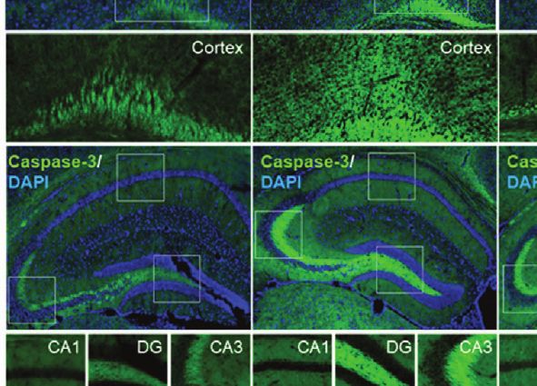
Kim et al.                                                                                          The Effects of Sorafenib on LPS-Induced Neuroinflammation

TABLE 1 | Sequences of primers used for RT-PCR.
                                                                                 5xFAD Mice
Gene name                                              Sequence                  5xFAD mice were used to determine the effects of sorafenib on
                                                                                 Ab-induced neuroinflammatory responses; these mice carry five
IL-1b               Sense            5’-AGC TGG AGA GTG TGG ATC CC-3’
                                                                                 familial AD mutations (APPSw, Lon, Flo, PS1M146L, L286V)
                    Antisense        5’-CCT GTC TTG GCC GAG GAC TA-3’
IL-6                Sense            5’-CCA CTT CAC AAG TCG GAG GC-3’
                                                                                 under the control of the Thy1 promoter, resulting in
                    Antisense        5’-GGA GAG CAT TGG AAA TTG GGG T-3’         overexpression of Ab. 5xFAD mice (Stock No. 34848-JAX;
COX-2               Sense            5’-GCC AGC AAA GCC TAG AGC-3’               B6.Cg-Tg (APPSwFlLon,PSEN1*M146L*L286V)6799Vas/
                    Antisense        5’-GCC TTC TGC AGT CCA GGT TC-3’            Mmjax) were purchased from Jackson Laboratory (Bar Harbor,
iNOS                Sense            5’-CCG GCA AAC CCA AGG TCT AC-3’
                                                                                 ME, USA). Genotyping of each mouse was performed using
                    Antisense        5’-GCA TTT CGC TGT CTC CCC AA-3’
GAPDH               Sense            5’-CAG GAG CGA GAC CCC ACT AA-3’            genomic DNA extracted from a tail snip. Only male mice were
                    Antisense        5’-ATC ACG CCA CAG CTT TCC AG-3’            used in this study.
                                                                                 Immunofluorescence Staining (IF)
TABLE 2 | Sequences of primers used for real time-PCR.                           The brains of wild-type and 5xFAD mice fixed as described above
Gene name                                              Sequence
                                                                                 were sectioned at a thickness of 30 mm with a cryostat microtome.
                                                                                 The sections were incubated with 10% normal goat serum (Vector
IL-1b               Sense             5’-TTG ACG GAC CCC AAA AGA TG-3’           Laboratories) for 1 h at room temperature, immunostained
                    Antisense         5’-AGG ACA GCC CAG GTC AAA G -3’
                                                                                 overnight at 4°C with primary antibodies (Iba-1, GFAP, COX-2,
IL-6                Sense             5’-CCA CGG CCT TCC CTA CTT C-3’
                    Antisense         5’-TTG GGA GTG GTA TCC TCT GTG A-3’
                                                                                 p-AKT, p-STAT3), and incubated with secondary antibodies for
COX-2               Sense             5’-CCA CTT CAA GGG AGT CTG GA -3’          2 h at room temperature. Images of sections mounted on glass
                    Antisense         5’-AGT CAT CTG CTA CGG GAG GA-3’           slides with DAPI (Vector Laboratories) were acquired by
iNOS                Sense             5’-GGA TCT TCC CAG GCA ACC A-3’            fluorescence microscopy (DMi8, Leica Microsystems, Wetzlar,
                    Antisense         5’-TCC ACA ACT CGC TCC AAG ATT-3’
                                                                                 Germany) and analyzed by ImageJ (NIH). Quantification was
GAPDH               Sense             5’-TGG GCT ACA CTG AGG ACC ACT-3’
                    Antisense         5’-GGG AGT GTC TGT TGA AGT CG-3’           performed using 2-3 brain slices per mouse and a total of 18-24
                                                                                 brain images/per group (4 mice/group). The primary and
                                                                                 secondary antibodies are listed in Table 3.
neuroinflammatory responses, wild-type mice were administered                     Western Blotting (WB)
LPS (10 mg/kg, i.p.) or PBS, and 30 min later, sorafenib (10 mg/kg,              The potential effects of sorafenib on LPS-mediated AKT and P38
i.p.) or vehicle was administered three times at 2-h intervals (i.e.,            signaling were assessed in BV2 microglial cells treated with 200
sorafenib was injected 30 min, 2.5 h, and 4.5 h after LPS or PBS                 ng/ml LPS or PBS for 45 min followed by 5 mM sorafenib or
injection). Eight hours after LPS injection, the mice were                       vehicle (1% DMSO) for 5.5 hr. The cells were then incubated in
anesthetized and transcardially perfused. The in vivo                            lysis buffer (ProPrep, iNtRON Biotechnology, Inc., Seongnam,
experimental design is summarized in Figures 3A, 6A, 8A.                         Korea) supplemented with protease and phosphatase inhibitor

TABLE 3 | List of antibodies used in this study.

Primary antibodies

Antigen                            Host species                   Dilution              Manufacturer                    Catalog no.            Analysis

Iba-1                              Rabbit polyclonal              1:500                     Wako                         019-19741                 IF
GFAP                               Rabbit polyclonal              1:500                  Neuromics                        RA22101                  IF
IL-1b                              Rabbit polyclonal              1:200                    Abcam                          AB9722                   IF
COX-2                              Rabbit polyclonal              1:500                    Abcam                          AB15191                  IF
p-AKTS473                          Rabbit polyclonal              1:500                 Cell Signaling                      9271                   WB/IF
AKT                                Rabbit polyclonal              1:500                 Cell Signaling                     9272S                   WB
p-STAT3S727                        Rabbit polyclonal              1:500                    Abcam                          AB86340                  ICC/IF
p-NF-kBS536                        Rabbit polyclonal              1:500                 Cell Signaling                     3033S                   ICC
p-P38T180/Y182                     Rabbit polyclonal              1:1000                   Abcam                            9211                   WB
P38                                Rabbit polyclonal              1:1000                   Abcam                            9212                   WB
CD11b                              Rat monoclonal                 1:200                    Abcam                          AB8878                   ICC
Secondary antibodies
Antibody                                                          Dilution              Manufacturer                    Catalog no.              Analysis
Goat anti-rabbit IgG, Alexa Fluor 488                              1:200                 Invitrogen                       A11008                   IF
Goat anti-rabbit IgG, Alexa Fluor 555                              1:200                 Invitrogen                       A28180                   IF, ICC
Goat anti-chicken IgG, Alexa Fluor 488                             1:500                 Invitrogen                       A11001                   IF
Goat anti-rat IgG, Alexa FITC                                      1:200                 Invitrogen                       A18866                   ICC
Goat anti-rabbit IgG, HRP conjugate                               1:10000                   Enzo                       ADI-SAB-300-J               WB
Goat anti-mouse IgG, HRP conjugate                                1:10000                   Enzo                       ADI-SAB-100-J               WB

Frontiers in Immunology | www.frontiersin.org                                4                                       May 2021 | Volume 12 | Article 684344
Kim et al.                                                                               The Effects of Sorafenib on LPS-Induced Neuroinflammation

for 5 min, followed by centrifugation at 12,000 rpm. The                DMSO) or 5 mM sorafenib for 5.5 h (Figure 1G). Sorafenib
supernatants were collected, and protein concentrations were            posttreatment significantly reduced the increase in COX-2 and
measured by the BSA protein assay. The quantified proteins were          IL-1b mRNA levels under LPS stimulation but did not alter IL-6
mixed with 4x SDS sample buffer and separated by                        and iNOS levels (Figures 1H, I and Supplementary Figure 1B).
electrophoresis on an 8% SDS-PAGE gel for 2 h. The proteins             Thus, either pre-treatment or post-treatment with sorafenib can
were then transferred to a polyvinylidene difluoride (PVDF)              regulate the LPS-evoked increase in proinflammatory cytokines in
membrane, followed by blocking with 5% skim milk (for total             microglial cells.
AKT and total P38) or 5% BSA (for p-AKT and p-P38) for 1 h.
Next, the membranes were incubated with primary antibodies at
                                                                        Sorafenib Reduces LPS-Induced COX-2
4°C overnight, washed for 5 min four times with TBST, and
incubated with peroxidase-conjugated secondary antibodies for
                                                                        mRNA Levels in Primary Astrocytes
                                                                        The effects of sorafenib on the proinflammatory response were
1 h at RT. Finally, the membrane was washed with TBST for
                                                                        further investigated in primary astrocytes. The cells were first
5 min, and the target proteins were visualized using ECL
                                                                        treated with 200 ng/ml LPS or PBS for 30 min and then vehicle
Western Blotting Detection Reagent (GE Healthcare, Chicago,
                                                                        (1% DMSO) or 5 mM sorafenib for 5.5 h. Real-time PCR was
IL, USA). Fusion Capt Advance software (Vilber Lourmat) was
                                                                        performed to assess proinflammatory cytokine levels. Sorafenib
used for image analysis. The primary and secondary antibodies
                                                                        posttreatment significantly decreased the LPS-induced increase
are listed in Table 3.
                                                                        in COX-2 mRNA expression but not IL-1b, IL-6 and iNOS
Statistical Analysis                                                    mRNA levels (Figure 1J). In summary, sorafenib appears to
Comparisons of two groups were performed with unpaired two-             selectively regulate the LPS-induced increase in COX-2 mRNA
tailed t-tests with Welch’s correction; multiple comparisons were       expression in primary astrocytes.
performed by one-way ANOVA (parametric or non-paramatric)
(Prism 7, GraphPad Software, USA). Post hoc analysis                    Sorafenib Suppresses LPS-Induced AKT/
was conducted with Tukey’s or Dunn’s multiple comparison                P38 Phosphorylation and Nuclear STAT3/
test; p < 0.05 was considered significant. The normal distribution
                                                                        NF-kB Phosphorylation
of data from in vitro and in vivo experiments was verified using
                                                                        AKT and the MAPK signaling kinase P38 play important roles
the Kolmogorov-Smirnov or Shapiro-Wilk normality test
                                                                        in glial cell activation by modulating the secretion of
(Supplementary Tables 2, 3). Data are presented as the mean
                                                                        proinflammatory cytokines (19). We recently reported that in
± SD (*p < 0.05, **p < 0.01).
                                                                        BV2 microglial cells, another anti-cancer drug and multikinase
                                                                        inhibitor, regorafenib, alters AKT and P38 signaling and LPS-
                                                                        induced neuroinflammation (8). To assess the ability of sorafenib
RESULTS                                                                 to regulate AKT and/or P38 signaling in vitro, BV2 microglial cells
                                                                        were treated with 200 ng/ml LPS or PBS for 45 min
Sorafenib Decreases LPS-Induced COX-2                                   before treatment with 5 mM sorafenib or vehicle (1% DMSO)
and IL-1b mRNA Levels in Microglial Cells                               for 45 min. AKT and P38 phosphorylation were measured by
The anti-cancer drug sorafenib is a multi-target kinase inhibitor       Western blotting of total extracted proteins with anti-p-AKTS473/
(Figure 1A). In the present study, we examined the effects of           AKT and anti-p-P38T180/Y182/P38 antibodies (Figure 2A).
sorafenib on neuroinflammation. First, mitochondrial arrest              Sorafenib posttreatment significantly decreased the LPS-induced
resulting from sorafenib cytotoxicity was assessed in vitro in          increases in p-AKTS473 and p-P38T180/Y182 without changing the
BV2 microglial cells using the MTT assay. No cytotoxicity was           total levels of AKT and P38 induced by LPS in this cell line
observed after 6 or 24 h at sorafenib concentrations of 0.1 to          (Figures 2B, C and Supplementary Figure 2).
10 mM, but cytotoxicity was evident at 20–25 mM sorafenib                   We then investigated the potential involvement of the
(Figures 1B, C). Based on these data, we selected a sorafenib           transcription factors STAT3 and NF-kB in sorafenib-associated
concentration of 5 mM as an intermediate concentration with no          neuroinflammatory responses. BV2 microglial cells were first
apparent cytotoxicity for all subsequent in vitro experiments.          treated with 200 ng/ml LPS or PBS for 30 min before treatment
   To investigate the effects of sorafenib on the proinflammatory        with 5 mM sorafenib or vehicle (1% DMSO) for 5.5 h
response induced by LPS, BV2 microglial cells were exposed first         (Figure 2D). Subsequent immunocytochemistry analysis with
to vehicle (1% DMSO) or 5 mM sorafenib for 30 min and then              anti-CD11b and anti-p-STAT3 S727 or anti-p-NF-kB S536
200 ng/ml LPS or PBS for 5.5 h. Proinflammatory cytokine levels          antibodies revealed that sorafenib posttreatment significantly
were assessed by RT-PCR (Figure 1D). Sorafenib pretreatment             reduced the LPS-induced increase in nuclear p-STAT3S727
prevented the increase in COX-2 and IL-1b mRNA levels evoked            (Figure 2E). Interestingly, sorafenib posttreatment also
by LPS but did not alter IL-6 and iNOS mRNA levels                      significantly decreased LPS-induced nuclear p-NF-kBS536 levels
(Figures 1E, F and Supplementary Figure 1A).                            (Figure 2F). In summary, sorafenib affects LPS-induced p-AKT/
   To examine the potential therapeutic influence of sorafenib on        p-P38-linked signaling and its associated transcription factors, p-
the proinflammatory response, BV2 microglial cells were exposed          STAT3 and p-NF-kB, to modulate neuroinflammatory responses
first to 200 ng/ml LPS or PBS for 30 min and then to vehicle (1%         in microglial cells.

Frontiers in Immunology | www.frontiersin.org                       5                                     May 2021 | Volume 12 | Article 684344
Kim et al.                                                                                                 The Effects of Sorafenib on LPS-Induced Neuroinflammation

                                A

                                B

                                C

                                D

                                E

                                F

 FIGURE 2 | Sorafenib downregulates the LPS-induced increases in p-AKT/p-P38 and nuclear p-STAT3/p-NF-kB levels. (A) Scheme for sequential treatment of BV2
 cells with LPS and sorafenib. (B, C) Western blotting analysis of BV2 cells treated as described in (A) with anti-p-AKTS473, anti-AKT, anti-p-P38T180/Y182, and anti-
 P38 antibodies (n=6/group). (D) Scheme for sequential treatment of BV2 cells with LPS and sorafenib. (E, F) Immunocytochemistry analysis of BV2 cells treated as
 described in (D) with anti-CD11b and anti-p-STAT3S727 antibodies (number of cells (n); Vehicle, n=428; LPS, n=357; LPS+sorafenib, n=361) or anti-CD11b and anti-
 p-NF-kBSer536 antibodies (Vehicle, n=461; LPS, n=584; LPS+sorafenib, n=416). *p < 0.05, **p < 0.01, Scale bar = 20 mM.

Sorafenib Pretreatment Inhibits LPS-                                                   consecutive days. Thirty minutes after the final injection of
Induced Microgliosis in Wild-Type Mice                                                 sorafenib, LPS (10 mg/kg, i.p.) or PBS was administered, and
To examine the effects of sorafenib on LPS-evoked gliosis in vivo,                     immunofluorescence staining of brain sections was performed
we selected a sorafenib dose of 10 mg/kg based on previous                             with an antibody against caspase-3, a marker of apoptotic cell
studies (20–22). To assess the potential toxicity of this dose of                      death (Figure 3A). We found that sorafenib pretreatment
sorafenib in vivo, wild-type mice were intraperitoneally (i.p.)                        significantly decreased the LPS-induced increase in caspase-3
injected with sorafenib (10 mg/kg/day) or vehicle daily for 3                          levels in the cortex but had no significant effect in the

Frontiers in Immunology | www.frontiersin.org                                      6                                           May 2021 | Volume 12 | Article 684344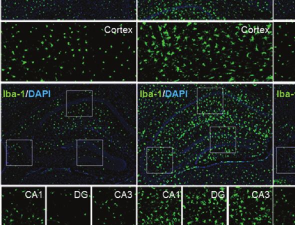
Kim et al.                                                                                                 The Effects of Sorafenib on LPS-Induced Neuroinflammation

                                                  A

                        B

                                                                                           C

                        D                                                                  E

 FIGURE 3 | Pretreatment of sorafenib inhibits LPS-induced microgliosis in wild-type mice. (A) Scheme for treatment of wild-type mice with sorafenib followed by
 LPS. (B, D) Immunofluorescence staining of brain slices from wild-type mice treated as described in (A) with an anti-caspase-3 and anti-Iba-1 antibody.
 (C, E) Quantification of the data in (B, D) (analyzed number of brain slices/images (n); Caspase-3: Vehicle, n=21; LPS, n=18; Sorafenib + LPS, n=21, Iba-1: Vehicle,
 n=18; LPS, n=19; Sorafenib + LPS, n=19). *p < 0.05, **p < 0.01. Scale bar = 200 mM.

hippocampus (Figures 3B, C). According to these results, 10 mg/                       induced increases in microglial kinetics, morphological activity,
kg sorafenib does not appear to have cytotoxic effects in                             and migration to sites of inflammation in the brain.
the brain.
    Next, to determine the effects of sorafenib on LPS-induced                        Pretreatment of Sorafenib Suppresses
microglial activation in vivo, we assessed levels of ionized                          LPS-Induced Astroglial Kinetics,
calcium-binding adapter molecule 1 (Iba-1), a critical marker of                      Morphological Activity, and Migration and
microglial activation in vivo that initiates neuroinflammation                         COX-2 Levels in Wild-Type Mice
defense mechanisms (23). Pretreatment of sorafenib                                    Astrocytes are neuron-supporting cells that modulate the LPS-
significantly decreased the LPS-induced increase in Iba-1                              mediated neuroinflammatory response by regulating nervous
immunofluorescence intensity in the cortex and hippocampus                             system repair (24). Astrocyte activation in response to
(CA1, DG, and CA3) (Figures 3D, E). Consistent with this                              infection or inflammation cues is essential for pathogen
finding, the number of Iba-1-positive cells and the percentage                         clearance and proinflammatory cytokine release and involves
of the area that was stained in the cortex and hippocampus were                       changes in astrocyte kinetics, morphology, and migration (25).
significantly reduced in mice administered sorafenib after LPS                         To determine if sorafenib alters LPS-evoked astrogliosis in vivo,
induction (Figures 3D, E). Thus, sorafenib downregulates LPS-                         glial fibrillary acidic protein (GFAP) immunofluorescence

Frontiers in Immunology | www.frontiersin.org                                     7                                           May 2021 | Volume 12 | Article 684344
Kim et al. The Effects of Sorafenib on LPS-Induced Neuroinflammation
intensity, the number of GFAP-activated cells, and the These data confirm that sorafenib regulates LPS-mediated
percentage of GFAP-stained area were measured in brain COX-2 levels in wild-type mice.
sections from wild-type mice injected as described above with
10 mg/kg sorafenib or vehicle followed by 10 mg/kg LPS or PBS.
Pretreatment of sorafenib significantly reduced the LPS-induced Pretreatment of Sorafenib Decreases
increases in GFAP immunofluorescence intensity and area of AKT and STAT3 Phosphorylation in
staining in the cortex and hippocampus (CA1, DG, and CA3) Wild-Type Mice
(Figures 4A, B). However, the LPS-induced increase in the The induction of AKT and STAT3 signaling by LPS has been linked
number of GFAP-labeled cells was only decreased in the cortex to the regulation of microglial and astrocyte activation in vivo (26).
by sorafenib treatment (Figures 4A, B). These data indicate that The effects of sorafenib on neuroinflammation-mediated
sorafenib reduces LPS-induced activation of astrocyte kinetics, phosphorylation of AKT and STAT3 were assessed in wild-type
morphological changes, and atrocytic migration in the wild-type mice treated as described in Figure 3A with 10 mg/kg sorafenib or
mouse brain. vehicle followed by 10 mg/kg LPS or PBS. Immunofluorescence
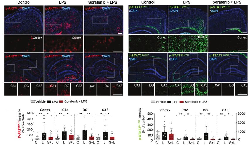
To verify the effects of sorafenib on the LPS-induced increase staining of brain sections was performed using anti-p-AKTS473 and
in COX-2 levels observed in Figure 1, brain sections from wild- anti-p-STAT3S727 antibodies. Pretreatment of sorafenib significantly
type mice treated as described in Figure 3A were subjected to reduced p-AKTS473 levels in the cortex and hippocampus stimulated
immunofluorescence staining with an anti-COX-2 antibody. by LPS (Figures 5A, B). In addition, pretreatment of sorafenib had
Pretreatment of sorafenib significantly reduced the LPS-induced no effect on LPS-induced p-STATS727 levels in the cortex but
increase in COX-2 immunofluorescence intensity in the cortex significantly reduced hippocampal LPS-induced p-STATS727 levels
and hippocampus (CA1, DG, and CA3) (Figures 4A–C). compared with LPS treatment (Figures 5C, D). Thus, sorafenib
A
B C
FIGURE 4 | Pretreatment of sorafenib suppresses LPS-induced astrogliosis and COX-2 levels in wild-type mice. (A) Immunofluorescence staining with anti-GFAP
and anti-COX-2 antibodies of brain slices from wild-type mice treated with sorafenib followed by LPS. (B, C) Quantification of the data in (A) (analyzed number of
brain slices/images (n); GFAP: Vehicle; n=16; LPS, n=18; Sorafenib + LPS, n=17, COX-2: Vehicle, n=16; LPS, n=18; Sorafenib + LPS, n=17). *p < 0.05, **p < 0.01,
Scale bar = 200 mM.
Frontiers in Immunology | www.frontiersin.org 8 May 2021 | Volume 12 | Article 684344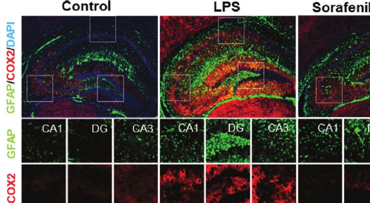
Kim et al.                                                                                             The Effects of Sorafenib on LPS-Induced Neuroinflammation

         A                                                                      C

         B                                                                      D

 FIGURE 5 | Pretreatment of sorafenib decreases LPS-mediated AKT and STAT3 phosphorylation in wild-type mice. (A, C) Immunofluorescence staining with anti-p-
 AKTS473 and anti-p- STAT3S727 antibody of brain slices from wild-type mice treated with sorafenib followed by LPS. (B, D) Quantification of the data in (A, C)
 (analyzed number of brain slices/images (n); p-AKT S473: Vehicle, n =16; LPS, n=16; Sorafenib + LPS, n=14, p-STAT3 S727, Vehicle, n=23; LPS, n=22; Sorafenib +
 LPS, n=20). *p < 0.05, **p < 0.01, Scale bar = 200 mM.

pretreatment modulates the phosphorylation of AKT and STAT3 in                      the cortex and hippocampus (Figures 6D, E). In addition, the
a brain region-specific manner in wild-type mice injected with LPS.                  LPS-mediated increase in GFAP-labeled cells and the percent
                                                                                    area of staining were significantly decreased in the hippocampus
                                                                                    but not the cortex (Figures 6D, E) in the presence of sorafenib.
Posttreatment of Sorafenib Inhibits LPS-                                            These data indicate that posttreatment with sorafenib
Induced Microgliosis and Astrogliosis in                                            downregulates the neuroinflammatory response by inhibiting
Wild-Type Mice                                                                      glial activation in the brain.
Since exposure to sorafenib before LPS injection regulates LPS-
mediated gliosis in vivo, we investigated whether posttreatment                     Posttreatment of Sorafenib Regulates
of sorafenib alters LPS-evoked neuroinflammation. As described                       LPS-Induced AKT Phosphorylation in
in Figure 6A, wild-type mice were i.p. injected with 10 mg/kg                       Wild-Type Mice
LPS or PBS followed 30 min later by three i.p. injections of 10                     To determine if posttreatment of sorafenib affects LPS-evoked
mg/kg sorafenib or vehicle at 2 h intervals. Eight hours after the                  neuroinflammatory-associated signaling, wild-type mice were
initial LPS or PBS injection, the mice were sacrificed, and                          injected with 10 mg/kg LPS or PBS and then injected with 10
immunofluorescence staining of brain sections was performed                          mg/kg sorafenib or vehicle as described in Figure 6A.
with an anti-Iba-1 or anti-GFAP antibody (Figure 6A).                               Immunofluorescence staining of brain sections from the mice
Posttreatment of sorafenib significantly decreased the LPS-                          was performed with anti-p-AKTS473 and anti-p-STAT3S727
induced increase in Iba-1 immunofluorescence intensity in the                        antibodies. Posttreatment with sorafenib significantly decreased
cortex and hippocampus (CA1, DG, and CA3) (Figures 6B, C).                          LPS-induced increase in AKT phosphorylation in cortex and
The number of Iba-1-positive cells and the percentage of the                        DG but not CA1 and CA3 (Figures 7A, B). Moreover,
stained area in the cortex and hippocampus were also                                posttreatment of sorafenib had no effect on LPS-induced p-
significantly decreased when LPS injection was followed by                           STATS727 levels in the cortex and hippocampus (Figures 7C, D).
sorafenib treatment in wild-type mice (Figures 6B, C). Similar                      These data indicate that posttreatment with sorafenib modulates
to Iba-1, posttreatment of sorafenib significantly reduced the                       AKT signaling but not STAT3 to alter LPS-mediated
LPS-induced increase in GFAP immunofluorescence intensity in                         neuroinflammation in wild-type mice.

Frontiers in Immunology | www.frontiersin.org                                  9                                         May 2021 | Volume 12 | Article 684344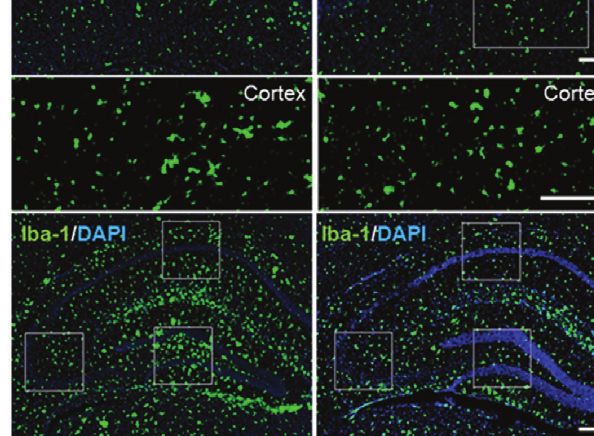
Kim et al.                                                                                                 The Effects of Sorafenib on LPS-Induced Neuroinflammation

                                           A

                        B                                                                   C

                        D                                                                  E

 FIGURE 6 | Posttreatment of sorafenib inhibits LPS-induced microgliosis and astrogliosis in wild-type mice. (A) Scheme for the treatment of wild-type mice with LPS
 followed by sorafenib. (B, D) Immunofluorescence staining with anti-Iba-1 and anti-GFAP antibodies of brain slices from wild-type mice treated as described in (A).
 (C, E) Quantification of the data in (B, D) (analyzed number of brain slices/images (n); Iba-1: Vehicle, n=21; LPS, n=23; LPS + Sorafenib, n=18, GFAP, Vehicle, n=23;
 LPS, n=22; LPS + Sorafenib, n=20). **p < 0.01. Scale bar = 200 mM.

Sorafenib Upregulated the LPS-Mediated                                                mice treated with LPS (Supplementary Figures 3A, B), and a
Decrease in Shank-1 Intensity in                                                      trend toward increased PSD-95 immunofluorescence intensity
Wild-Type Mice                                                                        was observed (Supplementary Figures 3C, D). Interestingly,
Systemic inflammation and neuroinflammation can impact                                  pre-exposure to sorafenib rescued the LPS-induced decrease in
cognitive and synaptic function (27). Since pre-and                                   shank-1 immunofluorescence intensity in the cortex and
posttreatment of sorafenib decreased LPS-mediated gliosis in                          hippocampal DG (Supplementary Figures 3E, F). These data
wild-type mice, we examined whether sorafenib modulates                               suggest that sorafenib pretreatment may positively or negatively
learning and memory-related proteins. For these experiments,                          modulates synaptic function in LPS-induced wild-type mice.
wild-type mice were injected with sorafenib followed by
LPS as described in Figure 3A, and brain sections were                                Sorafenib Suppresses Ab-Mediated
immunostained with anti-synaptophysin (a presynaptic                                  Astrogliosis in 5xFAD Mice
marker), anti-PSD-95 (a postsynaptic marker), or anti-shank-1                         Both pre- and post-treatment with sorafenib effectively
antibodies. Sorafenib pretreatment did not alter the                                  downregulated LPS-mediated neuroinflammation in vitro
immunofluorescence intensity of synaptophysin in wild-type                             and in vivo. To determine the effects of sorafenib on

Frontiers in Immunology | www.frontiersin.org                                    10                                           May 2021 | Volume 12 | Article 684344
Kim et al. The Effects of Sorafenib on LPS-Induced Neuroinflammation
A B
C D
FIGURE 7 | Posttreatment of sorafenib diminishes LPS-evoked AKT phosphorylation in wild-type mice. (A, C) Immunofluorescence staining with anti-p-AKTS473 and
anti-p-STAT3S727 antibodies of brain slices from wild-type mice treated with LPS followed by sorafenib. (B, D) Quantification of the data in A, C (analyzed number of
brain slices/images (n); p-AKT S473: Vehicle, = 14; LPS, n=24; LPS + Sorafenib, n=18, p-STAT3 S727, Vehicle, n=13; LPS, n=14; LPS + Sorafenib, n=20). *p < 0.05,
**p < 0.01, Scale bar = 200 mM.
neuroinflammatory responses in a mouse model of Alzheimer’s activation was also reduced by pre- or posttreatment of
disease (AD), 5xFAD mice were treated with sorafenib (10 mg/ sorafenib, although pretreatment of sorafenib effect on
kg/day, i.p.) or vehicle daily for 3 consecutive days, and astrocyte number. In wild-type mice, sorafenib administration
immunofluorescence staining were conducted with an anti-Iba- significantly reduced the LPS-induced increase in COX-2 levels
1 or anti-GFAP antibody (Figure 8A). Three consecutive days of by altering AKT/STAT3 signaling. Moreover, in 5xFAD mice,
sorafenib administration did not alter Iba-1 immunofluorescence sorafenib treatment (daily for 3 days) significantly suppressed
intensity and the percent area stained (Figures 8B, C). However, astrogliosis but not microgliosis. Taken together, our findings
Ab-induced GFAP immunofluorescence intensity in the cortex suggest that sorafenib could be a novel therapy for relieving
and hippocampus was significantly reduced (CA1, DG, and neuroinflammatory response-associated glial activation in
CA3) (Figures 8D, E). Consistent with these observations, the brain.
sorafenib administration significantly decreased the percent We and others have recently found that several small
area of GFAP in the hippocampus (CA1, DG, and CA3) but compounds and herbal extracts can modulate LPS-evoked
not the cortex (Figures 8D, E). These data suggest that 3 neuroinflammatory responses in vitro and in vivo. For
consecutive days of sorafenib administration selectively instance, the multitarget kinase inhibitor dasatinib, whose
modulates Ab-mediated astrogliosis in the brains of targets include Bcr-Abl and the Src kinase family, decreases
5xFAD mice. LPS-induced COX-2 and IL-6 levels via the AKT/STAT3
signaling pathway (6). Apamin (APM), a selective antagonist
of small conductance calcium-activated potassium (SK)
channels, inhibits LPS-stimulated TLR4 activation throughout
DISCUSSION CaMKII/ERK and NF-kB/STAT3 phosphorylation in BV2 and
primary microglial cells (28). In addition, ALWPs, a mixture of
In this study, we demonstrated that sorafenib, a multikinase Antler and LWPs, suppresses the LPS-induced increase in IL-1b
inhibitor and anti-cancer drug, decreases the levels of by downregulating FAK/NF-kB signaling pathways (29). These
proinflammatory cytokines as well as microglial and astrocyte findings suggest that LPS-mediated neuroinflammation could be
activation induced by LPS. In BV2 microglial cells, sorafenib regulated by various signaling pathways linked to drug targets.
diminished the LPS-induced increases in COX-2 and IL-1b levels In the present study, we investigated the effects of sorafenib
by modulating P38/AKT and NF-kB/STAT3 signaling. Sorafenib on neuroinflammation. The kinase targets of sorafenib include
also decreased the LPS-induced increase in COX-2 levels in Raf-1, VEGFR2 (Flk1), and PDGFRb (30), which are all potent
primary astrocytes. In vivo, pre- and posttreatment of inducers of proinflammatory cytokine release from microglial
sorafenib decreased LPS-induced changes in microglial cells and play significant roles in microglial-associated
kinetics, number, and morphology. LPS-induced astrocyte neuroinflammatory responses (31–33). For instance, high-dose
Frontiers in Immunology | www.frontiersin.org 11 May 2021 | Volume 12 | Article 684344Kim et al. The Effects of Sorafenib on LPS-Induced Neuroinflammation
A
B C
D
E
FIGURE 8 | Sorafenib suppresses Ab-mediated astrogliosis in 5xFAD mice. (A) Scheme for the treatment of 5xFAD mice with sorafenib. (B, D) Immunofluorescence
staining with anti-Iba-1 and anti-GFAP antibodies of brain slices from 5xFAD mice treated as described in (A). (C, E) Quantification of the data in (B, D) (analyzed
number of brain slices/images (n); Vehicle: =21, Sorafenib: n= 18). **p < 0.01. Scale bar = 200 mM.
infusion of VEGF-A, which binds VEGFR2, leads to the release The effects of multikinase inhibitors, including those targeting
of the proinflammatory cytokine MIP-1 alpha in the adult rat VEGFRs and PDGFRb (e.g., axitinib, nintedanib, dabrafenib,
cortex (34). In cultured T cells, VEGF treatment induces the regorafenib), are known to be associated with proinflammatory
secretion of interferon gamma (IFN-g) (35). In addition, oral cytokine release (38). For example, treatment of primary motor
administration of didymin (an activator of Raf-1 kinase inhibitor cortical neurons with dabrafenib (an inhibitor of C-Raf/Raf-1
protein (RKIP)) increases levels of TNF-a, IL-6, and IL-1b in and B-Raf) reduces TNF-a and IL-12 levels (39). Regorafenib,
liver tissues and RAW 264.7 cells (36). In the brains of mice, another multikinase inhibitor that inhibits VEGFR2 and
intra-ipsilateral and contralateral infusion of the PDGFRb PDGFRb, strongly reduces the LPS-induced increases in COX-
inhibitor Greevec decreases the intracerebral hemorrhage- 2, IL-1b, IL-6, and TNF-a mRNA expression in BV2 microglial
induced increase in TNF-a levels (37). Overall, these findings cells (8). IL-6, TNF-a, and IFN-g expression in melanoma cells
suggest that the effects of sorafenib on LPS-induced are also suppressed by axitinib, a selective inhibitor of VEGFRs
proinflammatory cytokine release may occur via modulation of and PDGFRs (40). In addition, the VEGFR, PDGFR and
Raf-1, VEGFR and/or PDGFRb signaling. fibroblast growth factor receptor (FGFR) inhibitor nintedanib
Frontiers in Immunology | www.frontiersin.org 12 May 2021 | Volume 12 | Article 684344Kim et al. The Effects of Sorafenib on LPS-Induced Neuroinflammation
reduces the secretion of several proinflammatory and fibrotic induction of IL-1b and COX-2 mRNA expression (50). Here,
cytokines (IL-1b, IL-8, IL-10 and CXCL13) in M1 macrophages we showed that sorafenib, an inhibitor of VEGFRs and PDGFRb,
(41). Here, we found that pre- and post-treatment with sorafenib also significantly reduces AKT phosphorylation in BV2 cells
significantly reduced the increases in COX-2 and IL-1b mRNA (Figure 2B), suggesting that sorafenib modulates LPS-mediated
levels in BV2 microglial cells treated with LPS (Figure 1). Taken AKT signaling to alter neuroinflammatory responses in microglia.
together, the present findings and previous work suggest that After identifying the involvement of LPS-linked AKT signaling
sorafenib downregulates proinflammatory cytokine mRNA in the effects of sorafenib in microglia, we next investigated
levels by inhibiting VEGFR2 and/or PDGFRb activation. In whether sorafenib modulates P38 signaling, a key pathway in
future work, we will determine whether sorafenib regulates the production of inflammatory mediators (51). For instance,
LPS-induced proinflammatory cytokine release by inhibiting activation of p-P38T180/Y182 by Ras-Raf kinases stimulates the
Raf-1, VEGFR2, and PDGFRb simultaneously or individually. release of proinflammatory cytokines from microglia (52, 53),
Astrocytes control multiple processes in the CNS, including and the VEGFRs inhibitor nintedanib decreases p-P38
synaptogenesis, neuronal differentiation, neuronal survival and immunoreactivity in GC7901 and MKN45 cells (54). Co-
neuroinflammation (42). Under conditions of inflammation, treatment of non-small cell lung carcinoma (NSCLC) cells with
astrocytes communicate with microglia and regulate extracellular VEGF and vandetanib or axitinib, both VEGFR inhibitors,
proinflammatory cytokine homeostasis (42). Previous studies have significantly suppresses the phosphorylation of P38 in a dose-
reported that proinflammatory cytokine levels are elevated in LPS- dependent manner (55). In addition, the PDGFRb inhibitor
induced rat and primary astrocytes (6, 7). Interestingly, the mRNA AG1295 decreases P38 phosphorylation in aortic vascular
and protein levels of VEGFRs are elevated in glioblastoma cells, and smooth muscle cells (56). Interestingly, in the present study,
soluble VEGFR1 is increased in astrocytic tumor cells compared sorafenib suppressed the LPS-induced increase in p-P38T180/Y182
with normal astrocytes (43, 44). Similarly, phosphorylation of in BV2 cells (Figure 2C). These data indicate that sorafenib
PDGFRb (tyrosine 75) is significantly increased in astrocytes regulates P38 phosphorylation by inhibiting VEGFRs and/or
located near breast cancer cells compared with normal astrocytes PDGFRb-linked P38 signaling in microglial cells. Further studies
(45). Moreover, we recently demonstrated that regorafenib, an will reveal whether sorafenib affects other signaling pathways
inhibitor of VEGFRs and PDGFRb, suppresses the LPS-induced linked to VEGFRs and/or PDGFRb in response to
increase in COX-2 mRNA expression in primary astrocyte culture neuroinflammatory responses in microglia.
(8). However, the functions of VEGFR2 and/or PDGFRb remain to The expression of proinflammatory cytokines is
be verified using target-specific blockade or epigenetic knockdown transcriptionally regulated by phosphorylated STAT3 and NF-
in astrocytes. In the current study, sorafenib only decreased the LPS- kB in the nucleus in microglia (57). Several studies have
induced increase in mRNA levels of COX-2 mRNA and not other demonstrated that activated P38 and AKT phosphorylate
proinflammatory cytokines in primary astrocytes. Why do the STAT3 at S727 and/or NF-kB at S536 (6, 7). Phosphorylation
multikinase inhibitors sorafenib and regorafenib affect only LPS- at S727 enhances the transcriptional activity of STAT3 as well as
induced COX-2 mRNA levels? It is possible that multikinase the transcript levels of several proinflammatory cytokines (i.e.,
inhibitors mainly inhibit the activation of VEGFRs and PDGFRb TNF-a, IL-1b, IL-6, and COX-2) in microglial cells (6, 7).
in astrocytes and critically regulate COX-2 gene expression in Similarly, phosphorylation of p65, a component of NF-kB, at
response to LPS. Thus, sorafenib may modulate LPS-induced glial S536 induces NF-kB import into the nucleus and activation of
proinflammatory cytokine expression by regulating the activities of LPS-mediated proinflammatory cytokine transcription (58). A
VEGFRs and/or PDGFRb. PDGFb-specific inhibitor, TKI258, reduces the PDGF-b-induced
LPS stimulates Toll-like receptor 4 (TLR4) signaling pathways increase in STAT3 phosphorylation in MiaPaCa2 pancreatic
to induce proinflammatory cytokine release by microglia and cancer cells and endothelial cells (59). Interestingly, VEGFR2
astrocytes (5). AKT and MAPK signal transduction are among overexpression increases the DNA binding affinity of NF-kB,
the main signaling pathways activated by TLR4 and share signals whereas the VEGFR2 inhibitors sunitinib and bevacizumab
with VEGFR2 and/or PDGFRb stimulation (5, 46, 47). AKT is a suppress DNA binding by NF-kB in endothelial cells (60).
serine/threonine-specific protein kinase that is phosphorylated at Moreover, the VEGFRs and PDGFRb inhibitor vorolanib
S473 in the C-terminus or T308 in the kinase domain and plays a diminishes STAT3 and NF-kB phosphorylation in a dose-
key role as a multiple activator of LPS-induced signaling in dependent manner in NSCLC cells and xenograft mice (49).
microglia (48). Vorolanib, an inhibitor of VEGFRs and We recently demonstrated that the multikinase inhibitor
PDGFRs, significantly reduces p-VEGFR2 and p-AKT levels in a regorafenib decreases nuclear p-STAT3S727 and p-NF-kBS536
dose-dependent manner in human umbilical vein endothelial cells levels in LPS-treated BV2 cells (8). Consistent with these
(HUVECs) (49). Interestingly, combination treatment with observations, in the present study, sorafenib significantly
vorolanib and gefitinib (an EGFR inhibitor) increases EGFR decreased the LPS-stimulated increases in p-STAT3S727 and
mutation and inhibits angiogenesis by downregulating VEGFR- p-NF-kBS536 levels in BV2 cells. Thus, it is possible that
linked AKT-STAT3 signaling (49). In addition, the VEGFRs and sorafenib suppresses LPS-evoked p-STAT3 and p-NF-kB levels
PDGFRb inhibitor regorafenib significantly reduces the increase by inhibiting VEGFRs and PDGFRb signaling in microglia.
in AKT phosphorylation in LPS-stimulated BV2 cells (8), and the In vivo, neuroinflammation is initiated by the activation of
resulting decrease in AKT activation in turn decreases the microglia and astrocytes to protect damaged neurons (2). Iba-1 is
Frontiers in Immunology | www.frontiersin.org 13 May 2021 | Volume 12 | Article 684344Kim et al. The Effects of Sorafenib on LPS-Induced Neuroinflammation specifically expressed in microglia and is upregulated during activation by inhibiting VEGFRs and PDGFRb. Conversely, it microglial activation (i.e., changes in microglial morphology and is also possible that sorafenib posttreatment inhibits LPS- location) in the brain (2). Similar to microglia, astrocytes can induced TLR4 activation to prevent VEGFRs and PDGFRb change their morphology, size, and mobility and become signaling and alter LPS-mediated neuroinflammatory hypertrophic and hyperplastic upon LPS injection or other responses. Interestingly, we observed that pretreatment but not injury (61). In assessing the effects of sorafenib pre- or posttreatment of sorafenib altered the number of astrocytes in posttreatment on glial cell activation in wild-type mice in vivo, the hippocampus in wild-type mice (Figures 4, 6). One we found that pre- and posttreatment of sorafenib decreased the possibility is that VEGFRs and PDGFRb have limited increases in Iba-1 immunofluorescence intensity, Iba-1-positive involvement in astrocyte migration, and thus pretreatment of cell number, and percent of staining area induced by LPS sorafenib has less of an effect on LPS-induced astrocytic (Figure 3, 6). Pretreatment with sorafenib had similar effects neuroinflammation than posttreatment in vivo. In future work, on GFAP immunofluorescence intensity and percent of stained we will further examine whether pre- and posttreatment of area (Figure 4). Additionally, posttreatment of sorafenib sorafenib alters LPS-evoked astrocytic neuroinflammatory significantly suppressed GFAP immunofluorescence intensity, responses in VEGFRs- and/or PDGFRb-dependent manner in cell number, and percent of stained area in LPS-treated wild-type wild-type mice. mice (Figure 6). How does pre- or posttreatment with sorafenib COX-2 is a typical proinflammatory marker and is released by modulate the neuroinflammatory responses induced by LPS in activated microglia and astrocytes at the beginning of vivo? Several studies have reported that multikinase inhibitors neuroinflammation (64). COX-2 expression supports the targeting VEGFRs and PDGFRs downregulate glial activation (8, inflammatory process and has a significant role in cell 62). For instance, regorafenib suppresses the LPS-induced proliferation, macrophage, and synoviocyte activation (2). increases in Iba-1 and GFAP immunofluorescence intensity in Systemic exposure to LPS significantly increases COX-2 wild-type mice (8), and in RIP-Tag2 mice, injection with mRNA and protein expression in the hippocampus and cortex VEGFRs and PDGFRb shRNAs significantly decreases Iba-1 of mice (6, 7). In a mouse model of angiogenesis, injection of a and GFAP immunointensity (62). Another VEGFRs and VEGFR2 inhibitor, microRNA-101, diminishes COX-2 PDGFRs inhibitor, dabrafenib, significantly increases LPS- expression (65), and in human intestinal microvascular induced neuroinflammatory response-linked cell survival by endothelial cells, curcumin reduces COX-2 mRNA levels by inhibiting the hyperpermeability and leukocyte migration of inhibiting VEGF (66). In addition, in rat smooth muscle cells, blood cells in C57BL/6 mice (63). Given these previous PDGF-induced expression of COX-2 is reduced by PDGFRb observations, our findings suggest that pre- and posttreatment inhibitors, and recombinant rat COX-2 cDNA is directly with sorafenib affects LPS-mediated microglial and astrocyte required for PDGFR-dependent stabilization of COX-2 mRNA, FIGURE 9 | Sorafenib affects LPS-stimulated neuroinflammatory responses in vitro and in vivo. In BV2 microglial cells and wild-type mice, sorafenib reduces the effects of LPS on the mRNA levels of the proinflammatory cytokines IL-1b and COX-2 in microglia and COX-2 in astrocytes by modulating AKT/P38-associated NF- kB/STAT3 signaling pathways. Accordingly, sorafenib may have therapeutic potential for neuroinflammation-related diseases. Frontiers in Immunology | www.frontiersin.org 14 May 2021 | Volume 12 | Article 684344
Kim et al.                                                                                    The Effects of Sorafenib on LPS-Induced Neuroinflammation

suggesting that PDGFR is required for regulating COX-2                       microglial activation (Figure 8). Further studies are needed to
expression (67). Importantly, we previously demonstrated that                determine if longer treatment periods (i.e., daily for 2 weeks or 4
the VEGFRs and PDGFRb inhibitor regorafenib significantly                     weeks) and/or higher doses of sorafenib are able to alter Ab-
decreases the increase in COX-2 levels induced by LPS in wild-               induced microgliosis.
type mice (8). Consistent with these findings, sorafenib treatment
reduced the LPS-induced increase in COX-2 levels in wild-type
mice (Figure 4). It is possible that multikinase inhibitors like             CONCLUSION
sorafenib and regorafenib regulate COX-2 expression via
inhibition of VEGFR2 and PDGFRb in response to                               In summary, sorafenib, a multikinase inhibitor whose targets
neuroinflammation in vivo.                                                    include VEGFR2 and PDGFRb, reduces the effects of LPS on
    Studies in LPS-injected mouse models have shown that AKT-                proinflammatory cytokine levels in BV2 cells and primary
linked STAT3 signaling contributes to the regulation of                      astrocytes (Figure 9). In addition, sorafenib suppresses the
proinflammatory responses in the brain (6, 7, 68). Xanthatin, a               AKT/P38-linked STAT3/NF-kB signaling pathway, which plays
VEGFRs and PDGFRb inhibitor, significantly reduces                            a role in proinflammatory cytokine release, in BV2 cells. In wild-
neuroinflammation by inhibiting AKT/PI3K/STAT3 signaling                      type mice, pre- and posttreatment of sorafenib significantly
in a rat corneal alkali burn model (69). TKI258, another VEGFRs              reduces the stimulation of microglial and astrocyte activation
and PDGFRs inhibitor, reduces p-AKT and p-STAT3 levels in                    and COX-2 levels by LPS by inhibiting AKT signaling, and
tumor xenograft nude mice (59). Similarly, we found that                     sorafenib suppresses Ab-mediated astrogliosis but not
sorafenib pretreatment decreased p-AKTS473 and p-STAT3S727                   microgliosis in a mouse model of AD. Thus, we suggest that
levels in LPS-injected wild-type mice (Figure 5). Interestingly,             sorafenib holds potential as a drug for protecting against acute
sorafenib posttreatment reduced p-AKTS473 levels in LPS-                     neuroinflammation in the brain.
injected wild-type mice but not p-STAT3 S727 levels (Figure 7).
Together, our findings and previous work suggest that sorafenib
pre-or posttreatments differently regulate VEGFRs- and/or                    DATA AVAILABILITY STATEMENT
PDGFRb-linked AKT and STAT3 signaling to modulate
                                                                             The original contributions presented in the study are included in
neuroinflammation in LPS-induced wild-type mice.
                                                                             the article/Supplementary Material. Further inquiries can be
    Neuroinflammation may impact learning and memory as well
                                                                             directed to the corresponding author.
as synaptic function both directly and indirectly (70). Conflicting
effects of sorafenib on cognitive/synaptic function have been
reported. In APPswe mice (a mouse model of AD), sorafenib                    ETHICS STATEMENT
treatment modulates neuroinflammatory responses to restore
working memory (15). On the contrary, negative effects of                    The animal study was reviewed and approved by Korea Brain
sorafenib on cognitive function via disruption of metabonomic                research institute IACUC.
pathways have been observed in cancer patients (71, 72).
Therefore, here we examined whether sorafenib alters LPS-
mediated pre- or postsynapse-linked proteins. In wild-type mice,             AUTHOR CONTRIBUTIONS
sorafenib treatment significantly reversed the LPS-mediated
                                                                             Study Conception and Design: JK and H-SH. Acquisition of data:
alteration of shank-l fluorescence intensity but had no effects on
                                                                             JK, J-HP, and SKP. Preparation of figures: JK. Preparation of
synaptophysin and PSD-95 (Supplementary Figure 3). Thus, the
                                                                             tables: JK. Writing of manuscript: JK and H-SH. All authors
present and previous findings indicate that sorafenib can regulate
                                                                             contributed to the article and approved the submitted version.
synaptic/cognitive function positively and/or negatively in LPS-
treated mice and/or mouse models of AD. A limitation of this
study is that mice were only i.p. treated with sorafenib daily for 3
                                                                             FUNDING
days at a dose of 10 mg/kg. This may not have been a sufficient
duration of treatment and/or dose to alter synaptic and cognitive            This work was supported by the KBRI basic research program
function. The effects of longer treatment periods and/or higher              through KBRI funded by the Ministry of Science, ICT & Future
doses of sorafenib on LPS-mediated synaptic and/or cognitive                 Planning (grant numbers 21-BR-02-11, 21-BR-03-05, H-SH) and
function will be assessed in future work.                                    the National Research Foundation of Korea (grant number
    Despite the availability of several models for evaluating the            2019R1A2B5B01070108, H-SH).
therapeutic potential of molecules (73, 74), the effects of sorafenib
on neuroinflammation have rarely been studied in non-LPS
models. To address this gap, we examined the effects of                      ACKNOWLEDGMENTS
sorafenib on neuroinflammation in 5xFAD mice, a model of
AD, which revealed that 3 consecutive days of treatments                     Confocal microscopy (Nikon, TI-RCP) data were acquired
significantly reduced Ab-mediated astroglial activation but not               in the Advanced Neural Imaging Center at the Korea

Frontiers in Immunology | www.frontiersin.org                           15                                     May 2021 | Volume 12 | Article 684344
You can also read


