Chemical fingerprints and microbial biomineralization of fish muscle tissues from the Late Cretaceous Múzquiz Lagerstätte, Mexico
←
→
Page content transcription
If your browser does not render page correctly, please read the page content below
Revista Mexicana de CienciasChemical
Geológicas, v. 30 núm.
fingerprints and2,microbial
2013, p. 417-435
biomineralization of fish muscle tissues 417
Chemical fingerprints and microbial biomineralization of fish muscle
tissues from the Late Cretaceous Múzquiz Lagerstätte, Mexico
Francisco Riquelme1, *, Jesús Alvarado-Ortega2, José Luis Ruvalcaba-Sil1,
Manuel Aguilar-Franco1, and Héctor Porras-Múzquiz3
1
Instituto de Física, Universidad Nacional Autónoma de México, A. P. 20-364, México, D.F., Mexico.
2
Instituto de Geología, Universidad Nacional Autónoma de México, Circuito de la Investigación S/N,
Ciudad Universitaria, Coyoacán, 04510 México, D.F., Mexico.
3
Museo Histórico de Múzquiz, Hidalgo 205, Centro, 26340 Múzquiz, Coahuila, Mexico.
* riquelme.fc@gmail.com
ABSTRACT
Fossil fish specimens from the Múzquiz Lagerstätte (Late Cretaceous) of northern México have
been analysed using UV light-induced visible fluorescence microscopy, Particle-induced X-ray Emission
(PIXE), X-Ray Diffraction (XRD), and Scanning Electron Microscopy (SEM). Specimens examined
with UV light microscopy show tightly packed trunk muscle tissues and digestive tract contents, as
well as a color gradient from pink to orange to brown associated with the chemical state of the muscle
tissues. PIXE analysis shows a 0.346 P/Ca ratio in muscle tissues, as well as a phosphorus increase by
a factor of more than four compared to surrounding sediment. Quantitative XRD analysis shows that
cryptocrystalline flourapatite (FAP) is the predominant mineral phase and calcite is complementary in
the muscle tissues. Nucleation of FAP and calcite may have occurred simultaneously with organic decay,
forming adhesive pellets in the soft watery carbonate mud, and caused immobilization of the carcasses.
Electron microscope scans show muscle tissues preserved with cellular and subcellular features as well
as digestive tract contents with calcareous nanoplankton. Fossil biofilms with bacteria have also been
exceptionally preserved as intact cells, casts and molds. This cell-specific, rapid mineralization can be
explained by a crystal seed process, which is discussed here.
Key words: fossil fish, exceptional preservation, PIXE spectrometry, biomineralization, Cretaceous,
Múzquiz, Mexico.
RESUMEN
Ejemplares de peces del Lagerstätte de Múzquiz (Cretácico Tardío) del norte de México han
sido analizados usando microscopía de fluorescencia visible inducida por luz UV, Emisión de Rayos X
Inducida por Partículas (PIXE), Difracción de Rayos X (DRX), y Micoscopía Electrónica de Barrido
(MEB). Los peces examinados preliminarmente por microscopía de luz UV muestran tejidos musculares
del torso y contenido del tracto digestivo, así como un gradiente de color de rosa-anaranjado-parduzco
asociado con el estado químico de los tejidos musculares. El análisis por medio de PIXE muestra
una relación de P/Ca de 0.346 en tejido muscular, así como un incremento en el contenido de fósforo
Riquelme, F., Alvarado-Ortega, J., Ruvalcaba-Sil, J.L., Aguilar-Franco, M., Porras-Múzquiz, H., 2013, Chemical fingerprints and microbial biomineralization
of fish muscle tissues from the Late Cretaceous Múzquiz Lagerstätte, Mexico: Revista Mexicana de Ciencias Geológicas, v. 30, núm. 2, p. 417-435.
418                                                                           Riquelme et al.

                         por un factor de más de cuatro en comparación con los sedimentos. Análisis cuantitativos de DRX
                         muestran que fluourapatita (FAP) criptocristalina es la fase mineral predominante, mientras calcita es
                         complementaria en los tejidos musculares. La nucleación de FAP y calcita pudo ocurrir simultáneamente
                         a la degradación orgánica y causó la inmovilización de los cadáveres formando pellets adhesivos en un
                         lodo de carbonatos blando y acuoso. Análisis de microscopía electrónica muestran tejidos musculares
                         con rasgos celulares y subcelulares, así como contenido del tracto digestivo con nanoplancton calcáreo.
                         Biopelículas fósiles con bacterias han sido también preservadas como células intactas, costras y moldes.
                         Esta rápida mineralización, celular-específica, puede explicarse por el proceso de semilla cristalina, el
                         cual es discutido aquí.

                         Palabras clave: peces fósiles, preservación excepcional, espectrometría PIXE, biomineralización,
                         Cretácico, Múzquiz, México.

INTRODUCTION                                                                            Ortega et al., 2006; Vega et al., 2007; Alvarado-Ortega and
                                                                                        Porras-Múzquiz, 2009). Typically, the fishes are preserved
      A diverse fossil assemblage has been collected sys-                               three-dimensionally and are well-articulated, exhibiting
tematically over the past ten years from a series of sites in                           preservation of soft tissue anatomy, including parts of the
northern Coahuila, Mexico, which are referred to here as the                            digestive tract and muscle mass, with exquisite detail, and
Múzquiz Lagerstätte type localities (late Turonian - early                              evidence of microbially mediated organic decay (Alvarado-
Coniacian; Figure 1). These sites yield exceptional fossil                              Ortega and Porras-Múzquiz, 2009, Riquelme et al., 2010).
preservation in fishes, including soft parts and tissues,                                      The fossils occur in limestone-marl alternations, a
as shown for example by Seilacher et al. (1985), Allison                                particular type of fine-grained calcareous rythmite (Figure
(1988), and Botjer et al. (2002). The fossil assemblage                                 2). The two conspicuous attributes of the rythmites is
includes marine invertebrates and vertebrates, such as                                  their cyclical nature with apparently different diagenetic
inoceramid bivalves, ammonoids, crustaceans, reptiles,                                  histories, and the occurrence of well-preserved fossils on
actinopterygians, and chondrichthyans (Blanco-Piñón and                                 the laminar interfaces. Little is known about the genesis
Alvarado-Ortega, 2005; Buchy et al., 2005; Nyborg et al.,                               of these rythmites and the fossilization process involved
2005; Stinnesbeck et al., 2005; Frey et al., 2006; Alvarado-                            in preservation of the Múzquiz biota, for which there are
                                                                                        no published taphomic studies. Preliminary interpretations
                                                                                        regarding the depositional environment of the Múzquiz
                                                                                        Lagerstätte are found in paleontological and biostratigraphic
          102°                                                       98°                studies of a number of small quarries within an area of ap-
                                             N                                          proximately 170 km2, such as El Rosario (Stinnesbeck et al.,
Big Bend NP
                                 Val Verde                           Austin
                                  county                                                2005); La Mula (Blanco-Piñón and Alvarado-Ortega, 2005);
                                     Del Rio                                            Venustiano Carranza, Los Temporales, El Pilote (Alvarado-
 29°

                                                                               29°

                              Cd. Acuña          Kinney                                 Ortega et al., 2006; Vega et al., 2007; Alvarado-Ortega
                 3       4                                   TEXAS
                          5           8
                                                 county                                 and Porras-Múzquiz, 2009), Jaboncillos, and Piedritas
             2            6                                                             (Alvarado-Ortega and Riquelme, 2010; pers. obs.). The
         1           7                Piedras     Maverick                              depositional environment associated with these sediments
 28°

                                                                               28°

                     Múzquiz          Negras       county
                                                                                        has been tentatively interpreted as an open marine shelf
        COAHUILA                      Nueva                                             with hostile, oxygen-deficient, bottom waters (e.g., Blanco-
                                      Rosita                                            Piñón and Alvarado-Ortega, 2005; Nyborg et al., 2005;
                                                   Río

                                                                                        Stinnesbeck et al., 2005, Alvarado-Ortega et al, 2006; Vega
                                                                               27°
  27°

                     Monclova
                                                     Bra

                                                               USA                      et al., 2007).
                                                       vo

                                                                                               The aim of the present work is to provide microscopic
                                                             MEX                        and biogeochemical data on the exceptionally preserved
                                                                ICO
                                                                                        fossils using a combination of high-resolution scanning
 26°

                          Saltillo                                                      electron microscopy (SEM), UV light microscopy, X-ray
                                                                           MEXICO

                                                                                        diffraction (XRD), and particle induced X-ray emission
                                                                            GULF
                                                                             OF

          102°                                    100 km             98°                spectrometry (PIXE). This work represents a first approach
                                                                                        to understanding the fossilization process that occurred in
Figure 1. Location of Múzquiz Lagerstätte type localities, in northern
Coahuila. 1: Piedritas, 2: Jaboncillos, 3: La Mula, 4: Venustiano Carranza,             the Múzquiz Lagerstätte through a more detailed documen-
5: Los Temporales, 6: El Rosario, 7: El Pilote, 8: Palestina.                           tation of its biogeochemical pathways.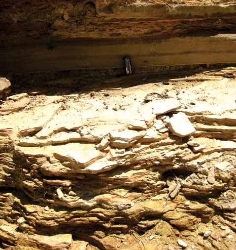
Chemical fingerprints and microbial biomineralization of fish muscle tissues                            419

Múzquiz Lagerstätte type localities                                                   CHALKY LIMESTONE

       The outcrops of Upper Cretaceous marine strata
exposed near the municipality of Múzquiz and in the sur-
rounding area of northern Coahuila provide some of the
most spectacular fossil-rich deposits in Mexico (Figure 1).
These deposits accumulated in diverse marine environments
that were related to the Western Interior Seaway, which ex-
tended from the northern Tethys Ocean into North America
(Goldhammer, 1999; Goldhammer and Johnson, 2001), and
are usually regarded as correlative with the Austin Chalk
and Eagle Ford Groups exposed in northern Coahuila and
Texas (Sohl et al., 1991; Eguiluz de Antuñano, 2001). These
strata consist mainly of chalk, limestone, and organic-rich                                                          2 cm
mudstone deposited during the peak transgression of the
Late Cretaceous epeiric sea in North America (Smith, 1981;
Sohl et al., 1991; Young, 1985; Young, 1986).
       The Eagle Ford sequence includes black shale, yel-
lowish-gray limestone, and interbedded marlstone dated                                                  LIMESTONE
as Cenomanian-Turonian in age (Myers, 2010). The Eagle
Ford strata conformably overlie the Buda Limestone and
are conformably overlain by a sequence of recrystallized
limestone, chalk, and marl of Coniacian-Santonian age
correlative with the Austin Chalk (Dravis, 1980; Larson
et al., 1991).
       The stratigraphic nomenclature changes in southwest-
ern Texas from Eagle Ford/Austin to Boquillas (Lock and
Peschier, 2006); for example, in the Big Bend region (Figure
1), where the overlying lower unit of the Austin Chalk is
                                                                                                       MARL
included as the San Vicente Formation within the Boquillas
Group (e.g., Freeman, 1961, Donovan and Staerker, 2010,
                                                                                                                     40 cm
Lock et al., 2010). The Eagle Ford and Austin Chalk terms
are generally used for correlative strata exposed on the cen-
                                                                    Figure 2. Limestone-marl rythmites of the Múzquiz Lagerstätte type
tral and eastern Texas coastal plain; however, in Coahuila it       locality (below), and a detailed view of the typical laminated chalky
is difficult to differentiate the Eagle Ford from other units       limestone (above). Palestina quarry.
or to recognize its members. The strata exposed in Coahuila
span an age range different from the Boquillas, San Vicente,
or Austin Chalk formations (e.g., Eguiluz de Antuñano,              including three-dimensionally preserved morphology,
2001; SGM, 2008a, 2008b). In the present paper, the term            articulated skeletons, muscle tissue remains, and evidence
Eagle Ford (Boquillas) is used.                                     of restricted organic decay.
       The Múzquiz Lagerstätte occurs in a series of re-
stricted outcrops within a repetitive sequence of fine-grained
calcareous rythmites. The lithology consists of platy lime-         Palestina quarry
stone alternating with impure chalk and marlstone with
earthy texture. Most of the argillaceous material is found as              Palestina quarry is located 290 km northeast of
stratified millimeter-thick layers of volcaniclastic bentonitic     Múzquiz, near the town of Acuña, at latitude 29°11’57’’
clay. Some exposures also exhibit irregular lenses and flaky        N, longitude 100°53’20’’ W (Figure 1). The average alti-
nodules of iron hydroxides, as well as minor thin layers            tude here is 300 m above sea level, in an area mapped as
of indurated non-fissile siltstone and grayish-black, thinly        the Coniacian-Santonian Austin Chalk Formation (SGM,
laminated calcareous shale.                                         2008a). Palestina is a very restricted outcrop composed
       Two localities are highlighted here due to the remark-       predominantly of creamy-yellow, platy, chalky limestone in-
able preservation of fish specimens: El Pilote and Palestina        tercalated with marl (Figure 2). Minor indurated non-fissile
quarries (Alvarado-Ortega et al., 2006; Vega et al., 2007;          siltstone and unconsolidated iron oxides, with volcaniclastic
Riquelme et al., 2010). Although the two sites show differ-         bentonitic clay, is periodically exposed as 3 to 5 cm thick
ences in their taphonomic features and variations in lithol-        layers in the upper beds. Neither the base nor the top of the
ogy, they exhibit comparable preservation in fossil fishes,         section are exposed.
420                                                      Riquelme et al.

       The upper fossiliferous chalky beds are finely crystal-            The El Pilote fossiliferous section has been assigned
line, and frequently contain invertebrates such as inoceramid      to upper part of the Eagle Ford Formation (Alvarado-Ortega
bivalves and gastropods with well-preserved calcareous             et al., 2006; Vega et al., 2007; Alvarado-Ortega and Porras-
shells. Microscopic analyses also reveal that the content          Múzquiz, 2009); the lithology and fossil assemblage here
of foraminifera, calcareous filamentous algae, and calcare-        are comparable with those of the Turonian fossiliferous
ous nanoplankton is significantly greater in these layers,         beds exposed at the La Mula and Los Temporales quarries.
with coccoliths being very abundant. Vertebrate fossils are        Nyborg et al. (2005) and Stinnesbeck et al. (2005) also
absent in the upper layers, whereas the chalky limestones          suggested that the El Pilote outcrops are equivalent to the
in the lower beds yield abundant articulated teoleostean           lower sequence of Upper Turonian strata exposed in the El
fishes including Pachyrhizodus sp., Ichthyodectoidea,              Rosario quarry. However, more supporting stratigraphic
and Clupeoidea with remarkably preserved muscle tissues            work is required to provide a certain correlation.
(Alvarado-Ortega, 2012, pers. obs.).                                      Eguiluz de Antuñano (2001) has described limestone,
       Small exposures of Palestina-like strata are found          marl, and shale beds about 300 m in thickness belonging
within 5 km of the quarry, mostly in road cuts and stream-         to the Eagle Ford Formation that are widespread across
beds where soil is absent. On the basis of recent geological       several municipalities in northwestern Coahuila, as well
fieldwork in northern Coahuila (SGM, 2008a; 2008b),                as in western, central, and northeastern Texas (Jiang, 1989;
and comparative analysis of Austin Chalk outcrops in               and Peschier, 2006, Donovan and Staerker et al., 2010).
South Texas given by Young and Marks (1952), Paulson               The Eagle Ford strata were deposited during a time of
(1968), Dravis (1980), Smith (1981), and Young (1985),             exceptionally high sea level (Lock et al., 2010). Dawson
the Palestina fossiliferous platy beds may be assigned to          (1997) and Eguiluz de Antuñano (2001) suggest that these
the lower section of the Austin Chalk (Early Coniacian             sediments accumulated in a quiet open marine environment
to Santonian; see additionally Hancock and Walaszczyk,             in deep waters. Additionally, stratigraphic studies of sev-
2004; Myers, 2010). According to Freeman (1961), Lock              eral members of the Eagle Ford exposed in central Texas
and Peschier (2006), and Donovan and Staerker (2010),              show successive transgressive, condensed, and highstand
among others, strata equivalent to the Austin Chalk are            sequences (Freeman, 1961, Donovan and Staerker, 2010,
included as the San Vicente Formation (Boquillas Group)            Lock et al., 2010).
in southwest Texas (Big Bend National Park, Val Verde,                    Currently, there is a little consensus about the strati-
Kinney, and Maverick counties; see Figure 1), in the vicinity      graphic relationships between the quarries where the
of the Múzquiz Lagerstätte localities.                             Múzquiz Lagerstätte are preserved (Blanco-Piñón and
       Wright (1987) and Young (1985) indicate that sea level      Alvarado-Ortega, 2005; Stinnesbeck et al., 2005; Alvarado-
rose during deposition of the Austin Chalk, coinciding with        Ortega et al.; 2006, Vega et al., 2007); the stratigraphy is
the maximum extent of the Cretaceous Interior Seaway,              complicated due to extensive lithofacies variation. These
and submerging the large carbonate platforms in northern           facies changes may have been caused by sea level shifts in
Mexico and Texas. It has been suggested that deposition of         the manner shown by Hancock and Walaszczyk (2004). The
the Austin Chalk may have occurred in water depths of up           change from Eagle Ford siliciclastic facies to pelagic chalk
to 250 m (Young and Marks, 1952)                                   of the Austin Chalk is probably a consequence of rising
                                                                   sea level as described by Young (1986) and Goldhammer
                                                                   and Johnson (2001). Thus, the Múzquiz marl-limestone
El Pilote quarry                                                   rythmites could be the result of an alternating sedimentary
                                                                   regime within the boundary between the two major facies.
       El Pilote quarry is located 140 km northwest of
Múzquiz, at latitude 102°29’51’’ W, longitude 28º41’50’’
N (Figure 1), within an area mapped as the Eagle Ford              MATERIALS AND METHODS
Formation (SGM, 2008b). The El Pilote section exposes
recrystallized, fossiliferous, marly limestone, interbed-                Samples. Specimens described here are housed in the
ded with grayish-brown marl and minor calcareous shale.            Museo Histórico de Múzquiz, A. C. (Aguilar and Porras-
Distinctive millimeter-scale interstratified non-fissile silt-     Múzquiz, 2009), institutional abbreviation MUZ.
stone layers and iron oxides are also present.                           The following specimens have been analyzed from
       The marly limestone beds contain bivalves, am-              the Austin Chalk Formation, Palestina quarry, Acuña
monites, and fishes with distinctive fossil preservation.          Municipality, Coahuila: Pachyrhizodus sp. –MUZ 341;
Although fossils occur here sporadically, this locality shares     clupeid fish – MUZ 602; clupeid fish – MUZ 603; clupeid
with Palestina quarry a similar style of preservation of the       fish – MUZ 607, Pachyrhizodus sp. – MUZ 609, almost
fish specimens. El Pilote fishes include teleosts (Clupeoidea,     complete and articulated specimens showing vertebrate
Pachyrhizodontidae, Ichthyodectiodea, Tselfatiiformes,             column contracted, digestive tract and muscle mass patches
Enchodontidae, and abundant isolated scales) and teeth of          preserved; clupeid fish. – MUZ 596 A and B (part and
the elasmobranch Ptychodus (Alvarado-Ortega et al., 2006).         counterpart), complete and articulated specimen, showing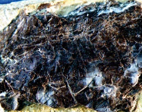
Chemical fingerprints and microbial biomineralization of fish muscle tissues                        421

digestive tract and muscle mass patches preserved.                 NIST: SRM 2704, SRM 2711, SRM 1880a, SRM 1400, and
        The following specimens have been analyzed from            analytic grade CaCO3 from Sigma Aldrich. The AXIL code
the Eagle Ford Formation: El Pilote quarry, Múzquiz                and PIXEINT program were used to measure the elemental
Municipality, Coahuila: Pachyrhizodus sp. – MUZ 73,                concentrations expressed as wt% for eight major elements:
Head, and trunk part with digestive tract and well-preserved       Al, Si, P, K, Ca, Mn, Fe; and also for nine trace elements at
muscle mass patches; clupeid fish – MUZ 602; clupeid               >100 µg/g: S, Ti, Ni, Cu, Zn, As, Sr, Y, U. A review of the
Fish – MUZ 325 A and B (part and counterpart), almost              PIXE technique is given by Johansson et al. (1995).
complete and articulated specimens showing digestive tract               Scanning Electron Microscopy (SEM). Muscle
and muscle mass patches preserved.                                 tissues and fossiliferous rock samples were examined using
        The soft parts and muscle tissues in the fishes were       JEOL JSM-6360 LV and JEOL JSM-5310 scanning electron
carefully extracted using surgery needles under a micro-           microscopes. Samples were coated with graphite or gold-
scope. Subsequently, these were exposed to preliminary             palladium and photomicrographs were taken in high vacuum
cleaning and demineralization with deionized water and,            (Riquelme et al., 2010).
for SEM analysis, partially dissolved using a 5% EDTA                    X-Ray Diffraction (XRD). Muscle tissues and
carbonate digestive solution (Riquelme et al., 2010). All          fossiliferous rock samples were analyzed in air at room
specimens and samples used for biogeochemical analysis             temperature using a Bruker AXS™ D8 Advanced equipment
(PIXE and XRD) were not exposed to solvents to avoid               with Bragg-Brentano θ-θ geometry, Cu Kα radiation, a
contamination or deposition of chemical residues.                  Ni 0.5 % Cu- Kβ- filter in the secondary beam and one
        Muscle tissue samples: M7 and M7a were extracted           dimensional position sensitive silicon strip detector (Bruker,
from MUZ 596 A and B; M8, M8a and M8b are from MUZ                 Linxeye). The diffraction intensity as a function of the
341; M11, M11a, and M11b were extracted from MUZ                   angle 2θ was measured between 20° and 110°, with a 2θ
73; M12 and M12b are from MUZ 325 A. Digestive tract               step of 0.01945°, for 53 seconds per point. The crystalline
samples: M15 was extracted from MUZ 73. Rock samples:              structure, phase composition, and lattice cell parameters
M5 is from MUZ 596 A; M6 is from MUZ 341; M9 is from               of solids were refined with the Rietveld method using
MUZ 607; M10 and M13 are from MUZ 73; M14 is from                  the FULLPROF code. Peak profiles were modelled with
MUZ 325 A.                                                         Pseudo-Voigt functions as shown in Thompson et al. (1987),
        UV light. A diagnostic profile of whole specimens was      which contained average crystallite size and microstrain as
performed using visible fluorescence induced by ultraviolet        characteristic parameters.
(UV) light with a range of 254 nm (short wavelength, SW)
to 365 nm (long wavelength, LW), and exposure time of
less than 3 minutes from 70° to 90° angle of incidence.            RESULTS
Microimaging of isolated muscle tissues and sediment
samples was acquired with a Edmond E-Zoom 6V video                 UV light microimaging
system with EO-3112 Digital CCD camera without any
filter. A zoom range of 50X to 480X with oblique/incident                 Fish body parts including bones, scales, muscle patch-
UV light was applied for dynamic relief contrast. Corel®           es, and digestive tracts, are clearly more fluorescent than sur-
Photo-Paint®X 4 software was used for image processing.            rounding sediments under UV light (Figure 3). The Múzquiz
The biogenic pattern, mineral phases, and hot spots for            fossil-bearing rocks show poor fluorescence in contrast to
subsequent chemical analysis were characterized in bones,          the enclosed fossils (Figure 3a, 3b). This is mostly observed
soft parts, anatomical impressions, as well as in the fossil-      by a color change associated with flourapatite and calcite as
bearing rock (Riquelme et al., 2010).                              dominant mineral phases in bones, scales, digestive tracts,
        Particle Induced X-ray Emission spectrometry               and muscle tissues. In a clupeid fish (MUZ 596 A and B;
(PIXE). Zones of mineralogical and biological interest,            Figure 3a, 3b) the color varies under UV light exposure from
detected in the preliminary study of unprepared fish               a saturated white color preliminarily linked to higher phos-
specimens with UV light, were measured using PIXE based            phorus content in bones; whereas pink, orange, and brown
on the external proton beam of 3MeV Tandem Pelletron               colors are observed in the muscle tissues (Figures 3c, 3d).
accelerator of Instituto de Física, UNAM (Ruvalcaba-Sil,           Additionally, a grayish-white to pale-yellow color is pre-
2008). The beam spots were between 1 mm and 0.5 mm                 liminarily linked to calcium-rich bones and scales, whereas
in diameter (Riquelme et al., 2009). Light elements were           bluish, gray, and yellow are observed in the calcium-rich
detected using a Si-PIN Amptek detector (150 eV resolution         muscle tissues (Figures 3a, 3b). In contrast, dissolved and
for Mn-Kα line) with a 1 mm diameter Ta collimator, and            recrystallized bones and unconsolidated material enriched
a Helium jet to improve detection of low energy X rays.            with siliciclastic minerals are less fluorescent (or notably
Heavier elements were detected using a LEGe detector               opaque) under UV light (Figures 3a, 3b).
with a thick Al filter (155µm). The PIXE spectra were                     Although poorly revealed in visible light, the diges-
collected for 5 minutes and the X-ray detection system was         tive tracts become conspicuous under UV light coupled to
calibrated with pellets of standard reference material from        microscopy. Results show digestive tracts slightly preserved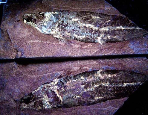
422                                                                Riquelme et al.

                                                                     a)                                                                      c)

                                                            5 cm                                                                    1 mm

                                                                     b)                                                                      d)

                                                     3 cm                                                    0.5 mm

Figure 3. UV light imaging of clupeid fish, (a) part and counterpart (MUZ 596 A and B), Palestina quarry, Austin Chalk, UV light at 365 nm LW; (b)
a closer view of same clupeid (MUZ 596 part A), UV light at 254 nm SW; (c) micrograph of trunk muscle tissue near vertebrae of Pachyrhizodus sp.
(MUZ 73), El Pilote quarry, Eagle Ford Formation, UV light at 254 nm SW (scale bar: 1mm); (d) micrograph of trunk muscle tissue of Pachyrhizodus
sp. (MUZ 609), Palestina quarry, UV light at 254 nm SW. SW: short wavelength; LW: long wavelength.

(Figure 4), with organic material linked to discernible food                 levels average 21.850 wt%, phosphorus 7.625 wt%, and both
remains showing a white, yellow, to brownish color (Figure                   strontium (10784 µg/g) and sulfur (2657 µg/g) levels are
4b). The surrounding sediment is less fluorescent, but the                   high (Figures 6, 7). In contrast, the same set of elements in
thin, more calcareous layers are well defined under UV light                 sediments show significantly lower values: calcium levels
(Figure 5). The results show a microstratigraphy of episodic,                average 16.924 wt%, and phosphorus 1.552 wt%, whereas
lime mud layers including shells, calcareous inclusions, and                 the concentrations of some metals increase, such as copper
fecal pellets with a white to bluish color (Figures 5b, 5d).                 (391 µg/g) and zinc (746 µg/g). This may suggest separate
                                                                             mineralization pathways or incorporation of foreign ions
                                                                             during fossilization. For instance, the average P/Ca ratio
Nondestructive X-ray spectroscopy and mineralogy                             of 0.346 in muscle tissue is notably higher than in sedi-
                                                                             ments with 0.092. The average phosphorus concentration
       On the basis of color variations observed with UV                     in sediment is 1.55 µg/g, but 7.63 µg/g in muscle tissue, an
light, certain hot spots were identified for non-destructive                 increase in phosphorus content by nearly a factor of five;
PIXE measurements in order to obtain chemical analyses                       this is consistent with precipitation and/or recrystallization
of muscle tissues and sediments avoiding bones and scales.                   of phosphate within the primary structure of the muscle
The results of these analyses are summarized in Table 1, and                 tissues.
the ratios of diagnostic elements such as P/Ca, Sr/Ca, and                          Bivariate plots of Al/P, Si/P, K/P, Mn/P, and Fe/P
S/Ca, may be determined. In the muscle tissues, calcium                      indicate differential ion exchange and rearrangement of ele-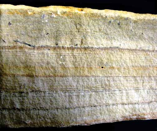
Chemical fingerprints and microbial biomineralization of fish muscle tissues                                      423

                                                                     a)                                                             UV           b)

        5 mm                                                                        5 mm

Figure 4. Micrographs of isolated digestive tract of Pachyrhizodus sp. (MUZ 73), El Pilote quarry, Eagle Ford Formation. Comparative analysis under
regular light (a) and UV light (b) at 365 nm LW.

                                                                     a)                                                                          c)

                               3 mm                                                               400 µm

 top                                                                 b)                                                                           d)

 bottom                        3 mm                                 UV                               400 µm                                     UV
Figure 5. Cross-sectional analysis of the rock containing Pachyrhizodus sp. (MUZ 341), Palestina quarry, Austin Chalk. Comparative views under regular
light (a) and UV light (b) at 365 nm LW; note the episodic, thin, lime mud layers (arrows) separated by sparry calcite enriched with calcareous fossils
(b). Micrographs using regular light (c) and UV light (d); note features that are poorly visible under regular light (c), however, lime mud arrangement
and calcareous gastropod shell are clearly visible under UV light (d).
424                                                               Riquelme et al.
      Table1. PIXE multi-elemental analysis. Major element concentrations are expressed in weight percent (wt%) and trace elements in µg/g.

Sample Spot Deposit         Al      Si      P       K      Ca     Mn     Fe         S    Ti      Ni     Cu    Zn      As      Sr     Y         U

                                                   wt%                                                        µg/g
Sediment
 M5         1   Palestina   0.93   4.29    0.49    1.07   13.34   0.07   0.43    1596    ---    501     284    859     14 8192       ---      ---
            2               2.02   6.78    0.69    1.25   11.65   0.13   0.87    1474    595    1274    530   1915   113 8666        ---      ---
            3               1.08   4.88    0.60    1.10   13.32   0.04   0.31    1340    ---     604    300   1522    ---  9752      ---      ---
M6          1   Palestina   1.59   5.69    0.51    1.21   12.87   0.10   0.41     982    137     213    495    502    --- 10010      ---      ---
            2               1.48   5.50    0.48    1.18   13.21   0.07   0.25    1071      60    439    273    360    ---  8798      ---      ---
            3               0.73   3.07    4.25    0.84   12.69   0.08   0.12    3151    133     198    634    514    --- 16098      ---      ---
M10         1   El Pilote   1.69   16.56   6.05    1.28   26.25   0.03   1.17    1882    857      ---   259    706     27 6131      322       ---
            2               1.47   20.00   0.35    1.20   21.04   0.02   2.87    1182    ---      ---   336    473    ---  2345      ---      ---
            3               1.75   23.50   1.74    1.28   27.98   0.10   0.98    1593    ---      ---   471    101    ---  5835     201       ---
            4               1.76   14.08   0.38    1.37   16.88   0.02   3.43    1000    ---      ---   325    504     34 1052        38      ---
Muscle tissue
M7          1   Palestina   0.96   3.01    3.62    0.89   13.42   0.34   0.60    2830    190    534     268   488     ---   12300   ---        ---
            2               0.80   2.98    3.54    0.98   12.78   0.04   0.29    3320    240    533     256   478     ---    9800   ---        ---
            3               0.89   3.45    4.23    0.87   12.76   0.05   0.24    1780    320    239     239   455      98    8900    97        ---
            4               0.99   2.80    4.19    0.96   12.89   0.04   0.32    1780    280    347     298   598      78   10500   ---        ---
M8          1   Palestina   0.70   2.90    3.78    0.95   12.34   0.06   0.34    1570    340    389     305   673      65   12800   ---        ---
            2               0.80   2.55    3.58    0.89   12.68   0.06   0.32    1900    290    498     309   887     ---   20500   ---        ---
            3               0.99   2.65    4.21    0.96   12.01   0.06   0.25    2340    320    299     198   498     ---   10000 145          ---
            4               0.97   2.99    3.89    0.95   13.24   0.04   0.28    3420    410    344     188   587     ---   10900   ---        ---
M11         1   El Pilote   0.87   4.78    13.57   0.18   39.80   0.03   0.36    4428   1103     ---    335   657      31    6590 367          ---
            2               0.90   9.46    14.74   0.15   38.57   0.03   0.28    2946    ---     ---    339    51     ---    9020 457          ---
            3               0.62   1.45    15.81   0.20   40.15   0.02   0.11    2728    ---     ---    287   441      11    8975 1720        1021
            4               0.38   2.31    16.34   0.15   41.55   0.02   0.11    2844    ---    ---     388   122     23     9128 1757        1134

ments in carbonates from the fossil-bearing rock (Figure 6).                  The calcium carbonate content of the fossil-bearing rocks
Aluminum, silicon, and potassium are associated with silici-                  from El Pilote (marly limestone) varies from 90 wt% to
clastic material accumulated during initial deposition of the                 93.90 wt%, whereas rocks from Palestina (chalky limestone)
sediment. In contrast, the manganese and iron content might                   show values ranging from 83.64 wt% to 86.99 wt%. The
reflect ionic substitution in secondary minerals precipitated                 quartz/calcite ratio (SiO2/CaO3) in rocks from Palestina
during carbonate diagenesis. According to Boggs (2009),                       show values ranging from 0.150 to 0.196, whereas rocks
foreign ions such as Mn and Fe are typically concentrated                     from El Pilote show values from 0.065 to 0.110 (Figure 9),
during ion exchange and recrystallization of carbonates as                    suggesting a different input of siliciclastic materials during
a result of burial diagenetic processes.                                      deposition.
      Complementary to PIXE analyses, the quantitative
XRD results show that flourapatite (FAP) and calcite are
the major components in the crystalline phase of muscle                       Muscle tissues and microbial biofilms
tissues (Table 2). Muscle tissues from Palestina quarry
exhibit higher concentrations of FAP than of calcite (Figure                        Electron micrographs show trunk muscle fiber bundles
8). Although sample M7a from Palestina shows high quartz                      with distinctive fibrillar structure (Figures 10, 11, and 12).
content, the rest of the samples display lower quartz content,                Cellular features are preserved, such as highly vascularized
which is linked to post-burial recrystallization. In contrast,                areas, and biogenic forms interpreted by their shape, size
muscle tissues from El Pilote show calcite amounts that are                   and location as nucleus-like structures (Figures 10b, 10c),
higher than that of phosphates. In addition, quartz content                   and fragments of sarcoplasmic reticulum that wrap around
is higher in muscle tissues from El Pilote (Figure 8). The                    muscle fibers (Figures 11a, 11b, 11c). The muscle bundles
quartz content may be related to the quality of fossil pres-                  preserved in the Múzquiz fishes are strongly associated with
ervation; specimens from Palestina show a higher quality                      fossil microbes (Figures 11 and 12). A particular network
of preservation accompanied by minor quartz content and                       identified among the microbes was interpreted to be organic
less diagenetic recrystallization.                                            as they show a spherulitic to spider web-like arrangement,
      For both Múzquiz marl and chalky limestone the                          and are linked to the extracellular polymer substance (EPS),
precursor sediment is composed of a mixture of biogenic                       which is secreted by microbes as the attachment mechanism
carbonates with variable argillaceous material (Table 2).                     in organic or inorganic substrates (Krumbein et al., 2003).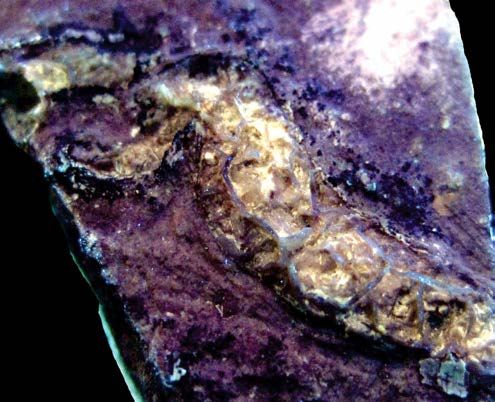
Chemical fingerprints and microbial biomineralization of fish muscle tissues                                               425

                        Sediment                          Muscle tissue                                      Sediment                         Muscle tissue
                                                                                               1.6
         0.35
                                                                                               1.4
         0.30
                                                                                               1.2
         0.25
                                                                                               1.0
Mn wt%

                                                                                      K wt%
         0.20
                                                                                               0.8
         0.15                                                                                  0.6

         0.10                                                                                  0.4

         0.05                                                                                  0.2

         0.00                                                                                  0.0
          3.5                                                                                  24
                                                                                               22
         3.0                                                                                   20
          2.5                                                                                  18
                                                                                               16
         2.0
Fe wt%

                                                                                      Si wt%
                                                                                               14
          1.5                                                                                  12
                                                                                               10
         1.0
                                                                                                 8
          0.5                                                                                    6
                                                                                                 4
         0.0
                                                                                                 2
         -0.5                                                                                    0
                                                                                               2.2
          40                                                                                   2.0
                                                                                               1.8
          35
                                                                                               1.6
          30                                                                                   1.4
                                                                                      Al wt%
Ca wt%

          25                                                                                   1.2
                                                                                               1.0
          20
                                                                                               0.8

          15                                                                                   0.6
                                                                                               0.4
          10
                0   1   2   3   4   5   6   7    80   2    4   6   8 10 12 14 16 18                  0   1   2   3   4   5   6   7   80   2    4   6   8 10 12 14 16 18
                                                P wt%                                                                            P wt%
                                                                         Palestina                   El Pilote
Figure 6. Graphs of element concentrations obtained with PIXE, contrasting muscle tissues and sediments for both Palestina and El Pilote localities.
Note the higher concentration of P and Ca in muscle tissues, as well as higher amounts of Al, Si, K, Fe, and Mg in sediments showing differential ion
exchange and rearrangement into carbonates that compose the fossil-bearing rocks. Values are expressed in weight percent (wt%); uncertainties are as
large as the symbol.

The size and shape of these are consistent with EPS (Figure                             Some trunk muscle tissue samples were isolated (Figures
11b, 11c).                                                                              12a, 12b), which show vascular vessels with bacterial
       At higher magnification, bacterial cells are more                                spherulitic web-like biofilms within them (Figures 12c,
easily distinguished, and linked to the mineralized EPS                                 12d). Complementary evidence of microbial biofilms
surrounding the muscle fibers and sarcoplasmic reticulum                                consists of abundant calcite botryoids in the fossil-bearing
(Figure 11). Bacterial cells consist of coccoid (Figures 11b,                           rocks (Figure 14c). These crystalline microstructures sur-
11c) and bacilliform morphotypes (Figures 11d, 11f). The                                rounding the fossils might have formed around bacterial
spheroidal structure of coccoid cells displays a cryptocrys-                            cell-like aggregates.
talline texture on the surface, which is primarily related to                                  Cross-sections of the digestive tract show a secondary,
cell walls; here tiny crystallites that nucleated around the                            continuous growth of crystals that used the organic material
cell walls are clearly observed (Figure 11c). Micro-fractured                           as a template. Disordered crystals have developed inside
crystalline crusts, flakes, and large calcite crystals also oc-                         and outside of the biogenic filaments, probably microbial
cur among the bacilliform bacteria cells (Figure 11d, 11e).                             (Figure 13a); whereas capillary aggregates of pseudomor-
426 Riquelme et al.
22000
M8 Mt=10784 µg/g
5500
Mt=2657 µg/g
In contrast, the chalky limestone from Palestina shows
a greater diversity of well-preserved coccoliths and less
20000 5000
Sed=7688 µg/g Sed=1527 µg/g
18000
dissolution of the microspar crystals. Also, the microfabric
4500 M11
M6
16000 4000
is considerably less interlocking, resulting in increased po-
Sr µg/g
14000 M8
S µg/g
M8 3500
M7 M6
12000
M7 M5
M7
M11
3000 M7
M11 rosity (Figure 14d, 14e, 14f). These ultra-structures reflect
different degrees of diagenetic overprinting, similar to that
10000 M6 M8 2500
M8
8000 M7 M11
shown by Munnecke and Westphal (2005), Westphal (2006),
M5 M10 2000 M7
M10
6000 M8
and Munnecke et al. (2008).
1500
M10 M10
4000
M10 1000 M5
2000 M10
500
M6 The El Pilote and Palestina rocks also show differ-
ences in the size and composition of the micrite grains.
0
0
1000
The chalky limestone of Palestina shows a fabric composed
2400
2200
Sed=746 µg/g Sed=391 µg/g
Mt=495 µg/g 900 Mt=284 µg/g
2000 M5
800
mostly of calcite crystals larger than 4 µm ('microspar' crys-
1800
1600
M5 700
tals sensu Folk, 1959); as well as a minor content of micrite
Cu µg/g
Zn µg/g
crystals of ≤ 4 µm size (Figures 14d, 14e). In contrast, the
M6
1400 600
M5
1200
500
M6
M10 marly limestone from El Pilote shows a significant content
of true micrite and only minor microspar crystals (Figures
1000 M5
800 M10 M11 400 M11
M7 M8 M8
14a, 14b, 14c). Such crystalline microfabric variability also
M8 M5 M7
M10 M5
600 300
M6 M10
M10
suggests that the carbonate source is variable, as interpreted
400 M8 M10 M6 M11
M7 M11 200 M7
200 M11 M8
0 M10 M11 100
in Flügel (2004) and Munnecke et al. (2008).
-200 0
Cross-sectional views of the chalky limestone from
Samples Samples
Palestina show an upper layer composed of profuse calcar-
Muscle tissue Sediment
eous nanoplankton, calcispheres, and foraminifera, which
Figure 7. Graphs of element concentration obtained with PIXE, contrasting
muscle tissues and sediments, showing higher Sr and S in muscle tissues
are cemented with sparry calcite (Figure 15a). This layer (a
whereas zinc and copper are lower. Average values for sediment (Sed) in Figure 15c) is the horizon where fishes are found in the
and muscle tissue (Mt) are included in each graph. Values are expressed laminated sediments. The layer below the fish-bearing layer
in µg/g and uncertainties are as large as the symbol.
(b in the Figure 15c), has less nanoplankton, calcispheres,
and foraminifera cemented with sparry calcite (gray), as
well as a very thin argillaceous layer with Fe hydroxides
phic carbonates growing alongside the biogenic structures
are also observed (Figure 13a, small rectangle). Such crystal
growths may be linked to a reducing microenvironment as
described by Boggs (2009). There are also significant con- Table 2. Quantitative X-ray diffraction analysis; values are expressed in
centrations of calcareous nanoplankton within the digestive weight percent (wt%). FAP: fluoroapatite.
tract (Figures 13b, 13c, 13d). These are also a main compo-
Sample FAP Calcite Quartz
nent of the associated sediments. However, the calcareous
nanoplankton found within the digestive tract show a much Palestina
higher quality of preservation, including entirely preserved M7 94.42 5.58 ---
M7a 78.34 13.73 7.93
a. Muscle tissue
cocospheres (Figure 13d), compared to those within the
M8 90.30 8.50 1.20
sediments (Figures 13b, 13c, 14).
M8a 93.13 6.97 ---
M8b 82.05 6.97 ---
Pilote
Ultrastructural analysis of the fossil-bearing rocks M11 61.26 37.65 1.09
M11a 54.30 43.61 2.09
Although individual beds of the Múzquiz Lagerstätte M11b 68.10 30.70 1.20
show great lateral continuity and an apparent monotonous M12 63.10 35.90 1.00
lithology, there are significant differences in the rock M12b 62.25 35.80 1.95
composition as seen in their ultrastructural arrangement
Sample Calcite Quartz SiO2/CaCO3
(Figure 14) and in the microstratigraphy of oriented cross
sections (Figure 15). Electron photomicrographs show that Palestina
coccoliths and elongated calcareous grains in the marly M5 86.99 13.01 0.150
b. Sediment
limestone from El Pilote are oriented parallel to the bedding M6 83.64 16.36 0.196
planes (Figure 14a). These rocks have low porosity, and M9 85.20 14.80 0.174
are composed of calcite crystals with a tightly interlocking Pilote
M10 90.17 9.83 0.109
texture, abundant botroyidal microaggregates, recrystallized
M13 93.90 6.10 0.065
and dissolved coccoliths, large calcite flakes, with a pitted
M14 90.08 9.92 0.110
microspar fabric (Figure 14a, 14b, 14c).Chemical fingerprints and microbial biomineralization of fish muscle tissues 427
(reddish; Figure 15b). Such alternating lamination suggests 0.22 0.22
Palestina
episodic deposition with changes in the sedimentary regime. 0.20 0.20
As shown in Flügel (2004) and Boggs (2009), episodic
deposition of calcareous nanoplankton can be consistent 0.18 0.18
with open basin deposition. 0.16 0.16
SiO2 /CaCO 3
0.14 0.14
DISCUSSION 0.12 0.12
The fossil fishes from Múzquiz exhibit structural pres- 0.10 0.10
ervation as seen in a “picture frozen in time”. The analysis 0.08 0.08
presented here shows that the fossil fishes have a complex
mineralogical arrangement in their muscle tissues, which are 0.06 0.06
dominantly preserved by both cryptocrystalline flourapatite 0.04
El Pilote
0.04
(FAP) and semicrystalline calcite in variable concentrations Sediment
(see PIXE and XRD values, Table 1, Figure 8). Nucleation Figure 9. Graph of SiO2/CaCO3 ratio in sediments obtained from XRD
of phosphate and calcite within the muscle tissues must have analysis.Differences in SiO2/CaCO3 ratio (quartz/calcite) as shown for
occurred simultaneously with organic decay during early the Palestina and El Pilote localities suggest significant variations during
depositional input.
burial. Nucleation of FAP and calcite occurring on a small-
scale may have caused the immobilization of carcasses and
skeletons forming adhesive pellets within and around them Alvarado-Ortega et al., 2007; Trinajstic et al., 2007). There
in the soft, watery carbonate mud. In soft-bodied fossils, has been some discussion as to whether calcite or apatite
both calcium phosphate and calcite can be present in soft- is the primary mineral phase involved in the preservation
tissue mineralization as shown by Briggs and Wilby (1996). of ancient soft tissues from marine sediments. Maisey
Accordingly, authigenic mineral crystallization eventually (1991, p. 80-88) and Schultze (1989, fig. 21) suggested
preserves the organic material and may also disrupt organic that muscle tissue in fishes of the Cretaceous Santana
decay. This implies that for fossil preservation in the marine Formation of Brazil are preserved in calcite because the
Múzquiz deposits, the mineralization process may be the tissues disappear with acid preparation, whereas gut contents
critical factor, instead of anoxic or hypersaline conditions, and those muscles in connection with the digestive tract
or immediate burial events (which are hard to demonstrate are truly phosphatized. In contrast, Martill (2012, pers.
by direct measurements or unambiguous data). comm.) suggests that when the muscle tissues are only
Preserved muscle tissues and digestive tract contents lightly phosphatized, they fall apart when calcite is removed.
in fossil fishes from a variety of carbonate sediments have As for the apatite, this has long been considered the most
been reported previously (e.g., Martill, 1988; Schultze, significant mineral in soft tissue preservation from marine
1989; Martill, 1990, Maisey, 1991, Wilby and Martill, 1992; deposits as shown for example by Martill (1988), Allison
and Briggs (1991), Lucas and Prèvôt (1991), Briggs et al.
(1993), and Martill (2003).
The physicochemical mechanism for soft tissue pres-
Palestina El Pilote ervation in marine deposits is also not fully understood.
100 Martill (1989) proposed the "Medusa Effect", which is a
rapid lithification process. It seems that this can be pos-
Muscle tissue total sample wt %
90
80
70
sible only at a small-scale and organ level; however, the
60 actual petrification mechanism remains unexplained. This
50 hypothesis is similar to the cellular permineralization theory
40 presented by Schopf (1975). Although challenged by Maisey
30 (1991), the Medusa Effect is typically based on phosphatiza-
20
tion process (Martill, 1988; Martill, 2003).
According to Briggs (2003a), the quality of preser-
vation induced by authigenic phosphatization is primarily
10 linked to crystallinity: the size and arrangement of phos-
phate-based crystallites, as well as to the sediment input.
0
Briggs (2003b) suggests that preservation of muscle tissues
FAP CALCITE QUARTZ FAP CALCITE QUARTZ requires the replication of their morphology by rapid in situ
growth of minerals, i.e., authigenic mineralization, which
Figure 8. Graph of X-ray diffraction results for muscle tissues. Note the
higher content of flourapatite (FAP) in samples from Palestina quarry, depends on several factors including the nature of microbial
whereas calcite is significantly higher in samples from El Pilote. activity, organic decay gradients, availability of ions, and the428 Riquelme et al.
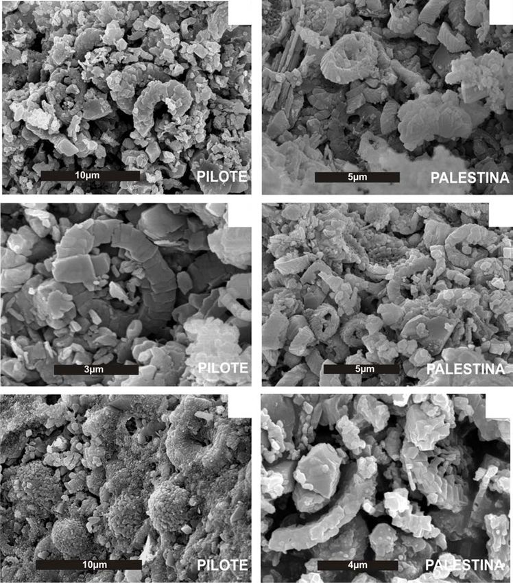
a) d)
400 µm 10 µm
b) e)
200 µm 2 µm
c)
50 µm 200 µm f)
Figure 10. Electron micrographs of (a) muscle tissue extracted from Pachyrhizodus sp. (MUZ 73), El Pilote quarry, Eagle Ford Formation; (b) isolated
thick muscle fibers of the previous sample after partial carbonate dissolution by EDTA; (c - e) closer views of same muscle fibers showing biogenic form
interpreted by shape, size, and location as nucleus-like structure; (f) muscle tissue extracted from clupeid fish MUZ 596 A.
biology of the organisms that are fossilized. Additionally, interrupt organic decay (Martill, 1988; Allison and Briggs,
Dornbos (2010) considered that selective phosphatization 1991; Lucas and Prèvôt, 1991; Briggs et al., 1993; Martill,
is controlled by a microbial environment at limited depths 2003; Dornbos, 2010), rather than a long-term fossiliza-
on the seafloor, with bacterial cells operating as phosphate tion mechanism governed by predominantly sedimentary
nucleation points. It is generally accepted that such chemi- processes, as in the classical concepts of preservational
cal and structural transformation in a short period of time modes (Schopf, 1975).
(hours or days) implies rapid mineralization, which can Mineralization of cells and subcellular structures oc-Chemical fingerprints and microbial biomineralization of fish muscle tissues 429
a) d)
20 µm 5 µm
b) e)
5 µm 5 µm
c) f)
2 µm 2 µm
Figure 11. Muscle tissue of Pachyrhizodus sp. (MUZ 73), El Pilote quarry, Eagle Ford Formation. Electron micrographs of (a - c) biogenic form interpreted
by shape, size and location as sarcoplasmic reticulum remain; (c) microbial biofilm showing characteristic arrangement of spherulitic to spider-web-like
form; (d - f) bacilliform bacteria in same muscle tissue surrounded by thick calcite flakes.
curs at a molecular level, induced by spontaneous chemical growth (secondary nucleation) using the organic material
reactions in supersaturated solutions (Weiner and Dove, as a template (see Figure 11). Depending on the chemical
2003; Qiu and Orme, 2008; Weiner and Addadi, 2011). As composition of the biomolecules and the reactive mineral
documented by De Yoreo and Vekilov (2003), Weiner and solution involved, this mineralized organic template may
Dove (2003), and Weiner and Addadi (2011), the onset of gain or lose fidelity compared with the original morphotype
this process occurs as a mineral nucleation spot (crystal (Mann, 2001).
'embryo' or crystal 'seed'), is followed by intense crystal Accordingly, it seems that the detail-rich preserva-430 Riquelme et al.
a) c)
300 µm 20 µm
b) d)
100 µm 10 µm
Figure 12. Electron micrographs of (a) muscle tissue from clupeid fish (MUZ 596 A), Palestina quarry, Austin Chalk; (b) muscle tissue
from Pachyrhizodus sp. (MUZ 609); (c) vascular vessels observed in same muscle tissues (small rectangle scale bar = 10 µm); (d) bacte-
rial biofilm with spherulitic web-like form within it.
tion of muscle tissues at Múzquiz occurred as a result of important source of microbial fossilization. Biofilms associ-
mineral nucleation and subsequent crystal growth at a ated with fossil soft tissues in vertebrates and invertebrates
macromolecular level. The crystal growth here may have from marine Lagerstätten have been reported elsewhere
been a consequence of natural occurring dissolved calcium (Wilby et al., 1996; Toporski et al., 2002; Krumbein et
phosphate and semicrystalline carbonate in the seafloor al., 2003; Liebig, 2003; Briggs et al., 2005, among oth-
microenvironment; and the seawater is the saline solution ers). According to Sanderman and Amundson (2003), and
that acted as the crystallization precipitant factor. Increased Briggs et al. (2005), the occurrence of biofilms within
temperature during early burial may also accelerate min- soft tissues is strongly associated with an assemblage of
eralization. Mineral precipitation in this supersaturated heterotrophic microorganisms that degrade carcasses.
system may induce the crystal growth in cells and rapid Microbes interact with their environment through surface
biomineralization of soft tissues, biofilms, and microbes reactions and metabolic activities (Gall, 2003; Krumbein
(see additionally Frankel and Bazylinskn, 2003). et al., 2003). It is likely that microbial growth on carcasses
The "crystal seed" process presented above is prob- generates a closed environment that fixes P and Ca cations
ably the mineralization pathway that explains soft tissue (Visscher and Stolz, 2005); this is known as bacterial seal-
preservation at the cellular and subcellular level. The physi- ing (Krumbein et al., 2003). The interaction of microbes
cochemical dynamics of the crystal seed process are largely and minerals can generate a sequence of biogeochemical
known and thoroughly investigated in biomineralization reactions that facilitate biomineralization of organic mat-
research as shown by Mann (2001), De Yoreo and Vekilov ter during burial in deposits such as those observed in the
(2003), Weiner and Dove (2003), and Weiner and Addadi fossiliferous Múzquiz beds (Gall, 2003; Flügel, 2004,
(2011), among others. Westphal, 2006; Munnecke et al., 2008). Eventually degrad-
Another critical aspect of the present work concerns ing microbes drive mineralization on themselves through
the microbial preservation. Fossil soft tissues represent an fixing P and Ca cations on their cell walls (Frankel andChemical fingerprints and microbial biomineralization of fish muscle tissues 431
a) c)
15 µm 5 µm
b) d)
10 µm 4 µm
Figure 13. Electron micrographs of digestive tract contents as observed in Pachyrhizodus sp. (MUZ 341), Palestina quarry, Austin Chalk.
(a) Disordered crystals growing inside and outside of biogenic filaments, likely microbial, as well as capillary aggregates of pseudomorphic
carbonates growing alongside same filaments (small rectangle scale bar 3 µm); (b - c) well-preserved coccoliths and enclosed calcareous
fragments linked to the chalky sediments; (d) intact cocosphere surrounded by coccolith fragments as preserved within digestive tract.
Bazylinskn, 2003; Krumbein et al., 2003; Liebig, 2003). (2003), and Boggs (2009), an important source comes
Finally, the source and mobilization of dissolved from carbonate precipitation that formed the seafloor sedi-
phosphate and semicrystalline carbonate in the Múzquiz ments. Emerson and Bender (1981) also report that another
Lagerstätte is linked to the decaying organisms and mi- significant source of Ca, P, Na cations and CO2 molecules
crobial mats. As shown by Allison and Briggs (1991), may be calcareous nanoplankton deposited on the seafloor.
Briggs et al. (1993), Dornbos (2010), a significant source This is consistent with the episodic deposition of calcareous
of dissolved P and Ca cations in marine burials is released nanoplankton observed in the Múzquiz sediments (Figures
from decaying carcasses, whereas microbial metabolism is 14, 15).
implicated in their mobilization through the water column
(Krumbein et al., 2003). Fish biology might also contribute
as intrinsic factor that increases their preservation potential. CONCLUSIONS
Schultze (1989), Martill (1990), Maisey (1991), and Wilby
and Martill (1992) have suggested that dietary intake can be Examination of fossils under UV light has previ-
a natural source of limiting elements such as phosphorus. ously been undertaken with vertebrates from Lagerstätten
Shewfelt (1981) demonstrated that muscle tissues and soft sediments preserved elsewhere (Tischlinger and Frey, 2002;
parts of extant fishes show high concentrations of available Hone et al., 2010, Kellner et al., 2010; among others). Such
Ca and P cations as micro-bioelements related to dietary analyses have focused on detailed morphological examina-
habits. tion of the specimens as preserved on the bedding plane
The availability of Ca, P, S, and Na cations, and in the fossiliferous rock. In the present work, UV-light
CO 2 molecules, associated with biomineralization of microscopy was supplemented with PIXE as a useful tool
soft tissues in marine burials is critical. As described by to extract biogeochemical information from exceptionally
Lucas and Prèvôt (1991), Sanderman and Amundson preserved fossils. This typically non-destructive technique432 Riquelme et al.
a) d)
10 µm 5 µm
b) e)
3 µm 5 µm
c) f)
10 µm 4 µm
Figure 14. Ultrastructural analysis of rocks contrasting the El Pilote locality (left) and Palestina locality (right); (a) poorly
preserved coccoliths and dissolved calcareous debris oriented parallel to the bedding planes; (b) tightly interlocking texture
and microspar fabric; (c) botryoidal microaggregates of calcite crystallites, dissolved coccoliths, carbonate flakes, and pit-
ted microspar; (b - d) Palestina sediments in contrast show less interlocking microspar crystals, well-preserved coccoliths,
and less dissolution.
can be carried out on unprepared samples without causing composites including sulfates, silicates, iron oxides, and
any damage or alteration to the fossil material or matrix others authigenic minerals.
(Riquelme et al., 2009).
Accordingly, results have shown different major
and trace element abundances for the matrix sediment ACKNOWLEDGEMENTS
and muscle tissues of the fossil fishes. These chemical
signatures may serve as diagnostic fingerprints that allow This work is part of the postgraduate-granting pro-
for interpretation of the fossil preservation process. The gram of Biological Sciences at the UNAM, financially
"crystal seed" process discussed here is a coherent theory supported by CONACYT. The authors thank Karim López
that may explain the physicochemical reactions that oc- and Francisco Jaimes for technical support during experi-
curred during rapid mineralization of ancient soft tissues. mental runs at the Pelletron, IF-UNAM. Special thanks for
This might also apply to non-carbonate or non-phosphate SEM imaging and analysis go to Yolanda Hornelas fromChemical fingerprints and microbial biomineralization of fish muscle tissues 433
a) Top c)
500 µm
b)
500 µm Bottom
Figure 15. Thin-sections of Palestina quarry sediment showing (a) profuse well-preserved calcare-
ous nanoplankton, calcispheres, and foraminifera; this represents the zone where the fossil fishes
occur; (b) layer below fish-bearing horizon showing notably less nanoplankton, calcispheres, and
poor foraminiferal remains cemented with sparry calcite (gray), as well as thin, reddish argillaceous
layers shown by arrows; (c) fossil-bearing rock, a thinly laminated chalky limestone; the letters a
and b represent the position of the previous thin sections.
ICMYL-UNAM, Silvia Espinosa FC-UNAM, and Jaqueline La Mula, Grupo Eagle Ford (Cretácico Superior: Turoniano),
Cañetas IF-UNAM. We would also like to thank people from Múzquiz, Estado de Coahuila, México: Revista Mexicana de
the limestones quarries in Múzquiz and Acuña area for help Ciencias Geológicas, 23(1), 107-112.
Alvarado-Ortega, J., Espinosa-Arrubarrena, L., Blanco-Piñón, A., Vega, F.,
in fossil collection: Juan Manuel Santos, Juan Quintana, Benammi, M., Briggs, D.E.G., 2007, Exceptional preservation of
Tania Aguirre, Maestra Esperanza, El Pilo, and El Diablo. soft tissues in Cretaceous fishes from the Tlayúa Quarry, Central
Also thanks to Drs. David Martill, Joseph Peterson, and Mexico: Palaios, 22, 682-685.
Thomas Lehman for valuable comments that improved the Blanco-Piñón, A., Alvarado-Ortega, J., 2005, Fishes from La Mula
quarries, a new Late Cretaceous locality from the vicinity of
manuscript. This research has also been partially supported Múzquiz, Coahuila, NE Mexico, in Poyato-Ariza , F.J. (ed.),
by PAPIIT-UNAM grants IN403210 and IN225008, as well Extended Abstracts, Fourth Internacional Meeting on Mesozoic
as CONACYT endowment U49839-R. Fishes –Systematics, Homology, and Nomenclature, Miraflores
de la Sierra, Madrid, August 8–14: Madrid, Spain, Universidad
Autónoma de Madrid, UAM Ediciones, 37-41.
Boggs, S., 2009, Petrology of sedimentary rocks: New York, Cambridge
REFERENCES University Press, 311-526.
Botjer, D.J., Etter, W., Hagadorn, J.W., Tang, C.M., 2002, Fossil-
Aguilar, F., Porras-Múzquiz, H., 2009, Los fósiles del Museo de Múzquiz Lagerstäten: jewels of the fossil record, in Botjer D.J., Etter W.,
A. C. y su resguardo patrimonial por el Instituto Nacional de Hagadorn J.W., Tang C.M. (eds.), Exceptional fossil preservation,
Antropología e Historia: Boletín de la Sociedad Geológica a unique view of the ervolution of marine life: New York,
Mexicana, 61(2), 147-153. Columbia University Press, 1-10.
Allison, P.A, Briggs D.E.G., 1991, Taphonomy of nonmineralized tissues, Briggs, D.E.G., 2003a, Exceptionally preserved fossils, in Briggs, D.E.G.,
in Allison, P.A., Briggs, D.E.G. (eds.), Taphonomy, Releasing Cowther, P.R. (eds.), Palaeobiology II: Malden, USA, Blackwell
the Data Locked in the Fossil Record: New York, Plenum Press, Publishing, 328-332.
26-58. Briggs, D.E.G., 2003b, The role of decay and mineralization in the
Allison, P.A., 1988, Konservat-Lagerstätten: cause and classification: preservation of soft bodied fossils, Annual Review of Earth and
Paleobiology, 14(4), 331-344. Planetary Sciences, 31, 275-301.
Alvarado-Ortega, J., Porras-Múzquiz, H., 2009, On the occurrence of Briggs, D.E.G., Wilby, P. R., 1996, The role of the calcium carbonate/
Gillicus arcuatus (Cope, 1875) (Pisces, Ichthyodectiformes) calcium phosphate switch in the mineralization of soft-bodied
in Mexico: Boletín de la Sociedad Geológica Mexicana, 61(2), fossils: Journal of Geological Society of London, 153, 665-668.
215-224. Briggs, D.E.G., Kear, A.J., Martill, D.M., Wilby, P.R., 1993, Phosphatization
Alvarado-Ortega, J., Blanco-Piñón, A., Porras-Múzquiz, H., 2006, Primer of soft-tissue in experiments and fossils: Journal of the Geological
registro de Saurodon (Teleostei: Ichthyodectiformes) en la cantera Society, 150(6), 1035-1038.You can also read