DIAGNOSING AN MI ECG: Tips and Tricks - Dr Ahmed Vachiat - SA Heart Congress
←
→
Page content transcription
If your browser does not render page correctly, please read the page content below
DIAGNOSING AN MI
  ECG: Tips and Tricks

                Dr Ahmed Vachiat
   MBBCh (Wits) FCP(SA) MMed Cert Cardiology (SA)

   Wits Donald Gordon Medical Centre
   Sunninghill and Sunward Park Hospitals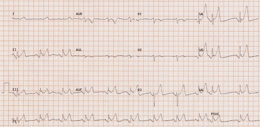
CLINICAL CRITERIA FOR MI

“The clinical definition of MI denotes the
presence of acute myocardial injury detected by
abnormal cardiac biomarkers in the setting of
evidence of acute myocardial ischaemia”
Fourth Universal definition of myocardial infarction, European Heart Journal 2018
Fourth Universal definition of myocardial infarction, European Heart Journal 2018
Normal ECG
Normal ≤100ms
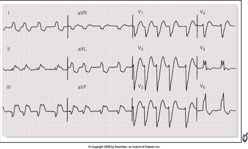
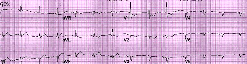
Case Study 1
65y old male patient
Typical chest pain at home @ 18h00
Presents to Casualty @ 20h30

Haemodynamically stable
ECG @ 21h00
Thrombolytic?

Primary PCI?
Chest pain with ST elevation

▪ Give thrombolytic within 30 minutes
  (if no contraindications within 12 hours)

▪ Primary PCI
  (If door to wire time less than 90 minutes)
Tip 1
Look at entire ECG for clues !
 • Rate
    Massive anterior MI ➔ tachycardia
    Pulmonary embolism ➔tachycardia
    Inferior MI with heart block ➔ bradycardia

 • Rhythm
    Arrhythmias can also raise troponin

 • Shape of ST elevation
    Concave up : myocardial infarction
    Convex up : pericarditis
Differential diagnosis of ST elevation
Myocardial infarction
Prinzmetal angina, Takotsubo syndrome
Postmyocardial infarction
Acute pericarditis
Normal variant (“early repolarization”)
LBBB, LVH
Myocardial contusion, Myocarditis
Hypothermia
Post DC cardioversion
Intracranial haemorrhage
Hyperkalaemia
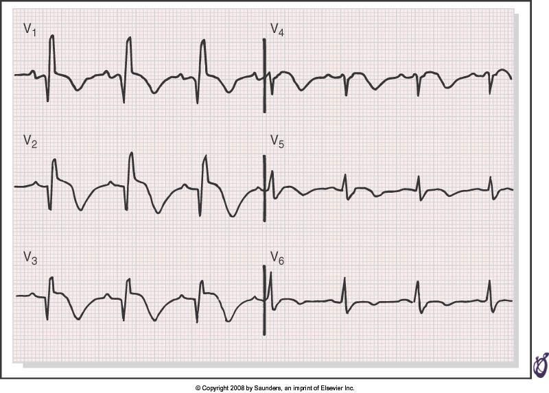
HypercalcaemiaCase Study 2 • 70y old male • Atypical chest pain after collapsing at home @18h00 • Presents to Casualty @ 20h30
ECG Changes : STEMI
All leads 1.0mm (besides Lead V2-3)
Lead V2-3
Male Female
>1.5mm
40y
>2.5mm >2.0mmTip 2 Look at the ECG with the company it keeps! Not all chest pain is myocardial infarction
Case Study 3
• 80y female
• Chest pain 1 day after total hip replacement
• Diabetic and hypertension
Tip 3
Use other investigations to come to
           final diagosis!
            Bloods
            Exercise stress tests
            Echocardiography
            CT scans
            MRI
            Nuclear Medicine studies
+ Troponins
2015 ESC Guidelines for the management of ACS in patients without persistent ST-segment elevation
>50% >20%
Anterior STEMI
VERY NB
TIME Goals
First medical contact (FMC) to first ECG                                ≤ 10 min

FMC to fibrinolysis                                                     ≤ 30 min

FMC to PPCI (PCI capable)                                               ≤ 90 min

FMC to PPCI (PCI capable and within 2 hours of symptom onset)           ≤ 60 min

Primary PCI rather than fibrinolysis                                    ≤ 120 min

Primary PCI rather than fibrinolysis (if large area at risk)            ≤ 90 min

Successful fibrinolysis t0 angiography                                  3-24hr

                                                         ESC STEMI guidelines 2015
Atypical presentations that deserve
prompt management
LBBB
Ventricular paced rhythm
Persistent ischaemic symptoms
Posterior MI
ST elevation aVRAnterior STEMI
Acute inferior STEMI
RBBB and anterior MI
Diagnosis of STEMI in the presence of
              LBBB (Sgarbossa et al)

            ST segment elevation ≥ 1 mm concordant with QRS (5)

            ST segment depression ≥ 1 mm in V1, V2 or V3 (3)

            ST segment elevation ≥ 5 mm discordant with QRS (2)

Score of ≥ 3 – Spec > 90% and PPV of 88%   .
LBBB and inferior MI
Acute MI: ECG subsets, correlation with artery and mortality
Category         Anatomy of         ECG                  30 day          1 year
                 occlusion                               mortality (%)   mortality (%)
Proximal LAD     Proximal to first ST ↑ V1-6, I, aVL     19.6            25.6
                 septal perforator and fasicular or
                                   BBB
Mid LAD          Proximal to       ST ↑ V1-6, I, aVL     9.2             12.4
                 diagonal but
                 distal to first
                 septal perforator
Distal LAD or    Distal to          ST ↑ V1-4, or I,     6.8             10.2
diagonal         diagonal           aVL, V5-6
Moderate to      Proximal RCA or    ST ↑ II, III, aVF and 6.4            8.4
large inferior   cicumflex          any below:
(posterior,                         a. V1, V3R, V4R
lateral, RV)                        b. V5-6
                                    c. R>S in V1,V2
Small inferior   Distal RCA or cx   ST ↑ II, III, aVF    4.5             6.7
                                    only
Echocardiography
Risk scores
Case study 4

55 year old male
Coughing for 2 weeks
Presents with chest pain for 12 hours
a)   Give IV streptokinase
b)   Do emergency PCI (percutaneous coronary intervention)
c)   Give paracetamol tablets
d)   Give sublingual nitrates
e)   Start antibiotics
Typical ECG for acute pericarditis

◆ PR segment elevation in aVR,
◆ depression in inferior leads
◆ together with widespread ST elevation.
a)   Give IV streptokinase
b)   Do emergency PCI (percutaneous coronary intervention)
c)   Give paracetamol tablets
d)   Give sublingual nitrates
e)   Start antibiotics
ECG Tips for diagnosing myocardial infarction

1) Look at entire ECG for clues

1) Look at the ECG with the company it keeps!

1) Use other investigations to come to final diagosis
You can also read