Dual Activation of Phosphodiesterase 3 and 4 Regulates Basal Cardiac Pacemaker Function and Beyond
←
→
Page content transcription
If your browser does not render page correctly, please read the page content below

International Journal of
               Molecular Sciences

Review
Dual Activation of Phosphodiesterase 3 and 4 Regulates Basal
Cardiac Pacemaker Function and Beyond
Tatiana M. Vinogradova * and Edward G. Lakatta

                                          Laboratory of Cardiovascular Science, Intramural Research Program, National Institute on Aging,
                                          National Institute of Health, 251 Bayview Boulevard, Baltimore, MD 21224, USA; lakattae@grc.nia.nih.gov
                                          * Correspondence: vinogradovat@grc.nia.nih.gov

                                          Abstract: The sinoatrial (SA) node is the physiological pacemaker of the heart, and resting heart
                                          rate in humans is a well-known risk factor for cardiovascular disease and mortality. Consequently,
                                          the mechanisms of initiating and regulating the normal spontaneous SA node beating rate are of
                                          vital importance. Spontaneous firing of the SA node is generated within sinoatrial nodal cells
                                          (SANC), which is regulated by the coupled-clock pacemaker system. Normal spontaneous beating
                                          of SANC is driven by a high level of cAMP-mediated PKA-dependent protein phosphorylation,
                                          which rely on the balance between high basal cAMP production by adenylyl cyclases and high
                                          basal cAMP degradation by cyclic nucleotide phosphodiesterases (PDEs). This diverse class of
                                          enzymes includes 11 families and PDE3 and PDE4 families dominate in both the SA node and cardiac
                                          myocardium, degrading cAMP and, consequently, regulating basal cardiac pacemaker function and
                                          excitation-contraction coupling. In this review, we will demonstrate similarities between expression,
                                          distribution, and colocalization of various PDE subtypes in SANC and cardiac myocytes of different
                                species, including humans, focusing on PDE3 and PDE4. Here, we will describe specific targets

                                          of the coupled-clock pacemaker system modulated by dual PDE3 + PDE4 activation and provide
Citation: Vinogradova, T.M.; Lakatta,     evidence that concurrent activation of PDE3 + PDE4, operating in a synergistic manner, regulates the
E.G. Dual Activation of                   basal cardiac pacemaker function and provides control over normal spontaneous beating of SANCs
Phosphodiesterase 3 and 4 Regulates
                                          through (PDE3 + PDE4)-dependent modulation of local subsarcolemmal Ca2+ releases (LCRs).
Basal Cardiac Pacemaker Function
and Beyond. Int. J. Mol. Sci. 2021, 22,
                                          Keywords: sinoatrial node; phosphodiesterase; adenylyl cyclase; cardiac pacemaker cells; local Ca2+
8414. https://doi.org/10.3390/
                                          release; sarcoplasmic reticulum; phospholamban
ijms22168414

Academic Editor: Robert David

Received: 28 June 2021                    1. Introduction
Accepted: 2 August 2021                         The sinoatrial (SA) node, the primary physiological pacemaker of the heart, is respon-
Published: 5 August 2021                  sible for generation of every normal heartbeat, and during a human lifespan, the SA node
                                          creates more than 2.8 billion heartbeats. The SA node automaticity is generated within SA
Publisher’s Note: MDPI stays neutral      node pacemaker cells (SANCs), which fire spontaneous action potentials (APs) because of
with regard to jurisdictional claims in   gradual depolarization of the membrane potential called diastolic depolarization (DD) [1].
published maps and institutional affil-   Despite many achievements in the last 20 years, many mysteries about mechanisms of
iations.                                  cardiac pacemaking and its intrinsic regulation remain unsolved. Several features separate
                                          cardiac pacemaker cells from atrial or ventricular myocytes. Morphologically, SANC are
                                          mostly spindle-shaped and much smaller (cell capacitance ~30–70 pF) compared to atrial
                                          or ventricular myocytes, and they do not have t-tubules. SANC do not have the inward
Copyright: © 2021 by the authors.         rectifier potassium current IK1 as well as its stabilizing effect on the resting membrane
Licensee MDPI, Basel, Switzerland.        potential. As a result, compared to atrial or ventricular myocytes, SANC have high mem-
This article is an open access article    brane resistance, which allow small ion currents to produce a substantial effect on the
distributed under the terms and           membrane potential. Nowadays, it is well recognized that spontaneous beating of cardiac
conditions of the Creative Commons        SANC is governed by a coupled-clock system (discussed below), which has overlapping
Attribution (CC BY) license (https://     and redundant systems to create robust and reliable automaticity [2–5]. Within the SA
creativecommons.org/licenses/by/
                                          node, individual SANC continually respond to intrinsic signals generated at the subcellular,
4.0/).

Int. J. Mol. Sci. 2021, 22, 8414. https://doi.org/10.3390/ijms22168414                                          https://www.mdpi.com/journal/ijms
Int. J. Mol. Sci. 2021, 22, 8414                                                                                          2 of 33

                                   cellular, and tissue levels, as well as extrinsic signals from the autonomic nervous system
                                   and various circulating and locally released factors.
                                         Since its discovery, the cAMP signaling pathway has emerged as an evolutionarily
                                   highly conserved mechanism, involved in regulation of multiple physiological responses
                                   across various cell types. Phosphodiesterases (PDEs) are the only enzymes that degrade
                                   cAMP and terminate cyclic nucleotide signal, controlling cAMP levels and spatial dis-
                                   tribution within the cell. Both PDE3 and PDE4 preferentially hydrolyze cAMP and are
                                   among the most widely distributed and abundant PDE isoforms [6]. Regulation of cAMP
                                   by synergistic dual PDE3 and PDE4 activation has been recognized in many different cell
                                   types, as diverse as cardiac myocytes, SANC, brown adipose tissue, and smooth muscle
                                   cells [7–11], suggesting conservation of dual PDE3 and PDE4 function among various cell
                                   types of multiple species. Currently, dual PDE3 + PDE4 inhibitors are accepted to treat
                                   patients with allergic rhinitis, asthma, and chronic obstructive pulmonary disease (COPD).
                                   In clinical trials, dual PDE3/PDE4 inhibitor (RPL554: Verona Pharma) improved lung
                                   function without undesirable side effects of “classical” PDE4 inhibitors [12,13]. Synergis-
                                   tic effects of dual PDE3 + PDE4 inhibition markedly increases drug efficacy, improving
                                   conditions of patients with asthma and COPD [13,14].
                                         The aim of this review is to highlight one of the essential intrinsic mechanisms, i.e.,
                                   the role of dual PDE3 and PDE4 activation in the regulation of the basal cardiac pacemaker
                                   function. Here, we first provide a brief overview of the “coupled-clock” pacemaker system
                                   that drive SA node automaticity, to introduce the primary targets for modulation of the
                                   SA node beating rate by basal PDE activation. This is followed by description of high
                                   basal level of cAMP in cardiac pacemaker cells created by constitutive adenylyl cyclase
                                   (AC) activation and consequent augmentation of phosphorylation by protein kinase A
                                   (PKA) and Ca2+ /calmodulin-dependent protein kinase II (CaMKII) of proteins involved
                                   in the generation of SA node automaticity. Then we introduce PDEs, which constantly
                                   degrade cAMP to keep cAMP and cAMP-mediated PKA-dependent phosphorylation under
                                   control, and synergistic effect of dual PDE3 + PDE4 activation on PKA-dependent protein
                                   phosphorylation and spontaneous beating rate of SANC. Finally, we will describe how dual
                                   PDE3 + PDE4 activation, working in a synergistic manner, modulates specific targets of the
                                   “coupled-clock” system and the critical role of LCRs in PDE-dependent regulation of the
                                   basal cardiac pacemaker function. We will also discuss evidence for compartmentalization
                                   of cAMP signaling in cardiac pacemaker cells under basal conditions.
                                         Though our review will be focused on the regulation of basal cardiac pacemaker
                                   function by dual PDE3 + PDE4 activation, we will also discuss how dual PDE3 + PDE4
                                   activation regulates Ca2+ cycling in cells from the atria and ventricle, which benefited
                                   from more extensive experimental studies, including results obtained from the transgenic
                                   mouse models as well as human studies. It should be mentioned that there are important
                                   differences between cell types in various regions of the heart (e.g., SANC vs. atrial or
                                   ventricular myocytes) based on their diverse cardiac function, which can lead to variations
                                   in the PDE-dependent modulation of ion channels and SR Ca2+ cycling.

                                   2. Coupled-Clock System Regulates Spontaneous Firing of Cardiac Pacemaker Cells
                                        The generation of repetitive, rhythmic action potentials is the main responsibility
                                   of the sinoatrial node, specifically sinoatrial node cells (SANC). The spontaneous DD
                                   is the essence of cardiac pacemaker cell automaticity; it produces a slow spontaneous
                                   increase in the membrane potential toward an excitation threshold to fire an action po-
                                   tential. Initially, it was assumed that surface membrane ion channels were sufficient at
                                   generating spontaneous DD and spontaneous beating of cardiac pacemaker cells. The
                                   cardiac pacemaker field adopted the idea of DD based on voltage- and time-dependent
                                   surface membrane channels, envisioned as the “membrane clock”, which regulates normal
                                   automaticity of cardiac pacemaker cells (Figure 1A). Several voltage-gated ion channels
                                   and transporters in the cell membrane have important contribution in the ‘membrane
                                   clock’, including the hyperpolarization activated “funny” current If , L-type and T-type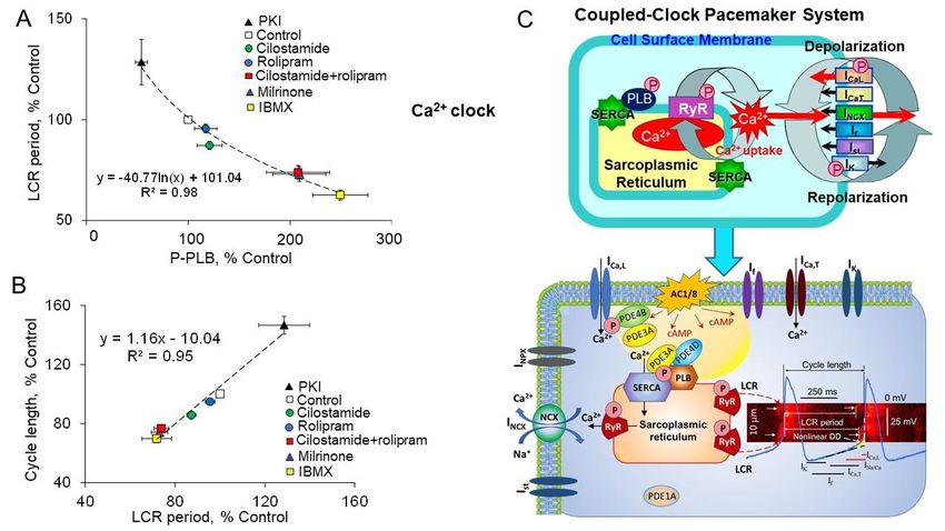
Int. J. Mol. Sci. 2021, 22, x FOR PEER REVIEW 3 of 33
Int. J. Mol. Sci. 2021, 22, 8414 3 of 33
porters in the cell membrane have important contribution in the ‘membrane clock’, in-
cluding the hyperpolarization activated “funny” current If, L-type and T-type Ca2+ cur-
Ca2+(Icurrents
rents Ca,L, ICa,T),(Idelayed
Ca,L , ICa,T ), delayed
rectifier rectifiercurrent
potassium potassium current
(IK), Na +/Ca2+(Iexchange +
K ), Na /Ca
2+ exchange
current (INCX),
current (INCX ), Na + +
/K exchange
Na +/K+ exchange current (INaK), etc.current (INaK
(only the ), etc.
most (only the
important most important
currents are showncurrents are
in Figure
shown in Figure
1A) [1,4,15–17]. 1A) [1,4,15–17].
Figure
Figure1.1.AAschematic
schematicillustration
illustrationofofthethecoupled-clock
coupled-clockpacemaker
pacemakersystem.
system.(A)
(A)Schematic
Schematicpresentation
presentationofofion
ionchannels
channels
“membrane clock” (including the hyperpolarization activated “funny” current I f, L-type and T-type Ca2+
“membrane clock” (including the hyperpolarization activated “funny” current If , L-type and T-type Ca currents (ICa,L ,
2+ currents (ICa,L,
ICa,T), ),
ICa,T delayed
delayed rectifier potassium
rectifier potassium current (IK),(INa
current
+/Ca2+ exchange current (INCX), Na+/K+ exchange current (INaK), sustained
+ 2+ exchange current (I + +
K ), Na /Ca NCX ), Na /K exchange current (INaK ),
current Ist, etc.) and (B) “Ca2+ clock” in cardiac 2+ pacemaker cells. Note that both L-type Ca2+ channels and Na+-Ca 2+
2+ exchanger
sustained current Ist , etc.) and (B) “Ca 2+clock” in cardiac pacemaker cells. Note that both L-type Ca channels and
are part of the “membrane clock” and “Ca clock”. Panel (C) illustrates the “coupled-clock” system with complex inter-
Na+ -Ca2+ exchanger are part of the “membrane 2+clock” and “Ca2+ clock”. Panel (C) illustrates the “coupled-clock” system
actions between the “membrane clock” and “Ca clock” (see text for details). (D) Schematic illustration of spontaneous
with complex interactions 2+ clock” (see text for details). (D) Schematic illustration
SANC action potentials, Cabetween the “membrane
2+ transients, clock” and
LCRs, and several major“Caion currents involved in generation of the diastolic de-
of spontaneous SANC action potentials, 2+
polarization (DD). LCR-induced increaseCain local
transients,
[Ca2+] LCRs,
beneath and several major
sarcolemma ion currents
activates involved
an inward NCX incurrent
generation of the
creating
diastolic depolarization (DD). LCR-induced increase in local [Ca 2+ ] beneath sarcolemma activates an inward NCX current
exponential increase in the DD rate (nonlinear DD). The LCR period represents the essence of the “coupled-clock” system,
which
creating comprises complex
exponential interactions
increase in the DDbetween cell membrane
rate (nonlinear DD). Theelectrogenic
LCR period molecules
represents and intracellular
the essence of theSR Ca2+ cycling
“coupled-clock”
(see text for details).
system, which comprises complex interactions between cell membrane electrogenic molecules and intracellular SR Ca2+
cycling (see text for details).
Similar to other cardiac cells, SANCs have the sarcoplasmic reticulum (SR) and are
Similar
equipped to other
to cycle Ca2+ cardiac
via SR Ca cells, SANCs
2+-ATPase have the and
(SERCA2) sarcoplasmic
Ca2+ release reticulum
channels,(SR) and are
ryanodine
equipped(RyR). 2+
to cycleConfocal
Ca via microscopy 2+
SR Ca -ATPase 2+ release channels, ryanodine
receptors and (SERCA2)
Ca -sensitive
2+ and Ca fluorescent probes allowed to
receptors
identify (RyR). Confocal
spontaneous, roughly microscopy and Ca
periodic, local Ca2+
2+ -sensitive
releases (LCRs) fluorescent
duringprobes
late DD allowed
beneathto
identify spontaneous, roughly periodic, local 2+ releases (LCRs) during late DD beneath
the sarcolemma of cardiac pacemaker cells [18]Ca
(Figure 1B). Further studies confirmed the
the sarcolemma
presence of rhythmicof cardiac pacemaker
spontaneous LCRs cells
under [18]normal
(Figurephysiological
1B). Further studies confirmed
conditions in SANC the
presence of rhythmic
of different species [19–23].spontaneous LCRs under normal physiological conditions in SANC
of different species
During each [19–23]. cycle, Ca2+ influx through L-type Ca2+ channels, triggered
spontaneous
2+ 2+ channels,
by theDuring each spontaneous
AP upstroke, produces acycle,
globalCa Ca2+influx through
transient, L-typeof
emptying Cathe triggered
SR Ca2+ store and
by the AP upstroke, produces 2+ Ca2+
averting LCR generation. When athe global
SR CaCa transient,
2+ content emptying
is refilled of the SR
by SERCA, which store and
constantly
averting
pumps CaLCR generation.
2+ back into the SR,When the SR
LCRs Ca2+
begin tocontent
occur, andis refilled
the cycleby SERCA,
begins which constantly
once more. The
pumps Ca 2+ back into the SR, LCRs begin to occur, and the cycle begins once more. The
restitution time, the time from the AP triggered global Ca transient to the onset of LCRs
2+
restitution time, the time from the AP triggered global Ca 2+ transient to the onset of LCRs
during DD is the LCR period (Figure 1D). LCRs do not require change in the membrane
during DD
potential andiscontinue
the LCRto period
occur(Figure
during 1D).
voltage LCRsclampdo not require
of the change in or
cell membrane theinmembrane
saponin-
permeabilized SANCs [2,20,24], manifesting intracellular SR Ca2+ cycling of or
potential and continue to occur during voltage clamp of the cell membrane “Cain2+saponin-
clock”
permeabilized SANCs [2,20,24], manifesting intracellular SR Ca 2+ cycling of “Ca2+ clock”
in the absence of the “membrane clock” (Figure 1B). Saponin treatment partially removes
in the
cell absence ofand
sarcolemma themakes
“membrane clock” (Figure
it permeable to small1B). ionsSaponin treatment
and molecules partially
without removes
disrupting
cell sarcolemma and makes it permeable to small ions and molecules
SR function. When the cell membrane is permeabilized to remove membrane currents,2+ without disrupting
SR function. When the cell membrane is permeabilized to remove membrane currents, Ca
cycling by the SR becomes “free running” and is controlled mostly by the concentration of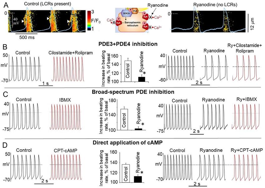
Int. J. Mol. Sci. 2021, 22, 8414                                                                                            4 of 33

                                   free cytosolic Ca2+ and the kinetics of Ca2+ pumping into and releasing from the SR. At
                                   the same physiological Ca2+ concentration, permeabilized SANC cycled Ca2+ beneath the
                                   sarcolemma more efficiently compared to permeabilized ventricular myocytes. Specifically,
                                   permeabilized SANC generated larger and more rhythmic spontaneous SR Ca2+ releases
                                   than ventricular myocytes at the similar SR Ca2+ content in both cell types [24].
                                         To generate an AP, the “membrane clock” interacts with the “Ca2+ -clock” via multiple
                                      2+
                                   Ca and voltage-dependent mechanisms creating a “coupled-clock” system (Figure 1C).
                                   There are many points where the function of the two clocks overlaps, e.g., both L-type
                                   Ca2+ channels and the Na+ /Ca2+ exchanger have dual affiliation as members of both
                                   “membrane clock” and “Ca2+ clock”. In intact SANC L-type Ca2+ channels provide Ca2+
                                   supply to pump into SR, while the LCR occurrence beneath sarcolemma activates an inward
                                   Na+ -Ca2+ exchange current (INCX ), which produces an exponential increase of the late DD
                                   rate (nonlinear DD) prompting the “membrane clock” to generate the next AP upstroke
                                   and, thus, modulating the spontaneous SANC beating rate (Figure 1B). Colocalization of
                                   Na+ /Ca2+ exchanger and RyRs beneath the sarcolemma of rabbit SANC [25] permits a
                                   quick conversion of LCRs into changes in the inward INCX current that depolarizes the
                                   membrane potential. Though the contribution of the L-type Ca2+ channels and Na+ -Ca2+
                                   exchanger for intracellular [Ca2+ ]i balance and the LCR period in SANC seems obvious,
                                   other channels, e.g., potassium channels, could also indirectly participate in adjusting
                                   the intracellular [Ca2+ ]i balance. Indeed, potassium channels repolarize the membrane
                                   potential and, therefore, inactivate L-type Ca2+ channels indirectly affecting [Ca2+ ]i balance
                                   and the LCR period. The coupled-clock system function together on a beat-to-beat basis
                                   and comprises complex crosstalk between the two clocks via signaling pathways, which can
                                   modulate each other to safeguard robustness and reliability of function (Figure 1C,D) [2–5].
                                   A perturbation of one clock inevitably affects the other due to subsequent indirect effects,
                                   resulting in mutual entrainment, e.g., inhibition of the If current by ivabradine also slows
                                   spontaneous SANC firing, leading to a decrease in the SR Ca2+ load and suppression of
                                   LCRs [26].
                                         The coupled-clock system is regulated not only by Ca2+ or voltage-dependent mech-
                                   anisms, but also by phosphorylation status of multiple proteins, which comprise both
                                   “membrane clock” and “Ca2+ clock” (Figure 1) (discussed below). Important phospho-
                                   rylation sites exist on phospholamban (PLB), which regulates activity of SERCA, L-type
                                   Ca2+ channels (modulating ICaL ), potassium channels (modulating IK ), and RyR (likely
                                   increasing its calcium sensitivity [27]). Phosphorylation acts on both clocks and results of
                                   model simulations demonstrated that changes in the phosphorylation status are linked to
                                   changes in the degree of synchronization of the coupled-clock system [28].

                                   3. Ca2+ -Activated ACs, cAMP Synthesis, and Its Relevance for the Spontaneous
                                   Beating Rate of SANC
                                        cAMP is a universal second messenger that coordinates a multitude of downstream
                                   intracellular signaling, and synthesis of cAMP from adenosine triphosphate (ATP) is
                                   regulated by the enzyme adenylyl cyclase (AC). The AC family consists of nine membrane-
                                   bound isoforms (AC1–9) and one soluble isoform [29]. All AC isoforms, except AC8, could
                                   be found in adult ventricular myocytes with AC5 and AC6 as major AC isoforms with
                                   lower levels of AC2, AC4, and AC9 [30]. Though AC5 and AC6 are closely related isoforms,
                                   they seem to play distinct roles in the regulation of cardiac function [30,31]. AC5 has
                                   been shown to be the dominant isoform in the heart and has the highest enzyme catalytic
                                   activity among AC isoforms [32]. The most notable effect of AC5 deletion is elimination of
                                   parasympathetic control of cAMP levels in AC5-KO mice, as well as protection of the heart
                                   against chronic β-AR stimulation, suggesting that the β1-adrenergic receptor selectively
                                   couples to AC5 [33,34]. Deletion of AC6 was associated with reduced left ventricular
                                   contractile function, decreased PKA activity, and marked abnormalities in Ca2+ transients
                                   of cardiac myocytes due to decreased PLB phosphorylation and impaired activity of SR
                                   Ca2+ -ATPase [35]. In cardiomyocytes, cAMP is produced by ACs generally in response to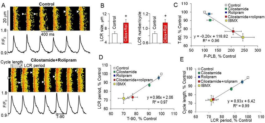
Int. J. Mol. Sci. 2021, 22, 8414                                                                                           5 of 33

                                   stimulation of catecholamine- or hormone-activated receptors; cAMP has several major
                                   downstream targets, including protein kinase A (PKA), exchange protein activated by
                                   cAMP (EPAC), nucleotide gated ion channels, and PDEs, which degrade cAMP into 50 -
                                   AMP terminating its ability to modulate downstream targets.
                                         Though all ACs are inhibited by high intracellular Ca2+ [Ca2+ ]i [36], AC5 and AC6
                                   are inhibited at the physiological range (0.1–1µmol/L) of [Ca2+ ]i . In contrast, AC1 and
                                   AC8 are stimulated by [Ca2+ ]i in a CaM-dependent manner, and AC 2/4/7/9 are Ca2+
                                   insensitive. Different AC isoforms localize to distinct membrane compartments: Ca2+ -
                                   sensitive ACs (AC1/3/5/6/8) were found in lipid rafts structures, while Ca2+ insensitive
                                   ACs (AC 2/4/7/9) were excluded [29]. Therefore, lipid rafts and by extension caveolae,
                                   likely represent specific cellular regions that can promote and strengthen interactions
                                   between multiple signaling targets [29].
                                         The idea that intracellular cAMP is required for cardiac pacemaker function was
                                   first proposed more than four decades ago and based on the observation that exposure to
                                   AC inhibitors suppressed spontaneous beating of isolated rabbit SA node [37], and this
                                   effect was reversed by dibutyryl cAMP [38]. Though inhibition of ACs in this original
                                   study was indirect, further experiments using an iontophoretic injection of cAMP into
                                   Purkinje fibers, or superfusion of isolated rabbit SA node with a dibutyryl cAMP containing
                                   solution, produced a marked increase in the DD slope, and a concomitant increase in the
                                   spontaneous beating rate [38,39], confirming the link between changes in intracellular
                                   cAMP and spontaneous firing of cardiac pacemaker tissue.
                                         Direct measurements of cAMP in isolated rabbit SANC were made three decades later
                                   and revealed that basal level of cAMP in intact isolated SANC is ~3-fold higher compared
                                   to ventricular myocytes (Figure 2A,B (left)). This high basal level of cAMP (in the absence
                                   of any membrane receptor agonists) in SANC is due to constitutive activation of ACs and
                                   is markedly suppressed by the AC inhibitor, MDL-12,330A (Figure 2B (right)) [40,41]. Con-
                                   stitutive AC activation could not be explained by constitutive activation of beta-adrenergic
                                   receptors (β-AR), because either β1-AR antagonist, CGP-20712A, or the β2-AR subtype in-
                                   verse agonist, ICI 118,551, failed to alter the normal spontaneous SANC firing rate [2,40,42].
                                         An examination of this ongoing basal activity of ACs demonstrated association be-
                                   tween Ca2+ and cAMP indicating a possible link to Ca2+ -activated type of ACs. SANCs,
                                   like ventricular myocytes, express AC5/6 but they also express Ca2+ -activated AC1 and
                                   AC8: AC1 was reported in the guinea pig SA node [45], and both AC1 and AC8 were found
                                   in rabbit SA node and SANC [42] (Figure 2C). The latter study also showed that Ca2+ -
                                   activated AC isoforms localize to membrane lipid microdomains and are activated by Ca2+
                                   (Figure 2D), increasing cAMP production over the wide physiologic range (0.2–1 µmol/L)
                                   of intracellular [Ca2+ ]i [42]. This association between Ca2+ and cAMP was further con-
                                   firmed in permeabilized SANC (in the absence of functional ion channels), i.e., an increase
                                   in cytosolic [Ca2+ ]i was accompanied by an increase in the cAMP level, indicating activa-
                                   tion of ACs and consequently an increase in the cAMP production [24]. Transgenic mice
                                   overexpressing AC8 have increased level of cAMP and a faster heart beating rate [46,47].
                                         PKA is the main downstream target of cAMP. Since mechanisms of PLB phosphoryla-
                                   tion via PKA are similar to mechanisms responsible for phosphorylation of other major
                                   proteins regulating SANC automaticity, the phosphorylation status of PLB could be used
                                   as an index of global phosphorylation status of SANC [2,40]. Intact SANC have high basal
                                   level of PLB phosphorylation at PKA-dependent Ser16 site, which by ~10-fold exceeds
                                   that in ventricular myocytes (Figure 2E), indicating high basal level of cAMP-mediated
                                   PKA-dependent phosphorylation of multiple proteins in cardiac pacemaker cells. Inhibi-
                                   tion of ACs not only reduced the level of cAMP (Figure 2B, right), but also suppressed
                                   PKA-dependent protein phosphorylation, confirming a direct link between AC activation
                                   and basal protein phosphorylation in cardiac pacemaker cells [40]. Although an increase in
                                   cytosolic [Ca2+ ]i had no effect on PKA-dependent PLB phosphorylation in permeabilized
                                   ventricular myocytes, it markedly increased PLB phosphorylation in permeabilized SANC,
and spontaneous firing of cardiac pacemaker tissue.
                                         Direct measurements of cAMP in isolated rabbit SANC were made three decades
                                   later and revealed that basal level of cAMP in intact isolated SANC is ~3-fold higher com-
                                   pared to ventricular myocytes (Figure 2A,B (left)). This high basal level of cAMP (in the
Int. J. Mol. Sci. 2021, 22, 8414   absence of any membrane receptor agonists) in SANC is due to constitutive activation   6 of of
                                                                                                                               33
                                   ACs and is markedly suppressed by the AC inhibitor, MDL-12,330A (Figure 2B (right))
                                   [40,41]. Constitutive AC activation could not be explained by constitutive activation of
                                   beta-adrenergic receptors (β-AR), because either β1-AR antagonist, CGP-20712A, or the
                                   indicating that high basal PKA-dependent protein phosphorylation in SANC is due to
                                   β2-AR subtype inverse agonist,    ICI 118,551, failed to alter the normal spontaneous SANC
                                   constitutive activation of Ca2+ -activated ACs [24].
                                   firing rate [2,40,42].

      Figure 2.
      Figure  2. Constitutive
                 Constitutive AC   activation, high
                              AC activation,   high basal
                                                    basal level of cAMP,
                                                          level of cAMP, and
                                                                           and PKA-dependent
                                                                               PKA-dependent protein
                                                                                                protein phosphorylation
                                                                                                         phosphorylation is
                                                                                                                         is central
                                                                                                                            central
      for spontaneous firing of cardiac pacemaker cells. (A) Cartoon representing constitutive activation of ACs, high basal level
      for spontaneous firing of cardiac pacemaker cells. (A) Cartoon representing constitutive activation of ACs, high basal
      of cAMP, and PKA-dependent phosphorylation in SANC. (B) Left, average basal cAMP in SANCs or ventricular myocytes
      level of cAMP, and PKA-dependent phosphorylation in SANC. (B) Left, average basal cAMP in SANCs or ventricular
      (modified from [41]); right, AC inhibitor MDL-12,330A markedly decreases cAMP in SANC (modified from [40]). (C)
      myocytes (modified from [41]); right, AC inhibitor MDL-12,330A markedly decreases cAMP in SANC (modified from [40]).
      (C) Abundance of AC isoform transcripts in the rabbit SA node or SANC relative to brain tissues (from [42]). (D) Presence of
      Ca2+ -stimulated AC activity in whole cell lysates of intact rabbit SANC (from [42]). (E) Basal level of PLB phosphorylation
      at PKA-dependent Ser16 site is significantly higher in SANC compared to ventricular myocytes (VM) (modified from [40]).
      (F) Time course of the effect of AC activation by cholera enterotoxin on cyclic AMP content (left) and pacemaker rate (right)
      of the rabbit SA node preparation (with permission, from [43]). (G–J) Ca2+ chelation, AC or PKA inhibition suppresses
      spontaneous SANC beating rate; representative examples of: (G) effects of Ca2+ chelation by 25 µmol/L BAPTA-AM in
      rabbit SANC (from [42]) and (H) 10 µmol/L BAPTA-AM in guinea pig SANC (with permission from [44]); (I) effects of
      AC inhibition by MDL-12,330A (400 µmol/L) and (J) PKA inhibition by specific PKA inhibitory peptide PKI (15 µmol/L)
      (I–J from [42]).

                                        Maneuvers known to regulate basal cAMP-mediated, PKA-dependent signaling also
                                   affect the normal SA node spontaneous beating rate. Indeed, stimulation of AC by cholera
                                   enterotoxin time-dependently increased level of cAMP, which was paralleled by an acceler-
                                   ation of spontaneous SA node beating rate [43] (Figure 2F). The dependence of spontaneous
                                   SANC firing rate on intracellular [Ca2+ ]i was further confirmed by the observation that
                                   spontaneous activity of single SANC isolated from either rabbit or guinea pig SA node
                                   is abrogated by chelation of cytosolic Ca2+ with intracellular BAPTA (Figure 2G and 2H,
                                   respectively). This effect of Ca2+ buffering happened, in part at least, via a reduced ability
                                   of Ca2+ to activate AC1/8, leading to a local reduction of cAMP and PKA-dependent
                                   protein phosphorylation.
                                        A key role of constitutive AC activation and PKA-dependent phosphorylation for
                                   normal spontaneous beating of single isolated SANC was demonstrated using selective
                                   AC inhibitor MDL-12,330A, which reduced the level of cAMP (Figure 2B, right) and con-
                                   currently decreased and finally abrogated spontaneous SANC firing (Figure 2I) [42]. This
                                   effect was partly due to the decrease in the PKA-dependent protein phosphorylation, since
                                   the membrane-permeable PKA inhibitor peptide (PKI) slowed and abolished spontaneous
                                   SANC beating; this effect was reversible on PKI washout (Figure 2J) [42]. Finally, numerical
                                   model simulations supported hypothesis that there is a direct link between changes in Ca2+ -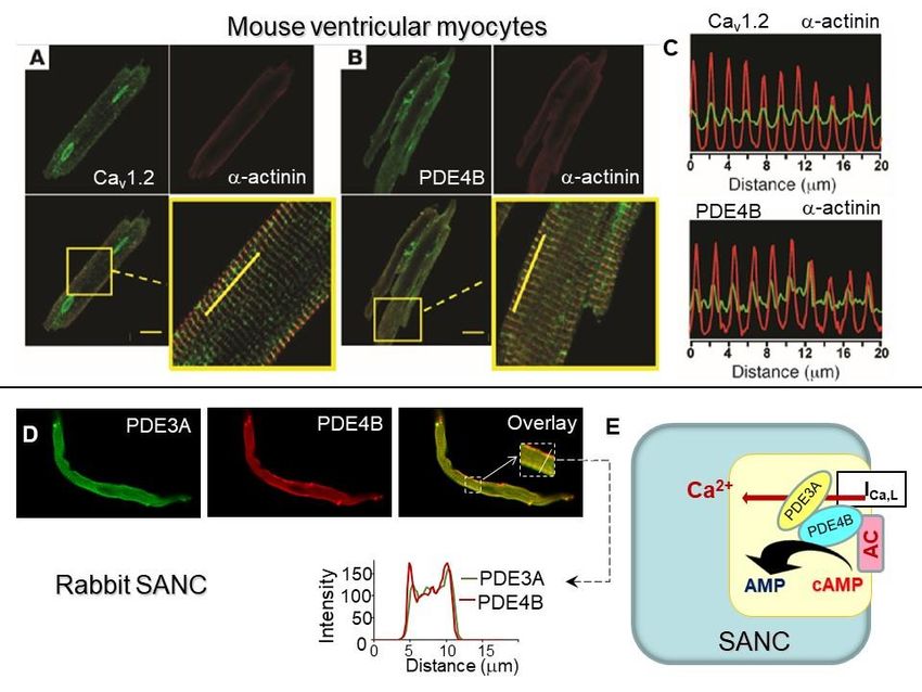
Int. J. Mol. Sci. 2021, 22, 8414                                                                                             7 of 33

                                   activated AC-cAMP-PKA signaling and spontaneous beating rate of cardiac pacemaker
                                   cells [48].

                                   4. Phosphorylation of “Membrane Clock” and “Ca2+ Clock” Proteins in SANC
                                   Safeguards Function of the Coupled-Clock System
                                          Basal AC activity and high level of cAMP increases PKA-dependent protein phospho-
                                   rylation in SANC (Figure 2) amplifies Ca2+ influx through L-type Ca2+ channels [49] and
                                   AP-induced Ca2+ transients, increasing local and global Ca2+ concentrations. Signals that
                                   increase [Ca2+ ]i activate CaMKII [50]. Active (autophosphorylated) CaMKII is located in
                                   microdomains of the surface membrane of SANC [51] in close proximity to L-type Ca2+
                                   channels and RyR [25,51], and it can retain its activity even in the absence of further increase
                                   in [Ca2+ ]i [52]. Based on its “memory” properties [53] CaMKII could serve as a “frequency
                                   detector” and integrate local Ca2+ signals, reflecting performance of SANC: the faster SANC
                                   beats and more frequent are local Ca2+ releases, the greater is the CaMKII activity. The
                                   basal level of activated (autophosphorylated) CaMKII in rabbit SANC by ~2-fold exceeds
                                   that in ventricular myocytes [54]. Close connection between PKA- and CaMKII-dependent
                                   phosphorylation has been recently confirmed in mice expressing a PKA inhibitor peptide in
                                   cardiomyocytes (cPKAi) with almost complete inhibition of cardiac PKA activity [55]. The
                                   reduction of PKA activity led to a markedly decreased level of CaMKII phosphorylation in
                                   cardiomyocytes as well as CaMKII-dependent phosphorylation of PLB and RyR2 [55].
                                          The high basal protein phosphorylation by both PKA and CaMKII is required for
                                   normal coupled-clock pacemaker function because spontaneous AP firing ceases when
                                   either PKA- or CaMKII-dependent phosphorylation is inhibited [40,51,54,56]. Both PKA
                                   and CaMKII share the same phosphorylation targets, including L-type Ca2+ channels, PLB
                                   and RyR2 (Figure 1). L-type Ca2+ channels in SANC are part of both “membrane clock” and
                                   “Ca2+ clock”, since they generate action potential upstroke and at the same time provide
                                   Ca2+ supply for pumping into SR. L-type Ca2+ channels in SANC are highly phosphory-
                                   lated by both PKA and CaMKII in the basal state, as specific PKA inhibitor peptide, PKI, or
                                   CaMKII inhibitors, KN-93 or AIP, suppressed ICaL by ~80% [49] and ~50% [51], respectively.
                                   Delayed rectifier potassium channels are also phosphorylated by PKA, i.e., stimulation of
                                   PKA with the membrane-permeable cAMP analog by ~2-fold increased the amplitude of
                                   the delayed-rectifier potassium current (IK ) in guinea pig ventricular myocytes [57]. In rab-
                                   bit SANC, β-AR stimulation increased IK current amplitude by ~70%, markedly shortened
                                   the decay of IK , and all effects were reversed by PKA inhibitor H-89 [58], indicating that IK
                                   is a target of PKA-dependent phosphorylation (Figure 1).
                                          Similar to ventricular myocytes, the SR “Ca2+ clock” in rabbit SANC is wired to cycle
                                       2+
                                   Ca , and it is tightly regulated by both PKA- and CaMKII-dependent protein phosphory-
                                   lation in the basal state. The SR Ca2+ -ATPase (SERCA) pumps Ca2+ , entering through the
                                   L-type Ca2+ channels, back into SR to refill the SR Ca2+ content and prepare for the next
                                   spontaneous cycle. Activation of SERCA accelerates re-uptake of Ca2+ into SR, shortens
                                   duration of AP-induced Ca2+ transients, and reduces duration of Ca2+ sparks [59]. Trans-
                                   genic mice overexpressing SERCA2a protein showed increased SR Ca2+ uptake function,
                                   i.e., an increase in SERCA2a protein levels by ~1.5-fold in transgenic mice was associated
                                   with acceleration of the maximum velocity of SR Ca2+ uptake by ~40% [60]. Moreover,
                                   hearts from mice overexpressing SERCA2a showed significantly higher myocardial con-
                                   tractility and slightly increased spontaneous beating rate [61], signifying SERCA as a key
                                   determinant of both cardiac contraction and spontaneous SANC firing.
                                          Similar to atrial myocytes [62], an expression of SERCA protein in rabbit SANC is
                                   ~1.5-fold higher compared to ventricular myocytes [54]. In line with increased amount of
                                   SERCA protein, the total LCR signal mass released by permeabilized SANC (in the absence
                                   of functional ion channels) at physiological cytosolic [Ca2+ ] (150–250 nmol/L) was ~2-fold
                                   larger compared to permeabilized ventricular myocytes despite similar SR Ca2+ content
                                   in both cell types [24,56]. Moreover, the elevated Ca2+ release from the SR produced no
Int. J. Mol. Sci. 2021, 22, 8414                                                                                           8 of 33

                                   detectable depletion of the SR Ca2+ content indicating more efficient SR Ca2+ pumping in
                                   SANC [24].
                                         PLB is a functional “brake” on SERCA, and abundance of PLB protein in many species,
                                   including humans, rabbits, guinea pigs, mice, and rats is ~2–3-fold less in atria compared
                                   to ventricle [62,63]. Abundance of PLB protein in rabbit SANC is ~2-fold less than in
                                   ventricular myocytes, indicating that inhibition of SERCA by PLB in cardiac pacemaker
                                   cells is lower compared to ventricular myocytes. Considering that the amount of SERCA in
                                   SANC is ~1.5-fold higher than in ventricular myocytes, the SERCA/PLB ratio could be at
                                   least ~3-fold larger in SANC than in ventricular myocytes, which should result in increased
                                   Ca2+ pumping into the SR required to support robust SR Ca2+ release [24]. Phosphorylation
                                   of PLB by PKA or CaMKII (at Ser16 or Thr17 sites, respectively) disengage an inhibitory
                                   action of PLB on SERCA and release SERCA inhibition, elevating SERCA activity by
                                   ~2–3-fold in ventricular myocytes [64,65]. In rabbit SANC, basal PLB phosphorylation
                                   at Ser16 and Thr17 sites was respectively ~10-fold (Figure 2D) and ~3-fold [54] greater
                                   compared to ventricular myocytes, signifying high level of protein phosphorylation by
                                   both PKA and CaMKII in the basal state.
                                         During every heartbeat, a small influx through L-type Ca2+ channels activate Ca2+ -
                                   induced Ca2+ release (CICR) mechanism to generate Ca2+ release from RyR, SR Ca2+
                                   release channels, amplifying the inward Ca2+ signal by ~10–20-fold [66,67]. The cardiac
                                   RyR2 is a large macromolecular complex connected to PKA, CaMKII, phosphatases, and
                                   PDE4D, which are hitched to the channel by A-kinase-anchoring proteins (AKAPs) [68–71].
                                   RyR, can be phosphorylated by PKA at Ser2030 , by CaMKII at Ser2814/2815 and by both
                                   PKA and CaMKII at Ser2809 . In isolated ventricular myocytes, activation of CaMKII was
                                   associated with increased Ca2+ spark frequency [72]. CaMKII dependent phosphorylation
                                   substantially modifies RyR function in cardiomyocytes, increasing Ca2+ sensitivity of RyR
                                   and enhancing Ca2+ release [71]. Transgenic mice overexpressing CaMKIId isoform showed
                                   elevated Ca2+ spark frequencies (despite lower SR Ca2+ content), pronounced SR Ca2+ leak
                                   and a susceptibility for arrhythmias linked to altered phosphorylation levels of proteins
                                   involved in Ca2+ handling [71,73].
                                         Basal RyR phosphorylation at CaMKII-dependent Ser2815 site in rabbit SA node is
                                   ~10-fold higher compared to ventricle [54], which could be partly due to similar distri-
                                   bution and likely association of activated autophosphorylated CaMKII and RyR beneath
                                   sarcolemma of rabbit SANC [25,51]. Basal RyR phosphorylation at Ser2809 site, which is
                                   phosphorylated by both PKA and CaMKII, is also ~2-fold higher in rabbit SANC compared
                                   to ventricular myocytes [54]. Robust, rhythmic LCRs in SANC require high basal PKA- and
                                   CaMKII-dependent protein phosphorylation since inhibition of either PKA- or CaMKII-
                                   dependent phosphorylation results in small, stochastic Ca2+ events that resembles Ca2+
                                   sparks in ventricular myocytes [24]. The coordinated and synchronized phosphorylation-
                                   driven increases in both Ca2+ release through RyRs and reuptake by the SR could sustain
                                   large and rhythmic spontaneous LCRs in SANC.

                                   5. Requirement of PDE Activation in Cardiac Pacemaker Cells in the Basal State
                                        The level of cAMP in the cell is determined not only by synthesis by ACs, but by
                                   cyclic nucleotide PDEs, which constantly degrade cAMP. High basal level of cAMP in
                                   SANCs could be a result of low cAMP degradation and negligible PDE activity in the basal
                                   state. On the other hand, high basal PKA- and CaMKII-dependent protein phosphorylation
                                   in SANC creates positive feedback system (Figure 3A), which requires strong feedback
                                   regulation. Indeed, basal PKA- and CaMKII-dependent phosphorylation promotes Ca2+
                                   influx via L-type Ca2+ channels, boosts phosphorylation of PLB, and increases SERCA
                                   activity, accelerating kinetics of SR replenishment with Ca2+ . At the same time, an increased
                                   RyR phosphorylation synchronizes RyR [74] and likely decreases the threshold for RyR
                                   Ca2+ release, elevating Ca2+ release from the SR via spontaneous LCRs. Higher levels
                                   of intracellular Ca2+ further stimulate Ca2+ -activated ACs, which generate more cAMP
                                   further activating PKA and CaMKII. This positive feedback system in cardiac pacemaker
Int. J. Mol. Sci. 2021, 22, 8414                                                                                         9 of 33

                                   cells, when Ca2+ release creates more Ca2+ release, elevating cAMP and amplifying the
                                   original action, is unstable and requires robust feedback regulation (Figure 3A), provided
                                   by PDEs. Indeed, there is a high basal level of PDE activity in rabbit SANC, since broad-
                                   spectrum PDE inhibitor IBMX produces ~9-fold increase in the cAMP level, an effect larger
                                   than that of a saturating concentration of β-AR agonist isoproterenol (Figure 3B), [41].
                                   Therefore, constitutively active ACs in the basal state coexist with high basal PDE activity
                                   in SANCs, and the latter perform a negative feedback regulation to limit and fine-tune the
                                   basal cAMP level.
                                         PDEs represent a highly diverse superfamily of enzymes encoded by 21 genes and
                                   divided into 11 families that give rise to over 100 PDE isozymes [75,76]. Three PDE
                                   families, PDE4, PDE7, and PDE8 specifically hydrolyze cAMP, three PDE families PDE5,
                                   PDE6, and PDE9 hydrolyze cGMP and five PDE families PDE1–PDE3, PDE10, and PDE11
                                   hydrolyze both cAMP and cGMP. Although numerous PDEs, except PDE6 and PDE10,
                                   were found in myocardial tissue of different species, including humans, PDE1, PDE3, and
                                   PDE4 families are the main PDEs that hydrolyze cAMP in the heart [77]. While PDE3 family
                                   dominates in larger mammals including dog, bovine, rabbit, and human myocardium,
                                   PDE4 predominates in rodent myocardium [77]. Though PDE4 is also expressed in the
                                   human heart, it accounts for only ~10% of the total cAMP-PDE activity (vs. 40–60% in rat
                                   and mouse) [77,78].
                                         PDE1 is Ca2+ /calmodulin-activated isoenzyme, hydrolyzing both cAMP and cGMP
                                   with similar substrate specificity, and PDE1 subfamily includes three genes (PDE1A, PDE1B,
                                   and PDE1C). Though PDE1 isoforms are highly expressed and have high activity in human
                                   myocardium [79,80], the physiologic role of PDE1 isoforms in the heart remains unclear,
                                   since inhibition of PDE1 activity produces a decrease rather than increase in contraction
                                   amplitude of human ventricular myocytes [80]. Experiments in cell culture suggest that
                                   PDE1 may not be active under basal conditions but becomes active when intracellular
                                   [Ca2+ ]i concentration is increased [81].
                                         PDE2 is cGMP-activated PDE and it can hydrolyze both cAMP and cGMP. Although
                                   PDE2 exhibits similar cAMP and cGMP substrate specificity in rat, guinea pig, and dog
                                   ventricular tissue, it preferentially hydrolyses cGMP in rabbit, porcine and human my-
                                   ocardium [77]. PDE2 is considered to act primarily as a signal integrator between cGMP
                                   and cAMP signaling, and most studies support the idea that under normal conditions
                                   PDE2 is responsible for relatively small fraction of total cAMP hydrolyzing activity in the
                                   myocardium [75].
                                         PDE3 family is divided in two subfamilies PDE3A and PDE3B, and both are expressed
                                   in the heart of different species [77]. Genetic manipulations of mice demonstrated that
                                   PDE3A modulates basal excitation-contraction coupling, SR Ca2+ content and contractility
                                   in cardiac myocytes [77,82]. Ablation of PDE3A eliminated >85% of the PDE3 activity and
                                   increased contractility in hearts from mice lacking PDE3A, but not PDE3B. The enhanced
                                   cardiac contractility in (PDE3A KO) hearts was associated with phosphorylation of key
                                   proteins involved in the regulation of SR Ca2+ cycling in cardiac myocytes PLB and RyR [83].
                                   Specifically, there was a 2-fold increase in PLB phosphorylation at PKA-dependent Ser16
                                   site accompanied by a 2-fold increase in RyR phosphorylation at PKA-dependent Ser2808
                                   and Ser2830 sites [83]. The role of PDE3A in the modulation of the SR Ca2+ cycling protein
                                   phosphorylation is likely linked to its regulation of cAMP in microdomains containing
                                   macromolecular complexes of SERCA2a-PLN-PDE3A [83,84].
                                         The PDE4 family consists of four subfamilies, PDE4A to PDE4D, but only PDE4A,
                                   PDE4B, and PDE4D are expressed in rodents’ hearts [85]. Although PDE4 has little effect
                                   in the resting state, it becomes active upon β-AR stimulation and starts to regulate global
                                   cAMP level in cardiac cells. For example, pharmacological inhibition of PDE4 in rat
                                   ventricular myocytes increases inotropic effects of β-AR stimulation and spontaneous
                                   diastolic Ca2+ waves [86]. Moreover, inhibition of PDE4 in the presence of isoproterenol
                                   potentiates phosphorylation of RyR2 and PLB, not only at specific PKA-dependent sites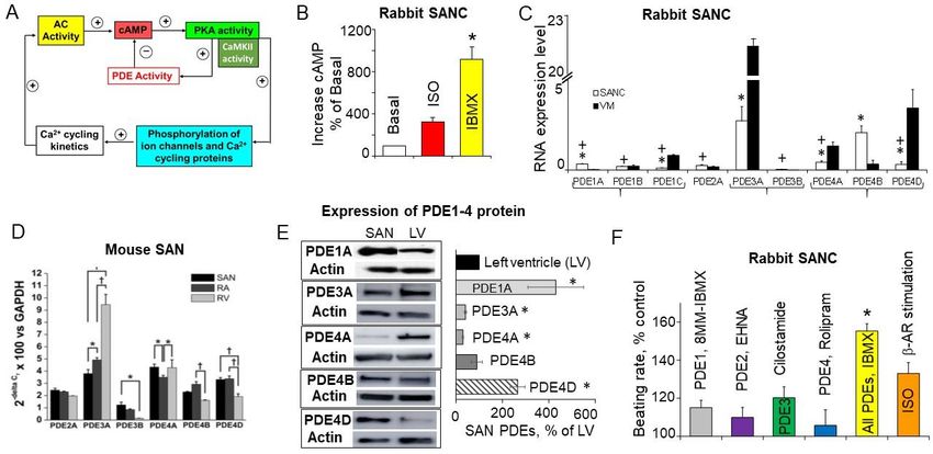
Int. J. Mol. Sci. 2021, 22, x FOR PEER REVIEW 10 of 33
Int. J. Mol. Sci. 2021, 22, 8414 10 of 33
(RyR at Ser2808 and PLB at Ser16), but also at CaMKII-dependent sites (RyR Ser2814 and PLB
(RyR 2808 and PLB at 2+ 16 ), but also at 2+ sites (RyR Ser2814 and PLB
at Thrat17),Ser
increasing SR Ca Ser load and SR Ca CaMKII-dependent
leak [86].
at Thr 17 ), increasing 2+ 2+
Genetic ablationSR of Ca
PDE4D load and SR Ca
enhanced leak [86].
the susceptibility to stress-induced ventricular
Genetic ablation of PDE4D enhanced the susceptibility
tachycardia, which was explained by PKA-dependent hyperphosphorylation to stress-induced ventricular
of RyR2
[69]. Baseline cardiac contractility in PDE4D-KO mice was markedly elevatedofand
tachycardia, which was explained by PKA-dependent hyperphosphorylation RyR2 [69].
cardi-
Baseline cardiac contractility in PDE4D-KO mice was markedly
omyocytes isolated from PDE4D-KO hearts showed increased Ca transient amplitudes elevated
2+ and cardiomy-
ocytes isolated from 2+ transient amplitudes with
with preserved ICa,L, PDE4D-KO
compared tohearts showed increased
WT cardiomyocytes CaThese
[87]. functional changes in
preserved ICa,L
PDE4D-KO , comparedwere
myocardium to WT cardiomyocytes
associated [87]. These
with increased PLBfunctional changes likely
phosphorylation in PDE4D-
due
KO myocardium were associated with increased PLB phosphorylation
to association of PDE4D with the PLB-SERCA2A complex. Both PDE4B and PDE4D can likely due to associ-
ation of PDE4D
associate with the with the Ca
L-type PLB-SERCA2A
2+ channel, but complex.
only PDE4B Bothregulates
PDE4B and ICa,LPDE4D
during canβ-ARassociate
stimu-
with the L-type Ca 2+ channel, but only PDE4B regulates I during β-AR stimulation in
lation in mouse ventricular myocytes. It was concluded that Ca,L
PDE3 and PDE4 families rep-
mouse ventricular myocytes. It was concluded that PDE3 and
resent major PDE families to degrade cAMP and regulate excitation-contraction coupling PDE4 families represent
major
in the PDE families to
myocardium degrade
[77,78], withcAMPPDE3and regulate excitation-contraction
dominating in larger mammals and coupling in the
in humans
myocardium [77,78], with PDE3 dominating in larger mammals and in humans [77,78].
[77,78].
Figure
Figure 3. Constitutive
Constitutive basal
basal PDE
PDE activation,
activation, PDE
PDE isoforms,
isoforms, andand their
their role
role in
in the
the regulation
regulation of normal
normal cardiac
cardiac pacemaker
pacemaker
function.
function. (A)
(A) Cartoon
Cartoon of of positive
positive basal
basal Ca
Ca2+/cAMP-PKA
2+
/cAMP-PKA“feed-forward”
“feed-forward”systemsystemkept
keptinincheck
checkby byhigh
highbasal
basalPDE
PDEactivity,
activity,
which
which acts
acts as
as aa negative
negative feedback
feedback mechanism restricting cAMP/PKA
mechanism restricting cAMP/PKA signaling
signaling and
and preventing
preventing an an excessive
excessive basal
basal beating
beating
rate. (B) Suppression of PDE activity in SANCs markedly increases the level of cAMP exceeding effect produced by satu-
rate. (B) Suppression of PDE activity in SANCs markedly increases the level of cAMP exceeding effect produced by
rating concentration of β-AR agonist isoproterenol (ISO). One-way ANOVA with Bonferroni post hoc test * pInt. J. Mol. Sci. 2021, 22, 8414 11 of 33
myocytes [89]. Expressions of PDE3A and PDE4B mRNA in rabbit SANC were markedly
higher than expression of other PDE subtypes (Figure 3C), while in the mouse SA node
mRNA transcript abundance for PDE2A, PDE3A, PDE4A, PDE4B, and PDE4D were similar
(Figure 3D) [88]. At the protein level, expression of PDE3A and PDE4A protein was less
abundant in the rabbit SA node compared to the left ventricle; expression of PDE4B protein
was similar in both tissues, while expression of PDE4D [11] and PDE1A protein [89] was
significantly higher in the rabbit SA node than in the ventricle (Figure 3E).
Compared to the effect of broad-spectrum PDE inhibitor IBMX or saturating concen-
tration of β-AR agonist ISO, the increase in the spontaneous beating rate produced by
selective inhibitors of PDE1, PDE2 or PDE3 activity was relatively small, while inhibition
of PDE4 had no noticeable effect on spontaneous SANC firing (Figure 3F). Despite elevated
basal activity of PDE1 in rabbit SANC [89], PDE1 inhibitor MIMX increased spontaneous
firing of rabbit SANC by ~15% (Figure 3F). It is possible that PDE1 activity might have a
greater impact at higher cAMP levels; e.g., stimulation of ACs with forskolin markedly
increases both the cAMP level and PDE1 activity in paced mouse ventricular myocytes,
suggesting that the contractility-coupled Ca2+ pool can activate PDE1 [90].
Effects of broad-spectrum PDE inhibition on the increase in cAMP level (Figure 3B)
and the spontaneous SANC beating rate (Figure 3F) exceeded those of the saturating
concentration of β-AR agonist isoproterenol (p < 0.05) [41], likely due to the more efficient
cAMP degradation by PDEs in the basal state compared to cAMP production triggered by
β-AR stimulation. Moreover, data in Figure 3F indicate that normal automaticity of cardiac
pacemaker cells is likely regulated, not by one individual PDE subtype, but combined
activity of several PDEs.
6. Basal Spontaneous Firing of Rabbit SANC Is Regulated by Dual
(PDE3 + PDE4) Activation
Previous studies in a variety of cell types indicated that while inhibition of PDE3 or
PDE4 alone have relatively small effects on their own, combined PDE3 + PDE4 inhibition
could produce a large synergistic response, creating effect which is greater than the simple
sum of separate PDE3 and PDE4 inhibition [7,14]. For example, under basal conditions,
effects of PDE3 or PDE4 inhibitor alone on lipolysis or glucose uptake or uncoupling
protein-1 expression were relatively small, and substantial effect was reached only with
combination of PDE3 + PDE4 inhibitors, which synergistically stimulated each of these
processes [7]. Multiple physiological functions as diverse as regulation of smooth muscle
cell motility [8] or excitation-contraction coupling in rat ventricular myocytes [9] or right
atrium contractility [10] are also regulated by dual (PDE3 + PDE4) activation.
Several studies investigated effects of separate or concurrent PDE3 and PDE4 inhi-
bition on intracellular levels of cAMP in cardiomyocytes. PDE3 inhibitors cilostazol or
milrinone produced modest dose-dependent increase in intracellular cAMP in rabbit ven-
tricular myocytes, but dual inhibition of PDE3 + PDE4 caused synergistic elevation of the
cAMP level [91] (Figure 4A). In isolated pig cardiomyocytes with expressed cAMP sensor
Epac-SH187 PDE3 inhibitor cilostamide or PDE4 inhibitor rolipram slightly, but signifi-
cantly (Int. J. Mol. Sci. 2021, 22, 8414 12 of 33
Int. J. Mol. Sci. 2021, 22, x FOR PEER REVIEW 12 of 33
Figure 4.
Figure 4. Dual
Dual PDE3
PDE3 ++ PDE4
PDE4 inhibition
inhibition increases spontaneous beating rate of isolated SANC and intact intact SA
SA nodes
nodes inin aa
synergistic manner. (A) Inhibition of PDE4 with rolipram (1 µ mol/L) in combination with PDE3
synergistic manner. (A) Inhibition of PDE4 with rolipram (1 µmol/L) in combination with PDE3 inhibitors cilostazol or inhibitors cilostazol or
milrinone (3 or 10 µ mol/L) causes synergistic increases in cAMP levels in rabbit ventricular myocytes.
milrinone (3 or 10 µmol/L) causes synergistic increases in cAMP levels in rabbit ventricular myocytes. # p < 0.05 vs. the # p < 0.05 vs. the
additiveeffects
additive effectsofofrolipram
rolipramandandcilostazol
cilostazol alone.
alone. @@pInt. J. Mol. Sci. 2021, 22, 8414 13 of 33
inhibition of PDE3 had almost no effect (Figure 4D). Nonetheless, concurrent inhibition of
PDE3 + PDE4 increased spontaneous firing of the isolated rabbit, rat, or mouse SA nodes
in the range of ~30–47% (Figure 4D), which surpassed added increases in the spontaneous
beating rates produced by inhibition of PDE3 and PDE4 alone by at least ~1.5-fold. These
results demonstrate that, like in rabbit SANC, dual PDE3 + PDE4 activation operates
synergistically to regulate basal spontaneous beating rate of isolated SA nodes of various
species. The robust modulation of the basal spontaneous beating rate of the cardiac
pacemaker by dual PDE3 + PDE4 activation both at the cell and tissue levels could be
accomplished only through synchronized PDE3 and PDE4 effects on specific targets of the
coupled-clock system (Figure 1), which are discussed below.
7. Synergistic Effect of Dual PDE3 and PDE4 Inhibition on L-type Ca2+ Current in
SANC and Atrial/ventricular Myocytes
It is well established that ICa,L in cardiac myocytes is modulated by cAMP-mediated
PKA-dependent phosphorylation: when L-type Ca2+ channels are phosphorylated, the
open probability of individual channels is increased leading to an increase in ICa,L ampli-
tude [96]. L-type Ca2+ channels are indirectly regulated by PDEs via cAMP-PKA-signaling,
and PDE inhibition by IBMX creates marked increases in the ICa,L amplitude in cardiac
myocytes of various species, including guinea pigs, rats, mice, rabbits, and humans (for
review see [77]). An impact of different PDE isoforms on cAMP-PKA-mediated modu-
lation of ICa,L , however, is not identical in different regions of the heart. For example,
in rat ventricular myocytes inhibition of PDE1, PDE2, PDE3, or PDE4 had no effect on
basal ICa,L amplitude and relatively small effect on cAMP level [97]. A marked stimulation
of basal ICa,L amplitude was produced by IBMX, which increases basal ICa,L by ~120%
(Figure 5A,B) and [cAMP]i by ~70%, or dual inhibition of PDE3 and PDE4, which increases
basal ICa,L by ~50% (Figure 5B) [97]. These results were further verified by a recent study
from the same group, which showed marked increases in the phosphorylation of L-type
Ca2+ channels in rat ventricular myocytes by dual PDE3 + PDE4 or broad-spectrum PDE
inhibition (Figure 5C). In contrast, selective PDE2, PDE3, or PDE4 inhibitors alone as well
as dual PDE2 + PDE3 or PDE2 + PDE4 inhibition had no effect on phosphorylation of
L-type Ca2+ channels in the basal state (Figure 5C) [9].
Broad-spectrum PDE inhibitor, IBMX, markedly increased basal ICa,L amplitude by
~130%, ~140% and ~185% in different regions of the mouse myocardium, including ventric-
ular, atrial myocytes, and SANC, respectively [88]. Consistent with previous results [97],
none of the selective PDE2, PDE3, or PDE4 inhibitors had any effect on basal ICa,L am-
plitude in mouse ventricular myocytes. In mouse atrial myocytes, however, selective
PDE2 or PDE4 inhibitors increased basal ICa,L by ~38% and ~72%, respectively, while
PDE3 inhibition was without effect [88]. In mouse SANC inhibition of PDE2, PDE3, or
PDE4 increased ICa,L by ~31%, ~66%, and ~93%, respectively [88], confirming differences
between ventricular, atrial myocytes, and SANC in terms of whether PDEs regulate ICa,L
amplitude in the basal state and, if so, what specific PDE isoforms modulate ICa,L in cells
from different regions of the heart. Dual PDE3 and PDE4 inhibition markedly increased
basal ICa,L in mouse ventricular myocytes by ~50%, while effect on ICa,L in mouse atrial
myocytes or SANC was markedly higher and comparable to that of IBMX [88].
In human atrial myocytes, only a small increase in the basal [cAMP]i level was ob-
served in response to PDE4 inhibition alone, while an increase in [cAMP]i was 2-fold larger
in response to PDE3 inhibition and 4-fold larger when both PDE3 and PDE4 were concur-
rently inhibited [99], indicating that concurrent activation of PDE3 and PDE4 controlled
basal cAMP level in human atrial myocytes in a synergistic manner. PDE4 inhibition in a
concentration-dependent manner moderately increased basal ICa,L amplitude, which was
further elevated by PDE3 inhibition [99]. Another study, however, failed to find any effect
of PDE4 inhibitor rolipram on basal ICa,L amplitude in human atrial myocytes, likely due
to differences in the experimental conditions [100].and [cAMP]i by ~70%, or dual inhibition of PDE3 and PDE4, which increases basal ICa,L by
~50% (Figure 5B) [97]. These results were further verified by a recent study from the same
group, which showed marked increases in the phosphorylation of L-type Ca2+ channels in
rat ventricular myocytes by dual PDE3 + PDE4 or broad-spectrum PDE inhibition (Figure
Int. J. Mol. Sci. 2021, 22, 8414 5C). In contrast, selective PDE2, PDE3, or PDE4 inhibitors alone as well as dual PDE2 +
14 of 33
PDE3 or PDE2 + PDE4 inhibition had no effect on phosphorylation of L-type Ca2+ channels
in the basal state (Figure 5C) [9].
Figure 5. Synergistic regulation of basal L-type Ca2+ current amplitude by dual PDE3 + PDE4 activation in rat ventricular
myocytes, human, or rabbit atrial myocytes and rabbit SANC. (A) Time-dependent effects of PDE4 inhibitor Ro20-1724 and
IBMX on ICa,L in a rat ventricular myocytes (with permission from [97]). (B) Both dual PDE3 + PDE4 inhibition (0.1 µmol/L
cilostamide and 0.1 µmol/L Ro20-1724) and IBMX (100 µmol/L) markedly increased ICa,L in rat ventricular myocytes, while
PDE3 or PDE4 inhibitors alone were without effect (calculated from Table 1 from [97]). (C) Effects of PDE inhibitors alone or
in combination on phosphorylation of CaV 1.2 in rat ventricular myocytes in basal conditions: PDE4 (Ro20-1724, 10 µmol/L),
PDE3 (cilostamide, 1 µmol/L), PDE3 + PDE4 (Ro20-1724 + cilostamide), PDE2 (Bay 60-7550 0.1 µmol/L), PDE2 + PDE3 (Bay
60-7550 + cilostamide), PDE2 + PDE4 (Bay 60-7550 + Ro20-1724), and IBMX (100 µmol/L). The membranes were stripped
and re-probed with calsequestrin (CSQ) antibodies used as a loading control (with permission from [9]). (D) Representative
examples of the enhanced effect caused by combined application of PDE3 (UD-CG 212) and PDE4 (rolipram) inhibitors on
ICa,L in a human (left) and rabbit (right) atrial myocytes (with permission from [98]). (E) Representative recordings of ICa,L
in rabbit SANC in response to inhibition of PDE3 (0.3 µmol/L cilostamide) or PDE4 (2 µmol/L rolipram) alone or dual
PDE3 + PDE4 inhibition (from [11]). (F) Average effects produced by inhibition of PDE3 or PDE4 alone or dual PDE3 +
PDE4 inhibition in rabbit SANC (from [11]) and human or rabbit atrial myocytes (from [98]). One-way ANOVA with Tukey
post hoc test adjusted, * p < 0.05 (rabbit SANC: PDE3 + PDE4 or IBMX vs. PDE3 or PDE4 alone); + p < 0.001 (rabbit or
human AM: PDE3 + PDE4 or IBMX vs. PDE3 or PDE4 alone).
There was substantial increase in the basal ICa,L amplitude in response to PDE3
inhibitor in rabbit and especially human atrial myocytes, but not to PDE4 inhibitor in both
species [98]. Dual PDE3 + PDE4 inhibition, however, increased ICa,L by ~215% and ~270% in
human or rabbit atrial myocytes respectively [98], creating synergistic amplification of ICa,L
amplitude. The effect of dual PDE3 + PDE4 inhibition markedly exceeded additive effects of
PDE3 or PDE4 inhibition alone (Figure 5D,F) and was comparable to the effect of IBMX [98].
L-type Ca2+ channels are essential for spontaneous firing of cardiac pacemaker cells, and
they are part of both “membrane clock” and “Ca2+ clock”: L-type Ca2+ current generatesYou can also read