Evaluation and optimisation of extraction methods suitable for the analysis of microplastic particles occurring in the edible part of seafood - EFSA
←
→
Page content transcription
If your browser does not render page correctly, please read the page content below
Evaluation and optimisation of extraction
methods suitable for the analysis of microplastic
particles occurring in the edible part of seafood
   Max Rubner-Institut, Federal Research Institute of Nutrition and Food
           Department of Safety and Quality of Milk and Fish

                                                                           Bild Dorade: © Peter Kirchhoff/PIXELIO
                             Julia Süssmann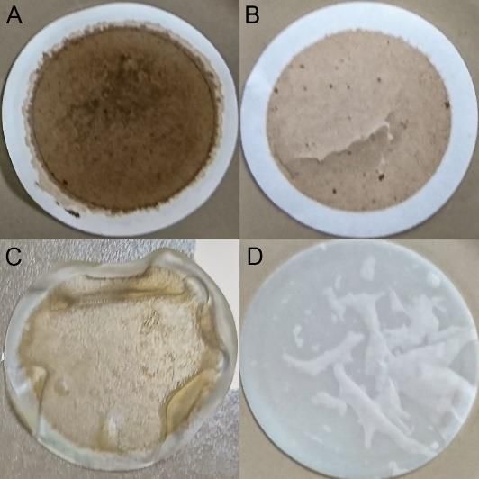
Microplastic in seafood: How much do we eat?

                                                Translocation

                                                 Pb

                                                             Migration         Cr
                                                                                   Leaching
                                                                                       Hg
                                                                                                    Cd

                                                                                                                                    ?
                                                              3700 ± 2500100[11]            0 - 24450[14]
                                                                                                     150 µm
                                                                              µm

                                                         400 - 8100100[6]µm 3000 ± 900
                                                                                       [3]           980 ± 266010[10]
                                                                                                                   µm
                                                                                     10 µm

    138000 ± 202300[16]                                     [13]                                           970 ± 261010[2]µm
                   10 µm                          700 - 290010 µm                      250 - 3605[4]µm

                                              1600 - 3500 [6]
                                                                                  0 - 680020[9]µm                               160 ± 13010[8]
   259400 ±     114100[16]
                                                        100 µm                                                                              µm
                                                                                                                                                   0 - 35020[5]µm
                         10 µm
                                                                                               650 -     1330[7] 10 µm
                                                    0 - 4280100[17]
                                                                 µm
                                                                                                                                                         [12]
                                                                                                                                              1600 - 270010
                                                                                        5300 ± 500[19]                                                      µm
              [15]
    560 - 1380100                                                     800[1]
                                                                          10 µm
                                                                                                  10 µm
                                                                                                                         1000 - 4000[18]
                  µm                                                                                                                 10 µm

Figure 1: Microplastic content in mussels (Mytilus spp.) in particle number per kg soft tissue (studies from 2014 – 2020).
No harmonised methods, limitation in comparison. Results are influenced by: resolution of analytical technique, possible polymer loss due to digestion
method ( , ), sub-optimal density separation ( ), incomplete ( ) or no identification ( ).

                                                                        illustrations: designed by freepik.com
  MRI – Institut für Sicherheit und Qualität bei Milch und Fisch        world map: https://mapswire.com/
                                                                                                                                             15.04.2021             2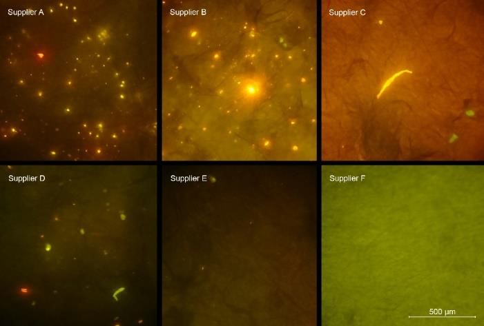
Microplastic extraction: What do we have to consider?

                 procedural contamination // Loss due to adsorption on labware surfaces

      insufficient digestion                                filter pore size                                     polymer identification

    degradation of plastics                                  filter material

              Digestion                                          Filtration                                              Analysis

                                                                 illustrations: designed by freepik.com
MRI – Institut für Sicherheit und Qualität bei Milch und Fisch   lab equipment: Landesbildungsserver Baden-Württemberg
                                                                                                                            15.04.2021    3
Evaluation of sample preparation protocols

                Literature research                                 efficiency
     Which protocols are regularly applied when
              digesting aquatic biota?                               integrity

                                                                                                 time
      Evaluation of digestion methods
 •     Are fish fillets, the soft tissue of mussels                                         steps
       and crustaceans digested sufficiently for
       filtration with pore size 1 µm?                                                           costs

 •     Are plastic particles not degraded?                                         alkaline (60 °C)        alkaline (25 °C)     acidic                 alkaline-acidic
 •     Is the method suited for routine analysis?                                  oxidative               alkaline-oxidative   enzymatic              enzymatic-alkaline

                                                                 Figure 2: Performance of digestion methods applied for isolating MP from fish fillet.

                     Optimisation                                                                 100.00
                                                                                                              fishes                     crustaceans         molluscs

                                                                      digestion efficiency [%]
 •     Which parameters have to be changed                                                         99.50
       for minimizing plastic degradation?                                                         99.00
 •     What measures have to be applied                                                            98.50
       regarding different analytical techniques                                                   98.00
                                                                                                   97.50
       or a broad range of sample matrices?
                                                                                                   97.00

                In-House-Validation
  Is the protocol suited for quantitative isolation
          of microplastics from seafood?                          Figure 3: Digestion efficiency of edible parts from different seafood species.
                                                                  Fishes are sorted according to their fat content (increasing).
MRI – Institut für Sicherheit und Qualität bei Milch und Fisch                                                                                  15.04.2021                  4
Optimisation: Towards a negligible impact on plastic particles
 polymer              recovery                 identification                                                     •    recovery based on weight
              weight [%]    area [%]    FTIR    Raman    py-GC/MS                                                      might not detect changes
 PA6          96 ± 2       104 ± 2       +        +         +                                                          in small surface layer
                                                                                                                  •    loss of small micro- &
      40 ºC   95 ± 1       not tested                                                                                  nanoplastics undetected
              98 ± 2
 PA12                            /       +        +         +
                                                                                                                  •    reduction of PET-particle
 PAN              -              -       -        +/        +/
                                                    -         -                                                        area at 60 ºC alkaline
 PC           96 ± 2        97 ± 1       +        +         +                                                          digestion but not at 40 ºC
                                                                    Figure 4: Photograph of a PET-particle before
      40 ºC 95 ±
Optimisation: The importance of filter choice
 •    improving filtration speed & preventing filter clogging, depending on…
         → pore size: larger pore size = less prone to clogging, but also loss of
                                                                                     cellulose nitrate         glass fiber
              smaller, probably more abundant, plastic particles
         → filter material: adsorption of matrix residues (e.g. proteins)
 •    compatibility of filter material and sample preparation, analytical methods
         → e.g. degradation of filter material by digestion solutions                 cellulose acetate polycarbonate
                                                                                    Figure 6: Photograph of membrane
         → e.g. inorganic filters for thermal analysis                              filters (pore size ~ 1 µm) after filtering
                                                                                    digested fish fillet.
 •    filter structure: impact on particle retention & detection[20]
                                                                                     knitted lattice        pressed fiber
         → missing fragments with multilayer/fiber-type (hidden between layers)
              (e.g. cellulose nitrate, cellulose fiber/paper, glass fiber)
         → loss of fibers with singlelayer-type (passing pores lengthwise)[20]
                                                                                           nylon              cotton fiber
              (e.g. polycarbonate, Al2O3)                                            multilayer-hole singlelayer-hole

                                                                                      mixed cellulose        polycarbonate
                                                                                    Figure 7: SEM-image of surface
                                                                                    morphology-types of membrane filters;
                                                                                    Cai et al. (2020).
MRI – Institut für Sicherheit und Qualität bei Milch und Fisch                                     15.04.2021                    6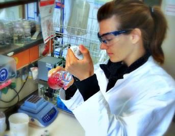
Optimisation: The importance of filter choice

                                                                                    Figure 8: Fluorescent PA12-particles on
                                                                                    glass fiber filters. Scan on same focal plane
                                                                                    (left) and stacked images of confocal scan
                                                                                    (range 100 µm).

         → consideration of focal plane of particles for imaging/filter scan

 •    perspective: adsorption of nanoparticles → incomplete separation
                                                                               Figure 9: SEM-image of polycarbonate filter
 → further research regarding filtration required                              (pore size 1 µm) with agglomerated Ø100 nm-PS
                                                                               adhering to the pores & matrix residues.

MRI – Institut für Sicherheit und Qualität bei Milch und Fisch                                     15.04.2021                7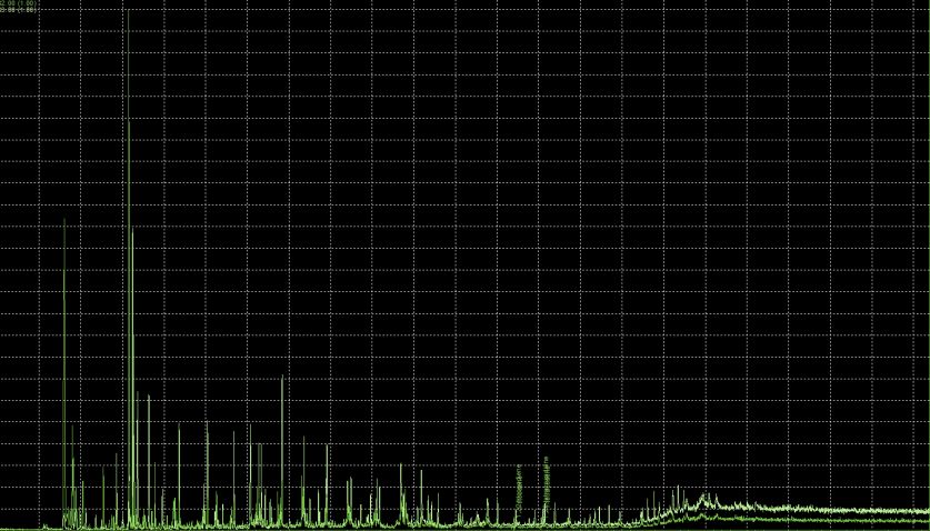
Optimisation: Preventing procedural plastic contamination

                                                                  particle number
•   small plastic particles are ubiquitous →                                        2500
                                                                                    2000
    monitoring & mitigation of contamination                                        1500

•   investigating probable sources                                                  1000
                                                                                     500
       → insufficiently cleaned glassware                                              0

       → reagents / solvents
       → exposure of samples to air
                                                                 Figure 10: Number of MP-suspect particles rinsed off glass
                                                                 flasks after application of different cleaning procedures.

                                                                 Figure 11: Photographs of Nile red-stained filters after
                                                                 filtration of pepsin from different suppliers. Particles with
                                                                 green, yellow or orange fluorescence are MP-suspect.

MRI – Institut für Sicherheit und Qualität bei Milch und Fisch                                      15.04.2021                   8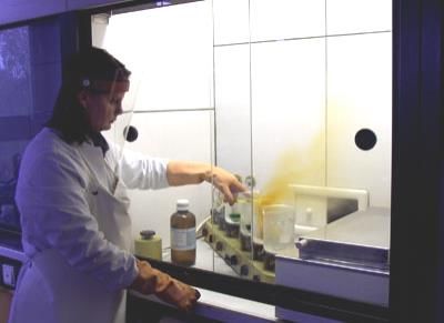
Optimisation: Preventing procedural plastic contamination
                                                                                   450

                                                                 particle number
                                                                                                      extraction                sedimentation
                                                                                   400

                                                                                   350                                       2 – 10 µm      11 – 20 µm

                                                                                   300                                       21 – 50 µm     51 – 100 µm

                                                                                   250                                       101 – 200 µm

                                                                                   200

                                                                                   150

                                                                                   100
•   current protocol for contamination prevention
                                                                                   50
       → cotton clothes, laminar flow workbench                                     0
                                                                                           heated fume hood   laminar    laboratory fume hood   laminar
       → pre-filtration (pore size < 1 µm) of all                                        glassware              flow                              flow

            reagents & solutions                                      Figure 12: Number of fluorescent particles (Nile red staining, FITC-filter) of
                                                                      heated glassware, a simulated extraction procedure and sedimented particles
       → cleaning of glassware [and filters]                          from air.

            dishwasher, heating (500 ºC), rinsing                                                                       30

       → rinsing of filtration apparatus between                                                                        20
                                                                                                                        10
             each sample (3x 10 mL filtered water)                                                                      0

•    monitoring of blank samples still required

                                                                                                     Figure 13: Number MP-suspect particles in blank samples.

MRI – Institut für Sicherheit und Qualität bei Milch und Fisch                                                                    15.04.2021              9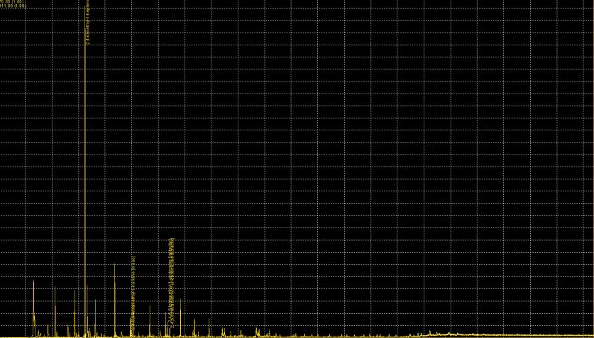
Validation of the optimised sample preparation protocol
                                                                                     m/z = 122 m/z = 113
  digestion

                                                                                     m/z1 = 130 m/z1 = 70                                        PA-6
                                                                                     m/z2 = 117 m/z2 = 111
                                                                                                                                                   PS
                                                                                     m/z1 = 82
                                                                                     m/z2 = 83
                                                                                                                                                   PE
  fitration & post-filtration treatment

                                                                                                                                                  PET

                                                                                                                                                   PP

                                                                                  Figure 15: Pyrogram of nine commercially relevant synthetic polymers
                                                                                  spiked to herring fillet and isolated with the optimized protocol. The filter
                                                                                  was silanized with TMCS before pyrolysis. The black chromatogram is
                                                                                  the TIC.
                                                                                                                           Recovery
                                                                                                                           n = 10        88 ± 16 %
                                                                                                                           n = 100       89 ± 12 %
                                                                                                                           n = 1000      103 ± 13 %

                                                                                                                           further research for
                                                                                                                           quantification of plastics
                                                                                                                           with py-GC/MS needed
 Figure 14: Schematic overview of optimised sample preparation protocol.
                                                                   illustrations: designed by freepik.com
MRI – Institut für Sicherheit und Qualität bei Milch und Fisch     lab equipment: Landesbildungsserver Baden-Württemberg
                                                                                                                                    15.04.2021            10
Prospective: Consideration of nanoplastics
Sample • concentration and separation of nano- and microplastics
preparation? • detection limit in field-flow-fractionation
• identification of plastic in the fractions
≥ 1 µm
< 1 µm
Detection limit?
Figure 16: AF4-separation of different amounts of nanoplastics.
MRI – Institut für Sicherheit und Qualität bei Milch und Fisch lab equipment: Landesbildungsserver Baden-Württemberg 15.04.2021 11Summary
• Optimised procedure for isolation of microplastics from edible part of seafood:
→ two-step digestion with pepsin (enzymatic) and KOH (alkaline) at ~ 37 ºC
→ filtration with filters of 1 µm pore size, Ø 47 mm (e.g. glass fiber, polycarbonate)
→ if required: filter bleaching with H2O2 (dark residues), degreasing with alcohol
• Necessity of blank samples even with thorough protocol for preventing microplastic
contamination; important aspects: purity of reagents, cleaning of glassware
• Choice of filter material has a great impact on filtration speed/matrix residues,
microplastic retention[20], and particle detection → more research required
• more research required regarding sample preparation for nanoplastics from seafood
details published in: Süssmann, Julia, et al. "Evaluation and optimisation of sample preparation protocols suitable for the
analysis of plastic particles present in seafood." Food Control 125 (2021): 107969.
Max Rubner-Institut – Bundesforschungsinstitut für Ernährung und Lebensmittel 15/04/2021 12Thank you for your support…
Federal Research Institute
of Nutrition and Food
Safety and Quality of Milk
and Fish
Jan Fritsche University of Hamburg
Torsten Krause Center for Earth System
Dierk Martin Research and Sustainability
Ute Ostermeyer Elke Fischer
Enken Jacobsen Matthias Tamminga
Björn Neumann …
Longina Reimann
Food Chemistry
Food Technology and
Bioprocess Engineering Technical University
Ralf Greiner Berlin
Elke Walz
Sascha Rohn
Birgit Hetzer
Andrea Tauer
Christian Geuter
Max Rubner-Institut – Bundesforschungsinstitut für Ernährung und Lebensmittel 15.04.2021 13Thank you for your
attention!
MRI – Institut für Sicherheit und Qualität bei Milch und Fisch 15.04.2021 14References [1] Abidli, Sami, Youssef Lahbib, and Najoua Trigui El Menif. "Microplastics in commercial molluscs from the lagoon of Bizerte (Northern Tunisia)." Marine pollution bulletin 142 (2019): 243-252. [2] Bråte, Inger Lise N., et al. "Mytilus spp. as sentinels for monitoring microplastic pollution in Norwegian coastal waters: A qualitative and quantitative study." Environmental Pollution 243 (2018): 383-393. [3] Catarino, Ana I., et al. "Low levels of microplastics (MP) in wild mussels indicate that MP ingestion by humans is minimal compared to exposure via household fibres fallout during a meal." Environmental pollution 237 (2018): 675-684. [4] Van Cauwenberghe, Lisbeth, and Colin R. Janssen. "Microplastics in bivalves cultured for human consumption." Environmental pollution 193 (2014): 65-70. [5] Cho, Youna, et al. "Abundance and characteristics of microplastics in market bivalves from South Korea." Environmental pollution 245 (2019): 1107-1116. [6] De Witte, B., et al. "Quality assessment of the blue mussel (Mytilus edulis): Comparison between commercial and wild types." Marine pollution bulletin 85.1 (2014): 146-155. [7] Digka, Nikoletta, et al. "Microplastics in mussels and fish from the Northern Ionian Sea." Marine pollution bulletin 135 (2018): 30-40. [8] Ding, Jinfeng, et al. "Detection of microplastics in local marine organisms using a multi-technology system." Analytical Methods 11.1 (2019): 78-87. [9] Fischer, Elke. "Distribution of microplastics in marine species of the Wadden Sea along the coastline of Schleswig- Holstein, Germany." Final Report University Hamburg (2019). [10] Iversen, Karine Bue. Microplastics in blue mussels (Mytilus edulis) from the marine environment of coastal Norway. MS thesis. Norwegian University of Life Sciences, Ås, 2018. MRI – Institut für Sicherheit und Qualität bei Milch und Fisch 15.04.2021 15
References
 [11] Karlsson, Therese M., et al. "Screening for microplastics in sediment, water, marine invertebrates and fish: method
 development and microplastic accumulation." Marine pollution bulletin 122.1-2 (2017): 403-408.
 [12] Li, Jiana, et al. "Microplastics in mussels along the coastal waters of China." Environmental pollution 214 (2016):
 177-184.
 [13] Li, Jiana, et al. "Microplastics in mussels sampled from coastal waters and supermarkets in the United Kingdom."
 Environmental pollution 241 (2018): 35-44.
 [14] Lusher, A. L., et al. "Sampling, isolating and identifying microplastics ingested by fish and invertebrates." Analytical
 methods 9.9 (2017): 1346-1360.
 [15] Mankin, Chloe, and Andrea Huvard. "Microfibers in Mytilus species (Mollusca, Bivalvia) from Southern California
 Harbors, Beaches, and Supermarkets.“
 [16] Murphy, Fionn, et al. "The uptake of macroplastic & microplastic by demersal & pelagic fish in the Northeast Atlantic
 around Scotland." Marine pollution bulletin 122.1-2 (2017): 353-359.
 [17] Reguera, Pablo, Lucía Viñas, and Jesús Gago. "Microplastics in wild mussels (Mytilus spp.) from the north coast of
 Spain." Scientia Marina 83.4 (2019): 337-347.
 [18] Li, Jiana, et al. "Microplastics in commercial bivalves from China." Environmental pollution 207 (2015): 190 -195.
 [19] Gomiero, Alessio, et al. "First occurrence and composition assessment of microplastics in native mussels collected
 from coastal and offshore areas of the northern and central Adriatic Sea." Environmental Science and Pollution Research
 26.24 (2019): 24407-24416.
 [20] Cai, Huiwen, et al. "Microplastic quantification affected by structure and pore size of filters." Chemosphere 257
 (2020): 127198.

MRI – Institut für Sicherheit und Qualität bei Milch und Fisch                                             15.04.2021            16
You can also read