Evaluation of acute and subacute toxicity of ethanolic extract and fraction of alkaloids from bark of Aspidosperma nitidum in mice
←
→
Page content transcription
If your browser does not render page correctly, please read the page content below
www.nature.com/scientificreports
OPEN Evaluation of acute and subacute
toxicity of ethanolic extract
and fraction of alkaloids from bark
of Aspidosperma nitidum in mice
Heliton Patrick Cordovil Brígido1, Everton Luiz Pompeu Varela2,3,
Antônio Rafael Quadros Gomes1,3, Mirian Letícia Carmo Bastos2,
Andre de Oliveira Feitosa4, Andrey Moacir do Rosário Marinho4, Liliane Almeida Carneiro5,
Márlia Regina Coelho‑Ferreira6, Maria Fâni Dolabela1,2 & Sandro Percário2,3*
This study investigated the acute and subacute toxicity of the ethanolic extract (EE) and alkaloid
fraction (FA) from A. nitidum. The EE was obtained from trunk bark with ethanol, FA was obtained
from the fractionation of EE. To test the acute toxicity, mice were divided into four groups, and the
negative controls received water or aqueous solution of dimethyl sulfoxide, whereas the others
received EE or FA (2000 mg/kg, orally, single dose). The same controls were used in the subacute
trial. However, the animals were treated for 28 days, and the dose used was 1000 mg/kg per day of
EE and FA. Daily clinical evaluations of the animals were performed. At the end of the experiment,
hematological, biochemical, and histopathological assessments (liver, lung, heart, and kidney) were
performed. In the acute and subacute toxicity studies, mice treated with EE and FA did not show any
clinical changes, there were no changes in weight gain, hematological and biochemical parameters
compared to the control groups (p > 0.05). In the histopathological examination, there was no
abnormality in the organs of the treated animals. Therefore, EE and FA did not produce toxic effects in
mice after acute and subacute treatment.
Aspidosperma nitidum Benth. Ex Müll. Arg (Apocynaceae), popularly known as Carapanaúba, is a plant found
in the Brazilian A mazon1 and widely used in local medicine to treat febrile illnesses, m
alaria2, uterus and ovary
inflammation, diabetes, cancer, contraception, stomach p roblems3, and r heumatism4. In addition, indigenous
people use its latex to treat leprosy3.
Species belonging to the genus Aspidosperma are chemically characterized by the occurrence of indolic
alkaloids5–7. In this context, the therapeutic properties of A. nitidum are mainly attributed to alkaloids8, with the
following compounds having been isolated: 10-methoxydihydrocorynantheol (Fig. 1-1), corynantheol (Fig. 1-2)9,
aspidospermine (Fig. 1-3), quebrachamine (Fig. 1-4), yohimbine (Fig. 1-5)10, carboxylic harman acid (Fig. 1-
6), 3-carboxylic ethylharman (Fig. 1-7)8, dihydrocorynantheol (Fig. 1-8), dehydrositsiriquine (Fig. 1-9), and
braznitidumine (Fig. 1-10)11.
A study demonstrated that the ethanolic extract from the stem bark of A. nitidum showed anti-inflammatory
activity in the carrageenan induced rat paw edema method, with braznitidumine being responsible for this
activity12.
Another study accessed the in vitro antimalarial activity of EE (IC50 = 3.6 µg/mL) and FA (IC50 = 2.32 µg/
mL), both displaying low cytotoxicity for HepG2 cells (EE, I C50 = 410.65 µg/mL; FA, IC50 = 346.73 µg/mL), and
highly selectivity for antimalarial activity (Selective Index—SI: EE = 114.07; FA = 149.45). The ethanolic extract
1
Post‑graduate Program in Pharmaceutical Innovation, Institute of Health Sciences, Federal University of
Pará, Belém, PA, Brazil. 2Post‑graduate Program in Biodiversity and Biotechnology (BIONORTE), Institute
of Biological Sciences, Federal University of Pará, Belém, PA, Brazil. 3Oxidative Stress Research Laboratory,
Institute of Biological Sciences, Federal University of Pará, Av. Augusto Corrêa, 01, Belém, PA 66075‑110,
Brazil. 4Post‑graduate Program in Chemistry, Institute of Exact and Natural Sciences, Federal University of
Pará, Belém, PA, Brazil. 5National Primate Center, Evandro Chagas Institute, Ananindeua, PA, Brazil. 6Botany
Coordination, Museu Paraense Emílio Goeldi, Ministério da Ciência, Tecnologia, Inovação e Comunicações, Belém,
PA, Brazil. *email: percario@ufpa.br
Scientific Reports | (2021) 11:18283 | https://doi.org/10.1038/s41598-021-97637-1 1
Vol.:(0123456789)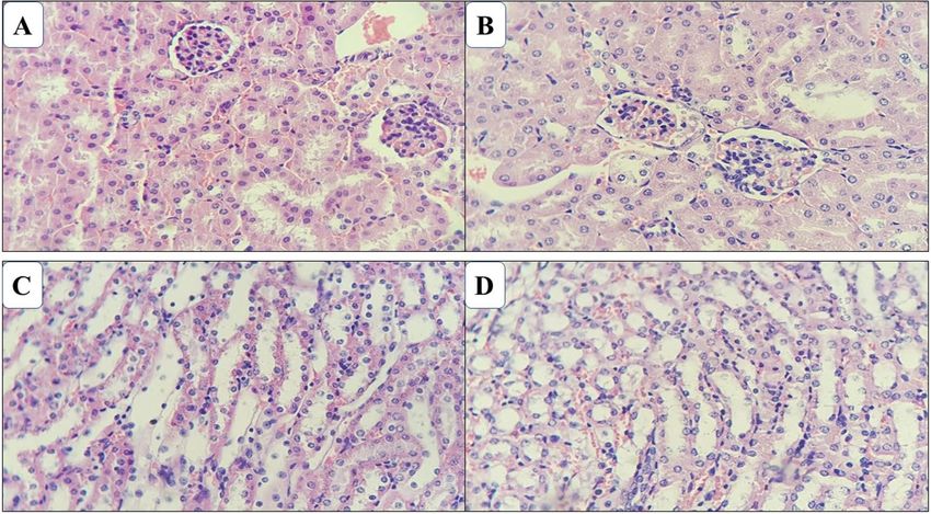
www.nature.com/scientificreports/
Figure 1. Chemical structure of compounds occurring in Aspidosperma nitidum.
10-methoxydihydrocorynantheol (1), corynantheol (2), aspidospermine (3), quebrachamine (4), yohimbine (5),
carboxylic harman acid (6), 3-carboxylic ethylharman (7), dihydrocorynantheol (8), dehydrositsiriquine (9),
braznitidumine (10).
and the alkaloid fraction reduced the parasitemia of mice infected with Plasmodium berghei (ANKA) by 80%
(dose of 500 mg/kg) on the 5th day. In preliminary studies of acute oral toxicity, the ethanolic extract (5000 mg/
kg; gavage) presented low toxicity, with no clinical or anatomopathological c hanges13.
Despite its wide use in popular medicine, its phytochemical diversity, and studies that validate its clinical use,
toxicity studies are still scarce, and the results are, at most, preliminary. Thus, one important component of this
study was the scientific assessment of the A. nitidum safety and toxicity.
This study result may fill the gap of previous studies about the species and provide some additional evidence
to recommend further studies to assess the toxicity profiles associated with the use of herbal preparations from
this plant as well. Thus, this study aimed to investigate the acute and subacute toxicity of the ethanolic extract
(EE) and alkaloid fraction (FA) obtained from trunk bark of A. nitidum.
Methods
Plant material. Trunk barks of A. nitidum were collected on the state highway PA-150 (coordinates S 02°
09′ 50.3″ and W 048° 47′ 56.9″), in the state of Pará-Brazil, during August 2017. The plant material was identi-
fied by Dr. Márlia Regina Coelho-Ferreira and the exsiccate was deposited at the Herbarium João Murça Pires
of the Museu Paraense Emílio Goeldi, under no. MG206608. In the present study, we used a wild plant collected
from a virgin forest of the Amazon, and our work posed no risk of extinction for the species. During the col-
lection, we took all care to remove the barks so as not to cause any damage to the species, in addition, only a
small proportion of the barks were collected. The species were kept integrated and survived the collection. The
project complies with national and international guidelines and legislation and is registered on the platform
of the National Management and Genetic Heritage System and Associated Traditional Knowledge (SISGEN),
whose provided license to collect the species under registration A2C3188. Moreover, according to the IUCN
2019 red list of endangered species, Aspidosperma excelsum, a synonymy of Aspidosperma nitidum is classified
oncern14.
as Least C
Preparation of extract and fraction of alkaloids. The barks of A. nitidum were washed under run-
ning water and dried in a circulating air oven (40 °C, for 7 days). The dry material was subjected to grinding in
a knife mill. The plant powder was subjected to maceration with ethanol at 96°GL (1:10 ratio). The ethanolic
solution was filtered and concentrated on a rotary evaporator under reduced pressure until total evaporation of
the alcohol, yielding the dry ethanolic extract (EE). The EE (5 g) was subjected to acid–base partition, being solu-
bilized in ethanol (4.0 mL), then 3% aqueous hydrochloric acid solution (7.5 mL) was added. This solution was
extracted with dichloromethane (250 mL for three times), yielding the neutral fraction (FN). The acidic aqueous
layer was made alkaline with 10% ammonium hydroxide (NH4OH) until pH 9, followed by a new extraction
with dichloromethane (250 mL for three times), yielding an alkaline aqueous layer and an organic layer (fraction
of alkaloids—FA).
High performance liquid chromatography coupled to a diode array detector (HPLC–DAD). The
HPLC–DAD analyzes of the ethanolic extract and FA were carried out according to the adapted methodology of
Coutinho et al.15. The extract and FA (1 mg) were solubilized in HPLC grade methanol (1 mL) under sonication
Scientific Reports | (2021) 11:18283 | https://doi.org/10.1038/s41598-021-97637-1 2
Vol:.(1234567890)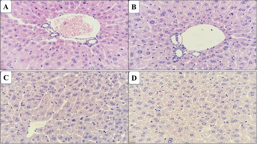
www.nature.com/scientificreports/
(ultrasound) for 15 min. The column used was LiChrospher 100 RP-18 (particles of 5 mm, 250 × 4 mm d.i.), UV
detection at 220–400 nm, flow of 0.5 mL/min, at 40 °C. Water (eluent A) and acetonitrile (eluent B) were used
as the mobile phase. A linear gradient was used: 70–30% of eluent B for 15 min, 60–40% of eluent B for 20 min,
50–100% of eluent B for 25 min.
Animals. Thirty-two (thirty-two) healthy Balb/c male mice (Mus musculus), adults, aged 6–8 weeks, weigh-
ing between 25 and 35 g, from the Vivarium of the Evandro Chagas Institute (Ananindeua-Pará, Brazil) were
used. The animals were housed in the Experimental Vivarium of the Oxidative Stress Research Laboratory of
ICB/UFPA, in polypropylene cages (30 × 19 × 13 cm), with a stainless-steel wire cover, containing a bed of Pine
shaving, with a maximum of five animals per cage and kept at room temperature (24 ± 2 °C) and light/dark cycle
every 12 h. Before and during the study period, the animals were kept with food (Presence, São Paulo-SP, Brazil)
and water ad libitum. Before any experimental procedure, the animals were acclimated to laboratory conditions
for 15 days. The experimental procedures with mice were performed at the Oxidative Stress Research Laboratory
(LAPEO/ICB/UFPA) and were performed according to the ethical standards of animal experimentation indi-
cated by the Brazilian Society of Laboratory Animal Science (SBCAL) and international s tandards16.
While under the effects of the anesthesia (9 mg/kg of ketamine 10% and 10 mg/kg of xylazine 2%), animals
underwent euthanasia through hypovolemia induction, after the collection of the total volume of blood available
from each animal by cardiac puncture.
Ethics declaration. All animal procedures were strictly in accordance with the National Institutes Guide
nimals16 and approved by the Animal Use Ethics Committee of the Evandro
for the Care and Use of Laboratory A
Chagas Institute (CEUA-IEC), under the number 38/2017. Furthermore, this study was conducted according to
ARRIVE guidelines17.
Experimental procedures. Acute toxicity assessment. The acute oral toxicity test was performed accord-
ing to the experimental protocol Guideline 423 of the Organization for Economic Cooperation and Development
(OECD)18, with an initial dose of 2000 mg/kg of EE or FA. The number of animals used in this evaluation fol-
lowed OECD (2001) guidance and reduction principle19, i.e., using the lesser possible number of animals to
obtain statistical relevance.
The animals were randomly divided into four groups (n = 3). The first group received orally a single dose of
EE (2000 mg/kg) dissolved in water. The second group received orally a single dose of FA (2000 mg/kg) dissolved
in an aqueous solution containing 99:1 (v/v) dimethyl sulfoxide. The last two groups (control groups) received
water (third group) and aqueous solution containing 99:1 (v/v) dimethyl sulfoxide (fourth group).
The following parameters were observed during the test; general activity, vocal frantic, irritability, touch
response, tail grip response, contortion, posterior train position, straightening reflex, body tone, force to grasp,
ataxia, auricular reflex, corneal reflex, tremors, convulsions, anesthesia, lacrimation, ptosis, urination, defecation,
piloerection, hypothermia, breathing, cyanosis, hyperemia, and death.
Subacute toxicity assessment. For the subacute toxicity assessment of repeated doses of EE and FA, the meth-
odology described in Guide 407 of the OECD g uidelines20 and Brito21 was used, using the limit test with a dose
of 1000 mg/kg of EE or FA.
The animals were randomly divided into four groups (n = 5). The first group received an oral dose of EE
(1000 mg/kg) dissolved in water for 28 days. The second group received orally a daily dose of FA (1000 mg/kg)
dissolved in an aqueous solution containing 99:1 (v/v) dimethyl sulfoxide for 28 days. The last two groups (con-
trol groups) received daily water (third group) and aqueous solution containing 99:1 (v/v) dimethyl sulfoxide
(fourth group) for 28 days. The animals were observed daily during the experiment to detect death or abnormal
clinical signs.
Observational parameters. After sample administration, the animals were kept under close observation con-
tinuously for 1 h and intermittently for the next 4 h and, thereafter, once every 12 h for the next 14 days for the
acute toxicity assessment, and for 28 days for the subacute toxicity study.
Throughout the study period, clinical observations were made for mortality, behavioral, neurological, or other
abnormalities, and their weight was measured weekly until the last day of experimentation.
The animals were evaluated at 30 min, 1 h, 2 h, 4 h, 6 h, 12 h, and 24 h and, from then on, daily, until the 14th
day after treatment. The following signs were evaluated following Hippocratic screening: general activity, vocal
frantic, irritability, touch response, tail grip response, contortion, posterior train position, straightening reflex,
body tone, force to grasp, ataxia, auricular reflex, corneal reflex, tremors, convulsions, anesthesia, lacrimation,
ptosis, urination, defecation, piloerection, hypothermia, breathing, cyanosis, hyperemia, and death. The signs
evaluated by behavioral observation and systematic clinical examination of the animals were recorded in a printed
protocol with the list of signs to be i nvestigated22,23.
Hematological parameters. At the end of the experiment, animals were anesthetized (9 mg/kg of ketamine 10%
and 10 mg/kg of xylazine 2%), and blood samples were drawn from each animal by cardiac puncture. The blood
was placed in two groups of test tubes, half of the tubes containing the anticoagulant ethylenediamine tetra acetic
acid (EDTA) and the other half without anticoagulant.
Blood samples in test tubes containing EDTA were used to determine hematological parameters: white blood
cell count (WBC), red blood cell count (RBC), hemoglobin (HGB), hematocrit (HCT), mean cell volume (MCV),
Scientific Reports | (2021) 11:18283 | https://doi.org/10.1038/s41598-021-97637-1 3
Vol.:(0123456789)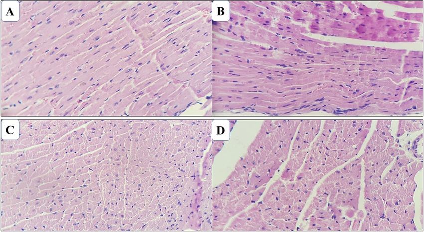
www.nature.com/scientificreports/
mean corpuscular hemoglobin concentration (MCHC), mean corpuscular hemoglobin (MCH), and platelet
count (PLT), determined by the automatic method using the BC-2800 VET/Mindray device (Mindray do Brasil
Ltda.; São Paulo, SP-Brazil). Differential counting was performed using a smear of blood stained by the panoptic.
Blood samples in test tubes without anticoagulant were left at room temperature to clot, and serum was
obtained by centrifugation (3000 rpm for 10 min). Subsequently, biochemical analyzes were performed on serum
to quantify aspartate aminotransferase (AST) and alanine aminotransferase (ALT) to assess liver damage and
creatinine and urea to assess kidney damage, using an automated biochemical system. Standard commercial
reagents (Labtest®, Labtest Diagnóstica SA, Lagoa Santa-MG, Brazil) were used, with kinetic, enzymatic, or col-
orimetric methods, at 37 °C, and the reading was performed on a semi-automatic spectrophotometer (Bio-Plus®
Biochemical Analyzer; Bioplus Produtos para Laboratórios Ltda.; Barueri-SP, Brazil).
Histopathological parameters. After euthanasia, liver, kidney, lung, and heart samples (1 cm thick) were col-
lected for histopathological examination21. The heart, liver, and kidneys were sectioned by sagittal incision. The
tissue sections were fixed in buffered formalin (10% formaldehyde) and after 24 h, cleaved for histopathologi-
cal processing: dehydration with increasing series of alcohol (70°–100°), followed by diaphanization in xylol,
impregnation and inclusion in paraffin, according to the usual methods24. In a microtome, tissue fragments
(3.0 µm) were sectioned with subsequent hematoxylin–eosin staining for microscopic examination (40× and
100×). Slides were evaluated by an independent certified histopathologist, and the results were confirmed by a
second independent certified histopathologist.
Statistical analysis. The results obtained in each experiment were compared to its matched control group
(i.e., EE-treated animals versus water-treated animals and FA-treated animals versus DMSO/water-treated ani-
mals) by Student’s T-test using Excel program, with a 95% confidence level and a significance level of α = 5%
(p < 0.05). The variables analyzed were expressed as mean ± standard deviation.
Results
HPLC–DAD analysis of A. nitidum extract and fractions. The EE chromatogram showed substances
of high, medium, and low polarity. The main peaks of 5.5 min (λmax 219.4, 272.6, and 364.1 nm), 5.7 min (λmax
219.0; 271.4 and 358.2 nm), 6.3 min (λmax 218.2; 272.6 and 376.1 nm), and 6.6 min (λmax 271.4 and 357.2) showed
ultraviolet spectra. The peaks with retention times of 9.8 min (λmax 221.8 and 272.5 nm) and 10.9 min (λmax 221.7
and 296.3 nm) showed absorbance (Fig. 2).
The FA chromatogram suggests that the main peaks identified at times of 4.9 min (λmax 222.9 and 298.7 nm),
6.7 min (λmax 220.6 and 272.6 nm), 7.5 min ( 220max 220.6 and 271.4 nm), 8.2 min (λmax 221.7 and 296.3 nm;
Fig. 3).
Acute toxicity assessment. The EE and FA orally administered at a dose of 2000 mg/kg in male mice did
not induce any death or toxic symptoms in treated mice. All animals displayed normal behavior throughout the
study and survived until the end of the 14-day experiment period. During the entire observation period, they
did not present any significant clinical alteration. Furthermore, no significant difference was observed between
the weight gain in these groups in relation to the controls (Table 1). Hematological analyzes showed that EE and
FA did not cause significant changes of any of the parameters (Tables 2 and 3) and did not alter renal or hepatic
function (Table 4), remaining within the reference range. As a result, the L D50 of EE and FA was greater than
2000 mg/kg of body weight.
Subacute toxicity studies. The ethanolic extract and the fraction of alkaloids orally administered to male
mice daily treated with 1000 mg/kg/28 days did not induce any death or toxic symptoms to the animals, whose
behaved normally throughout the study and survived until the end of the experiment (28 days). For the dura-
tion of the experiment, no significant clinical changes were observed, and the weight gain was also similar to
the controls (WCG and DCG; Table 5). There were no changes in the hematological parameters of the mice
(Tables 6 and 7). Moreover, no significant changes were observed in renal and hepatic function tests (Table 8).
These parameters were within the reference range.
Histopathological examination of the viscera of the animals surviving the oral treatment of repeated doses
of EE and AF obtained from the bark of A. nitidum at a dose of 1000 mg/kg/28 days, did not detect relevant
changes both in the animals of the control group and in those treated with the samples. In the kidneys, lobular
architecture was preserved with medullary pyramids covered with cortical tissue. The cortex presented regularly
distributed glomeruli, fine Bowman capsule. The proximal and distal contorted tubules and the segment of the
collecting duct did not evidence histological particularities, as did the Henle loops and collecting ducts of the
medullary pyramid. No inflammatory reaction was observed or fibrosis in the interstice (Fig. 4).
In the heart, the myocardium was represented by cardiac striated muscle cells, presenting transverse stria-
tions, and single or double nuclei, centrally positioned. The endocardium had endothelium supported by a thin
basal membrane and coated cavities and heart valves. The epicardium, coated with mesothelial pavement cells,
were fully preserved (Fig. 5).
The liver was observed that the hepatic parenchyma (consisting of the lobular center vein surrounded by
hepatocytes cords and sinusoid capillaries) were regularly distributed and with preserved structures. The hepato-
cyte presented a polygonal shape with spherical nucleus and centralized fully preserved (Fig. 6).
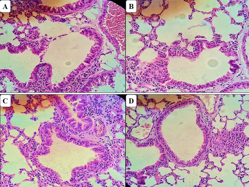
Histopathological evaluation of lung tissue sections in mice of all test groups showed a normal morphologi-
cal architecture without any pathological changes related to treatment and was similar to that of mice in the
control group (Fig. 7).
Scientific Reports | (2021) 11:18283 | https://doi.org/10.1038/s41598-021-97637-1 4
Vol:.(1234567890)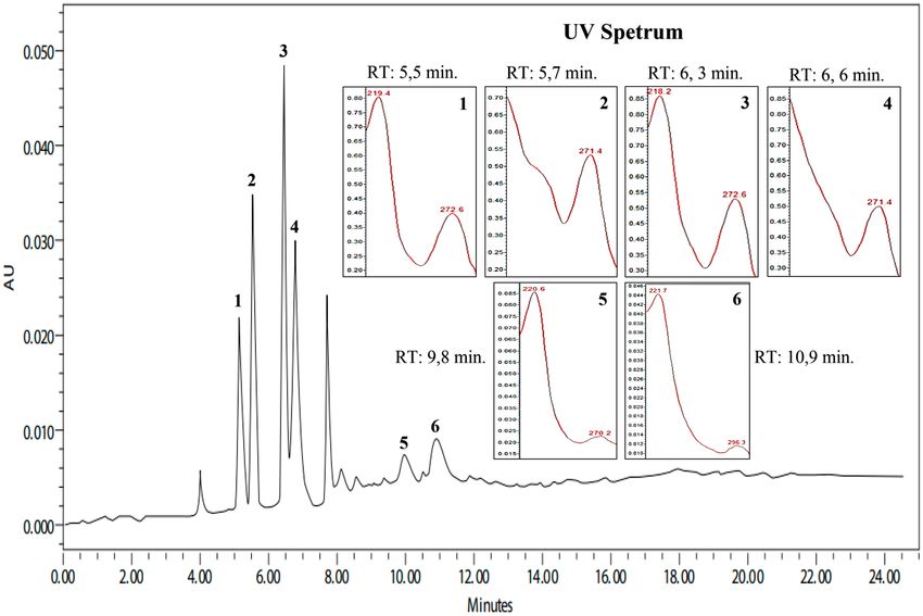
www.nature.com/scientificreports/

                                  Figure 2.  Chromatographic profile and UV spectra of the ethanolic extract of Aspidosperma nitidum. λ = 250–
                                  400 nm.

                                  Figure 3.  Chromatographic profile and UV spectra of the alkaloid fraction of Aspidosperma nitidum. λ = 250–
                                  400 nm.

                                  Discussion
                                  In this study we sought to investigate the acute and subacute toxicity of the ethanolic extract (EE) and the frac-
                                  tion of alkaloids (FA) obtained from A. nitidum, and our results showed that both the extract and the FA did
                                  not present toxicity.

Scientific Reports |   (2021) 11:18283 |               https://doi.org/10.1038/s41598-021-97637-1                                                 5

                                                                                                                                             Vol.:(0123456789)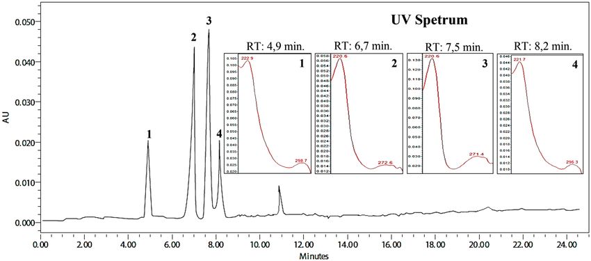
www.nature.com/scientificreports/
Weight (g)
Treatment duration (days) EE WCG p*
0 27.06 ± 0.75 26.63 ± 0.20 0.183
7 27.9 ± 1.15 27.73 ± 0.11 0.742
14 27.26 ± 1.20 28.63 ± 0.15 0.225
FA DCG (p)
0 26.23 ± 0.25 26.86 ± 0.58 0.183
7 27.06 ± 0.05 27.86 ± 0.35 0.423
14 28.1 ± 0.20 28.90 ± 0.17 0.230
Table 1. Weight of mice treated with ethanolic extract or fraction of alkaloids from Aspidosperma nitidum.
EE ethanolic extract, FA fraction of alkaloids, WCGwater control group, DCG DMSO control group (99%
water + 1% DMSO). *p value was obtained by Student’s T-test, comparing EE versus WCG, or FA versus DCG.
Groups RV25
Parameters EE WCG p* LABC CPqRR
Red blood cells (106/mm3) 6.60 ± 0.44 6.14 ± 1.13 0.251 7.4–11.1 5.8–10.5
Hematocrit (%) 35.36 ± 2.54 37.9 ± 0.56 0.647 34.3–51.3 28.3–68.3
Hemoglobin (g/dL) 13.1 ± 0.36 11.46 ± 2.72 0.346 11.5–15.9 10.1–18.4
MCV 53.5 ± 1.74 52.7 ± 0.49 0.851 46.8–48.0 47.0–51.0
MCH 19.86 ± 0.76 18.5 ± 1.58 0.275 14.1–16.9 14.6–18.5
MCHC 37.1 ± 1.57 35.3 ± 2.66 0.398 30.5–35.4 32.4–37.2
Platelet (103/mm3) 1329.3 ± 29.02 1342.5 ± 72.8 0.439 635–1118 179–1025
FA DCG P* LABC CPqRR
Red blood cells (106/mm3) 6.71 ± 1.38 6.77 ± 0.06 1.000 7.4–11.1 5.8–10.5
Hematocrit (%) 35.33 ± 9.84 37.93 ± 0.75 0.716 34.3–51.3 28.3–68.3
Hemoglobin (g/dL) 12.66 ± 2.40 12.8 ± 0.3 1.000 11.5–15.9 10.1–18.4
MCV 52.1 ± 4.01 55.7 ± 0.79 0.232 46.8–48.0 47.0–51.0
MCH 18.93 ± 2.07 18.5 ± 1.58 0.692 14.1–16.9 14.6–18.5
MCHC 36.4 ± 4.10 35.3 ± 2.66 0.763 30.5–35.4 32.4–37.2
Platelet (103/mm3) 1561 ± 42.4 1336.6 ± 52.4 0.704 635–1118 179–1025
Table 2. Erythrogram and platelet count of mice treated with ethanolic extract or alkaloid fraction from
Aspidosperma nitidum. EE ethanolic extract, FA fraction of alkaloids, WCGwater control group, DCG DMSO
control group (99% water + 1% DMSO), RV reference value, LABC Laboratory Animal Breeding Center,
CPqRR René Rachou Research Center. *p value was obtained by Student’s T-test, comparing EE versus WCG,
or FA versus DCG.
The phytochemical evaluation showed that EE and FA obtained from A. nitidum are basically constituted by
alkaloids. In the EE chromatogram, the four main peaks (TR = 5.5 min, 5.7 min, 6.3 min, and 6.6 min) presented
ultraviolet spectra suggestive of alkaloids, probably β-carbolinic a lkaloids26. The peaks with retention times of
9.8 min and 10.9 min showed absorbance suggestive of chromophores of indole a lkaloids27.
The FA chromatogram suggests that the main peaks identified at times of 4.9 min, 5.7 min, 7.5 min, and
8.2 min are suggestive of indole alkaloids with an aspidospermine n ucleus8. It is worth mentioning that studies
have demonstrated their antiplasmodial, antileishmanial, and antitrypanosomal activities28.
When comparing the chemical composition of EE in relation to FA, there was an absence of peaks suggestive
of β-carbolinic alkaloids in FA. This fact can be explained by the method used for fractioning. The acid–base
partition is more efficient for fractions containing indolic alkaloids, since β-carbolinic alkaloids, when in contact
with hydrochloric acid, tend to form phenol-harmol pairs, which can be precipitated and retained in alkaline
water solution29. Thus, only indole alkaloids, especially those containing the aspidospermine nucleus, are seen
in FA chromatograms.
Regarding to the toxicity of traditional plants used to treat diseases, the World Health Organization recom-
mends the development of scientific research on their toxic side e ffects30. Although A. nitidum has historically
been used in folk medicine to treat and prevent various diseases such as fever and m alaria15,31, to date, there are
no reports on its toxicity assessment.
Toxicological studies are necessary to determine safety, demonstrating the need to assess the toxicological
ose32. In this context, the acute and subacute oral toxicities of A. nitidum were
profile for the selection of a safe d
investigated.
Scientific Reports | (2021) 11:18283 | https://doi.org/10.1038/s41598-021-97637-1 6
Vol:.(1234567890)www.nature.com/scientificreports/
Groups RV25
Parameters EE WCG p* LABC CPqRR
Leukocytes (mm3) 2269.3 ± 0.07 2400 ± 0.15 0.423 2000–5900 1600–4100
Lymphocyte (%) 79.05 ± 12.65 76.66 ± 9.06 0.793 56–92 62.0–98
Lymphocyte (µL) 1801.8 ± 0.14 1860 ± 0.14 1.000 1280–4956 1050–3360
Neutrophil (%) 13.65 ± 11.24 15.46 ± 5.48 0.714 8.0–32.0 2.0–36
Neutrophil (µL) 300.3 ± 0.11 330 ± 0.03 0.635 288–1248 34.0–1050
Monocyte (%) 0.33 ± 0.57 0.06 ± 0.11 – 0.0–6.0 0.0–4.0
Monocyte (µL) 7.4 ± 0.87 1.44 ± 0.0 – 0.0–168 0.0–160
Eosinophil (%) 7.3 ± 1.41 7.8 ± 7.80 0.561 0.0–4.0 0.0–2.0
Eosinophil (µL) 150 ± 0.43 190.8 ± 0.07 0.633 0.0–96.0 0.0–44
FA DCG p* LABC CPqRR
Leukocytes (mm3) 3330 ± 0.42 2402.4 ± 0.06 0.021 2000–5900 1600–4100
Lymphocyte (%) 84.4 ± 2.36 74.46 ± 6.30 0.003 56–92 62.0–98
Lymphocyte (µL) 2820 ± 0.32 1920 ± 0.09 0.037 1280–4956 1050–3360
Neutrophil (%) 10.63 ± 2.25 15.5 ± 5.38 0.014 8.0–32.0 2.0–36
Neutrophil (µL) 360 ± 0.54 42 ± 0.42 0.023 288–1248 34.0–1050
Monocyte (%) 0.03 ± 0.05 0.1 ± 0.1 – 0.0–6.0 0.0–4.0
Monocyte (µL) 1.22 ± 0.34 2.4 ± 0.25 – 0.0–168 0.0–160
Eosinophil (%) 4.83 ± 2.85 7.86 ± 7.82 0.259 0.0–4.0 0.0–2.0
Eosinophil (µL) 165.5 ± 0.05 188.8 ± 0.09 0.139 0.0–96.0 0.0–44
Table 3. Leukogram of mice treated with ethanolic extract or alkaloid fraction from Aspidosperma nitidum.
EE ethanolic extract, FA fraction of alkaloids, WCGwater control group, DCG DMSO control group (99%
water + 1% DMSO), RV reference value, LABC Laboratory Animal Breeding Center, CPqRR René Rachou
Research Center. *p value was obtained by Student’s T-test, comparing EE versus WCG, or FA versus DCG.
Groups RV25
Parameters EE WCG P* LABC CPqRR
AST (U/L) 88 ± 4.35 77.33 ± 3.05 0.203 64–258 175–193
ALT (U/L) 44.66 ± 3.21 40 ± 3.46 0.034 75–193 32–178
UREA (mg/dl) 45.53 ± 4.29 49.26 ± 4.14 0.345 22–51 27–70
CREATININE mg/dl) 0.24 ± 0.06 0.31 ± 0.03 0.207 0.2–0.6 0.2–0.9
FA DCG p* LABC CPqRR
AST (U/L) 90 ± 7.93 83 ± 8.54 0.357 64–258 175–193
ALT (U/L) 86.66 ± 7.23 39.33 ± 4.72 0.000 75–193 32–178
UREA (mg/dl) 43.86 ± 1.92 49.4 ± 2.66 0.043 22–51 27–70
CREATININE (mg/dl) 0.27 ± 0.06 0.32 ± 0.04 0.378 0.2–0.6 0.2–0.9
Table 4. Biochemical parameters of mice treated orally with a single dose of ethanolic extract or alkaloid
fraction from Aspidosperma nitidum. EE ethanolic extract, FA fraction of alkaloids, WCGwater control
group, DCG DMSO control group (99% water + 1% DMSO), AST aspartate aminotransferase, ALT alanine
aminotransferase, RV reference value, LABC Laboratory Animal Breeding Center, CPqRR René Rachou
Research Center. *p value was obtained by Student’s T-test, comparing EE versus WCG, or FA versus DCG.
The acute toxicity test assesses the adverse effects that occur in a short time after the administration of a single
high dose of a substance. This test is performed mainly on rodents and is usually done at the beginning of the
development of a new substance to provide information about its potential toxicity33.
In this regard, the acute toxicity tests of EE and FA showed that, at the tested dose (2000 mg/kg), no toxic
sign, behavioral change or death was observed. These results show that both the extract and the FA obtained
from A. nitidum can be considered nontoxic based on the acute toxicity classification m ethod34, with the LD50
of EE and FA greater than 2000 mg/kg of body weight.
Toxicological assessments after repeated dosing provide evidence of dose response with possible health risks
after a 28-day subacute toxicity test. In the present study, a dose of 1000 mg/kg of EE or FA was administered
for 28 days and no toxic effects, death, or abnormal signs, nor changes in weight were observed in mice treated
with EE or FA. Such events indicate that these samples display no toxicity. Thus, EE and FA can be considered
relatively safe for acute or subacute exposure.
Scientific Reports | (2021) 11:18283 | https://doi.org/10.1038/s41598-021-97637-1 7
Vol.:(0123456789)www.nature.com/scientificreports/
Weight (g)
Treatment duration (days) EE WCG p*
0 28.62 ± 1.46 28.92 ± 1.13 0.713
7 29.38 ± 1.35 29.35 ± 1.12 0.903
14 30.14 ± 1.001 30.12 ± 1.41 0.713
28 30.96 ± 0.82 30.92 ± 1.51 0.806
FA DCG p*
0 28.27 ± 1.60 28.48 ± 1.42 0.903
7 29.45 ± 1.39 29.08 ± 1.22 0.807
14 30.2 ± 1.16 29.84 ± 1.20 0.540
28 30.95 ± 1.09 30.62 ± 1.31 0.540
Table 5. Weight of mice treated with repeated doses of ethanolic extract or alkaloid fraction from
Aspidosperma nitidum. EE ethanolic extract, FA fraction of alkaloids, WCGwater control group, DCG DMSO
control group (99% water + 1% DMSO). *p value was obtained by Student’s T-test, comparing EE versus WCG,
or FA versus DCG.
Groups VR25
Parameters EE WCG p* LABC CPqRR
Red blood cells (106/mm3) 8.56 ± 0.30 8.25 ± 0.20 0.391 7.4–11.1 5.8–10.5
Hematocrit (%) 39.27 ± 1.08 38.07 ± 0.61 0.462 34.3–51.3 28.3–68.3
Hemoglobin (g/dL) 14.12 ± 0.35 13.75 ± 0.33 0.624 11.5–15.9 10.1–18.4
MCV 46.15 ± 0.49 46.1 ± 0.54 0.462 46.8–48.0 47.0–51.0
MCH 16.5 ± 0.22 16.67 ± 0.22 0.270 14.1–16.9 14.6–18.5
MCHC 35.92 ± 0.35 36.15 ± 0.23 0.111 30.5–35.4 32.4–37.2
Platelet (103/mm3) 883.66 ± 24.13 765 ± 39.59 1.000 635–1118 179–1025
FA DCG p* LABC CPqRR
Red blood cells (106/mm3) 8.40 ± 0.19 8.64 ± 0.16 0.083 7.4–11.1 5.8–10.5
Hematocrit (%) 38.66 ± 0.37 38.4 ± 0.80 0.564 34.3–51.3 28.3–68.3
Hemoglobin (g/dL) 13.83 ± 0.15 13.6 ± 0.81 0.773 11.5–15.9 10.1–18.4
MCV 46.03 ± 0.45 45.9 ± 0.43 0.564 46.8–48.0 47.0–51.0
MCH 16.57 ± 0.40 16.75 ± 0.68 0.885 14.1–16.9 14.6–18.5
MCHC 35.53 ± 0.83 36.37 ± 0.85 0.061 30.5–35.4 32.4–37.2
Platelet (103/mm3) 849 ± 12.72 730 ± 36.42 0.563 635–1118 179–1025
Table 6. Erythrogram and platelet count of mice treated with repeated doses of ethanolic extract or alkaloid
fraction from Aspidosperma nitidum. EE ethanolic extract, FA fraction of alkaloids, WCGwater control group,
DCG DMSO control group (99% water + 1% DMSO), RV reference value, LABC Laboratory Animal Breeding
Center, CPqRR René Rachou Research Center. *p value was obtained by Student’s T-test, comparing EE versus
WCG, or FA versus DCG.
Regarding the physiological and pathological state in humans and animals, hematopoietic parameters are
considered the most sensitive markers to assess the toxic effects of substances35. It is known that variations in
this system are a sensitive index for human toxicity if the data obtained in animal studies are t ransposed36. In
the acute and subacute test of EE or FA, there was no noticeable change in the analyzed parameters, as well as in
liver and kidney function assessment of the mice used in this study.
In this regard, some enzymes and proteins, including ALT and AST, are known as sensitive biomark-
ers of hepatocellular f unction37, being ALT considered a marker with higher sensibility and specificity for
hepatotoxicity38, whereas AST responds very rapidly (24 h) to acute livre damage, with an increase up to ten-
fold of baseline values39,40. When there is liver damage, the serum levels of AST and ALT increase41. On the other
hand, renal function can be assessed by changes in urea nitrogen and creatinine, and an increase in these param-
eters indicates possible damage to renal f unction42. In this context, after treatment with EE or FA, plasma levels
of AST, ALT, urea, and creatinine remained within physiological limits, suggesting that the acute and subacute
administration of EE or FA does not interfere with hepatic metabolism, nor with renal excretion. Thus, it is safe
to consider that both EE and FA did not induce harmful effects on kidneys and l iver43,44.
Histopathological studies serve as supporting evidence for hematological and biochemical analyzes45. In the
present study, the histological evaluation performed in the subacute test showed that animals treated with EE or
FA did not show changes in color, shape, size, and texture of the liver, heart, lungs, and kidneys, when compared
to matched control groups. These findings are in accordance with the observed hematological and biochemical
Scientific Reports | (2021) 11:18283 | https://doi.org/10.1038/s41598-021-97637-1 8
Vol:.(1234567890)www.nature.com/scientificreports/
Groups RV25
Parameters EE WCG p* LABC CPqRR
Leukocytes (mm3) 2204 ± 515.9 2222 ± 632.75 0.711 2278–5900 1600–4100
Lymphocyte (%) 96.54 ± 2.83 96.07 ± 2.48 0.540 56–92 62.0–98
Lymphocyte (µL) 2126 ± 538.4 2137 ± 619.9 0.807 1280–4956 1050–3360
Neutrophil (%) 2.14 ± 1.82 2.25 ± 1.56 0.713 8.0–32.0 2.0–36
Neutrophil (µL) 42 ± 33.46 50.17 ± 35.39 0.807 288–1248 34.0–1050
Monocyte (%) 0.0 ± 0.0 0.0 ± 0.0 – 0.0–6.0 0.0–4
Monocyte (µL) 0.0 ± 0.0 0.0 ± 0.0 – 0.0–168 0.0–160
Eosinophil (%) 1.32 ± 1.01 1.67 ± 1.44 0.540 0.0–4.0 0.0–2.0
Eosinophil (µL) 18.5 ± 17.38 37.37 ± 33.79 0.602 0.0–96.0 0.0–44
FA DCG p* LABC CPqRR
Leukocytes (mm3) 2100 ± 234.9 2278 ± 561.7 1.000 2278–5900 1600–4100
Lymphocyte (%) 96.2 ± 2.54 95.66 ± 2.34 0.563 56–92 62.0–98
Lymphocyte (µL) 2027 ± 276.5 2180 ± 545.2 1.000 1280–4956 1050–3360
Neutrophil (%) 2.25 ± 1.50 2.5 ± 1.46 0.773 8.0–32.0 2.0–36
Neutrophil (µL) 45.25 ± 29.15 57.64 ± 34.9 1.000 288–1248 34.0–1050
Monocyte (%) 0.0 ± 0.0 0.2 ± 0.44 – 0.0–6.0 0.0–4
Monocyte (µL) 0.0 ± 0.0 5.0 ± 11.18 – 0.0–168 0.0–160
Eosinophil (%) 1.55 ± 1.16 1.74 ± 1.25 0.563 0.0–4.0 0.0–2.0
Eosinophil (µL) 27.5 ± 17.07 39.89 ± 29.8 0.194 0.0–96.0 0.0–44
Table 7. Leukogram in mice treated with repeated doses of ethanolic extract or alkaloid fraction from
Aspidosperma nitidum. EE ethanolic extract, FA fraction of alkaloids, WCGwater control group, DCG DMSO
control group (99% water + 1% DMSO), RV reference value, LABC Laboratory Animal Breeding Center,
CPqRR René Rachou Research Center. *p value was obtained by Student’s T-test, comparing EE versus WCG,
or FA versus DCG.
Groups RV25
Parameters EE WCG p* LABC CPqRR
AST (U/L) 76.6 ± 31.58 77.5 ± 2.06 0.624 64–258 175–193
ALT (U/L) 63.6 ± 39.84 42.5 ± 10.96 0.327 75–193 32–178
UREA (mg/dL) 51.68 ± 7.04 49.6 ± 3.02 1.000 22–51 27–70
Creatinine (mg/dL) 0.27 ± 0.02 0.32 ± 0.02 0.177 0.2–0.6 0.2–0.9
FA DCG p* LABC CPqRR
AST (U/L) 89.0 ± 10.63 80 ± 7.52 0.248 64–258 175–193
ALT (U/L) 65.75 ± 28.53 44 ± 3.74 0.773 75–193 32–178
UREA (mg/dL) 47.77 ± 4.37 49.3 ± 2.70 0.773 22–51 27–70
Creatinine (mg/dL) 0.32 ± 0.03 0.35 ± 0.29 0.563 0.2–0.6 0.2–0.9
Table 8. Biochemical parameters of mice treated orally with ethanolic extract or alkaloid fraction from
Aspidosperma nitidum for 28 days. EE ethanolic extract, FA fraction of alkaloids, WCGwater control
group, DCG DMSO control group (99% water + 1% DMSO), AST aspartate aminotransferase, ALT alanine
aminotransferase, RV reference value, LABC Laboratory Animal Breeding Center, CPqRR René Rachou
Research Center. *p value was obtained by Student’s T-test, comparing EE versus WCG, or FA versus DCG.
parameters, suggesting that EE or FA do not cause any harmful effects on vital organs even when administered
in repeated doses.
Other species of Aspidosperma have already been evaluated for toxicological potential. Gomes46, investigating
the toxicity of a hydroethanolic extract obtained from the bark of A. excelsum against Swiss mice, used the dose of
5000 mg/kg orally administered and found that there were no deaths nor toxicity signs. It is worth remembering
that A. nitidum is considered taxonomically synonymous with A. excelsum, and in this study a higher dose was
used in relation to the present study and yet, considered nontoxic in the tested doses.
Another study by Carvalho47 evaluated the acute and subacute toxicity of the ethanolic extract obtained from
the bark of A. subincanum, using a single dose of 300 mg/kg in Swiss mice (M. musculus). It showed no signs
of toxicity, death, or behavioral changes in mice. In the subacute trial, the ethanolic extract was administered
orally in doses of 75 mg/kg, 150 mg/kg, and 300 mg/kg, neither death nor any sign of toxicity was observed.
Scientific Reports | (2021) 11:18283 | https://doi.org/10.1038/s41598-021-97637-1 9
Vol.:(0123456789)www.nature.com/scientificreports/
Figure 4. Photomicrograph of the kidneys of mice treated with ethanol extract and alkaloid fraction of
Aspidosperma nitidum. (A) Group treated with ethanol extract; (B) control group (water:DMSO); (C) group
treated with alkaloid fraction; (D) control group (water).
Figure 5. Photomicrograph of the heart of mice treated with ethanol extract and alkaloid fraction of
Aspidosperma nitidum. (A) Group treated with ethanol extract; (B) control group (water:DMSO); (C) group
treated with alkaloid fraction; (D) control group (water).
Scientific Reports | (2021) 11:18283 | https://doi.org/10.1038/s41598-021-97637-1 10
Vol:.(1234567890)www.nature.com/scientificreports/
Figure 6. Photomicrograph of the liver of mice treated with ethanol extract and alkaloid fraction of
Aspidosperma nitidum. (A) Group treated with ethanol extract; (B) control group (water:DMSO); (C) group
treated with alkaloid fraction; (D) control group (water).
According to the author, all results corroborate the hypothesis that extracts of Aspidosperma species have low
toxic potential, similarly to the results obtained in our study.
In the extracts’ fractionation, there is the possibility of a higher concentration of a certain metabolite, accord-
ing to the chemical characteristics of the substance to be fractionated. In this sense, changes in the activity and
toxicity of substances can occur. In the present study, this fact was not observed since the FA did not show signs
of acute and subacute toxicity. In other words, the fractionation carried out on A. nitidum did not influence the
toxicity pattern of the fractions. Thus, the absence of toxicity from EE and FA suggests a possibility for the use
of these plant extracts for a longer period of treatment.
In phytochemical terms, a great diversity of compounds has already been isolated from A. nitidum, among
them, the indole alkaloids are the most identified. From the species, aspidospermine and yohimbine, which are
widespread in other representatives of the genus, are mostly present in the barks, leaves, and b ranches10. Aspi-
dospermine showed antimalarial activity against the chloroquine-resistant strain of Plasmodium falciparum48, and
yohimbine acts as a blocker of α2-adrenergic and serotoninergic receptors, causing central excitation, elevated
blood pressure, increased heart rate, and increased motor and antidiuretic activity49.
Nevertheless, there is still a lack of toxicity studies that evaluate the fractions of alkaloids obtained from
extracts of Aspidosperma bark. As the different pharmacological activities of species belonging to this genus have
been attributed to alkaloids, especially indole ones, it is important to assess whether obtaining fractions with
higher levels of this metabolite contributes to increased toxicity. In vitro studies have shown that fractionation
contributed to increased cytotoxicity in hepatoma cells (HepG2). The 50% cytotoxic concentration (CC50) of the
extract was 410.65 + 9.84 µg/mL, and the alkaloid fraction was 346.73 + 14.17 µg/mL50. However, this change in
toxicity was not observed in vivo.
Other studies have already evaluated the toxicity signs of extracts containing alkaloids belonging to species
of the same family as A. nitidum (Apocynaceae). The species Nerium oleander (active substances: oleandrin,
nerioside, and folineurin), and Thevetia neriifolia, in toxic doses, cause a clinical picture similar to digitalis, that
is, neurological and cardiovascular d isorders51. However, such signs were not observed in acute treatment or in
repeated doses of the fraction of alkaloids from A. nitidum.
Studies involving species used in traditional medicine in Amazon are essential for analyzing the potential of
the region’s flora, as well as justifying safe use by natives. The results obtained in this study show A. nitidum is a
promising plant, seen both by ethnobotanical studies and analysis of biological activity. Furthermore, our study
did not demonstrate acute and subacute toxicity at the concentrations and duration of the tests, in addition, it
is the first study that investigates the toxicity of A. nitidum.
Conclusion
The study of acute and subacute toxicity of ethanolic extract and fraction of alkaloids obtained from A. nitidum
was performed by oral administration using mice as an animal model. The results showed that both single-dose
and repeated doses did not lead to mortality or signs of toxicity in mice. Therefore, the L
D50 of the samples for
Scientific Reports | (2021) 11:18283 | https://doi.org/10.1038/s41598-021-97637-1 11
Vol.:(0123456789)www.nature.com/scientificreports/
Figure 7. Photomicrograph of the lungs of mice treated with ethanol extract and alkaloid fraction of
Aspidosperma nitidum. (A) Group treated with ethanol extract; (B) control group (water:DMSO); (C) group
treated with alkaloid fraction; (D) control group (water).
mice is greater than 2000 mg/kg in the acute test and greater than 1000 mg/kg in the subacute test, suggesting
a potential for safe use. Notwithstanding, further toxicological evaluations, including subchronic, chronic, and
genotoxicity assessments are required to stablish its real safety.
Data availability
The datasets generated during and/or analyzed during the current study are available from the corresponding
author on reasonable request.
Received: 20 May 2021; Accepted: 12 August 2021
References
1. Marcondes-Ferreira Neto, W. Aspidosperma Mart., nom. cons.(Apocynaceae): estudos taxonômicos. http://repositorio.unicamp.
br/jspui/handle/REPOSIP/315705 (1988). (Accessed 08 January 2021)
2. Oliveira, F. Q., Junqueira, R. G., Stehmann, J. R. & Brandão, M. G. L. Potencial das plantas medicinais como fonte de novos anti-
maláricos: espécies indicadas na bibliografia etnomédica brasileira. Revista Brasileira de Plantas Medicinais 5(2), 23–31 (2003).
3. Ribeiro, J. E. L. S. et al. Guía de identificação das plantas vasculares de uma floresta de terra-firme na Amazônia Central. Flora da
Reserva Ducke Manaus-AM INPA-DFID (1999).
4. Weniger, B. et al. Antiprotozoal activities of Colombian plants. J. Ethnopharmacol. 78(2–3), 193–200. https://doi.org/10.1016/
S0378-8741(01)00346-4 (2001).
5. Gilbert, B. Um estudo fitoquímico do gênero Aspidosperma. An. Acad. Bras. Ciênc. 38, 315–319 (1966).
6. Bolzani, V. D. S., Serur, L. M., Francisco, J. D. A. & Golieb, O. R. Indole alkaloid evolution in Aspidosperma. Biochem. System. Ecol.
15(2), 187–200. https://doi.org/10.1016/0305-1978(87)90019-6 (1987).
7. Schripsema, J., Dagnino, D. & Gosmann, G. Alcaloides indólicos in Aspidosperma. In Farmacognosia: da Planta ao Medicamento
3rd edn (ed. Simões, C. M. O.) 689–716 (Editora da Universidade de Florianópolis, 2001).
8. Pereira, M. D. M., Jácome, R. L. R. P., Alcântara, A. F. D. C., Alves, R. B. & Raslan, D. S. Alcalóides indólicos isolados de espécies
do gênero Aspidosperma (Apocynaceae). Quím. Nova 30(4), 970–983. https://doi.org/10.1590/S0100-40422007000400037 (2007).
Scientific Reports | (2021) 11:18283 | https://doi.org/10.1038/s41598-021-97637-1 12
Vol:.(1234567890)www.nature.com/scientificreports/
9. Arndt, R. R. et al. Alkaloid studies—LVIII: The alkaloids of six Aspidosperma species. Phytochemistry 6(12), 1653–1658. https://
doi.org/10.1016/S0031-9422(00)82898-8 (1967).
10. Marques, M. F. S., Kato, L., Leitão Filho, H. F. & de Reis, F. Indole alkaloids from Aspidosperma ramiflorum. Phytochemistry 41(3),
963–967. https://doi.org/10.1016/0031-9422(95)00660-5 (1996).
11. Nascimento, P. C., Araújo, R. M. & Silveira, E. R. Aplicação da CLAE na análise fitoquímica de Aspidosperma nitidum. In Reunião
Anual da Sociedade Brasileira de Química. (Águas de Lindóia, 2006). Available in: http://sec.sbq.org.br/cdrom/32ra/resumos/
T2285-2.pdf.
12. de Souza Lima, A. Avaliação da atividade anti-inflamatória de um alcalóide isolado da casca do caule de Aspidosperma nitidum
Benth. (Apocynaceae). http://riu.ufam.edu.br/handle/prefix/2196 (2011).
13. Brandão, N. D. L. et al. Anti-malarial activity and toxicity of Aspidosperma nitidum Benth: A plant used in traditional medicine
in the Brazilian Amazon. Res. Soc. Develop. https://doi.org/10.33448/rsd-v9i10.8817 (2020).
14. Botanic Gardens Conservation International (BGCI) & IUCN SSC Global Tree Specialist Group. Aspidosperma excelsum. IUCN
Red List Threatened Species 2019. https://doi.org/10.2305/IUCN.UK.2019-2.RLTS.T145684962A145684964.en (2019).
15. Coutinho, J. P. et al. Aspidosperma (Apocynaceae) plant cytotoxicity and activity towards malaria parasites. Part I: Aspidosperma
nitidum (Benth) used as a remedy to treat fever and malaria in the Amazon. Mem. Inst. Oswaldo Cruz 108(8), 974–982. https://
doi.org/10.1590/0074-0276130246 (2013).
16. NRC National Research Council. Guide for the Care and Use of Laboratory Animals (National Academy Press, 1996).
17. du Sert, P. N. et al. The ARRIVE guidelines 2.0: Updated guidelines for reporting animal research. PLoS Biol. 18(7), e3000410.
https://doi.org/10.1371/journal.pbio.3000410 (2020).
18. OECD Guidelines for Testing of Chemicals: Acute Oral Toxicity—Acute Toxic Class Method. Test No. 423, Adopted 22nd March
1996, and Revised Method Adopted 17th December 2001 (OECD, 2001).
19. Júnior, H. B. P. et al. Avaliação da toxicidade aguda do extrato hexânico de frutos de Melia azedarach (Meliaceae) em camundongos.
Ciência Animal Brasileira 13(4), 512–519 (2012).
20. OECD Guideline for testing of chemicals. Repeated Dose 28-day Oral Toxicity in Rodents, Test No. 407. (OECD, 2008).
21. Brito, A. S. Manual de ensaios toxicológicos in vivo. In Manual de ensaios toxicológicos in vivo. 122–122 (1994).
22. Malone, M. H. Pharmacological approaches to natural product, screening and evaluation. In Natural Products and Plant Drugs
with Pharmacological (eds Wagner, H. & Wolf, P.) 23–53 (Springer-Verlag, 1977).
23. Malone, M. H. & Robichaud, R. C. A hippocratic screening for pure or drug materials. Lloydia 25, 23–53 (1962).
24. Rapoport, H., Windgassen, R. J. Jr., Hughes, N. A. & Onak, T. P. Alkaloids of Geissospermum vellosii. Further studies on geis-
sospermine and the structures of the indolic cleavage products, geissoschizine1 and apogeissoschizine. J. Am. Chem. Soc. 82(16),
4404–4414. https://doi.org/10.1021/ja01501a069 (1960).
25. Araújo, F. T. M. Estabelecimento de valores de referência para parâmetros hematológicos e bioquímicos e avaliação do perfil
imunológico de linhagens de camundongos produzidas nos biotérios do Centro de Pesquisas René Rachou/FIOCRUZ—Minas e
do Centro de Criação de Animais de Laboratório/FIOCRUZ. Master thesis. https://www.arca.fiocruz.br/handle/icict/4280 (2012).
26. Pena, C. J. M. A. E., Medina, J. H., Novas, M. L., Paladini, A. C. & De Robertis, E. Isolation and identification in bovine cerebral
cortex of n-butyl beta-carboline-3-carboxylate, a potent benzodiazepine binding inhibitor. Proc. Natl. Acad. Sci. 83(13), 4952–4956.
https://doi.org/10.1073/pnas.83.13.4952 (1986).
27. Michalani, J. Técnica histológica em anatomia patológica com instruções para o cirurgião, enfermeira e citotécnico https://pesquisa.
bvsalud.org/portal/resource/pt/lil-252375 (1998).
28. Coatti, G. C. et al. Cytotoxicity, genotoxicity and mechanism of action (via gene expression analysis) of the indole alkaloid aspi-
dospermine (antiparasitic) extracted from Aspidosperma polyneuron in HepG2 cells. Cytotechnology 68(4), 1161–1170. https://
doi.org/10.1007/s10616-015-9874-9 (2016).
29. Perkin, W. H. & Robinson, R. Harmine and harmaline. Part IV. J. Chem. Soc. 115, 967–972 (1919).
30. World Health Organization. WHO guidelines on safety monitoring of herbal medicines in pharmacovigilance systems. (World
Health Organization, 2004). https://apps.who.int/iris/handle/10665/43034.
31. Ceravolo, I. P. et al. Aspidosperma pyrifolium, a medicinal plant from the Brazilian caatinga, displays a high antiplasmodial activity
and low cytotoxicity. Malaria J. 17(1), 436. https://doi.org/10.1186/s12936-018-2568-y (2018).
32. Elham, F. et al. Genotoxicity and acute and subchronic toxicity studies of a standardized methanolic extract of Ficus deltoidea
leaves. Clinics 68(6), 865–875. https://doi.org/10.6061/clinics/2013(06)23 (2013).
33. Chambers, F. L. A textbook of modern toxicology. In Trends in Pharmacological Sciences (eds Hodgson, E. & Levi, P. E.) 408
(Elsevier, 1987). https://doi.org/10.1016/0165-6147(87)90110-6.
34. Duan, W. L. & Liang, X. M. Technical Guidelines Assembly of Veterinary Medicine Research (Chemical Industry Press, 2011).
35. Li, X. R. et al. Acute and subacute toxicity of ethanol extracts from Salvia przewalskii Maxim in rodents. J. Ethnopharmacol. 131(1),
110–115. https://doi.org/10.1016/j.jep.2010.06.012 (2010).
36. Olson, H. Concordance of the toxicity of pharmaceuticals in humans and in animals. Reg. Toxicol. Pharmacol. 32(1), 56–67. https://
doi.org/10.1006/rtph.2000.1399 (2000).
37. Traesel, G. K. et al. Acute and subacute (28 days) oral toxicity assessment of the oil extracted from Acrocomia aculeata pulp in rats.
Food Chem. Toxicol. 74, 320–325. https://doi.org/10.1016/j.fct.2014.10.026 (2014).
38. Antoine, D. J., Mercer, A. E., Williams, D. P. & Park, B. K. Mechanism-based bioanalysis and biomarkers for hepatic chemical
stress. Xenobiotica 39(8), 565–577 (2009).
39. Al-Busafi, S. A., & Hilzenrat, N. Mild hypertransaminasemia in primary care. ISRN Hepatol. https://doi.org/10.1155/2013/256426
(2013).
40. Whitehead, M. W., Hawkes, N. D., Hainsworth, I. & Kingham, J. G. A prospective study of the causes of notably raised aspartate
aminotransferase of liver origin. Gut 45(1), 129–133 (1999).
41. Josef, O., Ratner, M., Shaw, M., Bailey, W. & Schomaker, S. The current state of serum biomarkers of hepatotoxicity. Toxicology
245(3), 194–205. https://doi.org/10.1016/j.tox.2007.11.021 (2008).
42. Ezeja, M. I., Anaga, A. O. & Asuzu, I. U. Acute and sub-chronic toxicity profile of methanol leaf extract of Gouania longipetala in
rats. J. Ethnopharmacol. 151(3), 1155–1164. https://doi.org/10.1016/j.jep.2013.12.034 (2014).
43. Chavalittumrong, P. et al. Chronic toxicity study of Portulaca grandiflora Hook. J. Ethnopharmacol. 90(2–3), 375–380. https://doi.
org/10.1016/j.jep.2003.10.018 (2004).
44. El Hilaly, J., Israili, Z. H. & Lyoussi, B. Acute and chronic toxicological studies of Ajuga iva in experimental animals. J. Ethnophar-
macol. 91(1), 43–50. https://doi.org/10.1016/j.jep.2003.11.009 (2004).
45. Traesel, G. K. et al. Oral acute and subchronic toxicity studies of the oil extracted from pequi (Caryocar brasiliense, Camb.) pulp
in rats. Food Chem. Toxicol. 97, 224–231. https://doi.org/10.1016/j.fct.2016.09.018 (2016).
46. Gomes, L. F. S. Abordagem fitoquímica, determinação da atividade antiplasmódica in vitro e avaliação preliminar da toxicidade
do extrato hidroetanólico das cascas de Aspidosperma excelsum Benth (Apocynaceae). 2011. Available in: http://repositorio.ufpa.
br/jspui/handle/2011/5622.
47. Carvalho, L. S. D. Efeito depressor e toxicidade do extrato etanólico da casca de Aspidosperma subincanum (apocynaceae) em camun-
dongos. Master Thesis (Universidade Federal de Goiás, 2013). http://repositorio.bc.ufg.br/tede/handle/tede/3202.
48. Mitaine-Offer, A. C. et al. Antiplasmodial activity of Aspidosperma indole alkaloids. Phytomedicine 9(2), 142–145. https://doi.org/
10.1078/0944-7113-00094 (2002).
Scientific Reports | (2021) 11:18283 | https://doi.org/10.1038/s41598-021-97637-1 13
Vol.:(0123456789)You can also read