EXACT: a collaboration toolset for algorithm aided annotation of images with annotation version control - Nature
←
→
Page content transcription
If your browser does not render page correctly, please read the page content below
www.nature.com/scientificreports
OPEN EXACT: a collaboration toolset
for algorithm‑aided annotation
of images with annotation version
control
Christian Marzahl1,2*, Marc Aubreville1,4, Christof A. Bertram3, Jennifer Maier1,
Christian Bergler1, Christine Kröger2, Jörn Voigt2, Katharina Breininger1,
Robert Klopfleisch3 & Andreas Maier1
In many research areas, scientific progress is accelerated by multidisciplinary access to image data
and their interdisciplinary annotation. However, keeping track of these annotations to ensure a high-
quality multi-purpose data set is a challenging and labour intensive task. We developed the open-
source online platform EXACT (EXpert Algorithm Collaboration Tool) that enables the collaborative
interdisciplinary analysis of images from different domains online and offline. EXACT supports
multi-gigapixel medical whole slide images as well as image series with thousands of images. The
software utilises a flexible plugin system that can be adapted to diverse applications such as counting
mitotic figures with a screening mode, finding false annotations on a novel validation view, or using
the latest deep learning image analysis technologies. This is combined with a version control system
which makes it possible to keep track of changes in the data sets and, for example, to link the results
of deep learning experiments to specific data set versions. EXACT is freely available and has already
been successfully applied to a broad range of annotation tasks, including highly diverse applications
like deep learning supported cytology scoring, interdisciplinary multi-centre whole slide image tumour
annotation, and highly specialised whale sound spectroscopy clustering.
The joint interdisciplinary evaluation of images is critical to scientific progress in many research areas. Special-
ised interpretation of images strongly benefits from cross-discipline cooperation among experts from different
disciplines such as the annotation of pathology microscopy slides with the aim of facilitating routine pathology
tasks. The strenuous annotation work can be greatly simplified by customised algorithmic support for medical
experts provided by engineers and computer scientists. However, this interdisciplinary cooperation has specific
demands on all parties involved. One important aspect to be observed is data privacy and protection. Regulations
must be put in place to control who is allowed to access which image set and which data are shared. Furthermore,
the tools for viewing and annotating images must be efficient and user-friendly in order to achieve a high level of
acceptance among medical professionals. Computer-scientists, however, require traceable high-quality and high-
quantity data sets which are essential for reproducibility when creating accurate machine learning algorithms.
In order to meet these diverse requirements for annotating image data, a wide variety of open-source software
solutions have been designed and published in recent years. These software solutions can be divided into three
groups: firstly, offline annotation tools like SlideRunner1, AnnotatorJ2, Icy3, or Q
uPath4. Secondly, web-based
solutions focusing on cooperation like C ytomine5 or O penHI6. And finally, platforms that combine established
solutions like Icytomine7 which combines both Icy and Cytomine. All these solutions support whole slide images
(WSIs) and provide open-source access for scientific research purposes.
In the following, we define a set of specific requirements for collaborative annotation software that are—in
this combination and at the time of this publication—not satisfied in open-source solutions. Furthermore, we
introduce annotation templates and annotation versioning as new requirements.
1
Pattern Recognition Lab, Friedrich-Alexander-Universität Erlangen-Nürnberg, Erlangen, Germany. 2Research
and Development, EUROIMMUN Medizinische Labordiagnostika AG, Lübeck, Germany. 3Institute of Veterinary
Pathology, Freie Universität Berlin, Berlin, Germany. 4Faculty of Computer Science, Technische Hochschule
Ingolstadt, Ingolstadt, Germany. *email: c.marzahl@euroimmun.de
Scientific Reports | (2021) 11:4343 | https://doi.org/10.1038/s41598-021-83827-4 1
Vol.:(0123456789)
www.nature.com/scientificreports/
Features Description
Application
Online EXACT is a Django-based server application with a browser client
Cross-platform The server can be installed on Windows, Linux, Mac and all other systems with Docker support
The multi-center support allows sharing data across multiple institutes with appropriate data privacy manage-
Multi-center
ment
The REST-API supports language independent create, read, update and delete (CRUD) operations on all data-
REST-API
base fields including image upload and download
Language The web-server is written in Python while the web-client is HTML and JavaScript based
Plugins Allow the frontend and backend integration of domain-specific features and analyses
Image-set administration Combining images to a folder like structure with team access rights and shared annotations scheme
Images: .tif, .png, bmp, .jpeg, .dcm (partially), .webp
File formats WSI scanner: .svs, .vms, .vmu, .ndpi, .scn, .mrxs, .iSyntax, .svslide, .bif, .czi, .tif
Time-Series: .avi, .mkt, .tif
User management Individual user or group rights for CRUD operations on the database
Annotation
Types Box, polygon, line, circle and per image (classification) annotations
Templates Define a framework for general annotation properties like type (box, polygon etc.), colour and default size
Single click Single click annotations with background knowledge provied by the templates
Guided screening A persistent screening mode in a user defined resolution which saves the progress
ML
Version control Enables the versioning of image sets with the corresponding image list and annotations
Inference Performing inference on client side via browser, on server side via Python or over the REST-API
Table 1. Features of EXACT regarding applications, data set annotation and machine learning.
The software should be usable online and offline, while providing multi-centre support for interdisciplinary
cooperation and an easy-to-use API to facilitate integration with existing software. Furthermore, an extensible
plugin system for easy adaptation to specific use cases should be included and an image-set administration aspect
to manage and group images with restricted access through a user management system. Bounding boxes and
polygon annotations as well as single click support are critical features for an efficient and flexible annotation
workflow. Annotation templates enforce a unique naming scheme essential for standardisation and allow the
incorporation of background knowledge. Additionally, guided screening to annotate WSIs systematically should
be supported. Finally, to achieve reproducible results in the machine learning algorithm development process, a
version control system for annotations and the possibility to perform inference of deep learning models is advan-
tageous. Based on these requirements, we introduce EXACT, a novel online open-source software solution for
massive collaboration in the age of deep learning and big data. EXACT was developed with seamless interaction
to offline clients in mind, and interoperates with the established SlideRunner s oftware1.
In the following section, we describe the architecture of EXACT with its key features (see Table 1) and the
design principles behind them. In the chapter "EXACT’s applications", we showcase four very different projects
where EXACT was applied to create high-quantity and high-quality data sets. Finally, we present a discussion
and outlook.
EXACT’s architectural design and features
The development of EXACT was based on the established online open-source software I mageTagger8, which was
developed for the RoboCup competition to create training data for machine learning projects. It already fulfils
many of our basic requirements. Due to its low complexity, it allows for fundamental changes to the software
design which are necessary to integrate functions like image set versioning. ImageTagger uses Django as its web
framework, a Postgres database system and hypertext markup language (HTML) with JavaScript as frontend user
interface. The following basic features and modules are substantially extended from or added to ImageTagger: We
have added the Docker encapsulation, implemented the complete REST-API and have changed the image viewer
to support the open-source software OpenSeadragon, which provides functionality to view WSIs in the browser.
In this context, we have extended the images module to handle WSIs and provide functions to convert images
into compatible WSI formats. Furthermore, we have made many performance adjustments to transfer annota-
tions in parallel, display multiple annotation types simultaneously and synchronise annotations of other users.
Also, we have completely redesigned the image viewer to display thumbnails of the image set and have created
the possibility to include plugins. In the following subsections, we will first describe the architecture including
the application and presentation tier. This is continued by introducing additional aspects of this software and
their specialised extensions, like inference, data privacy, annotation maps, image set versioning, crowd-sourcing
and annotation templates. Further implementation details are provided via videos, Jupyter notebooks or setup
and code files in the “Supplementary information” section (Table 2).
Scientific Reports | (2021) 11:4343 | https://doi.org/10.1038/s41598-021-83827-4 2
Vol:.(1234567890)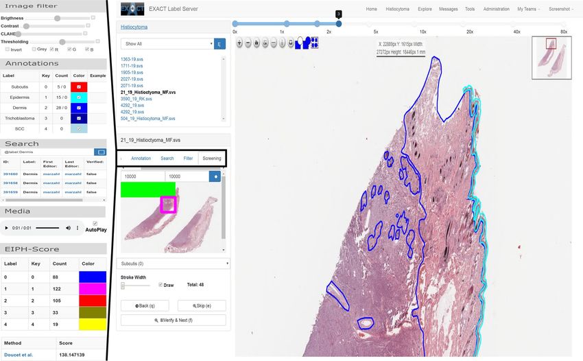
www.nature.com/scientificreports/
Section: Architecture
Setup EXACT via Docker with the command
docker-compose.prod.yml
docker-compose -f docker-compose.prod.yml up -d –build
Supplementary Video S10 EXACT installation guide with Docker
Section: Application tier
models.py Saves information to the database or file system
views.py Creates HTML views for the presentation tier
serializers.py Serialises data for the REST-API
api_views.py Provides database CRUD operations via the REST-API
doc/train_object_detection.ipynb Code to train an object detection model via the REST-API
urls.py Handles the mapping between URLs and python functions
The images module is responsible for all image-based CRUD opera-
exact/images
tions
Supplementary Video S7 How to create image sets and upload images
Supplementary Video S8 Explain image set details
The annotation module is responsible for all CRUD operations
exact/annotation regarding annotations, verification, media files and the annotation
versioning system
exact/plugin The plugin module handles analysis or visualisation plugins
exact/users The users module handles the CRUD operations for users and teams
Supplementary Video S14 How to setup user access rights
The datasets module provides features to automatically download and
exact/datasets
setup predefined data sets with their annotations
exact/datasets/templates Folder containing data set HTML templates
exact/datasets/views.py Implements functions to setup predefined data sets within EXACT
Supplementary Video S5 How to setup and use a demo data set
Section: Inference
doc/Inference Asthma.ipynb A REST-API inference example
Supplementary Video S9 Example for REST-API and JavaScript inference
Section: Annotation map screening mode
doc/AnnotationMap.ipynb Code to create annotation maps
Supplementary Video S2 How to create annotation maps
doc/ClusterCells.ipynb Code to cluster Asthma cells
Supplementary Video S4 How to cluster Asthma cells
Section: Image set versioning and machine learning support
doc/DownloadStudyAnnotations.ipynb Code to download annotations from EXACT
Supplementary Video S15 How to create a new image set version and track changes
Section: Annotation templates
Supplementary Video S3 How to create annotation templates with EXACT
Section: Pathology annotation study
How to download annotations and explanation of parts of the anno-
Supplementary Video S1
tation study
doc/DownloadStudyAnnotations.ipynb Code to download annotations via the REST-API
Section: Multi-species pulmonary hemosiderophages cytology data set
Supplementary Video S6 How to create density maps and explanation of the EIPH plugin
doc/Create_DensityWSI-Equine.ipynb Source code for density maps
Section: Skin tumour tissue quantification
doc/SyncImageAndAnnotations.ipynb Code to synchronise between SlideRunner and EXACT
Supplementary Video S12 How to synchronise between SlideRunner and EXACT
Supplementary Video S11 How to segment with EXACT
Code to download information from EXACT to train a segmentation
doc/Segmentation.ipynb
network
Code to download information from EXACT to train a patch clas-
doc/PatchClassifier.ipynb
sifier
Section: Clustering and visualisation of killer whale sounds
Supplementary Video S13 How to perform sound clustering and visualisation
Table 2. EXACT documentation and references for the corresponding sections and supplementary videos.
Scientific Reports | (2021) 11:4343 | https://doi.org/10.1038/s41598-021-83827-4 3
Vol.:(0123456789)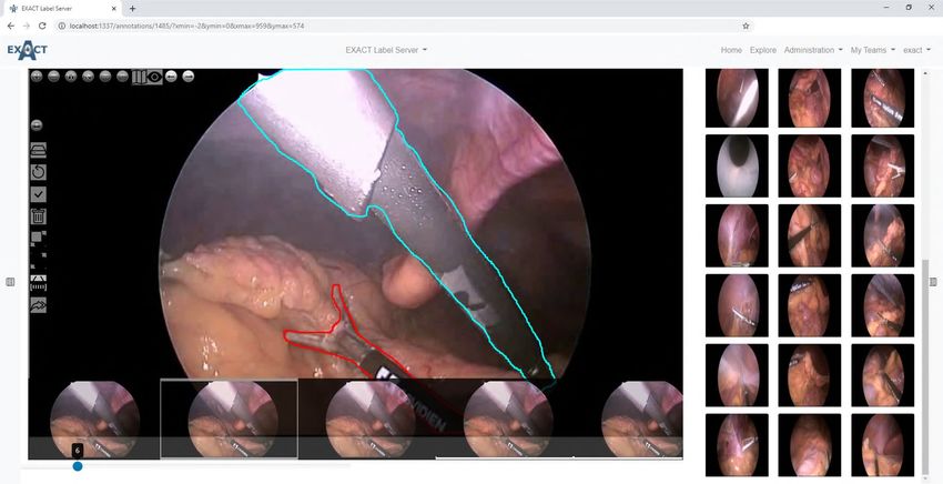
www.nature.com/scientificreports/
Data Application Presentation
EXACT Web-Clients
Web-Server NGINX
1..*
Django
Django
reverse proxy and load balancing
DB
Images
Users Annotations
Users Users
Users Plugins
Users
PostgreSQL Routing Routing Routing Routing
urls.py urls.py urls.py urls.py
Presentation
views.py
REST-API
api_views.py
Presentation
views.py
REST-API
api_views.py
Presentation
views.py
REST-API
api_views.py
Presentation
views.py
REST-API
api_views.py REST-API-Clients
Serialisation Serialisation Serialisation Serialisation
serializers.py serializers.py serializers.py serializers.py
Data Access Data Access Data Access Data Access
models.py models.py models.py models.py
Images
Figure 1. EXACT’s three-tier architecture. Left: The data tier contains the PostgresSQL Docker container and
the images, which can be saved within the Docker container or on the file system. Center: The application tier
with the web-server Docker container instantiating Django instances with the corresponding modules. These
modules handle images, annotations, users and plugin requests from the presentation tier and access the data
tier to retrieve the stored information. NGINX works as a reverse proxy and handles EXACT’s load balancing.
Right: The presentation tier contains the EXACT web client or third-party applications like SlideRunner, which
send requests via the provided REST-API.
Architecture. EXACT supports Docker to facilitate deployment and to enable a wide range of installation
scenarios ranging from single-user, single-computer setups to massive cloud deployment with modern load
balancing mechanisms (see Supplementary Video S10). EXACT is designed as a three-tier architecture contain-
ing the data, application, and presentation tier (Fig. 1). While the data and application tier are capsuled within
Docker containers, the presentation tier is executed at the client side in HTML and JavaScript. This tier-based
approach supports the development of secure applications by enforcing clearly defined interfaces between tiers
and ensures that data access pipelines can not bypass tiers. The data tier includes a Postgres database system and
the uploaded images and provides its content exclusively to the application tier.
Application tier. The application tier accesses the data tier to save information and to provide it to the presenta-
tion tier via a REST-API or as rendered HTML pages. EXACT uses Django as its web framework with four main
modules (see Fig. 1, namely the images, users, annotations, and plugins modules). Each module is responsible
for one group of tasks and is as independent as possible from the other modules. All modules implement func-
tions for saving information to the database or file system and for creating HTML views. Furthermore, the
modules define how to serialise data and provide a REST-API and a route request.
The images module is responsible for all image-based create, read, update and delete (CRUD) operations,
and provides the logic to save all supported image formats and to provide them as a complete image or in a tile-
based manner for WSIs. This multi type image support is implemented by converting all uploaded images that
are not compatible with O penSlide9 into an OpenSlide compatible format, if supported, and saving them as an
image pyramid. The formats and scanners that are supported by OpenSlide or our converter pipeline is listed
in table 1. EXACT’s open-source codebase allows developers to extend the list of supported image formats to
their requirements and image dimensions. An example for multi-dimensional image data support is the audio
video interleave (.avi) format. To support videos, EXACT converts each frame and handles the set of images as a
individual WSIs with OpenSlide (Fig. 3). Additionally, the images module contains the image sets functionality
which basically act as folders for the images and are assigned to teams to monitor user access rights. The Sup-
plementary Videos (S7, S8) describe the creation of image sets and the upload of images.
The annota tion module is responsible for all CRUD operations regarding annotations, verification, media files
and the annotation versioning system. The annotation model saves annotation information about the annotation
type, image, the creator and last editor with time stamps, JSON based meta data and the vector of coordinates
to the database. The vector information is saved as JSON and contains the image coordinates of the annotation.
The advantage of using JSON to store coordinates is the ability to search for annotations using vector coordinates
in SQL. Furthermore, JSON provides the flexibility to adapt the representation of the vector to the target image
format and dimensions.
The plugin module handles analysis or visualisation plugins which are specialised for specific research ques-
tions or data sets. One of these plugins is a persistent user-based screening mode which enables the user to
systematically screen a WSI or parts of it on a self-defined zoom level (Fig. 2). This plugin, which is crucial to
create high quality data sets, is implemented and used in the following manner: the user defines a zoom level and
the algorithm divides the WSI in equal-sized patches with an overlap of 15% and saves the calculated screening
map to the database. While the user is screening the WSI, the progress is constantly visualised at a thumbnail
view of the WSI and the user’s position on the WSI is saved to the database to recover the position if the screen-
ing has to be continued later.
Scientific Reports | (2021) 11:4343 | https://doi.org/10.1038/s41598-021-83827-4 4
Vol:.(1234567890)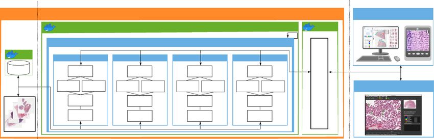
www.nature.com/scientificreports/
Figure 2. Left: Five examples of plugins (from top to bottom): The image filter plugin allows to make common
intensity adjustments to the image, the annotation plugin shows the available annotations and their frequency
of use. The search field allows to query the database for arbitrary annotation properties. The media plugin can
be used to play media files attached to an annotation. The EIPH-Score plugin is an example of a domain-specific
plugin, allowing to calculate the Doucet s core10. Right: A screenshot of the annotation view depicting a WSI
with polygon annotations, the list of images in the image set and the screening mode plugin, which enables the
user to screen the image persistently. The screening plugin visualise the screened area in green and a purple
rectangle for the current field of view.
Figure 3. Left: An example frame from a laparoscopic colorectal video with annotated surgical instruments.
Below the current frame (6), small previews of the previous and the next frames are displayed. Right: A
browsing view to provide an overview of different image sets from the Robust Endoscopic Vision Challenge
201911.
Scientific Reports | (2021) 11:4343 | https://doi.org/10.1038/s41598-021-83827-4 5
Vol.:(0123456789)www.nature.com/scientificreports/
The users module handles the CRUD operations for users and teams, and it further manages the user access
rights. It is therefore involved in every server request to check if the request has the necessary CRUD rights (see
Supplementary Video S14). To keep the annotations consistent, deleted users are anonymised and deactivated
while their annotations are left unchanged.
An additional module is the data sets module. It provides features to automatically download and setup
predefined data sets with their annotations from the EXACT user interface. The list of available data sets can be
extended by adding an HTML template, which provides background information like the number of images or
the data set source, and by implementing a download and setup function (see Supplementary Video S5).
Presentation tier. The presentation tier is programmed in HTML and JavaScript. All dynamic web-page con-
tents like annotations, images or sub-images (tiles) for WSIs are loaded via JavaScript over the REST-API. The
pagination-based REST-API implementation allow to load information chunk-wise from the server and there-
fore enable the transfer of huge quantities of data (e.g., hundreds of thousands of annotations per WSI) in par-
allel. We incorporated the open-source software OpenSeadragon as JavaScript-based image viewer with WSI
support. A visualisation of the presentation tier is shown in Fig. 2.
Inference. Different modes for inference of deep learning models are supported to match the requirements
across different use cases. In general, the inference can be performed directly on the server. For applications that
require fast response times, the execution of JavaScript-based TensorFlow models is implemented by initially
transferring the deep-learning model for the corresponding modality from the server via the REST-API to the
JavaScript client. Afterwards, the model is executed on the current field of view of the image. The resulting anno-
tations can then be rejected or confirmed and transferred to the server. For high-throughput applications, the
inference load can be distributed on multiple machines by downloading the model and the WSIs via the REST-
API and synchronising the results after performing inference. An inference example for equine asthma cytology
images can be accessed at doc/Inference Asthma.ipynb or as Supplementary Video S9.
Data privacy and multi‑centre support. Medical data should naturally be subject to the highest safety
standards possible. Despite that, in order to enable interdisciplinary medical research and cooperation between
different groups and locations, it can be necessary to share medical image data anonymously and in strict con-
sideration of data privacy. Therefore, EXACT ensures the original image data, which may contain patient infor-
mation (file name, metadata) to remain within the original institution while the actual data exchange between
experts and institutes is executed on small sub-images via decentralised image storage. Technically this was
implemented in several steps. Firstly, all server communication is protected with Hypertext Transfer Proto-
col Secure (HTTPS) and access is restricted via a user authentication system. Secondly, when transferring the
images to an EXACT server instance, a new private name derived from the file name and a pseudonymised
public name is generated. The pseudonymised public name is generated by the current date-time followed by
a four-digit hash function of the original name (yymmdd-hhmm-****). Thirdly, for cooperation between dif-
ferent institutes, virtual image sets are supported. Here the information (for example annotations) is imported
from several EXACT instances to a central server. However, access to the images themselves is always provided
by the institute owning the data in compliance with their respective data privacy policy for images. This means
that only the requested raw pixel data for the field of view is transferred to the collaborator, but not the image
container or any metadata.
Annotation map screening mode. For applications that focus on annotation quality1,7,8, a specialised
validation mode is implemented that allows for a verification of each individual annotation. For data sets with
hundreds or thousands of annotations, this is an important but error-prone, labour-intensive and time-consum-
ing task. This becomes even more complicated for usage scenarios where each cell can receive multiple labels by
one or multiple users. To make this validation process more convenient, we propose so-called annotation maps
which can be efficiently processed using the screening mode. Annotation maps visualise all annotations belong-
ing to one label in a matrix-like fashion which makes it easy to identify outliers. For efficient handling, a new
image is created for each class which consists of all corresponding annotations which can then be viewed in the
screening mode (Fig. 4 top and Supplementary Video S2). The annotation maps can be efficiently screened for
errors, while the users can define how many annotations they want to see simultaneously. Corrections made on
these screening images are synchronised with the original data.
An advanced extension of this method is the clustering of labelled and unlabelled images or image patches.
This manner of presentation allows the user to efficiently create initial labels or to quickly validate prior annota-
tions, since similar images which are likely to have similar labels are displayed closely together. The clustering
pipeline consists of three steps. Firstly, characteristic features are extracted from each image, for example, by deep
learning or classic image processing. Secondly, the extracted high-dimensional features are transformed into
two-dimensional features, for example, using t-SNE12, PCA13 or UMAP14. Finally, the extracted image patches
are drawn in a new image container according to their nearest two-dimensional feature representation, which
does not overlay any other image patches. The resulting image is visualised for labelling or validation (Fig. 4
bottom) in EXACT. A detailed code example can be accessed at doc/ClusterCells.ipynb in combination with a
Supplementary Video S4.
Image set versioning and machine learning support. In general, two main criteria in research and
medical applications are reproducibility and traceability of results and experiments. Especially reproducibility is
non-trivial in settings where researchers from different fields like medicine and computer science work together
Scientific Reports | (2021) 11:4343 | https://doi.org/10.1038/s41598-021-83827-4 6
Vol:.(1234567890)www.nature.com/scientificreports/
Figure 4. Top row: Supervised single-cell validation, with annotation maps generated from three labelled
equine asthma WSIs where each colour represents one class of cells. Bottom row: U MAP14 dimensionality
reduction approach in an unsupervised setting. The segmented equine asthma cells are first classified. Then,
features for each cell are extracted. Afterwards, the high-dimensional features are transformed into a two-
dimensional representation and visualised in a new image. Both approaches allow the user to verify and enhance
the automatic classification results.
and make adjustments to data sets over time. In software development, it is an established process to use version
control systems (such as git or subversion) for source code to coordinate the collaboration between software
developers and keep code changes traceable. Remarkably, this process is to our knowledge not provided by any
open source software for annotations on medical data sets. To implement this feature, we included a versioning
system with functions that support traceability of annotations and attach experimental results to versions. If a
version is added to a data set, the current annotation state, an optional description, and the current list of images
in the data set is saved. If a user leaves a project, he or she is not deleted from EXACT but only deactivated and
anonymised so that versioned annotations are not affected. In contrast, if an image is deleted from the image set,
all annotations are lost due to the impracticability of versioning WSIs with multiple gigabytes of size. For exam-
ple for training machine learning algorithms, the annotations can be filtered by versions and exported in user-
defined text formats or per script using the provided REST-API. This supports the users to perform experiments
on defined, reproducible data sets while providing the flexibility to export input data to a wide range of machine
learning frameworks. Additionally, training artefacts like performance metrics, annotations, or generated mod-
els can be uploaded and attached to a version. In combination with the virtual image set function introduced
previously in this article, it is possible to create virtual training, testing, and validation sets. This combination of
versions and virtual image sets helps to keep track of different experiment versions and supports the comparabil-
ity of results (see Supplementary Video S15).
Crowd‑sourcing and study support. One of the biggest challenges in developing, training, testing, and
validating state-of-the-art machine learning algorithms is the availability of high-quality, high-quantity labelled
image databases. Crowd-sourcing has numerous successful applications in the medical fi eld15 and crowd-algo-
16
rithm collaboration has the potential to decrease the human effort . EXACT supports this development by
providing multiple features for managing crowd-sourcing. Firstly, the user privilege system allows to set specific
rights like annotation or validation to users or user groups. Secondly, the crowd- or expert-algorithm collabora-
tion is assisted by importing pre-computed annotations or generating them on-premise with machine learning
models. Finally, EXACT supports multiple annotation modes like.
1. Cooperative One user can verify the image, and each user sees all other annotations.
2. Competitive or blind Every user must verify every image and cannot see other users’ annotations.
3. Second opinion A predefined number of the users must verify every annotation.
Scientific Reports | (2021) 11:4343 | https://doi.org/10.1038/s41598-021-83827-4 7
Vol.:(0123456789)www.nature.com/scientificreports/
Annotation templates. Standardisation is critical to encourage cooperation, interoperability and effi-
ciency. To support this, EXACT introduces annotation templates, which allow to define a set of properties of
annotations associated with a defined label. Annotation templates contain general information about the target
structure like a name, an example image, the sort order in which the annotation should be displayed on the user
interface, display colour, keyboard shortcuts to efficiently assign the label to an annotation, and default size.
Default sizes enable the user to introduce background knowledge into the annotation process; this allows for
efficient single click annotations and reduces the need to further adjust annotations. One or more annotation
templates are grouped to products with pieces of information like name or description and can be assigned to
image sets. The products in turn can be assigned to multiple image sets and support the reproducibility of the
annotation process by enforcing a standard naming and annotation schema (see Supplementary Video S3).
EXACT’s applications
In the following sections, we present several previously published usage scenarios using EXACT and describe
how they made use of EXACT’s features to increase efficiency and annotation quality.
Pathology annotation study. In a study by Marzahl et al.17, EXACT was used to investigate how the
efficiency of the pathology image annotation process can be increased with computer-generated pre-computed
annotations. The design and results of the published s tudy17 showcase a prominent EXACT use case and are
summarised in the following paragraphs. Ten pathologists had to perform three pathologically relevant diagnos-
tic tasks on 20 images each, once without algorithmic support and once with algorithmic support in the form of
pre-computed annotations which are visualised for the expert to review. Firstly, they had to detect mitotic fig-
ures on microscopy images. Each of the 20 images spanned ten high power fields (HPF, total area = 2.37 mm2).
The second task focused on performing a differential cell count in cytology of equine pulmonary fluid; a task
relevant for diagnosing respiratory disease. For this, five types of visually distinguishable cells (eosinophils, mast
cell, neutrophils, macrophages, lymphocytes) had to be labelled. The last task was to determine the severity of
pulmonary haemorrhaging by grading the amount of breakdown products of red blood cells (hemosiderin) in
alveolar macrophages according to the scoring scheme by Golde et al.18.
Several EXACT features were used for this study. First of all, we used the blind annotation mode for assign-
ing identical grading tasks to all pathology experts, which we then combined with the feature of importing pre-
computed annotation for the algorithmic support. The annotation templates enabled rapid single click annota-
tions by providing appropriate default annotation sizes for each cell type, which was particularly helpful for the
equine asthma task where the different cells types have notable size differences. The systematical grading of the
images was supported by the persistent screening mode plugin, which enables the expert to resume the grading
process at the previously selected position on the slide at any time. During the course of the study, the pathologists
annotated 26,015 cells on 1200 images. The algorithmic support with EXACT lead to an increase in accuracy
and a decrease of annotation time17 for all tasks. For detailed results, we kindly refer the reader to the original
study. A video showcasing this study can be viewed (see Supplementary Video S1) with related source code at
doc/DownloadStudyAnnotations.ipynb to download the annotations. Furthermore, we added the images and
ground-truth annotations from the study to the list of demo data sets which can be accessed and instantiated
from the EXACT user interface.
Multi‑species pulmonary hemosiderophages cytology data set. In our previous w ork19, 17 WSIs
with 78,047 pulmonary hemosiderophages were fully annotated by a veterinary pathologist and used to develop
a deep learning based object detection model. Pulmonary haemorrhage is diagnosed by performing a cytology
of bronchoalveolar lavage fluid (BALF). The basis for this scoring system from Golde et al.18 is that alveolar mac-
rophages degrade the red blood cells into an iron-storage complex called hemosiderin. After staining the sample
with Perls’ Prussian Blue or Turnbull’s Blue, the macrophages can be assigned a discrete grade from zero (low
hemosiderin content) to four (high hemosiderin content).
Building on this work, EXACT played an essential part in creating a large, fully annotated multi-species pul-
monary haemorrhage data set. For this project 40 additional equine WSIs, seven feline WSIs and twelve human
WSIs with evidence of chronic pulmonary haemorrhaging were annotated by expert-algorithm collaboration
using EXACT and the provided object detection model. In a first step, all WSIs were annotated automatically
with the deep learning model and afterwards a pathologist carefully reviewed whether all target objects were
annotated. Then, the pre-computed label class was verified separately by incorporating and modifying EXACT’s
novel annotation map feature based on a cell-based regression a pproach19 that reflects the continuous increase of
the hemosiderin content in the target cells. This approach assigns a continuous grade between zero and four to
each cell to create the annotation map for efficient manual validation (Fig. 5 bottom). This annotation map orders
the cells by score on the x-axis resulting in a density map of hemosiderin scores. By stacking the corresponding
cell images of the same score along the y-axis, the quantity of annotated cells across the different scores is visual-
ised (Fig. 5 bottom). This enables the trained pathologist to efficiently verify the computer-generated label class
by focusing on the cells which are located on the borders between two grades. Another specialised plugin was
developed to calculate the EIPH score over the current field of view in real-time (Fig. 2), according to Doucet
et al.10. Code to create density maps can be accessed at doc/Create_DensityWSI-Equine.ipynb in combination
with a Supplementary Video S6.
Skin tumour tissue quantification. This ongoing project aims to segment and classify nine of the most
common dog skin tumour types with deep learning algorithms. For this purpose, slides were scanned and partly
annotated using SlideRunner’s advanced tissue annotation tools. This project needs to synchronise the generated
Scientific Reports | (2021) 11:4343 | https://doi.org/10.1038/s41598-021-83827-4 8
Vol:.(1234567890)www.nature.com/scientificreports/
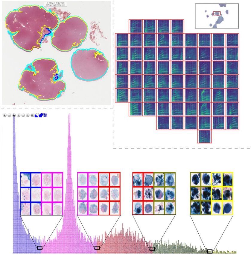
Figure 5. Top Left: Polygon annotations of a canine skin tumour tissue whole slide image. Top Right: Clustered
whale sound spectroscopy images with the option to listen to the attached waveform online. Bottom: Pulmonary
hemosiderophages, labelled according to their predicted class and arranged according to their predicted
regression score for efficient validation by human experts.
slides and annotations to EXACT for coordination and distribution between the participating pathology experts
and computer scientists for analysis at multiple institutes and locations. SlideRunner and EXACT communicate
via EXACT’s REST-API to synchronise annotations, images and annotation templates (see Supplementary Video
S12). EXACT’s novel feature of annotation templates plays an essential role in increasing standardisation and the
overall image set quality by ensuring standard annotation naming schemes and the use of polygon annotations
independent of the user or user application (Fig. 5 top left). While the project is actively being developed, 350
slides have already been fully annotated, resulting in 12,859 polygon annotations representing tissue layers. This
indicates that a combination of online and offline tools enables fast multi-expert annotations. Code to download
images, annotations and train a segmentation model can be found at doc/Segmentation.ipynb in combination
with a Supplementary Video S11.
Clustering and visualisation of killer whale sounds. While the EXACT platform is primarily devel-
oped for cooperative interdisciplinary research on microscopy images, its flexibility extends to other research
areas without adaptation. We therefore showcase its use in a project that aims at deepening the understanding
of killer whales (Orcinus Orca) and their large variety of different sound t ypes20. In this study, EXACT is used to
cluster and visualise the spectral shape of machine-pre-segmented killer whale audio samples (Fig. 5 top right).
Multiple EXACT features support this challenging undertaking: firstly, the support of viewing and annotating
gigapixel size images, which, in this use case, contain up to thousands of clustered spectrograms, where each
Scientific Reports | (2021) 11:4343 | https://doi.org/10.1038/s41598-021-83827-4 9
Vol.:(0123456789)www.nature.com/scientificreports/
spectrogram represents an individual killer whale sound. Secondly, grouped annotation assignments, which
enable the user to select numerous visually grouped spectrograms simultaneously by drawing a rectangle around
them in order to assign them to the same label. Finally, EXACT supports attaching media records like videos,
images or sound files to the respective annotations and plays them in a web browser (Fig. 2 left). These features
enable the user to see the grouped spectrograms and additionally listen to the attached killer whale sound (see
Supplementary Video S13).
Discussion
With the rapidly evolving digitisation of image data and the widespread use of machine learning algorithms, the
need for platforms that are able to organise and display large amounts of large image data while also managing
and keeping track of annotations is more crucial than ever. In this paper, we have introduced EXACT which is
an open-source online platform that enables the collaborative interdisciplinary analysis of images with annota-
tion version control.
EXACT has proven to satisfy these requirements in several different projects ranging from collaborative
tissue segmentation in the field of digital pathology to whale sound clustering. This diverse range of application
represents its primary advantage. It does not only allow to extend existing offline projects with cooperation
and synchronisation functions, but is also able to support researchers in various fields. Furthermore, EXACT’s
features provide computer scientist with version controlled annotations, advanced visualisation techniques like
annotation maps or clustering, and saving artefacts from experiments like trained models. With EXACT, it is
also possible to define reproducible training, validation and testing sets. Generally, all software solutions face the
issues of support, maintenance and handling future developments. To increase the chances of turning EXACT
into a successful project which offers added value for the community in the long term, EXACT will stay open-
source and focus on the compatibility and synchronisation with other image analysis software. The flexible open-
source software architecture allows for adaptation to future developments in digital pathology or other research
areas. In future releases, we are planning to support a higher amount of publicly available data sets. In addition,
we want to create specialised plugins exploring molecular pathology issues—an increasingly significant subdis-
cipline of the classic anatomical pathology. Also, valuable future extensions to EXACT include the integration
of servers (like Omero21), which are specialised in providing microscopic images, as well as exploring options to
connect EXACT with other established tools like Cytomine. Furthermore, we are investigating the integration
of gamification as a promising new method to annotate data at scale.
In summary, EXACT provides a novel feature set to boost the creation of high-quality big data sets in com-
bination with functions to develop state-of-the-art machine learning algorithms.
Code availability
Server: https://github.com/ChristianMarzahl/Exact Demo-Server: https://exact.cs.fau.de/ User: "Demo" PW:
"demodemo" REST-API Client: https://github.com/ChristianMarzahl/EXACT-Sync Notebooks: https://github.
com/ChristianMarzahl/Exact/tree/master/doc.
Received: 29 April 2020; Accepted: 2 February 2021
References
1. Aubreville, M., Bertram, C., Klopfleisch, R. & Maier, A. Sliderunner. In Bildverarbeitung für die Medizin 2018 (eds Maier, A.,
Deserno, T., Handels, H., Maier-Hein, K., Palm, C. & Tolxdorff, T.) 309–314 (Springer Vieweg, Berlin, Heidelberg, 2018).
2. Hollandi, R., Diósdi, Á., Hollandi, G., Moshkov, N. & Horvath, P. Annotatorj: An imagej plugin to ease hand-annotation of cellular
compartments. Mol. Biol. Cell 31(20), 2179–2186 (2020).
3. De Chaumont, F. et al. Icy: An open bioimage informatics platform for extended reproducible research. Nat. Methods 9, 690 (2012).
4. Bankhead, P. et al. Qupath: Open source software for digital pathology image analysis. Sci. Rep. 7, 1–7 (2017).
5. Marée, R. et al. Collaborative analysis of multi-gigapixel imaging data using cytomine. Bioinformatics 32, 1395–1401 (2016).
6. Puttapirat, P. et al. Openhi—An open source framework for annotating histopathological image. In 2018 IEEE International
Conference on Bioinformatics and Biomedicine (BIBM), 1076–1082 https://doi.org/10.1109/BIBM.2018.8621393 (2018).
7. Obando, D. F. G., Mandache, D., Olivo-Marin, J.-C. & Meas-Yedid, V. Icytomine: A user-friendly tool for integrating workflows on
whole slide images. In Digital Pathology (eds Reyes-Aldasoro, C. C., Janowczyk, A., Veta, M., Bankhead, P. & Sirinukunwattana,
K.) 181–189 (Springer International Publishing, Cham, 2019).
8. Fiedler, N., Bestmann, M. & Hendrich, N. Imagetagger: An open source online platform for collaborative image labeling. In Robo-
Cup 2018: Robot World Cup XXII (eds Holz, D., Genter, K., Saad, M. & von Stryk, O.) 162–169 (Springer International Publishing,
Cham, 2018).
9. Goode, A., Gilbert, B., Harkes, J., Jukic, D. & Satyanarayanan, M. Openslide: A vendor-neutral software foundation for digital
pathology. J. Pathol. Inform. 4(1), 27 (2013).
10. Doucet, M. Y. & Viel, L. Alveolar macrophage graded hemosiderin score from bronchoalveolar lavage in horses with exercise-
induced pulmonary hemorrhage and controls. J. Vet. Intern. Med. 16, 281–286 (2002).
11. Maier-Hein, L. et al. Heidelberg colorectal data set for surgical data science in the sensor operating room. arXiv:2 005.0 3501 (2020).
12. Maaten, L. V. D. & Hinton, G. Visualizing data using t-sne. J. Mach. Learn. Res. 9, 2579–2605 (2008).
13. Wold, S., Esbensen, K. & Geladi, P. Principal component analysis. Chemometr. Intell. Lab. 2, 37–52 (1987).
14. McInnes, L., Healy, J. & Melville, J. UMAP: Uniform manifold approximation and projection for dimension reduction. arXiv:1 802.
03426 (2018).
15. Ørting, S. N., Doyle, A., van Hilten, A., Hirth, M., Inel, O., Madan, C. R., Mavridis, P., Spiers, H. & Cheplygina, V. A survey of
crowdsourcing in medical image analysis. Hum. Comput. 7(1), 1–26 (2020).
16. Marzahl, C. et al. Is crowd-algorithm collaboration an advanced alternative to crowd-sourcing on cytology slides? In Bildverar-
beitung für die Medizin 2020 (eds Tolxdorff, T., Deserno, T., Handels, H., Maier, A., Maier-Hein, K. & Palm, C.) 26–31 (Springer
Fachmedien Wiesbaden, Wiesbaden, 2020).
Scientific Reports | (2021) 11:4343 | https://doi.org/10.1038/s41598-021-83827-4 10
Vol:.(1234567890)www.nature.com/scientificreports/
17. Marzahl, C. et al. Are fast labeling methods reliable? A case study of computer-aided expert annotations on microscopy slides. In
Medical Image Computing and Computer Assisted Intervention – MICCAI 2020 (eds Martel, A. L., Abolmaesumi, P., Stoyanov, D.,
Mateus, D., Zuluaga, M. A., Zhou, S. K., Racoceanu, D. & Joskowicz, L.) 24–32 (Springer International Publishing, Cham, 2020).
18. Golde, D. W., Drew, W. L., Klein, H. Z. et al. Occult pulmonary haemorrhage in leukaemia. Br. Med. J. 2, 166–168 (1975).
19. Marzahl, C. et al. Deep learning-based quantification of pulmonary hemosiderophages in cytology slides. Sci. Rep. 10, 1–10 (2020).
20. Bergler, C. et al. ORCA-SPOT: An automatic killer whale sound detection toolkit using deep learning. Sci. Rep. https://doi.org/
10.1038/s41598-019-47335-w. (2019).
21. Allan, C. et al. Omero: flexible, model-driven data management for experimental biology. Nat. Methods 9, 245–253 (2012).
Acknowledgements
CAB gratefully acknowledges financial support received from the Dres. Jutta & Georg Bruns-Stiftung für inno-
vative Veterinärmedizin.
Author contributions
C.M. developed the server, created the visualisation code and wrote the main part of the manuscript. M.A.
co-wrote the manuscript, provided code for the synchronisation with SlideRunner, provided expertise through
intense discussions. C.A.B. co-wrote the manuscript, provided expertise through intense discussions. J.M., J.V.,
C.B., C.K., K.B., R.K., A.M. provided expertise through intense discussions. All authors contributed to the
preparation of the manuscript and approved of the final manuscript for publication.
Funding
Open Access funding enabled and organized by Projekt DEAL
Competing interests
The authors declare no competing interests.
Additional information
Supplementary information The online version contains supplementary material available at https://doi.org/
10.1038/s41598-021-83827-4.
Correspondence and requests for materials should be addressed to C.M.
Reprints and permissions information is available at www.nature.com/reprints.
Publisher’s note Springer Nature remains neutral with regard to jurisdictional claims in published maps and
institutional affiliations.
Open Access This article is licensed under a Creative Commons Attribution 4.0 International
License, which permits use, sharing, adaptation, distribution and reproduction in any medium or
format, as long as you give appropriate credit to the original author(s) and the source, provide a link to the
Creative Commons licence, and indicate if changes were made. The images or other third party material in this
article are included in the article’s Creative Commons licence, unless indicated otherwise in a credit line to the
material. If material is not included in the article’s Creative Commons licence and your intended use is not
permitted by statutory regulation or exceeds the permitted use, you will need to obtain permission directly from
the copyright holder. To view a copy of this licence, visit http://creativecommons.org/licenses/by/4.0/.
© The Author(s) 2021
Scientific Reports | (2021) 11:4343 | https://doi.org/10.1038/s41598-021-83827-4 11
Vol.:(0123456789)You can also read