Expression and uptake of the thyroxine-binding protein transthyretin is regulated by oxygen in primary trophoblast placental cells
←
→
Page content transcription
If your browser does not render page correctly, please read the page content below
159

Expression and uptake of the thyroxine-binding protein transthyretin
is regulated by oxygen in primary trophoblast placental cells
J Patel1,2, K A Landers2, R H Mortimer1,2,3 and K Richard1,2
1
    School of Medicine, Royal Brisbane and Women’s Hospital, The University of Queensland, Herston, Brisbane, Queensland 4029, Australia
2
    Conjoint Endocrine Laboratory, Pathology Queensland and Department of Endocrinology, Royal Brisbane and Women’s Hospital, QIMR Bancroft Centre,
Herston, Brisbane, Queensland 4029, Australia
3
    Disciplines of Medicine, Obstetrics and Gynaecology, The University of Queensland, Herston, Brisbane, Queensland 4029, Australia
(Correspondence should be addressed to K Richard at Conjoint Endocrine Laboratory, Pathology Queensland and Department of Endocrinology, Royal Brisbane
   and Women’s Hospital; Email: kerry.richard@qimr.edu.au)

Abstract
Transplacental delivery of maternal thyroid hormones to the                       higher expression of TTR mRNA and protein levels at 1% O2
fetus, in particular thyroxine (T4), is critical in ensuring                      than at 8 and 21% O2. Significant increases were observed
normal fetal neurological development. The fetus relies on                        after culture at 3% O2 and following DFO treatment. We
maternal T4 till around 16 weeks gestation, but mechanisms                        observed significantly higher uptake of 125I-TTR and Alexa-
of placental T4 transport are not yet fully elucidated. Placenta                  594-TTR when cells were cultured at 1 and 3% O2 and in
produces, secretes and takes up the thyroid hormone-binding                       the presence of 200 mM DFO than at 8 and 21% O2. When
protein transthyretin (TTR). Many placental genes are                             JEG-3 choriocarcinoma cells were transfected with TTR
regulated by oxygen levels, which are relatively low (1%) in                      promoter reporter constructs, increased luciferase activity was
the early first trimester, rising to 3% in the mid first trimester                measured in cells cultured at 1 and 3% O2 in comparison to 8
and 8% in the early second trimester and thereafter. We                           and 21% O2. We conclude that placental TTR expression and
examined the expression and uptake of TTR in isolated                             uptake is increased by the relative hypoxia observed in the first
primary human placental cytotrophoblast cells cultured under                      trimester of pregnancy, a time when materno–fetal T4 transfer
different oxygen concentrations (1, 3, 8, 21% O2 and 200 mM                       is the sole source of fetal T4.
desferrioxamine (DFO)) for 24 h. We observed sevenfold                            Journal of Endocrinology (2012) 212, 159–167

Introduction                                                                      the components of this putative system and its possible
                                                                                  regulation (trophoblast cell membrane thyroid hormone
Thyroid hormone, in particular thyroxine (T4), plays an                           transporters, placental type III deiodinase and possibly
important role in the activation, differentiation and matu-                       transthyretin (TTR)) are known but the mechanisms of
ration of the fetal central nervous system (Bernal 2005, Patel                    thyroid hormone transfer and its regulation are yet to be
et al. 2011b). Before 16 weeks gestation, the fetus relies                        elucidated (Patel et al. 2010a).
solely on transplacental delivery of maternal T4 (Obregon                            We have recently reported that human trophoblast cells
et al. 2007). By 6 weeks gestation, measurable amounts of T4                      produce the T 4 and retinol-binding protein, TTR
and T3 have been detected in human fetal coelomic fluid and                       (McKinnon et al. 2005). Studies on polarised choriocarci-
there is evidence to suggest that even after the onset of fetal                   noma JEG-3 cells maintained in bicameral chambers indicate
thyroid function, transport of T4 from maternal to fetal                          that TTR is secreted apically and basally (Landers et al. 2009).
circulation continues (Vulsma et al. 1989, Patel et al. 2011a).                   Trophoblasts also take up TTR from culture medium and this
Rat experiments suggest that fetal brain development                              uptake is increased when T4 is bound to TTR, increasing
preferentially requires T4, which is locally deiodinated to T3                    TTR tetramer formation (Landers et al. 2009). These findings
(Calvo et al. 1990). Clinical studies suggest that even mild                      raise the possibility that placental TTR is involved in a
reductions in maternal T4 adversely affect the intellectual                       transplacental T4 shuttle system.
function of offspring, indicating that human brain develop-                          The early placenta is marked by low oxygen levels (1%) that
ment is very sensitive to mild maternal hypothyroidism                            increase as placental vascularisation is established so that by 12
(Haddow et al. 1999). The biological corollary of these                           weeks gestation placental oxygen levels reach 8%, a concen-
clinical and animal studies is that a maternal to fetal T4 transfer               tration that in normal pregnancies is maintained until term
system must operate in very early human pregnancy. Some of                        (Schneider 2011). Many placental genes and placental

Journal of Endocrinology (2012) 212, 159–167                                                                                   DOI: 10.1530/JOE-11-0348
0022–0795/12/0212–159 q 2012 Society for Endocrinology           Printed in Great Britain         Online version via http://www.endocrinology-journals.org

                                                                                                            Downloaded from Bioscientifica.com at 11/11/2021 05:56:08PM
                                                                                                                                                          via free access
160 J PATEL and others . Oxygen regulates TTR uptake in placental cells
development itself are regulated by changing oxygen levels similar concentrations without affecting cell viability (Patel
throughout gestation (Patel et al. 2010b). We have recently et al. 2010a). DFO is an iron chelator and mimics hypoxia by
demonstrated in JEG-3 cells that TTR mRNA expression, inhibiting prolyl hydroxylases, which are essential for the
protein levels and uptake are increased at the lower oxygen proteasomal degradation of hypoxia-inducible factor-1a
levels found in the early placenta (Patel et al. 2010a). (HIF-1a) during times of normoxia (O5% O2), resulting in
Therefore, the aim of this study was to investigate the TTR the nuclear accumulation of HIF-1a and downstream effects
expression and uptake by isolated human term trophoblasts (Ran et al. 2005). DFO has been used extensively in many cell
cultured under different oxygen concentrations reported in and tissue types and is recognised as the gold standard for
the developing human placenta to confirm our previous creating a hypoxic environment under normoxic conditions
findings in JEG-3 cells. We also measured TTR promoter (Ran et al. 2005, Takeda et al. 2007, Chu et al. 2010). Viability
activity in JEG-3 cells cultured under different oxygen of cells used in the experiment was assessed on day 4 by
concentrations. We postulate that TTR expression and uptake measurement of human chorionic gonadotrophin (hCG) and
in primary trophoblast cells will increase at low oxygen levels, lactate dehydrogenase (LDH) in cell culture medium. hCG is
as suggested by our previous work on JEG-3 cells. a marker of trophoblast function and LDH secretion is
elevated when cellular material breaks down. hCG and LDH
were measured by Chemical Pathology, Pathology Queens-
Methods and Materials land. hCG was measured using the Beckman Coulter Access
Immunoassay Total bhCG system (intra-assay coefficient of
Placental tissue variation (CV) 4.1%; inter-assay CV 4.6%). LDH was
measured using the Beckman Coulter SYNCHRON LX
Placentas from term (38–39 weeks; nZ12) normal healthy system with the SYNCHRON enzyme validator set (intra-
women undergoing caesarean section at the Royal Brisbane assay CV 3.5%; inter-assay CV 7.3%).
and Women’s Hospital were collected, with informed
consent, soon after delivery. This study was approved by the
Royal Brisbane and Women’s Hospital and Queensland Quantitative RT-PCR
Institute of Medical Research Human Research Ethics On day 4, following 24 h of culture at low oxygen or
Committees. treatment with DFO, total RNA was extracted from the
trophoblast cells using the RNAeasy Mini Kit (Qiagen). Total
Isolation of cytotrophoblast cells from placental tissue RNA (4 mg) was reverse transcribed to produce cDNA using
the Superscript III RT Kit (Invitrogen) and P(dt)15 primers
Villous cytotrophoblasts were isolated as described previously (Roche Applied Sciences) in a reaction volume of 20 ml.
(Kliman et al. 1986, Greenwood et al. 1996, Barber et al. Quantitative PCR was performed using 0.5 mM of forward
2005). Isolated cytotrophoblasts were cultured in and reverse primers with FastStart SYBR Green master-mix
DMEM:F12 nutrient mixture, 25 mM HEPES and 1% in a 15 ml reaction, which included 3 ml cDNA (Roche
L-glutamine (all from Sigma), supplemented with 1% Applied Sciences) in a Rotor Gene RG-3000 (Corbett
penicillin/streptomycin, 0.1% plasmocin and 10% FCS (all Research, Sydney, NSW, Australia). The real-time PCR
from Invitrogen). Cytotrophoblasts were seeded onto sterile method was validated by using serially diluted cDNA as a
coverslips in 24-well culture plates and fixed for immuno- standard curve. To quantify the mRNA expression profile in
cytochemical staining of cytokeratin-7 to determine purity of each sample, the efficiency of each standard curve was
the cell population. determined by its slope and comparative threshold according
to the manufacturer’s instructions. For each sample, the
Low oxygen experiments amount of targeted mRNA (AU) was normalised to the
housekeeping gene b-actin. mRNA quantification was
Isolated cytotrophoblasts were simultaneously seeded at 3! performed using the Pfaffl method (Pfaffl et al. 2002),
106 cells in a 6-well culture plate and cultured for 4 days. which makes no distributional assumptions. The primers
From days 1 to 3, the cells were grown under standard used to amplify specific mRNAs are listed in Table 1.
laboratory culture conditions at 21% O2. On day 3, the cells
were transferred to Heraeus HeraCell incubators (Thermo Table 1 Primer sequences
Scientific, Langenselbold, Germany) and maintained at
different oxygen concentrations of 1, 3 and 8% for 24 h. Gene Primers
Cells were also studied at 21% O2, which is standard
b-Actin (Forward) 5 0 -CATGTACGTTGCTATCCAGGC-3 0
laboratory practice and routine for placental cell function (Reverse) 5 0 -CTCCTTAATGTCSCGCACGAT-3 0
studies. Desferrioxamine (DFO; 200 mM), a hypoxic mimetic TTR (Forward) 5 0 -ATGGCTTCTCATCGTCTGCT-3 0
drug, was used as a positive control (Li et al. 2011). DFO, a (Reverse) 5 0 -TGTCATCAGCAGCCTTTCTG-3 0
hypoxia mimetic drug, was used in this study as a positive
control and has been used in JEG-3 cells in other studies at TTR, transthyretin.
Journal of Endocrinology (2012) 212, 159–167 www.endocrinology-journals.org
Downloaded from Bioscientifica.com at 11/11/2021 05:56:08PM
via free accessOxygen regulates TTR uptake in placental cells . J PATEL and others 161
All PCR samples were run from three separate cell culture Labelling Kit according to the manufacturer’s instructions
experiments in triplicate. Data from each experiment were (Invitrogen).
averaged and then compared to values obtained from cells
cultured at 8% O2 to determine fold changes in expression 125
I-TTR membrane binding and internalisation
and S.E.M. These data were used to carry out statistical analysis.
On day 3, isolated trophoblast cells were transferred to 1, 3, 8,
or 21 O2G 200 mM DFO for 24 h in 6-well plates prior to
Western blotting uptake studies on day 4; each experiment was carried out in
On day 4, following 24 h of culture at low oxygen or DFO triplicate. Binding and internalisation studies were performed
treatment, protein was extracted from the trophoblast cells as described previously (Landers et al. 2009). Cells were
using the M-PER Mammalian Protein Extraction Reagent washed with PBS and cultured in SF-DMEM for 4 h before
(Thermo Scientific, Rockford, IL, USA) and Protein incubation with 125 I-TTR (10 pM) in SF-DMEM
Inhibitor Cocktail (Sigma). HIF-1a westerns were performed containing 100 mM potassium perchlorate at 37 8C. Incu-
on nuclear extracts prepared using a NE-PER Nuclear bation with an excess of unlabelled TTR (1 mM) was used to
Extraction Kit (Thermo Scientific). Total protein (30 mg) was distinguish between specific/non-specific binding and
separated on 4–12% Novex NuPage Bis/Tris gels (Invitrogen) internalisation. T4 (10 mM) was used as described previously
and transferred onto nitrocellulose membrane (Bio-Rad (Landers et al. 2009). Cells were incubated in a 21% O2
Trans-Blot). Rabbit anti-TTR (Dako Australia Pty Ltd, incubator for 1 h to allow binding and internalisation to
Campbellfield, VIC, Australia), mouse anti-HIF-1a (Abcam) occur. After 1 h, the media was removed and cells washed in
and mouse anti-b-actin (Sigma) antibodies were used at ice-cold PBS. Surface bound proteins were removed using
1:1000 dilutions. HRP-conjugated secondary antibodies (GE ice-cold 0.025% trypsin/0.01% EDTA as described pre-
Healthcare, Buckinghamshire, UK) were used at 1:3000. viously (Divino & Schussler 1990, Landers et al. 2009).
Blots were developed using a Supersignal West Femto Kit Internalised TTR was collected by harvesting cells with 1 M
NaOH for 5 min at 37 8C. Samples were counted in a Packard
(Pierce Biotechnology, Rockford, IL, USA) following the
Cobra II series counter (Hewlett Packard, Blackburn, VIC,
manufacturer’s instructions and images captured by charge-
Australia), with a counting efficiency of 80.27%.
coupled device camera (Fuji image LAS-3000; FujiFilm,
Brookvale, NSW, Australia). Densitometric analysis was
performed using Multigauge v2.3 Software (Fuji, FujiFilm). Alexa-TTR internalisation
Each experiment was carried out in triplicate.
On day 3, isolated cytotrophoblast cells were transferred to
SF-DMEM for 16 h before incubating the cells with
Brefeldin A treatment 100 mg/ml Alexa594-TTR for 1 h. Uptake of Alexa-TTR
was evaluated in the presence of 1 mM unlabelled TTR and
On day 3, isolated trophoblast cells grown in 6-well cell 10 mM T4 (Landers et al. 2009). Cells were washed with cold
culture flasks were incubated overnight in serum-free (SF) PBS before fixation in 4% paraformaldehyde. Counter-
media. On day 4, the cells were then cultured in SF staining with DAPI and phalloidin were conducted to mark
mediaG10 mg/ml brefeldin A for 6 h. Brefeldin A inhibits nuclei and actin filaments respectively. Images were captured
the transport of proteins from the endoplasmic reticulum using Deltavision Core Technology (Applied Precision,
(ER) to the Golgi apparatus, leading to the accumulation of Washington, DC, USA). To ensure identical image analysis
protein with the ER, preventing secretory proteins such as for the comparison of TTR uptake was undertaken, all slides
TTR from being released (Klausner et al. 1992). SF media was from the same experiment were scanned using the same
used since serum contains TTR and other proteins that settings including u.v. light transmission and exposure time.
may interfere with the experiment. Total cell lysates were Captured images were displayed with the same intensity scale
collected and analysed by western blotting, with each for channel of Alexa-Fluor594 labelled TTR. Using ImageJ
experiment carried out in triplicate. Brefeldin A had no Software (National Institutes of Health, NIH), individual cells
adverse effects on cell viability as determined by trypan blue (nZ30) were randomly selected, magnified and the intra-
exclusion. This experiment was carried out under the same cellular TTR-labelled foci (red) counted.
oxygen conditions as described previously.
Preparation of TTR promoter constructs
TTR labelling
Upstream regions (0.11, 0.65 and 2.1 kb) of the human
TTR (Sigma) was labelled with 125-Iodine (125I) using the TTR gene (Mirkovitch & Darnell 1991) were ligated into
chloramine-T method by John Bellen, Department of the pGL3-basic vector between the KpnI and NheI
Nuclear Medicine, Royal Brisbane and Women’s Hospital sites (Promega). The following primers were used: 0.11 kb
(Greenwood et al. 1963). Fluorescent labelling of TTR (5 0 -GGCGGTACCTTGACTAAGTCAATA-3 0 ), 0.65 kb
(Alexa-TTR) was performed with an Alexa-Fluor-594 (5 0 -GGCGGTACCGAGAAATTGCTGACTAAGCAA-3 0 )
www.endocrinology-journals.org Journal of Endocrinology (2012) 212, 159–167
Downloaded from Bioscientifica.com at 11/11/2021 05:56:08PM
via free access162 J PATEL and others . Oxygen regulates TTR uptake in placental cells
A 120 B 700 multiple comparison post-hoc test using results from experi-
100 600
ments conducted at 8% O2 as the reference level. A P value
hCG in culture
LDH in culture
medium (IU/l)
*
medium (IU/l)
500
80
60
400 **
** ** of P%0.05 was considered statistically significant.
300
40 200
20 100
0
21% 1% 3% 8% DFO
0
21% 1% 3% 8% DFO
Results
Oxygen concentration Oxygen concentration
In the analysis of the data, we have focussed on the
Figure 1 hCG and LDH levels in cell culture medium from primary
cytotrophoblast cells cultured at different oxygen concentrations
physiologically relevant oxygen concentrations that are
(1, 3, 8 and 21% O2) and treated with 200 mM DFO. (A) LDH levels present in vivo during placental development: 1, 3 and 8% O2.
were not different. (B) Significant increases in hCG secretion were
observed from cells cultured at 8% O2 in comparison to all other
groups. All experiments were performed in triplicates. *P%0.05 vs Cell culture purity and viability
8%; **P%0.01 vs 8%.
Trophoblast cell population purity ranged from 90 to 100%, as
judged by cytokeratin-7 staining. Cell function and viability
and 2.1 kb (5 0 -GGCGGTACCTAACATATCTATGGGCT- were assessed on day 4 of culture by measuring hCG and
GATG-3 0 ). The following reverse primer was used for all LDH concentrations in culture media (Fig. 1). At 8% O2
three sequences (5 0 -TATCGCTAGCAGGAGCAGACGAT- (632.31G29.8 IU/l), the highest level of secreted hCG was
GAGAAGC-3 0 ). The constructs were designated pGL3- measured in comparison to all other experimental groups: 1%
0.11 kb (0.11 kb insert), pGL3-0.65 kb (0.65 kb insert) (291.51G24.79 IU/l, P%0.01), 3% (365.36G32.64 IU/l,
and pGL3-2.1 kb (2.1 kb insert). The TTR promoter P%0.01), 21% (502.64G26.57 IU/l P%0.05) and DFO
constructs were cloned using JM109 Escherichia coli and the (352.4G31.29, P%0.01 IU/l).
vector (plasmid) DNA was extracted using the Qiagen
Plasmid Midi Kit (Qiagen). Quality of plasmid DNA
(pDNA) extracted was measured for concentration and purity
TTR mRNA expression
using A260/A280 spectroscopy on the Nanodrop ND-1000 RT-PCR results revealed significant differences in TTR
machine (Thermo Scientific). mRNA expression in cells cultured under different oxygen
concentrations (Fig. 2). At 1 and 3% O2 and following
200 mM DFO treatment, there was a 5.54G0.61 (P%0.001),
Luciferase assays
4.84G0.71 (P%0.001) and 4.31G0.64 (P%0.001) respect-
JEG-3 cells were obtained from the American Type Culture ive fold increase in TTR mRNA expression in comparison
Collection (ATCC, Manassas, VA, USA) and maintained in to cells grown at 8% O2 (Fig. 2). This shows that TTR
DMEM (Sigma) with 1% penicillin/streptomycin, 0.1% mRNA levels are up-regulated under hypoxic conditions.
plasmocin and 10% fetal bovine serum. Once confluent,
JEG-3 cells were plated onto white flat bottom 96-well plates 7 ###
(Perkin Elmer, Glen Waverley, VIC, Australia) with regular *** ###
Relative expression – TTR
6
culture medium. On day 2, the cells were transfected with a *** ###
total of 0.33 mg pDNA per well. The transfection mixture 5 ***
contained 300 ng empty pGL3 vector or pGL3 containing 4
the TTR promoter insert with 30 ng of the internal control
vector (pRL-TK), at a ratio of 3:1 with FuGENE 6 3
transfection reagent (Roche), according to the manufactures’ 2
instructions. Transfection mixture (100 ml) was added to each
appropriate well. Transfected cells were cultured under the 1
same low oxygen conditions previously described for a 0
further 24 h. The luciferase reporter assay was conducted by 21% 1% 3% 8% DFO
using the Promega Dual-Glo Luciferase reporter assay kit Oxygen concentration
(Promega). Luminescence was measured using the POLAR-
Figure 2 TTR mRNA expression in primary cytotrophoblast cells
star OPTIMA luminometer (BMG Labtech, Mornington,
cultured at different oxygen concentrations (1, 3, 8 and 21% O2)
VIC, Australia). The ratio of firefly:Renilla luminescence for and treated with 200 mM DFO. All groups were normalised to
each well (nZ12) was calculated. 8% O2. Significant fold increases in TTR mRNA were observed
at 1% (5.54G0.61) O2, 3% (4.84G0.71) O2 and with 200 mM of
DFO (4.31G0.64) in comparison to 8% O2. Significant increases
Statistical analysis were also observed at 1 and 3% O2 and 200 mM DFO in
comparison to 21% O2. b-Actin was used as the housekeeping
All data sets were represented as meanGS.E.M. Significant gene. All experiments were performed in triplicates. ***P%0.001
differences were determined using ANOVA with Dunnett’s vs 8%; ###P%0.001 vs 21%.
Journal of Endocrinology (2012) 212, 159–167 www.endocrinology-journals.org
Downloaded from Bioscientifica.com at 11/11/2021 05:56:08PM
via free accessOxygen regulates TTR uptake in placental cells . J PATEL and others 163
A 5 ng C P%0.01) and following 200 mM DFO treatment (1.58-fold
21% 1% 3% 8% DFO TTR 21% 1% 3% 8% DFO increase, P%0.01) were also observed in comparison to cells
TTR HIF-1α cultured at 8% O2 (Fig. 4).
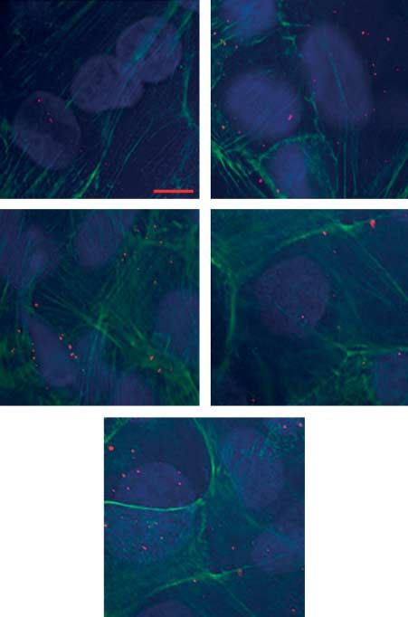
β-Actin β-Actin Internalisation of Alexa594-TTR by trophoblast cells was
visualised as small red foci bordered by phalloidin (green)-
B ### D ### ###
6 *** 12 stained actin filaments. Alexa594-TTR uptake was increased
expression – HIF-1α
expression – TTR
### *** *** ###
5 ### 10 in cells cultured at 1 and 3% O2 and following 200 mM DFO
*** ***
Relative
4 *** 8
Relative
3 6 treatment in comparison to 8% O2 (Fig. 5A). Using Image J
2 4 software, individual intracellular TTR foci were counted.
1 2
0 0
Significant increases in intracellular TTR foci were counted
21% 1% 3% 8% DFO 21% 1% 3% 8% DFO in cells that had been cultured at 1% O2 (12.9G0.8,
Oxygen concentration Oxygen concentration P%0.001), 3% O2 (11.8G1.1, P%0.001) and following
Figure 3 Western blots of TTR and HIF-1a. (A) Western blot DFO treatment (9.90G0.50, P%0.01) in comparison to
demonstrating changes in TTR protein production in cells cultured 8% O2 (Fig. 5B).
at different oxygen concentrations (1, 3, 8 and 21% O2) and treated
with 200 mM DFO. (B) All groups were normalised to 8% O2.
Significant fold increases in TTR protein expression were observed TTR promoter activity
at 1% (4.97G0.33) O2, 3% (3.64G0.31) O2 and 200 mM DFO
(3.13G0.36) in comparison to 8% O2. Significant increases were TTR promoter activity was measured via luciferase pro-
also observed at 1 and 3% O2 and 200 mM DFO in comparison to duction following transfection of reporter constructs into
21% O2. (C) and (D) Western blot and densitometry demonstrating
JEG-3 placental cells and cultured under low oxygen
the activation of HIF-1a protein collected from nuclear extracts in
cells cultured different oxygen concentrations (1, 3, 8 and 21% O2) conditions (Fig. 6). No significant differences in luciferase
and treated with 200 mM DFO. b-Actin was used as the house- activity were measured between experimental groups in
keeping gene for both TTR and HIF-1a. All experiments were JEG-3 cells transfected with the pGL3-0.11 kb TTR pro-
performed in triplicates. ***P%0.001 vs 8%; ###P%0.001 vs 21%. moter construct. A significant increase in luciferase activity
was observed in JEG-3 cells transfected with the pGL3-
TTR protein expression 0.65 kb TTR promoter construct cultured at 1% O2 in
comparison to 8% (P%0.01) and 21% O2 (P%0.01).
It is very difficult to directly measure TTR secreted by Significant increases were also observed in cells cultured at
cells into culture medium since the concentration of TTR 3% O2 and 200 mM DFO in comparison to 21% O2
diluted into the medium is below the limits of detection (P%0.05). Significant increases in luciferase activity were
of available assays. To overcome this, cells were incubated observed in JEG-3 cells transfected with the pGL3-2.1 kb TTR
with brefeldin A to prevent active secretion of TTR into promoter construct cultured at 1% O2 in comparison to both
the culture medium, therefore allowing cellular TTR protein 8% (P%0.01) and 21% O2 (P%0.01). A significant increase in
to accumulate sufficiently to be measured. In cells cultured luciferase activity was also observed at 3% O2 in comparison
at 1 and 3% O2 and following 200 mM DFO treatment, to 21% O2 (P%0.05). This demonstrates that the TTR pro-
there were 4.97G0.33 (P%0.001), 3.64G0.31 (P%0.001) moter is differentially activated under low oxygen conditions.
and 3.13G0.36 (P%0.001) fold increases, respectively,
in TTR protein expression in comparison to cells cultured
2·5
at 8% O2 (Fig. 3). ##
The effects of intracellular hypoxia were assessed by **
2·0 ## ##
measurement of trophoblast HIF-1a levels. In comparison ** **
Fold increase
to cells cultured at 8% O2, HIF-1a protein showed fold 1·5
increases of 9.97G0.34 (P%0.001) from cells cultured at
1% O2, 9.76G0.65 (P%0.001) at 3% O2 and 7.61G0.29 1·0
with 200 mM DFO treatment. This clearly demonstrates a
functioning in vitro hypoxia model (Fig. 3). 0·5
125 0·0
I-TTR and Alexa-Fluor594-TTR uptake 21% 1% 3% 8% DFO
Effect of T4 binding on TTR internalisation by trophoblast Oxygen concentration
cells was determined using 125I-TTR uptake assays. 125I- Figure 4 125I-TTR uptake in primary cytotrophoblast cells cultured
TTR uptake was measured in the presence and absence of at different oxygen concentrations (1, 3, 8 and 21% O2) and treated
1 mM TTR and 10 mM T4. Significant increases in 125I-TTR with 200 mM DFO. 125I-TTR uptake in the presence of 10 mM T4
significantly increased at 1 and 3% O2 and 200 mM DFO in
uptake were measured in cells cultured at 1% O2 in comparison to 8% O2. Significant increases were also observed in
comparison to 8% O2 (1.85-fold increase P%0.01). Increases comparison to 21% O2. All experiments were performed in
in TTR uptake in cells cultured at 3% O2 (1.56-fold increase, triplicates. **P%0.01 vs 8%; ##P%0.01 vs 21%.
www.endocrinology-journals.org Journal of Endocrinology (2012) 212, 159–167
Downloaded from Bioscientifica.com at 11/11/2021 05:56:08PM
via free access164 J PATEL and others . Oxygen regulates TTR uptake in placental cells
Discussion
A
21% 1%
Our results confirm our previous findings that the low
placental oxygen levels reported in very early pregnancy may
up-regulate TTR mRNA and protein levels and TTR
secretion by trophoblast cells (Patel et al. 2010a). Exposure
to low oxygen levels also resulted in increased cellular uptake
of TTR by primary trophoblasts, validating the use of JEG-3
cells as a model for examining interactions of TTR synthesis,
secretion and uptake when exposed to different oxygen levels
5 µm (Patel et al. 2010a).
TTR is a 56 kDa homotetrameric protein found in serum
where it transports T4 and retinol (Blake et al. 1971, Palha
3% 8%
2002). TTR is produced and secreted into the circulation by
liver (Hamilton & Benson 2001). TTR secretion by fetal
tissues begins very early in gestation, with TTR secretion
observed in the choroid plexus by week 8 and in the fetal liver
by weeks 16–20 (Jacobsson 1989). TTR is one of the three
major TH transport proteins in serum (thyroxine-binding
globulin (TBG) and albumin being the other two) and has a
relatively high binding affinity for T4 (7.0!107 M) and a
serum concentration of w4.6!10K6 M. Approximately 15%
DFO of circulating T4 is bound to TTR (Hamilton & Benson
2001). TTR is also synthesised and secreted by choroid plexus
and is the predominant T4 binding protein in cerebrospinal
fluid (CSF; Herbert et al. 1986). TTR may be involved in
transfer of serum T4 to CSF and distribution of CSF T4 into
brain (Schreiber et al. 1995). The retinal pigment epithelium
also strongly expresses TTR and a role for T4 delivery within
the eye has been proposed (Cavallaro et al. 1990). Previous
work from our group has demonstrated that the human
placenta is also capable of producing albumin but not TBG.
B ###
### However, the role of albumin, which has a low affinity but
14 *** high capacity for thyroid hormone binding, within the
***
12 ## placenta is not yet known (McKinnon et al. 2005).
Intracellular TTR+T4
** A number of placental cell membrane thyroid
10
hormone transporters have been described, including
vesicles
8 monocarboxylate transporters 8 and 10 (MCT8, MCT10);
6 l-amino acid transporters 1 and 2 (Lat1, Lat2); and organic
anion transporting polypeptide 1A2 and 4A1 (Oatp1A2,
4
Oatp4A1), which have been postulated to mediate T4
2 transport (Loubiere et al. 2010). Only Lat1 has been reported
0 to be affected (destabilised) by hypoxia (Boado et al. 2003).
21% 1% 3% 8% DFO We hypothesize that placental TTR is involved in transport
Oxygen concentration of maternal T4 to the fetus, as a carrier of T4 or by delivery
Figure 5 Alexa-594-TTR uptake quantification of TTR immuno- of T4 to trophoblast T4 membrane transporters or both.
fluorescence in primary cytotrophoblast cells cultured at different During the first trimester, the placenta and fetus are
oxygen concentrations (1, 3, 8 and 21% O2) and treated with exposed to relatively hypoxic conditions; oxygen con-
200 mM DFO. (A) Alexa-594-TTR uptake in the presence of 10 mM
T4 increased (red foci) at 1 and 3% and with 200 mM DFO in centrations early after implantation range from 1 to 3%
comparison to both 8 and 21% O2. Image magnified 40! (scale bar (15–18 mmHg). These are optimal conditions for early
5 mm). (B) Alexa-594-TTR foci were counted in randomly selected placental and embryonic development (Rodesch et al. 1992,
cells (nZ30) to quantify internalisation. Significant increases in Jauniaux et al. 2000). Low oxygen levels stimulate trophoblast
counted TTR foci were observed at 1 and 3% O2 and 200 mM
DFO in comparison to both 8 and 21% O2. All experiments invasion into the maternal decidua, leading to increased uterine
were performed in triplicates. **P%0.01 vs 8%; ***P%0.001 vs spiral artery remodelling and increased vascular compliance.
8%; ##P%0.01 vs 21%; ###P%0.001 vs 21%. By 8–10 weeks gestation, oxygen concentrations rise 3–5%
Journal of Endocrinology (2012) 212, 159–167 www.endocrinology-journals.org
Downloaded from Bioscientifica.com at 11/11/2021 05:56:08PM
via free accessOxygen regulates TTR uptake in placental cells . J PATEL and others 165
** treatment with 200 mM DFO, a hypoxia-mimicking agent.
35
21 ## During hypoxia, HIF-1a drives the transcriptional response
# ** to oxygen deprivation by binding to hypoxia response
30 1 #
## elements within the promoters or enhancers of genes involved
3
Luciferase production
25 8 # # in nutrient exchange and energy expenditure (Bunn &
(Firefly / Renilla)
DFO Poyton 1996, Wenger & Gassmann 1997). HIF-1a activity is
20 critical for normal placental development (Aplin 2000). We
have demonstrated up-regulation of HIF-1a in cells grown at
15 1 and 3% O2 and in cells grown at 21% treated with DFO,
suggesting that up-regulation of TTR at low oxygen
10
concentrations may be driven by HIF-1a.
5
TTR promoter activity was also increased in JEG-3 cells
cultured under low oxygen conditions, specifically in the
0 pGL3-0.65 kb and pGL3-2.1 kb constructs. Previous work
pGL3 0·11 0·65 2·1 on TTR promoter function in liver cells demonstrated that a
number of transcription factors must be bound for TTR
TTR construct
expression to be activated and binding sites for these are found
Figure 6 TTR promoter activity in JEG-3 placental cells cultured within the pGL3-0.65 kb and pGL3-2.1 kb constructs. These
at different oxygen concentrations (1, 3, 8 and 21% O2) and treated
with 200 mM DFO. Three TTR promoter constructs (pGL3-0.11, include hepatic nuclear factors 1, 3 and 4 (HNF-1, 3 and 4),
pGL3-0.65 and pGL3-2.1) were transfected into JEG-3 cells and CCAAT/enhancer binding protein (C/EBP) and activator
cultured for 24 h. Luciferase activity was measured to determine protein-1 (AP-1; Fung et al. 1988, Costa & Grayson 1991).
promoter activity using a ratio between Firefly/Renilla lumines- TTR expression has been detected in multiple tissue types
cence. No differences were observed in cells transfected with
pGL3-0.11 (nZ12). In cells transfected with pGL3-0.65 (nZ12),
including the placenta, choroid plexus and yolk-sac, and
significant increases in luciferase activity were measured in cells coordination of these transcription factors to illicit TTR
cultured at 1% O2 in comparison to 8 and 21% O2. Significant expression will differ between tissues (Costa et al. 1990, Qian
increases were also observed in cells grown at 3% O2 and et al. 1995). This is perfectly demonstrated in the liver, where
200 mM in comparison to 21% O2. In cells transfected with the during the acute-phase response, TTR expression is rapidly
pGL3-2.1 (nZ12) TTR construct, significant increase at 1% O2 in
comparison to 8 and 21% O2 was observed. Significant increases down-regulated (Qian et al. 1995). However, in the choroid
were also observed at 3% O2 and 200 mM DFO in comparison to plexus, there is no change in TTR expression under the same
21% O2. **P%0.01 vs 8%; #P%0.05 vs 21%; ##P%0.01 vs 21%. conditions (Costa et al. 1990). Additionally, early gestation
yolk-sac production of TTR clearly occurs under hypoxic
conditions and we have now also demonstrated this within
(18 mmHg). Towards the end of the first trimester (11–12
placental cells, albeit in vitro. Within the placenta, HNF-3
weeks gestation), oxygen concentrations rise 7–10%
(also known as FOXa2), C/EBP and AP-1 are found to be
(60 mmHg) and remain at this level until term (Rodesch et al.
highly expressed throughout gestation and play a key role in
1992). These changes in placental oxygen concentrations
activating the transcription of a number of important genes
are important for regulating trophoblast differentiation, for fetal development (Bamberger et al. 2004a, 2004b,
maturation and function throughout gestation (Genbacev Friedman & Kaestner 2006). It is also interesting to note
et al. 1997). that both C/EBP and AP-1 are up-regulated under hypoxic
Before experimentation could be conducted, it is needed conditions and closely interact with HIF-1a, leading to the
to be determined whether a functioning cell culture model transcription of specific gene targets (Cummins & Taylor
was present. Therefore, both hCG and LDH were measured 2005, Janardhan 2008). In this study, we have demonstrated
in cultured medium. hCG secretion was reduced at 1% O2, increased TTR promoter activity in cells cultured under
rising slightly at 3% O2 and DFO and peaking at 8% O2. low oxygen culture conditions; however, further in-depth
These findings are consistent with previous work in our analysis of placental transcription factors will increase
laboratory, where hCG secretion was measured from BeWo our understanding of placental specific regulation of TTR
placental cells cultured at low oxygen concentrations (Li et al. expression.
2011). No changes in LDH secretion between the There is also a growing body of evidence to suggest that
experimental groups demonstrated good cell viability, as was type 3 deiodinase (D3) activity is regulated by hypoxia in a
observed in our previous study using JEG-3 placental cells tissue-specific manner (Simonides et al. 2008). D3 is highly
(Patel et al. 2010a). expressed in the placenta, more so earlier in gestation to
In this study, TTR mRNA expression, protein levels and actively regulate the amount of maternal T4 being presented
uptake of 125I-TTR and Alexa-594-TTR in cells cultured in to the fetus (Chan et al. 2003). Therefore, we postulate that
1 and 3% O2 were significantly greater than in trophoblasts TTR binding may play a role in protecting maternal T4 from
cultured at 8%. In cells cultured at 21% O2, TTR mRNA and active deiodination within the placenta, allowing greater
protein expression and TTR uptake were increased by concentrations of T4 to enter the fetal circulation, particularly
www.endocrinology-journals.org Journal of Endocrinology (2012) 212, 159–167
Downloaded from Bioscientifica.com at 11/11/2021 05:56:08PM
via free access166 J PATEL and others . Oxygen regulates TTR uptake in placental cells
early in gestation where T4 is so critical for neurological Bernal J 2005 Thyroid hormones and brain development. Vitamins and
development. Hormones 71 95–122.
Blake CC, Swan ID, Rerat C, Berthou J, Laurent A & Rerat B 1971 An X-ray
Oxygen appears to be an important regulator of trophoblast study of the subunit structure of prealbumin. Journal of Molecular Biology 61
TTR expression in vitro. While enhancement of TTR in the 217–224. (doi:10.1016/0022-2836(71)90218-X)
early, relatively hypoxic placenta (at a time when the fetus is Boado RJ, Li JY, Tsukamoto H & Pardridge WM 2003 Hypoxia induces
absolutely dependent on maternal transfer of T4) has de-stabilization of the LAT1 large neutral amino acid transporter mRNA in
brain capillary endothelial cells. Journal of Neurochemistry 85 1037–1042.
teleological appeal, however, many other unknown in vivo
(doi:10.1046/j.1471-4159.2003.01757.x)
factors undoubtedly play a role in the regulation of TTR. Bunn HF & Poyton RO 1996 Oxygen sensing and molecular adaptation to
Therefore, this topic deserves further investigation. hypoxia. Physiological Reviews 76 839–885.
Calvo R, Obregon MJ, Ruiz de Ona C, Escobar del Rey F & Morreale de
Escobar G 1990 Congenital hypothyroidism, as studied in rats. Crucial role
of maternal thyroxine but not of 3,5,3 0 -triiodothyronine in the protection
Conclusion of the fetal brain. Journal of Clinical Investigation 86 889–899. (doi:10.1172/
JCI114790)
Our study has unequivocally demonstrated up-regulation of Cavallaro T, Martone RL, Dwork AJ, Schon EA & Herbert J 1990 The retinal
TTR expression and TTR uptake by primary cytotropho- pigment epithelium is the unique site of transthyretin synthesis in the rat
eye. Investigative Ophthalmology & Visual Science 31 497–501.
blast cells cultured at low oxygen concentrations. This may
Chan S, Kachilele S, Hobbs E, Bulmer JN, Boelaert K, McCabe CJ,
suggest increased transplacental delivery of T4 to the fetus Driver PM, Bradwell AR, Kester M, Visser TJ et al. 2003 Placental
during the first trimester of pregnancy. iodothyronine deiodinase expression in normal and growth-restricted
human pregnancies. Journal of Clinical Endocrinology and Metabolism 88
4488–4495. (doi:10.1210/jc.2003-030228)
Declaration of interest Chu PW, Beart PM & Jones NM 2010 Preconditioning protects against
oxidative injury involving hypoxia-inducible factor-1 and vascular
The author declares that there is no conflict of interest that could be perceived endothelial growth factor in cultured astrocytes. European Journal of
as prejudicing the impartiality of the research reported Pharmacology 633 24–32. (doi:10.1016/j.ejphar.2010.02.008)
Costa RH & Grayson DR 1991 Site-directed mutagenesis of hepatocyte
nuclear factor (HNF) binding sites in the mouse transthyretin (TTR)
Funding
promoter reveal synergistic interactions with its enhancer region. Nucleic
Acids Research 19 4139–4145. (doi:10.1093/nar/19.15.4139)
A University of Queensland Research Scholarship supported J P during the
Costa RH, Van Dyke TA, Yan C, Kuo F & Darnell JE Jr 1990 Similarities in
study.
transthyretin gene expression and differences in transcription factors: liver
and yolk sac compared to choroid plexus. PNAS 87 6589–6593. (doi:10.
Author contribution statement 1073/pnas.87.17.6589)
Cummins EP & Taylor CT 2005 Hypoxia-responsive transcription factors.
J P performed the laboratory work in this study as part of his PhD thesis. K A L Pflügers Archiv: European Journal of Physiology 450 363–371. (doi:10.1007/
designed and prepared the TTR reporter constructs. R H M and K R s00424-005-1413-7)
supervised the project. Divino CM & Schussler GC 1990 Receptor-mediated uptake and internalization
of transthyretin. Journal of Biological Chemistry 265 1425–1429.
Friedman JR & Kaestner KH 2006 The Foxa family of transcription factors
in development and metabolism. Cellular and Molecular Life Sciences 63
Acknowledgements 2317–2328. (doi:10.1007/s00018-006-6095-6)
Fung WP, Thomas T, Dickson PW, Aldred AR, Milland J, Dziadek M, Power B
We would like to thank all the mothers from the Royal Brisbane and Women’s , Hudson P & Schreiber G 1988 Structure and expression of the rat
Hospital who kindly agreed to the use of their placentas in our study and the transthyretin (prealbumin) gene. Journal of Biological Chemistry 263 480–488.
midwives who assisted in this. Genbacev O, Zhou Y, Ludlow JW & Fisher SJ 1997 Regulation of human
placental development by oxygen tension. Science 277 1669–1672.
(doi:10.1126/science.277.5332.1669)
Greenwood FC, Hunter WM & Glover JS 1963 The preparation of I-131-
References labelled human growth hormone of high specific radioactivity. Biochemical
Journal 89 114–123.
Aplin JD 2000 Hypoxia and human placental development. Journal of Clinical Greenwood SL, Clarson LH, Sides MK & Sibley CP 1996 Membrane
Investigation 105 559–560. (doi:10.1172/JCI9512) potential difference and intracellular cation concentrations in human
Bamberger AM, Bamberger CM, Aupers S, Milde-Langosch K, Loning T & placental trophoblast cells in culture. Journal of Physiology 492 629–640.
Makrigiannakis A 2004a Expression pattern of the activating protein-1 Haddow JE, Palomaki GE, Allan WC, Williams JR, Knight GJ, Gagnon J,
family of transcription factors in the human placenta. Molecular Human O’Heir CE, Mitchell ML, Hermos RJ, Waisbren SE et al. 1999 Maternal
Reproduction 10 223–228. (doi:10.1093/molehr/gah011) thyroid deficiency during pregnancy and subsequent neuropsychological
Bamberger AM, Makrigiannakis A, Schroder M, Bamberger CM, Relakis C, development of the child. New England Journal of Medicine 341 549–555.
Gellersen B, Milde-Langosch K & Loning T 2004b Expression pattern (doi:10.1056/NEJM199908193410801)
of the CCAAT/enhancer-binding proteins C/EBP-alpha, C/EBP-beta Hamilton JA & Benson MD 2001 Transthyretin: a review from a structural
and C/EBP-delta in the human placenta. Virchows Archiv 444 149–152. perspective. Cellular and Molecular Life Sciences 58 1491–1521. (doi:10.1007/
(doi:10.1007/s00428-003-0935-7) PL00000791)
Barber KJ, Franklyn JA, McCabe CJ, Khanim FL, Bulmer JN, Whitley GS & Herbert J, Wilcox JN, Pham KT, Fremeau RT Jr, Zeviani M, Dwork A,
Kilby MD 2005 The in vitro effects of triiodothyronine on epidermal Soprano DR, Makover A, Goodman DS, Zimmerman EA et al. 1986
growth factor-induced trophoblast function. Journal of Clinical Endocrinology Transthyretin: a choroid plexus-specific transport protein in human brain.
and Metabolism 90 1655–1661. (doi:10.1210/jc.2004-0785) The 1986 S. Weir Mitchell award. Neurology 36 900–911.
Journal of Endocrinology (2012) 212, 159–167 www.endocrinology-journals.org
Downloaded from Bioscientifica.com at 11/11/2021 05:56:08PM
via free accessOxygen regulates TTR uptake in placental cells . J PATEL and others 167
Jacobsson B 1989 In situ localization of transthyretin-mRNA in the adult human Patel J, Landers K, Li H, Mortimer RH & Richard K 2011a Delivery of
liver, choroid plexus and pancreatic islets and in endocrine tumours of the maternal thyroid hormones to the fetus. Trends in Endocrinology and
pancreas and gut. Histochemistry 91 299–304. (doi:10.1007/BF00493004) Metabolism 22 164–170. (doi:10.1016/j.tem.2011.02.002)
Janardhan HP 2008 The HIF-1 alpha-C/EBP alpha axis. Science Signaling 1 Patel J, Landers K, Li H, Mortimer RH & Richard K 2011b Thyroid
jc2. (doi:10.1126/scisignal.143jc2) hormones and fetal neurological development. Journal of Endocrinology
Jauniaux E, Watson AL, Hempstock J, Bao YP, Skepper JN & Burton GJ 2000 209 1–8. (doi:10.1530/JOE-10-0444)
Onset of maternal arterial blood flow and placental oxidative stress. A Pfaffl MW, Horgan GW & Dempfle L 2002 Relative expression software
possible factor in human early pregnancy failure. American Journal of tool (REST) for group-wise comparison and statistical analysis of relative
Pathology 157 2111–2122. (doi:10.1016/S0002-9440(10)64849-3) expression results in real-time PCR. Nucleic Acids Research 30 e36.
Klausner RD, Donaldson JG & Lippincott-Schwartz J 1992 Brefeldin A: (doi:10.1093/nar/30.9.e36)
insights into the control of membrane traffic and organelle structure. Qian X, Samadani U, Porcella A & Costa RH 1995 Decreased expression of
Journal of Cell Biology 116 1071–1080. (doi:10.1083/jcb.116.5.1071) hepatocyte nuclear factor 3 alpha during the acute-phase response
Kliman HJ, Nestler JE, Sermasi E, Sanger JM & Strauss JF III 1986
influences transthyretin gene transcription. Molecular and Cellular Biology
Purification, characterization, and in vitro differentiation of cytotrophoblasts
15 1364–1376.
from human term placentae. Endocrinology 118 1567–1582. (doi:10.1210/
Ran R, Xu H, Lu A, Bernaudin M & Sharp FR 2005 Hypoxia
endo-118-4-1567)
preconditioning in the brain. Developmental Neuroscience 27 87–92. (doi:10.
Landers KA, McKinnon BD, Li H, Subramaniam VN, Mortimer RH &
1159/000085979)
Richard K 2009 Carrier-mediated thyroid hormone transport into placenta
by placental transthyretin. Journal of Clinical Endocrinology and Metabolism 94 Rodesch F, Simon P, Donner C & Jauniaux E 1992 Oxygen measurements in
2610–2616. (doi:10.1210/jc.2009-0048) endometrial and trophoblastic tissues during early pregnancy. Obstetrics and
Li H, Landers K, Patel J, Richard K & Mortimer RH 2011 Effect of oxygen Gynecology 80 283–285.
concentrations on sodium iodide symporter expression and iodide uptake Schneider H 2011 Oxygenation of the placental–fetal unit in humans.
and hCG expression in human choriocarcinoma BeWo cells. American Respiratory Physiology & Neurobiology 178 51–58. (doi:10.1016/j.resp.2011.
Journal of Physiology. Endocrinology and Metabolism 300 E1085–E1091. 05.009)
(doi:10.1152/ajpendo.00679.2010) Schreiber G, Southwell BR & Richardson SJ 1995 Hormone delivery systems
Loubiere LS, Vasilopoulou E, Bulmer JN, Taylor PM, Stieger B, Verrey F, to the brain-transthyretin. Experimental and Clinical Endocrinology & Diabetes
McCabe CJ, Franklyn JA, Kilby MD & Chan SY 2010 Expression of 103 75–80. (doi:10.1055/s-0029-1211332)
thyroid hormone transporters in the human placenta and changes associated Simonides WS, Mulcahey MA, Redout EM, Muller A, Zuidwijk MJ,
with intrauterine growth restriction. Placenta 31 295–304. (doi:10.1016/ Visser TJ, Wassen FW, Crescenzi A, da-Silva WS, Harney J et al. 2008
j.placenta.2010.01.013) Hypoxia-inducible factor induces local thyroid hormone inactivation
McKinnon B, Li H, Richard K & Mortimer R 2005 Synthesis of thyroid during hypoxic-ischemic disease in rats. Journal of Clinical Investigation 118
hormone binding proteins transthyretin and albumin by human tropho- 975–983.
blast. Journal of Clinical Endocrinology and Metabolism 90 6714–6720. Takeda T, Sakata M, Isobe A, Yamamoto T, Nishimoto F, Minekawa R,
(doi:10.1210/jc.2005-0696) Okamoto Y, Tasaka K & Murata Y 2007 Hypoxia represses the
Mirkovitch J & Darnell JE Jr 1991 Rapid in vivo footprinting technique differentiation of Rcho-1 rat trophoblast giant cells. Gynecologic and Obstetric
identifies proteins bound to the TTR gene in the mouse liver. Genes and Investigation 63 188–194. (doi:10.1159/000097634)
Development 5 83–93. (doi:10.1101/gad.5.1.83) Vulsma T, Gons MH & de Vijlder JJ 1989 Maternal–fetal transfer of thyroxine
Obregon MJ, Calvo RM, Del Rey FE & de Escobar GM 2007 Ontogenesis of in congenital hypothyroidism due to a total organification defect or thyroid
thyroid function and interactions with maternal function. Endocrine agenesis. New England Journal of Medicine 321 13–16. (doi:10.1056/
Development 10 86–98. NEJM198907063210103)
Palha JA 2002 Transthyretin as a thyroid hormone carrier: function revisited.
Wenger RH & Gassmann M 1997 Oxygen(es) and the hypoxia-inducible
Clinical Chemistry and Laboratory Medicine 40 1292–1300. (doi:10.1515/
factor-1. Biological Chemistry 378 609–616.
CCLM.2002.223)
Patel J, Landers K, Li H, Mortimer RH & Richard K 2010a Oxygen
concentration regulates expression and uptake of transthyretin, a thyroxine
binding protein, in JEG-3 choriocarcinoma cells. Placenta 32 128–133. Received in final form 13 October 2011
(doi:10.1016/j.placenta.2010.11.016)
Patel J, Landers K, Mortimer RH & Richard K 2010b Regulation of hypoxia
Accepted 1 November 2011
inducible factors (HIF) in hypoxia and normoxia during placental Made available online as an Accepted Preprint
development. Placenta 31 951–957. (doi:10.1016/j.placenta.2010.08.008) 1 November 2011
www.endocrinology-journals.org Journal of Endocrinology (2012) 212, 159–167
Downloaded from Bioscientifica.com at 11/11/2021 05:56:08PM
via free accessYou can also read