How does access to this work benefit you? Let us know!
←
→
Page content transcription
If your browser does not render page correctly, please read the page content below
City University of New York (CUNY)
CUNY Academic Works

Dissertations, Theses, and Capstone Projects                                      CUNY Graduate Center

9-2021

Longitudinal Changes of Regional Myelin Water Fraction during
First Year after Moderate-to-Severe Diffuse Traumatic Brain Injury
Likowsky L. Desir
The Graduate Center, City University of New York

How does access to this work benefit you? Let us know!
More information about this work at: https://academicworks.cuny.edu/gc_etds/4578
Discover additional works at: https://academicworks.cuny.edu

This work is made publicly available by the City University of New York (CUNY).
Contact: AcademicWorks@cuny.edu
LONGITUDINAL CHANGES OF REGIONAL MYELIN

                   WATER FRACTION DURING FIRST YEAR AFTER

                MODERATE-TO-SEVERE DIFFUSE TRAUMATIC BRAIN

                                           INJURY

                                              by

                                     LIKOWSKY DESIR

   A master’s thesis submitted to the Graduate Faculty in Cognitive Neuroscience in partial

fulfillment of the requirements for the degree of Master of Science, The City University of New

                                            York

                                             2021
© 2021

LIKOWSKY DESIR

All Rights Reserved

                      ii
Longitudinal Changes of Regional Myelin Water Fraction During First Year After
                     Moderate-to-Severe Diffuse Traumatic Brain Injury

                                              by

                                       Likowsky Desir

       This manuscript has been read and accepted for the Graduate Faculty in Cognitive
       Neuroscience in satisfaction of the thesis requirement for the degree of Master of
                                              Arts.

Date                                    Dr. Junghoon Kim, PhD

                                        Thesis Advisor

Date                                    Dr. Tony Ro, PhD

                                        Executive Officer

                     THE CITY UNIVERSITY OF NEW YORK

                                                                                            iii
ABSTRACT

        Longitudinal Changes of Regional Myelin Water Fraction During First Year After
                      Moderate-to-Severe Diffuse Traumatic Brain Injury

                                                 by

                                          Likowsky Desir

Advisor: Dr. Junghoon Kim

       Traumatic brain injury (TBI) is a worldwide health issue with a highly heterogeneous

disease characterization, including widespread white matter damage in the brain. Most non-

invasive white matter imaging methods currently available, including diffusion tensor imaging

(DTI), cannot reliably measure the degree of neurodegeneration due to various confounds, making

it challenging to select an endpoint measure for neuroprotective clinical trials. The present study

investigates longitudinal white matter changes, measured by a novel imaging metric, apparent

myelin water fraction (aMWF), and its relationship with neuropsychological measures during the

first year after TBI. Data from 15 adult patients with moderate-to-severe TBI and 30 matched

healthy controls were analyzed in the study. Whole-brain aMWF from direct visualization of short

transverse relaxation time component (ViSTa) myelin water imaging (MWI) was evaluated in

patients with TBI at 3, 6 and 12 months post-injury. Longitudinal neuropsychological assessment

was done in the domains of executive function (EF), verbal learning (VL), and processing speed

(PS). Based on recent meta-analysis studies, we selected three regions known to have altered white

matter DTI metrics in TBI—i.e., splenium of corpus callosum, fornix, and superior longitudinal

fasciculus (SLF). Then aMWF values were extracted from these white matter regions at each time

                                                                                                      iv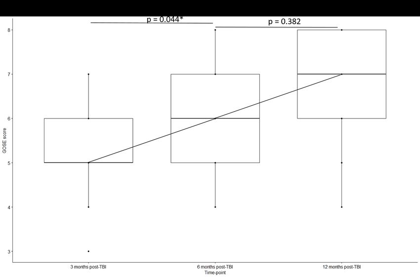
point. Among the three regions, we found that only the SLF showed a significant longitudinal

variation of aMWF in TBI patients (2.89%, 2.35%, and 2.53% at 3, 6, and 12 months post-injury

respectively; Friedman test P=5.748 X 10⁻⁶). A post hoc Wilcoxon signed-rank test revealed a

significant decline in aMWF from 3 to 6 months post-injury (P=6.1 X 10⁻⁵), but not from 6 to 12

months. Our results indicate that 1) there are significant regional variations in the temporal

dynamics of white matter degeneration after moderate-to-severe TBI and 2) the rate of

degeneration is steeper during the first half of the first year post-injury. We did not find significant

associations between regional aMWF and neuropsychological performance at any time points.

Potential explanations of the observed null findings were discussed.

                                                                                                       v
Acknowledgements

Special thanks to Naomi Gaggi, June Yul Choi, Dr. Valerie Shafer, my Mother, and most of all Nia for
introducing me to life’s joys.

                                                                                                       vi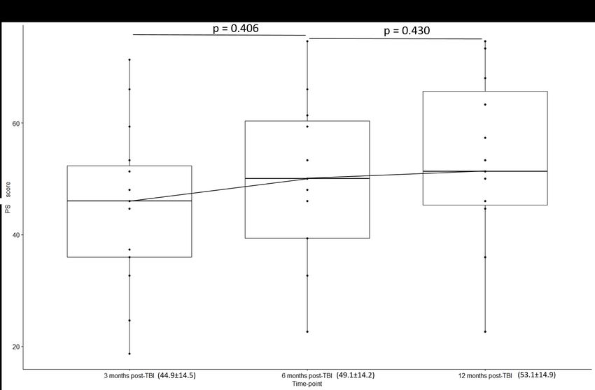
TABLE OF CONTENTS

Introduction ......................................................................................................................................... 1
   Epidemiology of Traumatic Brain Injury ........................................................................................ 1
   Neurocognitive Deficits after TBI................................................................................................... 1
   Diffuse Axonal Injury ..................................................................................................................... 3
   Neuropathology of White Matter Degeneration.............................................................................. 4
   Challenges in Monitoring TBI-induced White Matter Degeneration ............................................. 5
       Conventional Computed Tomography and Magnetic Resonance Imaging ................................. 5
       Structural Morphometry of White Matter .................................................................................... 6
       Diffusion Tensor Imaging ............................................................................................................ 6
       Myelin Water Imaging ................................................................................................................. 8
   Aims of the Study............................................................................................................................ 9
Methods............................................................................................................................................. 10
   Participants and Procedure ............................................................................................................ 10
   MRI Data Acquisition ................................................................................................................... 11
   A Priori Selection of White Matter Regions and Image Processing ............................................. 12
   Statistical Analysis ........................................................................................................................ 13
Results ............................................................................................................................................... 14
   Descriptive Analysis ..................................................................................................................... 14
   Inferential Analysis ....................................................................................................................... 14
       Longitudinal Variation in Neuropsychological and Functional Outcomes .............................. 14
       Longitudinal Variation in aMWF Values .................................................................................. 15
       Correlation between aMWF values and neuropsychological outcomes ................................... 15
       Comparison of aMWF Values between TBI Patients and Healthy Controls ............................ 16
Discussion ......................................................................................................................................... 22
   Conclusion..................................................................................................................................... 25
References ......................................................................................................................................... 27

                                                                                                                                                      vii
LIST OF FIGURES
Figure 1. Longitudinal variation of neuropsychological and functional outcome in TBI patients
(n=15). ........................................................................................................................................................... 18
Figure 2. Box plots showing the temporal variation of aMWF in selected brain regions. (A)
Splenium, (B) Fornix, (C) Superior longitudinal fasciculus ............................................................. 20
viiiLIST OF TABLES
Table 1. Baseline demographic and clinical characteristics of the study participants ..................... 14
Table 2. Neuropsychological test, functional outcome, and neuroimaging metrics at different time
points in TBI patients ........................................................................................................................ 15
Table 3. Correlation analysis between aMWF and neuropsychological and functional outcomes in
TBI patients....................................................................................................................................... 21
Table 4. Comparison of aMWF values between TBI patients and healthy controls …………...... 21
ixIntroduction
Epidemiology of Traumatic Brain Injury
Traumatic brain injury (TBI) is a leading cause of mortality and disability worldwide and
imposes a heavy burden on healthcare and social systems (Galgano et al., 2017; Maas et al., 2017;
Finkelstein et al., 2006). The epidemiology of TBI has been extensively investigated in the United
States. Approximately 2.5 million TBI-related injuries and hospitalizations occur annually in the
USA alone (CDC, 2003), of which about 50,000 result in death. Men are twice as likely as women
to suffer from TBI-related injuries (CDC, 2016). The leading causes of TBI include falls, external
insults such as car crashes and assault to the head. Mortality rates are highest in individuals who
sustained TBI classified as severe. Individuals with TBI are more likely to die from a range of
medical issues such as seizure, septicemia, respiratory conditions other than pneumonia, and
digestive conditions than the general population with similar demographic characteristics
(Harrison-Felix et al., 2006).
Neurocognitive Deficits after TBI
Following moderate-to-severe TBI, cognitive problems can result in difficulties in several
cognitive domains, namely attention, episodic memory, executive functions, working memory,
information-processing speed, language functions, and visuospatial processing (Dikmen et al.,
2009). Changes in emotional response may result in depression, anxiety, fear, anger and frustration.
Common behavior changes include apathy, irritability, aggressive and/or socially inappropriate
behavior, restlessness and agitation and difficulty relating to others (Ponsford et al., 1995). In the
longer term, TBI increases the risk of seizures, psychotic disorders and dementia (Gualtieri & Cox,
11991). Studies of long-term outcomes indicate that, in most cases, it is the cognitive and
behavioral, rather than the sensorimotor or physical impairments, which are the most disabling
(Hoofien et al., 2001; Ponsford et al., 1995).
In moderate-to-severe TBI, indicators of severity in the form of either duration of
unconsciousness, length of coma measured with the Glasgow Coma Scale (GCS; Jennett et al.,
1976), and duration of post-traumatic amnesia (PTA) are predictive of cognitive and functional
outcomes (Dikmen et al., 1995a; Dikmen et al., 1995b; Gordon et al., 2006; Ownsworth &
McKenna, 2004). In the study by Dikmen and colleagues (1995a), the authors compared
individuals with head injuries (TBI group) to that of general trauma group (control group), and they
found that the TBI group performed significantly worse on a range of neuropsychological
measures, and that the magnitude of impairments was dependent on the severity of the injury. Their
study assessed the severity of an injury by the length of coma on the GCS. The general trauma
group with less than 1-hour coma was comparable to the experimental group (TBI), without
significant differences between the assessment metrics, for example, motor functioning, attention,
executive functioning, and memory. Selective impairments on measures of attention and memory
were found in the group with coma of more than 1 hour but less than 24 hours. With a further
increase in injury severity, all measures were affected.
Diffusion axonal injury (DAI) is a type of primary TBI resulting from a non-penetrating
injury to the brain and results in a distinct neuropsychological impairment profile caused by
disruption of neural connections. Specifically, patients with DAI often show long-lasting
neuropsychological impairments characterized by predominantly executive and memory
dysfunction (Scheid et al., 2003).
2Diffuse Axonal Injury
The first mention of a DAI-related injury was in 1956, where the description of the clinical
case was diffuse degeneration of white matter (Strich, 1956), a part of central nervous system made
of myelinated axons. Ever since then, the disease has had several other descriptions, namely
shearing injury (Strich, 1970), diffuse white matter shearing injury (Zimmerman et al., 1978), inner
cerebral trauma (Grcevic, 1982), and presently as diffuse axonal injury (Adams, Graham, Murray,
& Scott, 1982). As the investigation into milder forms of TBI-induced DAI progressed, it became
evident that axonal damage is a distinct clinicopathological occurrence (Gennarelli, Adams, &
Graham, 1986).
Axons are the nerve fibers that help in the conduct of nerve impulses, electric signals
created by electrochemical gradient of ions across the nerve membrane. Myelin forms an insulating
layer of fatty tissues around the axons, aiding in the rapid conduct of such gradients from one axon
node to another. Any physiological disturbances in these ionic gradients as a result of
neurotransmitter imbalances, formed one of the earliest dominant theory behind acute TBI
pathophysiology (Giza & Havda, 2001). It was thought that biomechanical forces elongated,
sheared, and strained the axons, resulting in alterations of intracellular and extracellular ions, in
which an abnormal concentration of calcium and sodium ions would enter the cell, and in turn,
drive potassium out (Bigler & Maxwell, 2012). These molecular disturbances translate into clinical
symptoms of dizziness, fatigue, difficulty speaking, and nausea (Giza & Havda, 2014).
Furthermore, membranes that are altered due to TBI enter into a compensatory mode termed
energy crisis, in which sodium-potassium pumps are overclocked to restore ionic balance, moving
potassium in and sodium out of the cell (Giza & Havda, 2001). This compensatory mechanism
results in an increased demand for energy, that is, glucose, to drive ions against their gradients,
resulting in the depletion of cellular ATP. As the investigation into this matter expanded, recent
3research has gravitated towards axonal damage as a predominant mechanism of brain injury (Chen
et al., 2009; Johnson et al., 2013).
DAI occurs primarily as a result of rapid acceleration or deceleration forces that cause
shear-strain deformations in several cerebral areas, notably the cingulum, corpus callosum, and
other deep white matter regions (McAllister & Stein, 2010; Chatelin et al., 2011; Bigler &
Maxwell, 2012; Smith et al., 2003). Evidence suggests that these mechanical forces impair the
susceptible cerebral microstructure within the axons, particularly axonal cytoskeleton components,
including neurofilaments and microtubules (Povlishock, 1992; Saatman et al., 2008). Support for
microstructure alterations in DAI comes from studies using histological stains and procedures
pertaining to axonal structures, most notably antibodies on transport proteins that travel the axon
medium anterogradely (Gultekin & Smith, 1994; Scherriff et al., 1994). Focal or axonal swelling,
bulbs, or perturbances are distinct cellular features of DAI post-traumatic injury (Adams et al.,
1982; Christman et al., 1994).
Neuropathology of White Matter Degeneration
Several animal studies found that that demyelination and subsequent white matter damage
occurred following TBI (Bramlett & Dietrich, 2002; Hall et al., 2005; Pierce et al., 1998).
Pathophysiological mechanisms of white matter damage include traumatic axonal injury and
demyelination-induced apoptosis. Axonal demyelination may have a more prominent role in the
progressive, long-term consequences of moderate-to-severe TBI, showcasing its effect by altering
functional neuronal processes that rely on normal myelin structure and integrity (Fields, 2008). In
fact, several clinical studies have shown a correlation between a history of acute TBI and the
incidence of neurodegenerative diseases in later life stages (Goldman et al., 2006; DeKosky et al.,
2013; Plassman et al., 2000).
4A handful of studies have also shown relationships of white matter damage with protein
aggregation, amyloid accumulation, and tau cellular toxication (Graham et al., 1995; Hamberger et
al., 2003; Bramlett & Dietrich, 2003; Sherriff et al., 1994; Castillo-Carranza et al., 2013). The
studies, as mentioned earlier, focus mainly on mild TBI and long-term proteotoxicity. However, it
has yet to be determined whether or not a similar pathological mechanism exists for moderate-to-
severe forms of TBI. Furthermore, whether or not similar proteotoxicity manifestation is evident in
progressive TBI-induced neurodegeneration has garnered interest amongst researchers due to the
highly overlapping characteristics of TBI and neurodegenerative diseases.
There is an agreement among the literature that quantitative measurement of white matter
integrity captured by in vivo imaging can potentially serve as a valuable biomarker of TBI.
However, there are challenges in accurately assessing the degree of white matter integrity using
non-invasive neuroimaging. The following section will review the available methods that
attempted to measure TBI-induced white matter degeneration.
Challenges in Monitoring TBI-induced White Matter Degeneration
Conventional Computed Tomography and Magnetic Resonance Imaging
Several guidelines have suggested that any TBI patient regarded as moderate-to-severe on
the Glasgow Scale (scale from 3-15 measuring level of consciousness) should undergo a non-
contrast CT evaluation to rule out pathology or the need for more invasive interventions (Shetty et
al., 2016). Although useful as a preliminary measure, many argue that the limitations of non-
contrast CT can potentially underestimate other features of TBI, including diffuse axonal injury
(Servadei, 2000). Conventional MRI (T1-, T2-, proton density-weighted) reveals 10-30% more
abnormalities than CT. However, in many cases of TBI, conventional MRI also fails to detect
5diffuse and subtle damage, such as those consistent with axonal damage (Humble et al., 2018).
Because conventional MRI is inherently limited due to its low resolution, diagnosing diffuse
axonal injury becomes a clinical challenge.
Structural Morphometry of White Matter
Several white matter structures have been previously investigated and demonstrated
significant volumetric atrophy following moderate-to-severe TBI, with the most common studied
structures being the corpus callosum, capsule, brainstem, superior fasciculus, and fornix (Bendlin
et al., 2008; Ruet et al., 2018; Sidaros et al., 2009). Both atrophy and structural changes have been
previously noted in the literature, with longitudinal investigations showing a decrease of 5.8% in
whole white matter volume (Warner et al., 2010), and more specifically, 16% in the posterior
corpus callosum (Warner et al., 2010) and 14% in the fornix (Tomaiuolo et al., 2005). These results
suggest that white matter degeneration is a vital component of TBI, and quantitative measurement
of white matter morphology captured by in vivo imaging can potentially serve as a valuable
biomarker of TBI.
Diffusion Tensor Imaging
An advanced MRI technique that has recently garnered attention in TBI research is
diffusion tensor imaging (DTI). DTI can be utilized to monitor white matter damage by leveraging
the Brownian motion of water molecules within the region of interest, which can provide
information on both the range and directionality of diffusion (Pierpaoli et al., 1996). Two main
measures of DTI, fractional anisotropy (FA) and mean diffusivity (MD), examine the degree of
directionality (ratio of eigenvalues) and overall orientation of water molecules’ motion. These
6measures are believed to be associated with the integrity of cerebral fiber tracts, that is, axons and
myelin sheath (Beaulieu, 2002).
Some longitudinal DAI studies revealed partial normalization of DTI indices after different
periods (Arfanakis et al., 2002; Rutgers et al., 2008), while other investigations indicated traumatic
microstructural alteration to be more permanent (Bendlin et al., 2008). To date, the majority of
studies have reported lower functional anisotropy (FA), and a higher apparent diffusion
coefficient/mean diffusivity (ADC/MD), both being measures of white matter damage (Douglas et
al., 2015, Wallace et al., 2018a). Longitudinal decrease in FA has been reported in studies of up to
12 months (Sidaros et al., 2008), 24 months (Kumar et al., 2010), and 48 months (Farbota et al.,
2012). This decrease may be attributed to alterations in axial and radial diffusivity and correlate
with neuropsychological performance measures, such as paired-associate visual learning
(Newcombe et al., 2016). On the other hand, several studies have reported an increase in
longitudinal FA scores measured by DTI (Sidaros et al., 2008; Wu et al., 2010).
The conflicting results reviewed in the previous paragraph question DTI’s ability to reliably
detect longitudinal white matter degeneration that accompanies DAI. Regarding reported increases
in longitudinal FA in TBI patients, it is unclear whether the improvements can be attributed to true
neural recovery or other factors such as edema and swelling (Bazarian et al., 2007; Wu et al.,
2010). The inconsistency of longitudinal FA scores may result from poor distance coverage by the
fibers in the region of interest. This is partly due to the non-linear characteristics of the fiber tracts
leading to high amounts of crossing over with neighboring fibers (Douglas et al., 2015). Given the
nonlinearity of fibers and the low resolution of voxel scale unit in millimetres, diffusion intensity is
highly reduced, resulting in reduced anisotropy. These limitations during clinical assessment
reduce the reliability of this FA measure. A second limitation that merits discussion is the size of
acquired voxels (in millimetres), which can contain a plethora of axons with complex fiber
7orientations. Of importance are fibers that crossover, curve, and fan because their respective
geometric configuration has posed certain limitations to the underlying reconstruction algorithms
(Leergaard et al., 2010; Chamberland et al., 2019). This consequently produces a high degree of
uncertainty of the fiber orientation (Daducci et al., 2014), and as a result, the anatomical accuracy
of DTI and white matter tract reconstruction is hindered. Taken together, the DTI technique is
limited when it comes to characterizing longitudinal white matter changes, especially in an injured
brain.
Myelin Water Imaging
Recent literature has implicated a critical role of damage to myelin in the pathophysiology of
TBI (Jurick et al., 2016). Most recently, a novel imaging methodology, myelin water imaging (MWI)
has been developed that can directly measure the associated changes in myelin post-TBI (Prasloski
et al. 2012, Wright et al. 2016, Choi et al. 2019). Through this methodology, fraction of the total
brain water that can be attributed to myelin can be measured, also called myelin water fraction
(MWF) (Mackay et al. 2006). The methodology has led to important discoveries in delineating
pathophysiology of diseases of myelin damage, mainly multiple sclerosis (Laule et al. 2016).
This newer alternative to traditional DTI is direct visualization of short transverse component
(ViSTa) myelin water imaging (MWI). ViSTa MWI suppresses long T1 components in MWI,
leaving only the short T2* signal, a measure of myelin water. The decrease in apparent myelin water
fraction (aMWF), a quantitative measure of myelin damage, has been shown to reduce in proportion
to the severity of the DAI (Alappatt, 2018; Choi et al., 2019). This backs the notion that MWI can
serve as a biomarker for assessing severity in myelin and axonal damage in DAI patients. DTI metrics
and aMWF have shown moderate correlation, suggesting that perhaps aMWF can be complementary
to DTI measures (Choi et al., 2019).
8To date, we identified only a handful of studies performing a longitudinal assessment of
aMWF levels in specific brain regions (Jurick et al., 2018; Spader et al., 2019; Russell-Schulz et al.,
2021). Notably, all the earlier reports studied mild TBI patients (Jurick et al., 2018; Spader et al.,
2019; Russell-Schulz et al., 2021). In this study, ViSTa MWI is used to investigate changes in myelin
water signaling in moderate-to-severe TBI patients at 3, 6, and 12 months post-injury and healthy
controls. We plan to extend the findings of a previous study from our group (Choi et al., 2020) that
described the longitudinal pattern of whole-brain aMWF in the same cohort. The current study aims
to determine the longitudinal pattern of regional aMWF in TBI patients during the first year post-
injury compared to healthy controls and the relationship with neuropsychological performance. Since
consideration of all the white matter regions may lead to spurious associations due to multiple testing,
we restricted our hypothesis testing to the three most frequently damaged white matter tracts, namely
the splenium, fornix and superior longitudinal fasciculus, based on the recently published meta-
analysis studies (Wallace et al. 2018a, Wallace et al. 2018b).
Aims of the Study
A previous seminal study from our group has used aMWF to assess the injury severity of
white matter after moderate-to-severe TBI (Choi et al., 2019). However, the cross-sectional study
used only the baseline data (i.e., 3 months post-injury) without any longitudinal follow-up time
points. In addition, the study used only the whole-brain global aMWF metrics. The current study
aims to achieve the following three objectives: First, we aim to describe the pattern of longitudinal
changes in aMWF in a priori regions of interest (splenium, fornix, and superior longitudinal
fasciculus) as well as neuropsychological and functional outcomes over the first year after TBI. We
hypothesized that aMWF would exhibit a significant decline as the post-injury time increases,
9reflecting DAI-induced white matter degeneration. Our tentative working hypothesis is that the rate
of longitudinal decline will be uniform over the brain region analyzed. Second, we aim to correlate
regional aMWF values (splenium, fornix, and superior longitudinal fasciculus) with
neuropsychological outcomes at each time point. We hypothesize that aMWF correlates positively
with the neuropsychological outcome irrespective of brain region or post-injury time point analyzed.
Third, we aim to compare regional aMWF values (splenium, fornix, and superior longitudinal
fasciculus) between healthy controls and TBI patients at different time points. We hypothesize that
aMWF is significantly lower than healthy controls for all of brain region measures and post-injury
times.
Methods
Participants and Procedure
The data used in this study was acquired as part of a larger project investigating the
neuroimaging correlates of functional recovery after diffuse TBI (principal investigator: Junghoon
J. Kim). Data from a total of 15 TBI patients and 30 healthy controls were analyzed in this study.
Inclusion criteria were: a) age between 18 and 64 years, b) diagnosis of non-penetrating TBI with
either a "Glasgow Coma Scale (GSC score) < 13 in the emergency department (ED) after ruling out
sedation, paralysis or intoxication as the underlying causes" or loss of consciousness ≥12 h or
prospectively documented PTA≥24 h. Exclusion criteria were a) Poor English speaking skills, b)
history of prior TBI or neurological or psychiatric diseases, c) history of severe alcohol or drug
abuse, d) pregnancy, e) non-availability of MRI scanning data at baseline or the first follow-up, f)
large focal intraparenchymal lesions on acute CT or MRI (approximately>5 cm3 for subcortical
10lesions and 50 cm3 for cortical lesions judged by visual inspection). We also included age, gender
and education-matched controls in the study with additional exclusion criteria of absence of any
TBI history resulting in alteration or loss of consciousness.
All the patients were followed up at 3, 6 and 12 months to collect data on the study
outcomes described below. We further collected data on injury variables using the medical records,
including the mechanism of injury. The study was approved by the home institutional review board
(IRB) committee, and all the study participants gave their informed consent before inclusion in the
study.
Concerning cognitive outcomes, we collected longitudinal data on three neuropsychological
domains. First, executive function (EF) was assessed with five subtests that measured different
aspects of executive function (Rabinowitz et al., 2018). The working memory tests included
subtests of the Wechsler Memory Scale-IV (Wechsler et al., 2008), specifically the Letter-Number
sequencing and Digits Backwards sections. Cognitive flexibility was tested using the Controlled
Oral Word Association Test (Benton et al., 1983) and the Trail Making Test (part B) (Reitan &
Wolfson, 1985). Selective attention was evaluated using the Color Word section of the Color Word
Interference Test (CWIT) from the Delis-Kaplan Executive Function System (D-KEFS; Delis et
al., 2001). Second, mental processing speed (PS) was assessed using the Processing Speed Index
from the Wechsler Adult Intelligence IV (Wechsler et al., 2008). Lastly, verbal learning (VL) was
assessed using the Rey Auditory Verbal Learning Test (Lezak et al. 2004). The clinical outcomes
were assessed using the Disability Rating Scale (DRS) and Glasgow Outcome Scale – Extended
(GOSE).
MRI Data Acquisition
Participants with TBI were scanned using 3 T MRI (Trio, Siemens, Erlangen, Germany)
11with an 8-channel phased-array head coil. For ViSTa MWI, a 3D segmented EPI-based ViSTa
sequence (Oh et al., 2013) was implemented using the following parameters: repetition time
(TR) = 1160 ms; echo time (TE) = 4.5 ms; the duration between the first and second inversion
(TI1) = 560 ms; the duration between the second inversion and the excitation (TI2) = 220 ms; the
duration from the excitation and the first inversion (TD) = 380 ms; matrix size = 160 × 160; in-
plane resolution = 1.4 × 1.4 × 5.0 mm3; the number of slices = 26; partial k-space = 6/8 in the z-
direction, EPI factor = 15; bandwidth = 1008 Hz/px; scan time = 7 min 33 s. Fat and flow saturation
pulses were applied to prevent artefacts from those. To quantify aMWF, a PD- weighted GE
sequence based on the same EPI as the ViSTa sequence, but without the two inversion pulses
(TR = 97 ms; flip angle = 28°; scan time = 38 s).
A Priori Selection of White Matter Regions and Image Processing
To extend upon our groups’ results of longitudinal whole-brain aMWF in TBI patients
(Choi et al. 2020), we decided to investigate potential regional differences in the longitudinal white
matter aMWF changes after TBI. Since consideration of all the brain regions could lead to spurious
positive findings due to multiple testing, we restricted our statistical hypothesis testing to the three
most frequently damaged regions after moderate-to-severe TBI: the fornix, splenium, and superior
longitudinal fasciculus. These were selected as ROIs based on the following three criteria. First,
based on a recent meta-analysis (Wallace et al., 2018a), at least three previous studies must have
reported FA value reductions in that ROI. Second, a large Hedge’s effect size (g of less than -1.0)
must have been reported for that ROI based on the same meta-analysis (Wallace et al., 2018a).
Third, the white matter integrity in that ROI must show consistent associations with different
domains of cognition according to another comprehensive meta-analyses by Wallace and
colleagues (Wallace et al., 2018b). Based on the criteria above, the splenium, fornix and superior
12longitudinal fasciculus were selected and defined using the John Hopkins University (JHU) White
Matter Atlas (Mori et al., 2005; Oishi et al., 2008). To calculate aMWF in white matter across the
whole brain, all ViSTa MWI images from healthy controls and TBI patients at all time points were
registered non-linearly to a JHU FA map using SyN in ANTs (Avants et al., 2004, 2010). For
regional aMWF values, aMWF across voxels in the region mask was averaged for subsequent
analysis.
Statistical Analysis
Continuous demographic variables were compared between the TBI patients and healthy
controls using the Mann Whitney U test, and categorical demographic variables were compared
using the Chi-square test. For all analyses in Aim 1, we used the Friedman test with posthoc
Wilcoxon Signed Rank. We used these tests to determine the longitudinal pattern of regional
aMWF in the splenium, SLF, and fornix in patients with TBI at 3-, 6-, and 12-months post-injury.
We also used these tests to determine the longitudinal pattern of neuropsychological performance
in TBI patients at 3, 6, and 12 months post-injury. For Aim 2, we used Spearman’s correlation to
correlate regional aMWF in the splenium, SLF, and fornix with neuropsychological scores at 3-, 6-,
and 12-months post-injury TBI patients. We computed partial correlations adjusted for age and
gender. Lastly, we compared the aMWF in the splenium, SLF, and fornix between patients with
TBI and healthy controls in Aim 3 using the Mann Whitney U test. To address the issue of multiple
testing when comparing aMWF values, results were considered statistically significant if PResults
Descriptive Analysis
Selected demographic and clinical characteristics of control and patient participants are
shown in Table 1. Quantified neuropsychological outcomes of TBI patients 3, 6, and 12-months
post TBI are shown in Table 2. All the neuropsychological test scores showed a moderate increase
during the study duration. Concerning functional outcomes, while DRS scores decreased rapidly
with time, GOSE scores showed a gradual increase.
Inferential Analysis
Longitudinal Variation in Neuropsychological and Functional Outcomes
Statistical evaluation of changes in neuropsychological and functional outcomes during the
study duration showed a significant change in their distribution post-TBI period in TBI patients
(Friedman test P values: EF = 5.16 x 10⁻³, VL = 7.19 x 10⁻⁴, PS = 3.46 x 10⁻⁶, DRS = 0.017, GOSE
= 4.82 x 10⁻³). Furthermore, post-hoc comparisons showed significant differences in the scores
between 3 and 12 months post-TBI for DRS and GOSE outcomes (PPTA, days 21.9 (22.4) - -
GCS 5.67 (8.72) - -
PTA: Duration of post-traumatic amnesia in days; GCS: Glasgow Coma Scale; Comparison of
continuous variables (age, education, PTA and GCS) was done using the Mann-Whitney U test;
Comparison of categorical variables (gender, race) done using Chi-square test
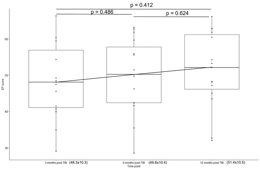
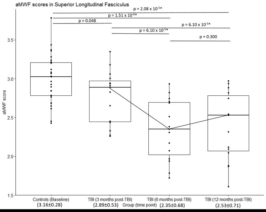
Longitudinal Variation in aMWF Values
Among all three brain regions examined for the aMWF levels, only the superior
longitudinal fasciculus levels showed a significant fluctuation in the values with time (Figure 2).
The Friedman test showed a significant change in aMWF in the SLF during the first year post-
injury (Median: 2.89 vs 2.35 vs 2.53; P=5.7 X 10⁻⁶). The post hoc Wilcoxon Signed Rank analysis
further confirmed that there was a significant difference between aMWF in the SLF between 3 and
6 months’ post-injury in TBI patients (P=6.1 X 10⁻⁵), as we failed to observe any statistical
difference between 6 and 12 months (P=0.3).
Correlation between aMWF values and neuropsychological outcomes
We did not observe any significant correlation between aMWF and any of the
neuropsychological outcomes, irrespective of time post-injury (Table 3).
Table 2. Neuropsychological test, functional outcome, and neuroimaging metrics at different time
points in TBI patients
3 months 6 months 12 months
Measure in TBI patients (N=15) post-TBI post-TBI post-TBI
Mean (SD) Mean (SD) Mean (SD)
Neuropsychological domains
EF 48.3 (10.3) 49.8 (10.4) 51.4 (10.5)
VL 38.1 (17.6) 36.6 (15.7) 46.1 (15.2)
PS 44.9 (14.5) 49.1 (14.2) 53.1 (14.9)
Functional outcome scales
DRS 2.20 (1.47) 1.40 (1.55) 0.867 (1.36)
GOSE 5.27 (1.10) 6.27 (1.28) 6.67 (1.35)
Neuroimaging metrics
aMWF (Splenium) 2.92 (0.37) 2.80 (0.36) 2.82 (0.44)
aMWF (Fornix) 1.99 (0.23) 1.94 (0.24) 1.94 (0.31)
aMWF (Superior longitudinal fasciculus) 2.77 (0.34) 2.34 (0.39) 2.39 (0.44)
15aMWF: apparent myelin water fraction, DRS: Disability Rating Scale, EF: Executive Function, GOSE: Glasgow
Outcome Scale – Extended, PS: Processing Speed, VL: Verbal Learning, SD: Standard Deviation.
Comparison of aMWF Values between TBI Patients and Healthy Controls
Both splenium and superior longitudinal fasciculus showed significantly lower aMWF
values in TBI patients compared to healthy controls at all-time points (Figure 2, Table 4).
However, the difference was much larger in the superior longitudinal fasciculus region at 6 and 12
months (P=5.1 x 10⁻⁶, P=2.1 x 10⁻⁵).
A
16B
C
17D
E
Figure 1. Longitudinal variation in neuropsychological and functional outcome in TBI patients (n=15). (A)
EF, (B) VL, (C) PS, (D) DRS, (E) GOSE. P computed using post hoc Wilcoxon Signed Rank tests. *PProcessing speed, VL: Verbal Learning.
A
B
19C
Figure 2. Box plots showing the temporal variation of aMWF in selected brain regions. (A)
Splenium, (B) Fornix, (C) Superior longitudinal fasciculus. Wilcoxon signed rank test for comparing
aMWF within the TBI group. Mann Whitney U test for comparing aMWF between the healthy
controls and TBI group. *Significant P
20Table 3. Correlation analysis between aMWF and neuropsychological outcomes in TBI patients
Correlation at 3 months showing rho coefficient (P) (Adjusted for age and gender)
Measure VL PS EF
aMWF (Splenium) -0.137 (0.655) 0.402 (0.172) 0.312 (0.299)
aMWF (Fornix) -0.055 (0.859) 0.281 (0.353) 0.206 (0.500)
aMWF (Superior longitudinal fasciculus) -0.223 (0.464) 0.306 (0.308) 0.177 (0.561)
Correlation at 6 months showing rho coefficient (P-values) (Adjusted for age and gender)
Measure VL PS EF
aMWF (Splenium) 0.046 (0.881) 0.388 (0.190) 0.301 (0.317)
aMWF (Fornix) 0.202 (0.509) 0.161 (0.600) 0.246 (0.417)
aMWF (Superior longitudinal fasciculus) -0.074 (0.809) 0.360 (0.226) 0.319 (0.288)
Correlation at 12 months showing rho coefficient (P-values) (Adjusted for age and gender)
Measure VL PS EF
aMWF (Splenium) -0.152 (0.619) 0.365 (0.220) 0.439 (0.133)
aMWF (Fornix) -0.036 (0.906) 0.217 (0.477) 0.305 (0.311)
aMWF (Superior longitudinal fasciculus) -0.347 (0.246) 0.134 (0.663) 0.179 (0.561)
aMWF: apparent myelin water fraction, EF: Executive Function, PS: Processing Speed, VL: Verbal Learning. P
computed using Spearman rank correlation test.
Table 4. Comparison of aMWF values between TBI patients and healthy controls.
Healthy
Time of assessment TBI Median
Brain region Median (IQR) controls P*
(post-injury) (N) (IQR)
(N)
Splenium 3 months 15 2.94 (0.43) 30 3.16 (0.28) 0.068
Splenium 6 months 15 2.83 (0.58) 30 3.16 (0.28) 3.5E-03**
Splenium 12 months 15 2.77 (0.41) 30 3.16 (0.28) 0.007**
Fornix 3 months 15 1.96 (0.40) 30 1.39 (0.32) 0.140
Fornix 6 months 15 1.99 (0.33) 30 1.39 (0.32) 0.025
Fornix 12 months 15 2.09 (0.48) 30 1.39 (0.32) 0.140
Sup. Long. fasciculus 3 months 15 2.89 (0.53) 30 3.02 (0.43) 0.048
Sup. Long. fasciculus 6 months 15 2.35 (0.68) 30 3.02 (0.43) 1.5E-06**
Sup. Long. fasciculus 12 months 15 2.53 (0.71) 30 3.02 (0.43) 2.1E-05**
*P computed using Mann-Whitney U test. ** Significant after controlling for multiple testing
21Discussion
Traumatic brain injury is recognized for its pervasive nature and long-lasting burden on the
patients and caregivers. Longitudinal investigation of the long-term consequences has been
relatively sparse, with available evidence illustrating functional as well as morphometrical changes
in various cortical and subcortical regions in the brain, that is, a hallmark of neurodegenerative
disease. In a previous MWI study comparing changes in myelin water in moderate-to-severe TBI
patients to that of healthy cohorts, our research group established reduced aMWF in moderate-to-
severe TBI patients at 3 months post-injury. Our findings suggested that TBI may lead to
demyelination of white matter and thus a subsequent decline in neurocognitive performance and
post-injury prognosis (Choi et al., 2019).
The present thesis expands these findings by longitudinally assessing the changes in aMWF
in the selected region of interest in a subset of the same patient cohort at 3, 6 and 12 months post-
injury. We observed significantly lower aMWF levels in the splenium and superior longitudinal
fasciculus regions in TBI patients compared to healthy controls at 6 and 12 months’ post-injury.
Further examination of intra-group longitudinal variation in TBI patients specifically showed a
sharp, significant decline in aMWF levels in the superior longitudinal fasciculus region between 3-
and 6-months post-injury. Although we also observed a declining trend throughout the study period
in the splenium region, it failed to reach statistical significance. On the other hand, aMWF levels in
the fornix region in TBI patients maintained a steady level throughout the study duration without
statistically significant longitudinal variation. These results suggest that different white matter
tracts may go through different rate of degeneration after moderate-to-severe TBI in our study
participants.
22To date, several studies have demonstrated TBI-induced region specific white matter
degeneration using different imaging approaches. Warner and colleagues (2010) prospectively
studied longitudinal changes in global and regional brain volumes of patients with TBI and sex-
matched controls. The study found significant whole white matter atrophy in TBI cohorts
compared to healthy controls; however, more importantly, the study also revealed the decreased
volume of specific subcortical structures while others maintained integrity. Similarly, Cole and
colleagues (2018) investigated spatial patterns of brain atrophy in moderate-to-severe TBI patients
and concluded that atrophy is most prominent in white matter (~ 84%) but also noted a higher
volume loss in cortical sulci compared to cortical gyri. Similar to the morphometry studies
reviewed above, studies employing DTI have also revealed regional specificities in the brain
damage in TBI patients by identifying anisotropic diffusion of water in white matter regions. Using
FA and ADC/MD as parameters to assess white matter damage, Wallace and colleagues
specifically reported the most significant white matter damage in the corpus callosum, internal
capsule, occipital white matter, centrum semiovale, fornix and thalamic radiations (Wallace et al.,
2018a).
The MWI methodology was recently applied to patients with mild TBI in a prospective
study (Wright et al., 2016), which demonstrated a significant decline in MWF at 2 weeks’ post-
injury stage. The study by Spader and colleagues (2019) compared MWF values in 12 mild TBI
athletes at 3 months’ post-injury to that at baseline stage, and observed reductions in the MWF
values in the peripheral white matter and bilaterally in the internal capsules (Pcompared three months MWF values in TBI patients with healthy controls. Contrary to our
findings of a declining trend in MWF values in TBI patients in two of the three investigated brain
regions (splenium and superior longitudinal fasciculus), the study by Spader and colleagues failed
to observe a decrease in MWF values in any of the brain regions.
Our results of reduced MWF in comparison to controls are in line with another recent report
by Russell-Schulz and colleagues (2021), which showed significantly reduced MWF in the
splenium (P=0.008, uncorrected) and superior longitudinal fasciculus region (left: P=0.03,
uncorrected; right: P = 0.02, uncorrected) between 15 adults with chronic mild TBI and 12 healthy
controls. However, the study failed to stratify patients according to post-TBI time, and included
patients with time-since injury from 6 months to 5 years. The study also observed a decline of
MWF in other brain regions, including genu and body of corpus callosum, minor forceps, right
anterior thalamic radiation, and corticospinal tract. However, the previous study's findings may be
considered exploratory in nature due to the failure to apply the correction for multiple testing.
In summary, survey of the literature regarding brain region-specific MWF values in mild
TBI patients and healthy controls returned mixed results. A large proportion of studies were
underpowered, followed heterogeneous study designs with limited follow-up period, and failed to
apply corrections for multiple testing, often making it hard to judge the validity and reliability of
the findings.
Unlike MWF values, the cognitive sequelae of TBI have been extensively studied. While
the methodology used to measure different cognitive functions differ drastically between each
study, there is substantial agreement to suggest that neuropsychological measures are predictive of
outcome after TBI. In the present study, we show significant changes in all of the investigated
neuropsychological and functional outcomes during the study duration post-TBI period in
moderate-to-severe TBI patients, namely EF, VL, PS, DRS, and GOSE. Hence, our findings are in
24strong agreement with the previous literature. In general, TBI resulted in poorer
neuropsychological outcomes. However, we failed to observe any significant correlation between
aMWF and neuropsychological outcomes which could be to be due to our small sample size.
The findings of the present study must be interpreted with caution on account of several
inherent limitations. One of the major limitations of the present study was the limited sample size,
which may not have been adequate to detect subtle differences between healthy control and TBI
patients. Due to the limited sample size, we could not adjust for several potential confounding
variables which may have influenced the longitudinal trajectory of neuropsychological and clinical
outcomes. The limited sample size may also provide overestimated or underestimated temporal
trends. Any trends reported in the present study require careful interpretation and will need to be
replicated with a larger sample. It is also possible that we failed to detect any curvilinear
trajectories at the earlier stages of post TBI recovery due to factors associated with logistics at the
time of hospitalization, such as delayed transport to the hospital and use of steroids for treating
patients (Brazinova et al., 2015). The possibility of some individuals in our study population
having suffered from previously mild episodes of TBI cannot be completely ruled out. We did not
collect data on other aspects of cognitive functioning, such as language and reasoning, which might
have given us a more complete picture of the effects of TBI on cognition. Lastly, we could not
consider all the white matter regions due to the potential Type I error associated with conducting
multiple tests. However, despite these limitations, our study is, to our knowledge, the first study
that demonstrated tract-specific longitudinal trajectories of aMWF changes post moderate-to-severe
TBI.
Conclusion
Traumatic brain injury imparts a huge burden on society, and even mild TBI can have long-
25term effects on patients. There is increasing evidence that TBI is not a ‘single event’ but rather an
ongoing process that unfolds across months, years, and possibly over a lifetime; thus, improved
monitoring of long-term effects of TBI is essential. White matter tracts are particularly vulnerable
in TBIs, and monitoring white matter degeneration of these tracts may facilitate the development of
diagnostic and prognostic biomarkers. Myelin water imaging is a powerful quantitative technique
to assess myelin water content and our results successfully demonstrated that it can reliably assess
the longitudinal degeneration after moderate-to-severe TBI, indicating that it may serve as a proxy
outcome metric of neuroprotective therapies in future clinical trials. Our results also highlight
significant regional variations in the temporal dynamics of white matter degeneration after
moderate-to-severe TBI, specifically during the first half of the first year post-injury. Although
there was a lack of relationship between longitudinal neuropsychological and aMWF changes, the
data are encouraging, and ViSTa MWI is a promising neuroimaging technique for assessing
region-specific progressive white matter degeneration in TBI patients. Future studies can include a
larger sample size to allow sufficient power for a greater confidence in our findings.
26References
Adams, J. H., Graham, D. I., Murray, L. S., & Scott, G. (1982). Diffuse axonal injury due to
nonmissile head injury in humans: An analysis of 45 cases. Annals of Neurology, 12(6), 557–563.
doi:10.1002/ana.410120610
Alappatt, J., Parker, D., Ould Ismail, A. A., Kim, J., & Verma, R. (2019). Free water elimination is
necessary to characterize Diffuse Axonal Injury in moderate Traumatic Brain Injury. Abstract
presented at ISMRM 27th Annual Meeting Exhibition. https://archive.ismrm.org/2019/0078.html
Alexander, A. L., Lee, J. E., Lazar, M., Field, A. S. (2007). Diffusion Tensor Imaging of the Brain.
Neurotherapeutics, 4(3), 316–329. doi: 10.1016/j.nurt.2007.05.011
Arfanakis, K., Haughton, V. M., Carew, J. D., Rogers, B. P., Dempsey, R. J., & Meyerand, M. E.
(2002). Diffusion tensor MR imaging in diffuse axonal injury. AJNR. American Journal of
Neuroradiology, 23(5), 794–802.
Avants, B., & Gee, J. C. (2004). Geodesic estimation for large deformation anatomical shape
averaging and interpolation. NeuroImage, 23 Suppl 1, S139–S150.
https://doi.org/10.1016/j.neuroimage.2004.07.010
Avants, B. B., Yushkevich, P., Pluta, J., Minkoff, D., Korczykowski, M., Detre, J., & Gee, J. C.
(2010). The optimal template effect in hippocampus studies of diseased populations.
NeuroImage, 49(3), 2457–2466. https://doi.org/10.1016/j.neuroimage.2009.09.062
Baddeley, A. D., & Hitch, G. (1974). Working Memory. Psychology of Learning and Motivation,
47–89. doi:10.1016/s0079-7421(08)60452-1
Bendlin, B. B., Ries, M. L., Lazar, M., Alexander, A. L., Dempsey, R. J., Rowley, H. A., Sherman,
J. E., & Johnson, S. C. (2008). Longitudinal changes in patients with traumatic brain injury
assessed with diffusion-tensor and volumetric imaging. Neuroimage, 2(2), 503-514. doi:
10.1016/j.neuroimage.2008.04.254.
Bendlin, B. B., Ries, M. L., Lazar, M., Alexander, A. L., Dempsey, R. J., Rowley, H. A., Sherman,
J. E., Bigler, E. D., & Maxwell, W. L. (2012). Neuropathology of mild traumatic brain injury:
relationship to neuroimaging findings. Brain Imaging and Behavior 6, 108–136. doi:
10.1007/s11682-011-9145-0.
Benton, A. L., Hamsher, S. K. de, & Sivan, A. B. (1983). Multilingual Aplasia Examination (2nd
ed.), AJA Associates, Iowa City, IA
Bishara, S. N., Partridge, F. M., Godfrey, H. P., & Knight, R. G. (1992). Post-traumatic amnesia
and Glasgow Coma Scale related to outcome in survivors in a consecutive series of patients
with severe closed-head injury. Brain Injury, 6(4), 373–380.
https://doi.org/10.3109/02699059209034952
27Blumbergs, P. C., Scott, G., Manavis, J., Wainwright, H., Simpson, D. A., & McLean, A. J. (1995).
Topography of axonal injury as defined by amyloid precursor protein and the sector scoring
method in mild and severe closed head injury. Journal of Neurotrauma, 12(4), 565–572.
https://doi.org/10.1089/neu.1995.12.565
Boake, C., Millis, S. R., High, W. M., Delmonico, R. L., Kreutzer, J. S., Rosenthal, M., …
Ivanhoe, C. B. (2001). Using early neuropsychologic testing to predict long-term productivity
outcome from traumatic brain injury. Archives of Physical Medicine and Rehabilitation, 82(6),
761–768. doi:10.1053/apmr.2001.23753
Bonds, B. W., Dhanda, A., Wade, C., Massetti, J., Diaz, C., & Stein, D. M. (2015). Prognostication
of Mortality and Long term Functional Outcomes Following Traumatic Brain Injury: Can We
Do Better? Journal of Neurotrauma. doi:10.1089/neu.2014.3742
Bramlett, H. M., & Dietrich, W. D. (2002). Quantitative structural changes in white and gray
matter 1 year following traumatic brain injury in rats. Acta Neuropathologica, 103(6), 607–614.
doi.org/10.1007/s00401-001-0510-8
Bramlett, H. M., & Dietrich, W. D. (2003). Synuclein aggregation: possible role in traumatic brain
injury. Experimental Neurology, 184(1), 27–30. doi:10.1016/s0014-4886(03)00401-1
Brazinova, A., Majdan, M., Leitgeb, J., Trimmel, H., Mauritz, W., & Austrian Working Group on
Improvement of Early TBI Care (2015). Factors that may improve outcomes of early traumatic
brain injury care: prospective multicenter study in Austria. Scandinavian Journal of Trauma,
Resuscitation and Emergency Medicine, 23, 53. https://doi.org/10.1186/s13049-015-0133-z
Castillo-Carranza, D. L., Lasagna-Reeves, C. A., & Kayed, R. (2013). Tau aggregates as
immunotherapeutic targets. Frontiers in Bioscience (Scholar edition), 5, 426–438.
doi.org/10.2741/s381
Centers for Disease Control and Prevention (CDC) (2003). Report to Congress on mild traumatic
brain injury in the United States: steps to prevent a serious public health problem.
Centers for Disease Control and Prevention (CDC) (2019). Surveillance Report of Traumatic Brain
Injury-related Emergency Department Visits, Hospitalizations, and Deaths—United States.
Centers for Disease Control and Prevention (CDC) (2016). Rates of TBI-related Deaths by Sex—
United States, 2001–2010. Available:
http://www.cdc.gov/traumaticbraininjury/data/rates_deaths_bysex.html. Accessed 7 August
2021.
Chamberland, M., Raven, E. P., Genc, S., Duffy, K., Descoteaux, M., Parker, G. D., Tax, C., &
Jones, D. K. (2019). Dimensionality reduction of diffusion MRI measures for improved
tractometry of the human brain. NeuroImage, 200, 89–100.
https://doi.org/10.1016/j.neuroimage.2019.06.020
Chartrand, G., Cheng, P. M., Vorontsov, E., Drozdzal, M., Turcotte, S., Pal, C. J., Kadoury, S., &
28Tang, A. (2017). Deep Learning: A Primer for Radiologists. Radiographics : a review
publication of the Radiological Society of North America, Inc, 37(7), 2113–2131.
doi.org/10.1148/rg.2017170077
Chatelin, S., Deck, C., Renard, F., Kremer, S., Heinrich, C., Armspach, J.P., & Willinger, R. (2011).
Computation of axonal elongation in head trauma finite element simulation. Journal of the
Mechanical Behavior of Biomedical Materials 4(8), 1905-1919. doi:
10.1016/j.jmbbm.2011.06.007.
Chen, X. H., Johnson, V. E., Uryu, K., Trojanowski, J. Q., & Smith, D. H. (2009). A lack of
amyloid beta plaques despite persistent accumulation of amyloid beta in axons of long-term
survivors of traumatic brain injury. Brain Pathology (Zurich, Switzerland), 19(2), 214–223.
https://doi.org/10.1111/j.1750-3639.2008.00176.x
Choi, J. Y., Hart, T., Whyte, J., Rabinowitz, A. R., Oh, S.-H., Lee, J., & Kim, J. J. (2019). Myelin
water imaging of moderate-to-severe diffuse traumatic brain injury. NeuroImage, 22, 101785.
doi: 10.1016/j.nicl.2019.101785
Choi, S. C., Barnes, T. Y., Bullock, R., Germanson, T. A., Marmarou, A., & Young, H. F. (1994).
Temporal profile of outcomes in severe head injury. Journal of Neurosurgery, 81(2), 169–173.
https://doi.org/10.3171/jns.1994.81.2.0169
Choi, J. Y., Hart, T., Whyte, J., Rabinowitz, A. R., Oh, S.-H., Lee, J., & Kim, J. J. (2020).
Longitudinal changes of myelin water fraction during the first year after moderate to severe
diffuse traumatic brain injury. Poster presented at ISMRM & SMRT Virual Conference and
Exhibition. https://archive.ismrm.org/2020/0054.html
Christman, C. W., Grady, M. S., Walker, S. A., Holloway, K. L., & Povlishock, J. T. (1994).
Ultrastructural Studies of Diffuse Axonal Injury in Humans. Journal of Neurotrauma, 11(2),
173–186. doi:10.1089/neu.1994.11.173
Cole, J. H., Jolly, A., Simoni, S. D., Bourke, N., Patel, M. C., Scott, G., & Sharp, D. J. (2018).
Spatial patterns of progressive brain volume loss after moderate-severe traumatic brain injury.
Brain, 141(3), 822–836. doi: 10.1093/brain/awx354
Crépeau, F., & Scherzer, P. (1993). Predictors and indicators of work status after traumatic brain
injury: A meta-analysis. Neuropsychological Rehabilitation, 3(1), 5–35.
doi:10.1080/09602019308401421
Daducci, A., Canales-Rodríguez, E. J., Descoteaux, M., Garyfallidis, E., Gur, Y., Lin, Y. C., Mani,
M., Merlet, S., Paquette, M., Ramirez-Manzanares, A., Reisert, M., Reis Rodrigues, P.,
Sepehrband, F., Caruyer, E., Choupan, J., Deriche, R., Jacob, M., Menegaz, G., Prčkovska, V.,
Rivera, M., Wiaux, Y., Thiran, J. P. (2014). Quantitative comparison of reconstruction methods
for intra-voxel fiber recovery from diffusion MRI. IEEE Transactions on Medical
Imaging, 33(2), 384–399. https://doi.org/10.1109/TMI.2013.2285500
Daniel, Wayne W. (1990). Spearman rank correlation coefficient. Applied Nonparametric Statistics
(2nd ed.). Boston: PWS-Kent. pp. 358–365.
29You can also read