Influence of adiponectin and inflammatory cytokines in fatty degenerative atrophic muscle - Nature
←
→
Page content transcription
If your browser does not render page correctly, please read the page content below

www.nature.com/scientificreports
OPEN Influence of adiponectin
and inflammatory cytokines
in fatty degenerative atrophic
muscle
Issei Shinohara, Takeshi Kataoka, Yutaka Mifune*, Atsuyuki Inui, Ryosuke Sakata,
Hanako Nishimoto, Kohei Yamaura, Shintaro Mukohara, Tomoya Yoshikawa, Tatsuo Kato,
Takahiro Furukawa, Takehiko Matsushita & Ryosuke Kuroda
Tendon rupture and nerve injury cause fatty infiltration of the skeletal muscle, and the adipokines
secreted from the infiltrated adipocytes are known to contribute to chronic inflammation. Therefore,
in this study, we evaluated the effects of the adipokines on chronic inflammation using a rat
sciatic nerve-crushed injury model. In vitro and in vivo experiments showed that the expression of
adiponectin was decreased (0.3-fold) and the expression of Il6 (~ 3.8-fold) and Tnf (~ 6.2-fold) was
increased in the nerve-crushed group compared to that in the control group. It was also observed
that the administration of an adiponectin receptor agonist decreased the levels of Il6 (0.38-fold) and
Tnf (0.28-fold) and improved cellular viability (~ 1.9-fold) in vitro. Additionally, in the fatty infiltrated
skeletal muscle, low adiponectin levels were found to be associated with chronic inflammation.
Therefore, the local administration of adiponectin receptor agonists would prevent chronic
inflammation.
Muscle volume is an important factor determining muscle f unction1. Muscle atrophy is characterized by a
decrease in the cross-sectional area of muscles, resulting in decreased muscle strength or endurance, or b oth2.
Additionally, muscle atrophy that is caused by a wide range of factors, such as tendon rupture or nerve damage-
inducing fat infiltration into atrophied muscles, leads to a decrease in mobility, a deterioration of the quality
of life, and reduced life e xpectancy3,4. The condition in which fat infiltrates atrophied muscle causing muscle
dysfunction is called fatty d egeneration5. Even though tendon rupture and nerve injuries have been repaired
successfully, fatty degeneration is irreversible in some c ases6.
Adipokine is a collective term of various cytokines secreted from adipose tissues7. Among them, inflammatory
cytokines, such as interleukin-6 (IL6) and tumor necrosis factor (TNF), which are secreted by adipocytes infiltrat-
ing the muscle, have been identified as factors that enhance inflammation as well as chronic p ain8. Conversely,
adiponectin, which is one of the adipokines, enhances fatty acid oxidation, insulin sensitivity, and glucose uptake,
while inhibiting hepatic gluconeogenesis and exerting anti-inflammatory effects9. Adiponectin was first identified
as a circulating adipokine in plasma; however, it was demonstrated that it originates from skeletal muscles10,11.
The level of adiponectin in muscle does not seem to be related to circulating levels, highlighting the possibility
of an independent process in skeletal muscles10. Furthermore, it is also attracting increasing attention owing to
its role as a locally expressed paracrine/autocrine factor11, and reportedly, its administration can reduce muscle
damage12. However, the expression of adiponectin and its receptors in muscles after muscle atrophy and fatty
infiltration has not been sufficiently studied. Muscle atrophy and fatty infiltration are known to cause chronic
inflammation8, and we hypothesized that decrease of adiponectin expression is related to expression of inflam-
matory cytokines after the nerve injury. Therefore, in this study, we evaluated the expressions of adipokines on
chronic inflammation using a rat sciatic nerve-crushed injury m odel13. In addition, few reports have shown the
effect of adiponectin receptor agonist administration on muscle atrophy and fatty infiltration. We also evaluated
the effects of AdipoRon, an adiponectin receptor agonist, on myotubes after the nerve injury in vitro.
Department of Orthopedic Surgery, Graduate School of Medicine, Kobe University, 5‑2, Kusunoki‑cho7, Chuo‑ku,
Kobe‑shi, Hyogo 650‑0017, Japan. *email: m-ship@kf7.so-net.ne.jp
Scientific Reports | (2022) 12:1557 | https://doi.org/10.1038/s41598-022-05608-x 1
Vol.:(0123456789)
www.nature.com/scientificreports/
Figure 1. (a) Procedure for establishing the nerve-crushed rat model. The sciatic nerve was clamped using
hemostatic forceps for one minute at a proximal distance of 5 mm from the bifurcation point of the peroneal
and tibial nerves. (b) Resected gastrocnemius muscle. (c) The weight of the gastrocnemius muscle on the
affected side was significantly decreased in the nerve-crushed group compared to the unaffected side (p < 0.001).
(d) The weight ratio of the muscle on the affected side and the healthy side in the nerve-crushed group
(57.9 ± 2.3%).
Results
Gastrocnemius muscle weight. The weight of the gastrocnemius muscle at 4 weeks post-operation was
compared between the nerve-crushed model (refer to the Methods section, Fig. 1a, b), in which the sciatic nerve
was crushed as described in a previous study13, and the control group, which had sham surgery wherein only a
skin incision was made. The gastrocnemius muscle weight was significantly decreased on the affected side in the
nerve-crushed group than on the unaffected side. The weight of the gastrocnemius muscle in the control group
was not significantly different from those of the affected and unaffected sides. There was no significant difference
in gastrocnemius muscle weight between the unaffected side of the nerve-crushed group and both sides of the
control group (Fig. 1c). The ratio of the muscle weight on the affected side to that on the unaffected side in the
nerve-crushed group was significantly lower than that in the control group, as shown in Fig. 1d.
Cell morphology evaluation. Morphological observation via microscopy and Hematoxylin–eosin (HE)
staining showed the formation of myotubes in the control group. Conversely, in the nerve-crushed group, the
cells appeared small, with poorly formed myotubes (Fig. 2a). The number of myotubes formed per field of view
(400 μm × 600 μm) was calculated as the average of four fields of view. The number of myotubes formed per field
of view was 24.8 ± 2.3 in the control group and 6.6 ± 1.9 in the nerve-crushed group, and the number of myotubes
formed was significantly lower in the nerve-crushed group (p = 0.0003).
Cell viability (cell proliferation assay). The water-soluble tetrazolium salt (WST) assay was used to
measure the absorbance of reduced formazan at 450 nm. Thus, it was observed that the absorbance correspond-
ing to the control group was significantly higher than that corresponding to the nerve-crushed group. Further,
in the nerve-crushed group treated with AdipoRon, the absorbance showed a significant increase compared with
the nerve-crushed group without AdipoRon treatment. However, the differences among all the AdipoRon doses
were not significant (Fig. 2b).
Fluorescent immunostaining. The cytoplasm of the adiponectin-positive cells was stained green, and the
quantitative evaluation of the ratio of adiponectin-stained cells to blue-stained 4’,6-diamidino-2-phenylindole
(DAPI)-positive cells was determined (Fig. 3a). Thus, we observed that the ratio of green adiponectin-stained
Scientific Reports | (2022) 12:1557 | https://doi.org/10.1038/s41598-022-05608-x 2
Vol:.(1234567890)
www.nature.com/scientificreports/

                                  Figure 2.  (a) Morphological observation based on microscopy and HE staining. The nerve-crushed group
                                  showed smaller cells and poorer myotube formation. (b) Fold change comparison (baseline: nerve-crushed
                                  group). Water-soluble tetrazolium salt (WST) assay showed that myocyte proliferation was significantly lower in
                                  the nerve-crushed group than in the control group (p = 0.08). AdipoRon treatment significantly improved cell
                                  viability in the nerve-crushed group (p < 0.05). The different AdipoRon doses showed no significant differences.

Scientific Reports |   (2022) 12:1557 |               https://doi.org/10.1038/s41598-022-05608-x                                                 3

                                                                                                                                            Vol.:(0123456789)
www.nature.com/scientificreports/

                                            Figure 3.  (a) Ratio of adiponectin staining evaluated based on the percentage of green adiponectin-stained
                                            cells relative to blue-stained DAPI-positive cells. (b) The ratio of green-stained adiponectin cells to blue-stained
                                            DAPI-positive cells (significantly lower in the nerve-crushed group (0.15) than in the control group (0.68)).

                                            cells to blue DAPI-positive cells. Evidently, that corresponding to the nerve-crushed group was significantly
                                            lower than that corresponding to the control group (Fig. 3b).

                                            Histological examination. HE staining showed that the diameter of muscle fibers corresponding to the
                                            rats in the nerve-crushed group was smaller than that corresponding to the rats in the control group. Further-
                                            more, the mean cross-sectional area of each muscle fiber was significantly decreased in the nerve-crushed group,
                                            indicating that the muscles were atrophied four weeks after the nerve crush injury (Fig. 4a, b). Oil Red-O posi-
                                            tive lipid droplets were observed in the muscle in both the control and nerve-crushed groups. The number of
                                            fat droplets quantitatively assessed in four non-overlapping areas by microscopy was significantly higher in the
                                            nerve-crushed group than in the control group (Fig. 5a, b). Furthermore, immunofluorescence staining showed
                                            that adiponectin was stained along the fascia, and the degree of staining corresponding to the nerve-crushed
                                            group was lower than that corresponding to the control group (Fig. 6a). Quantitative evaluation performed
                                            using the ratios of green adiponectin- and adiponectin receptor (AdipoR)-stained cells to blue DAPI-positive
                                            cells showed that the expression of adiponectin was significantly weaker in the nerve-crushed group than in the
                                            control group. Conversely, the two groups showed no significant differences with respect to the expression of
                                            AdipoR1 (Fig. 6b).

          Scientific Reports |   (2022) 12:1557 |                https://doi.org/10.1038/s41598-022-05608-x                                                        4

Vol:.(1234567890)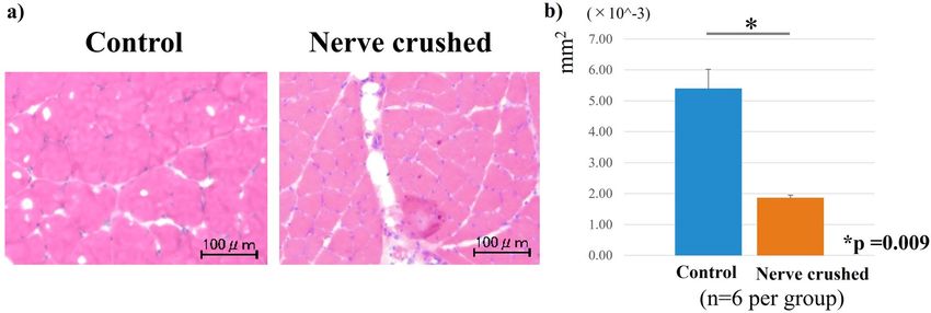
www.nature.com/scientificreports/

                                  Figure 4.  (a) Hematoxylin–eosin (HE) staining showing a smaller diameter for the muscle fibers corresponding
                                  to the neurolysis group than those corresponding to the control group. (b) The mean cross-sectional area of each
                                  muscle fiber was significantly reduced in the crushed nerve group compared to the control group. (p = 0.009).

                                  Figure 5.  (a) Oil red-O staining showed infiltration of fat droplets in the muscle. (b) The quantitative
                                  assessment of the number of fat droplets showed a significant increase in the nerve-crushed group (p = 0.0004).

                                  Real‑time PCR. Expression of the adiponectin (Adipoq) gene was significantly decreased in the nerve-
                                  crushed group. The gene expression levels of muscle markers, such as myogenin and Myod, were significantly
                                  lower in the nerve-crushed group, which also showed significantly higher expression levels of muscle atrophy
                                  markers (Atrogin1, Muscle RING-Finger Protein-1 (Murf1), Forkhead box O1 (Foxo1)) than the control group.
                                  Furthermore, the gene expression levels of Il6, Tnf, and cyclooxygenase1 (Cox1) were significantly higher in the

Scientific Reports |   (2022) 12:1557 |               https://doi.org/10.1038/s41598-022-05608-x                                                    5

                                                                                                                                            Vol.:(0123456789)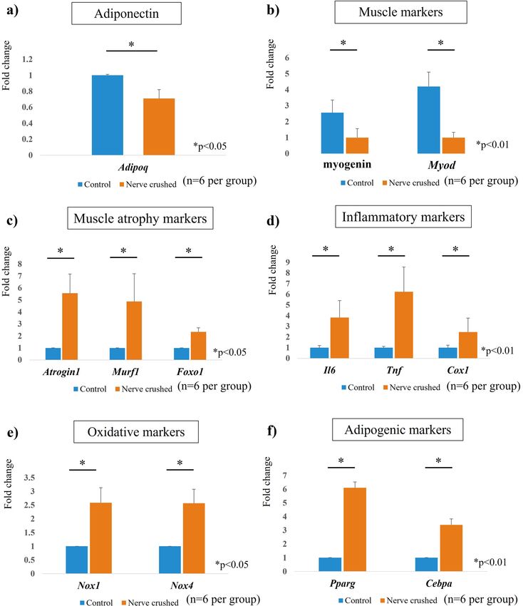
www.nature.com/scientificreports/

                                            Figure 6.  (a) Adiponectin and AdipoR1 stained along the fascia. (b) Quantitative analysis of adiponectin
                                            expression showed that adiponectin expression was significantly lower in the nerve-crushed group (p < 0.01)
                                            than in the control group. Conversely, AdipoR1 expression showed no significant difference between the two
                                            groups.

                                            nerve-crushed group than in the control group, indicating the occurrence of an inflammatory reaction in the
                                            muscles after nerve injury. The expression of oxidative markers, NADPH oxidase1 (Nox1) and Nox4, was sig-
                                            nificantly increased in the nerve-crushed group. Furthermore, the expression levels of adipogenic markers, such
                                            as Pparg and Cebpa were significantly higher in the nerve-crushed group than in the control group, as shown
                                            in Fig. 7.
                                                To evaluate the effect of AdipoRon administration in vitro, the nerve-crushed group received AdipoRon at
                                            concentrations of 0 μg/ml, 0.3 μg/ml, 1.0 μg/ml, and 2.0 μg/ml, respectively. Although the expression levels of
                                            myogenin and Myod, the markers of muscle atrophy, were not significantly different after the AdipoRon treat-
                                            ment, the expression levels of Atrogin1, Murf1, and Foxo1, the markers of muscle atrophy, were significantly
                                            decreased following AdipoRon treatment, the differences in group which were administered different concentra-
                                            tions were not significant. In terms of inflammatory cytokines, Il6 and Tnf were significantly downregulated after
                                            AdipoRon treatment. AdipoRon treatment did not bring about any significant changes in Cox1 expression. The
                                            expressions of Nox1 and Nox4, which are involved in oxidative stress, were significantly decreased by AdipoRon
                                            treatment. The groups that received different doses did not show significant differences (Fig. 8).

                                            Discussion
                                            In this study, we used a nerve-crushed rat model to verify the hypothesis that an imbalance in adipokine secre-
                                            tion owing to fatty degeneration associated with muscle atrophy triggers an inflammatory response. In the
                                            nerve-crushed group, the cross-sectional area of the muscle was decreased, fatty infiltration was observed by
                                            oil red staining, and the expression of adipogenic differentiation markers was increased. This was accompanied
                                            by decreased expression of Adipoq in the skeletal muscle and increased levels of inflammatory cytokines and
                                            Nox, which are involved in oxidative stress. However, the expression of adiponectin receptor, AdipoR1, was not
                                            significantly different between the control and nerve-crushed groups. It was also observed that in vitro agonist
                                            treatment significantly improved inflammatory response and increased cellular viability. To the best of our

          Scientific Reports |   (2022) 12:1557 |                https://doi.org/10.1038/s41598-022-05608-x                                                 6

Vol:.(1234567890)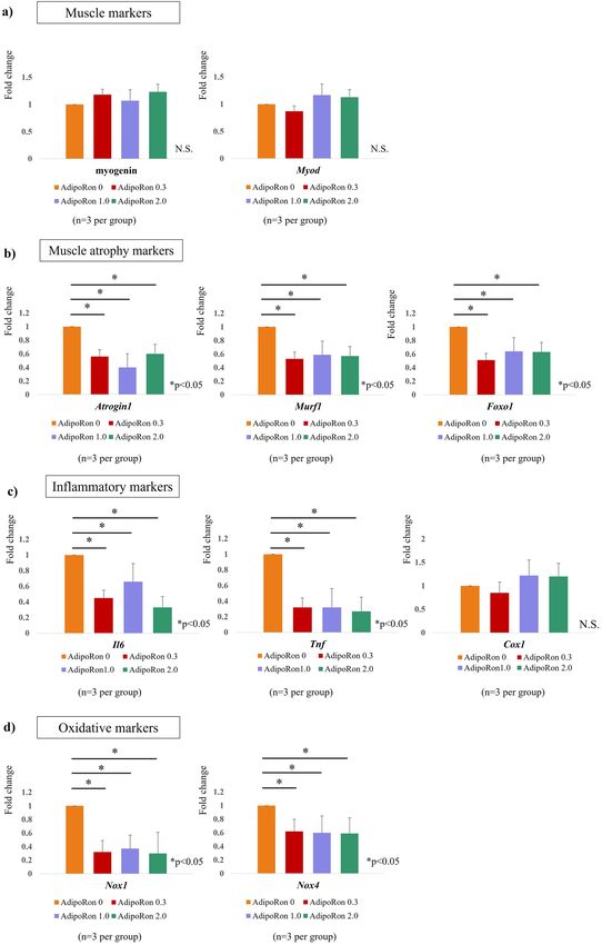
www.nature.com/scientificreports/
Figure 7. (a) Adipoq expression was significantly decreased in the nerve-crushed group. (b) Gene expression of
muscle markers (myogenin and Myod; significantly lower in the nerve-crushed group than in the control group)
and (c) muscle atrophy markers (Atrogin1, Murf1, Foxo1; significantly higher in the nerve-crushed group than
in the control group). (d) Gene expression of inflammation markers, Il6, Tnf, and Cox1, (e) oxidative markers,
Nox1 and Nox4, and (f) adipogenic markers, Pparg and Cebpa. (significantly higher in the nerve-crushed group
than in the control group).
knowledge, there is no report on the effect of AdipoRon administration on muscle atrophy. In this in vitro study,
AdipoRon administration showed anti-oxidant, anti-inflammatory, and anti-apoptotic effects.
Reportedly, muscle atrophy accompanied by the fatty degeneration of muscles is clinically observed after
nerve injury or tendon rupture4. However, despite recent improvements in nerve and tendon repair techniques,
chronic inflammation and muscle atrophy associated with fatty degeneration still limit postoperative function6,14.
Muscle atrophy occurs when the breakdown of proteins in muscle cells exceeds their synthesis, resulting in a
decrease in muscle mass15, and reportedly, inflammatory cytokines and oxidative stress enhance this muscle
degradation process.
In the skeletal muscle, myokines secreted by the muscle and adipokines secreted by the white adipose tissue
balance the m etabolism16. Among these cytokines, IL6 and TNF are considered inflammatory cytokines, while
Scientific Reports | (2022) 12:1557 | https://doi.org/10.1038/s41598-022-05608-x 7
Vol.:(0123456789)
www.nature.com/scientificreports/

                                            Figure 8.  (a) comparison of fold change (baseline: AdipoRon 0). AdipoRon treatment did not cause a
                                            significant difference in the expression of muscle markers (myogenin, Myod), but (b) significantly reduced
                                            muscle atrophy markers (Atrogin1, Murf1, Foxo1). (c) AdipoRon administration significantly reduced the levels
                                            of inflammatory cytokines, Il6 and Tnf. The AdipoRon treatment did not affect the level of the pro-inflammatory
                                            cytokine, Cox1. (d) The expression of oxidative markers was also significantly decreased by AdipoRon
                                            treatment. No significant differences were seen in groups treated with different AdipoRon doses.

          Scientific Reports |   (2022) 12:1557 |               https://doi.org/10.1038/s41598-022-05608-x                                               8

Vol:.(1234567890)
www.nature.com/scientificreports/
adiponectin is considered an anti-inflammatory c ytokine17–19. Although adiponectin was originally identified
as a secretory protein in adipose tissue, it is now known to be expressed in the skeletal muscle10. Adiponectin
in skeletal muscle has potent anti-inflammatory, antioxidant, and anti-apoptotic effects, as well as a significant
inverse correlation with pro-inflammatory cytokines, like IL6 and TNF20. These adipokines are tightly regu-
lated to maintain homeostasis in the skeletal muscle16,21; however, when adipose tissue proliferates under non-
physiological conditions, such as is the case with obesity and degeneration, the adipokine secretion balance is
disturbed22. Thus, the adipocytes that infiltrate skeletal muscle owing to the degeneration caused by trauma or
tendon rupture (ectopic fat) possibly disrupt adipokine homeostasis, resulting in chronic pain23. Under such
conditions, atrophied muscles with ectopic fat tissue secrete low amounts of a diponectin23, and the catabolism
induced by TNF is particularly e nhanced24,25. In addition, NOX-mediated activation of reactive oxygen species
causes oxidative stress, which induces apoptosis via the FoxO1/MuRF-1/Atrogin-1 signaling pathway and con-
tributes to muscle a trophy26,27. Adiponectin is thought to play an important role in antagonizing these inflam-
matory and oxidative s tresses20,28.
Two subtypes of adiponectin receptors (AdipoR1 and AdipoR2) have been identified via complementary DNA
expression cloning29. Specifically, AdipoR1 is predominantly expressed in skeletal muscles, while AdipoR2, which
functions as a major receptor for adiponectin in vivo, is predominantly expressed in the liver28. Additionally,
AdipoR1 activates the activated protein kinase (AMPK) pathway, which promotes fatty acid burning and reduces
inflammation30,31. Even though the anti-inflammatory and antioxidant effects of adiponectin via AdipoR1 have
been evaluated in some previous studies32, reports on the expression of adiponectin and AdipoR1 in atrophied
muscles are limited. In this study, the fluorescence immunostaining of muscle samples from the control group
showed that adiponectin and its receptor, AdupoR1, were expressed along the fascia. Conversely, the nerve-
crushed group showed a significant decrease in adiponectin expression, even though the AdipoR1 expression was
maintained. There was no significant difference in the expression of AdipoR1 between the control group and the
nerve injury group. These results suggest that AdipoRon administration is useful for correcting the imbalance of
adipokines after muscle atrophy. Furthermore, previous reports have demonstrated that it shows anti-inflamma-
tory and anti-fibrotic actions in the liver as well as anti-inflammatory actions in the myocardium33,34. In this study,
real-time PCR showed decreased Tnf and Il6 expression owing to AdipoRon treatment. However, this treatment
had no effect on Cox1 expression. Therefore, the balance between TNF and IL6 as inflammatory cytokines and
the secretion of adiponectin is important for the maintenance of skeletal muscle homeostasis. The expression
of muscle atrophy markers and oxidative markers also showed a significant decrease following AdipoRon treat-
ment. Furthermore, the anti-inflammatory and anti-apoptotic effects of adiponectin are expected to be sufficient
to maintain the cytokine balance, even at low concentrations given at high concentrations (20 μM). AdipoRon
administration to myocytes rather inhibits differentiation into myotubular cells, resulting in a decrease in muscle
mass35. AdipoRon treatment decreased inflammatory, oxidative, and degenerative markers and increased cellular
activity in the nerve-crushed group. These results suggest that AdipoRon has anti-inflammatory, antioxidant, and
anti-apoptotic effects in vitro. The doses of AdipoRon used in this study were based on previous reports36,37. No
cytotoxicity was observed within the range of doses administered. In addition, there was no significant differ-
ence in the effect of AdipoRon dose concentration. When examined in light of previous reports35,36, this may be
due to saturation of AMPK pathway activation. Furthermore, atrophied muscles were used in this study, which
may have lowered the response threshold to AdipoRon stimulation. The saturation of the AMPK pathway after
AdipoRon administration was thought to lead to a concentration-independent response of anti-inflammatory
and antioxidant effects. Therefore, administration of AdipoRon at a low dose concentration may be an effective
treatment option for fatty degeneration-associated chronic inflammation and muscle atrophy.
This study had several limitations. The first is that the evaluation period was limited to a single time point
(the subacute phase). This implies that the evaluation of the chronic phase needs to be considered in the future.
Second, in this study, we evaluated adiponectin receptor agonists only in vitro. However, in vivo expression of
adiponectin receptors was observed in the nerve-crushed group, suggesting that the administration of adiponec-
tin may be effective. Therefore, in the future, it would be necessary to examine the dosage concentrations as well
as other factors. Further research on the role of AdipoRon in chronic inflammation and muscle atrophy is also
required. In vivo evaluation of the effect of AdipoRon on the activation of the AMPK pathway in injured muscle
may lead to further confirmation of the therapeutic effect. Third, the present study did not measure differences
in the isoforms of adiponectin in gastrocnemius muscle. Since adiponectin physiologically forms m ultimers10,
further studies investigating isoforms are needed. Finally, adipokines from atrophied muscle and infiltrated
fat cannot be strictly evaluated separately. The balance of adipkines in muscle is important, and correcting the
imbalance is expected to prevent chronic pain and degeneration.
In this study, we observed muscle atrophy and fatty infiltration accompanied by a decreased expression of
adiponectin which had anti-inflammatory and anti-apoptotic effects in the nerve-crushed group. Conversely,
the secretion of inflammatory cytokines increased. This cytokine imbalance possibly resulted in chronic pain
owing to the induction of apoptosis and inflammation in the muscle cells. Even though adiponectin expression
was decreased in the nerve-crushed group, the expression of AdipoR1 was maintained. Furthermore, the in vitro
administration of the adiponectin receptor agonist showed anti-inflammatory and anti-apoptotic effects, sug-
gesting that the control of adipokines via the local administration of AdipoRon could lead to the prevention of
fatty degeneration-associated chronic pain.
Materials and methods
Animal model. This study was approved by the animal research committee of the Department of Ortho-
pedic Surgery, Graduate School of Medicine, Kobe University, Kobe, Japan. All experiments on animals were
conducted under the approval and guidance of the Animal Care and Use Committee of our institution. The
Scientific Reports | (2022) 12:1557 | https://doi.org/10.1038/s41598-022-05608-x 9
Vol.:(0123456789)www.nature.com/scientificreports/
experiments were conducted in accordance with the ARRIVE guidelines. Twenty-four SD rats (12-week-old)
with a mean weight of 250 g were used in this study (CLEA Japan, Inc., Tokyo, Japan).
Surgical procedure. All surgical interventions were performed under sterile conditions, with isoflurane,
the intraperitoneal injection of pentobarbital sodium (50 mg/kg; Kyoritsu Seiyaku, Tokyo, Japan), and the sub-
cutaneous injection of lidocaine (2.5 mg/kg, Xylocaine®; AstraZeneca, London, UK) at the surgical site as anes-
thesia.
The skin of the rats was incised at the right hind limbs, and the vastus lateralis and biceps femoris muscles
were separated to expose the sciatic nerve. To establish the sciatic nerve injury model, the sciatic nerve was
clamped using hemostatic forceps for one minute at a proximal distance of 5 mm from the bifurcation point of
the peroneal and tibial nerves (Fig. 1a)13. Twelve rats were included in the nerve-crushed group. For the control
rats, only a skin incision was made on the hind limbs. The skin incision was closed with a 4–0 nylon suture, and
4 weeks after the surgery, gastrocnemius muscle samples were harvested and analyzed (Fig. 1b).
Cell culture. After muscle weight measurement, the muscles were minced and treated with 0.4% collagenase
for 2 h. After this collagenase treatment, the cells were washed with phosphate-buffered saline (PBS) and centri-
fuged at 1200 rpm for 5 min. Thereafter, the cells were plated in 100-mm diameter culture dishes and cultured in
a monolayer mode using Dulbecco’s modified eagle medium (DMEM, HyClone, Logan, UT, USA) mixed with
10% fetal bovine serum (FBS, Cansera, Rexdale, Ontario, Canada), 100 U/ml penicillin, and 100 μg/ml strepto-
mycin. The cells were then evaluated after 2–3 passages and group comparisons were performed.
Protocol for in vitro AdipoRon administration. Myocytes corresponding to the nerve-crushed group
were treated with AdipoRon (AdipoGen Life Sciences, Liestal, Switzerland)36, a small molecule adiponectin
receptor (AdipoR) agonist. Specifically, the AdipoRon was first dissolved in DMSO to obtain a 2 mM stock
solution. Thereafter, it was administered at concentrations of 0.3, 1.0, and 2.0 μg/ml, as previously r eported36,37,
and the differences in the efficacy of this treatment among the treatment groups were determined. Specifically,
cell viabilities at 24 h after treatment administration were compared using the WST assay, and the expression
levels of Tnf, Il6, and Cox1 (inflammatory markers) and myogenin and Myod (muscle markers) and Murf1 and
Atrogin1 (muscle atrophy markers) were evaluated via real-time PCR.
Evaluation method. Cell morphology evaluation. After three passages, 1.0 × 10⁵ cells were seeded into
12-well plates, and the cell morphologies were evaluated via HE staining after 48 h.
Cell viability (cell proliferation assay). Cell viability was measured via a WST assay using a cell counting kit-8
(Dojindo, Kumamoto, Japan). A total of 5,000 cells were seeded into each well of a 96-well-plate and cultured in
a DMEM medium for 48 h.
Fluorescent immunostaining. The expression level of intracellular adiponectin in myocyte samples from the
different groups was detected using an anti-adiponectin polyclonal antibody (NB100-65810F; Bios, Boston, MA,
USA). The antibodies, which were used at a dilution ratio of 1:100, were incubated with myocytes (5 × 10⁴) for
60 min in the dark at 37 °C. Thereafter, they were washed twice with PBS and nuclear staining was performed
using DAPI solution for 10 min. The percentage of stained cells was then observed using a fluorescence micro-
scope (BZ-8000 confocal microscope; Keyence, Osaka, Japan). For quantification, the number of DAPI-positive
and Adiponectin-stained cells in five fields of view (0.75 mm × 1.0 mm) on each slide was counted, and the ratio
of the mean values was calculated. The measurements were performed on randomly selected areas by two inves-
tigators who were blinded to each other.
Histological examination. The weight ratio of the gastrocnemius muscle at the affected side to that at the healthy
side was measured. Specifically, gastrocnemius muscle samples were harvested and embedded in an optimal cut-
ting temperature (OCT) compound (Sakura Finetek USA, Inc., Torrance, CA), and stored at −80 °C for histo-
chemical and immunohistochemical staining. Specifically, the gastrocnemius muscle samples in the OCT blocks
were sectioned serially to have a thickness of 6 μm. Thereafter, they were was mounted on a silane-coated glass
slide, and air-dried for 1 h before fixation with 4% paraformaldehyde at 4 °C for 5 min. The tissue sections were
then stained with HE to observe the histological differences between the muscle samples from the control and
nerve-crushed groups. Fat droplets were also stained with Oil Red-O solution (Mutoh Pure Chemical, Tokyo,
Japan) to evaluate fat infiltration into the muscle. Gastrocnemius muscle was frozen using isopentane cooled
with liquid nitrogen and stored at −80 °C until needed. As described in the previously published protocol38, the
muscles were cryosectioned at a thickness of 10 μm and fixed in 4% paraformaldehyde. The sections were then
stained with Oil Red-O and counterstained with hematoxylin.
For immunofluorescence staining, adiponectin antibody and AdipoR1 antibody were used at a dilution ratio
of 1:100; the staining was performed at 25 ℃ room temperature for 1 h. To ensure nuclear staining, the DAPI
solution was applied for 5 min. All sections were visualized using a fluorescence microscope (BZ-8000 confo-
cal microscope; Keyence). Furthermore, the number of positively stained cells was counted in five randomly
selected fields (250 × 250 mm).
Real‑time PCR. Myocytes from both groups were seeded in 12-well culture plates at a density of 1.0 × 1 05 cells/
well and cultured in DMEM for 48 h. Thereafter, total RNA was extracted using an RNeasy Mini Kit (QIAGEN,
Scientific Reports | (2022) 12:1557 | https://doi.org/10.1038/s41598-022-05608-x 10
Vol:.(1234567890)www.nature.com/scientificreports/
Gene Forward primer (5’ to 3’) Reverse primer (5’ to 3’)
Adipoq AATCCTGCCCAGTCATGAAG CATCTCCTGGGTCACCCTTA
Il6 TCCTACCCCAACTTCCAATGCTC ACCCAGAGCGTATCATCCTTCAC
Tnf AAATGGGCTCCCTCTCATCAGTTC CCAACTGACTTTGAGCCAACGAG
Cox1 TGCCAGTATTAGCAGCAGGT GAATTGGGTCTCCACCTCCA
Nox1 CTTCCTCACTGGCTGGGATA TGACAGCATTTGCGCAGGCT
Nox4 AGTCAAACAGATGGGATA TGTCCCATATGAGTTGTT
Pparg TGTGGACCTCTCTGTGATGG CATTGGGTCAGCTCTTGTGA
Cebpa CCCGATGAGCAGCCACCTCCA TACCCCGCAGCGTGTCCAGT
Myogenin CCTTGCTCAGCTCCCTCA TGGGAGTTGCATTCACTGG
Myod GGAGACATCCTCAAGCGATGC AGCACCTGGTAAATCGGATTG
Atrogin1 GAACAGCAAAACCAAAACTCAGTA GCTCCTTAGTACTCCCTTTGTGAA
Murf1 TGTCTGGAGGTCGTTTCCG ATGCCGGTCCATGATCACTT
Foxo1 GAGGTGCAATGTGGGAGAAT TTGAATGAAATGGCAAAGCA
Gapdh TGGCCTCCAAGGAGTAAGAAAC GGCCTCTCTCTTGCTCTCAGTATC
Table 1. Primers for real-time PCR.
Valencia, CA, USA) according to the manufacturer’s protocol. Furthermore, oligo (deoxythymidine)-primed
first-strand cDNA was synthesized using a High Capacity cDNA Transcription Kit (Applied Biosystems, Foster
City, CA, USA), and quantitative real-time PCR was performed in a 20 μl reaction mixture using the SYBR
Green Master Mix reagent (Applied Biosystems) on an ABI Prism 7500 sequence detection system (Applied
Biosystems). The PCR conditions were as follows: 1 cycle at 95 °C for 10 min, followed by 40 cycles at 95 °C for
15 s, and 40 cycles at 60 °C for 1 min. The messenger RNA (mRNA) expression levels of Adipoq were evaluated.
As muscle-related markers, the expression levels of anabolic markers, myogenin and Myod, and muscle atrophy
markers, Atrogin1, Murf1, and Foxo1 were also monitored. Additionally, the mRNA expression levels of Il6, Tnf,
and Cox1 were analyzed as pro-inflammatory cytokines, Nox1 and Nox4 were analyzed as oxidative markers, and
Pparg and Cebpa were used as markers of adipose degeneration.
The primer sequences are shown in Table 1. The relative expression levels of the genes were determined using
the DD-Ct method and normalized to Gapdh.
Statistical analysis. All data are expressed as mean values ± standard deviations. Cell viability and real-
time PCR results were expressed as n-fold differences relative to the baseline control at the corresponding time
point. Student’s t-test was performed to compare two groups, and two-way ANOVA and Tukey’s posthoc test
were used to compare two or more AdipoRon treatment groups. Results with p < 0.05 were considered statisti-
cally significant. The data were analyzed using SPSS v23.0 (IBM Corporation, Armonk, NY, USA).
Data availability statement
The data presented in this study are available on request from the corresponding author. The data are not publicly
available because of confidentiality issues.
Received: 22 September 2021; Accepted: 12 January 2022
References
1. Nader, G. A. Molecular determinants of skeletal muscle mass: getting the “AKT” together. Int. J. Biochem. Cell Biol. 37, 1985–1996.
https://doi.org/10.1016/j.biocel.2005.02.026 (2005).
2. Jaitovich, A. & Barreiro, E. Skeletal muscle dysfunction in chronic obstructive pulmonary disease. What we know and can do for
our patients. Am. J. Respir. Crit. Care Med. 15, 175–186. https://doi.org/10.1164/rccm.201710-2140CI (2018).
3. Ding, S., Dai, Q., Huang, H., Xu, Y. & Zhong, C. An overview of muscle atrophy. Adv. Exp. Med. Biol. 11088, 3–19. https://doi.org/
10.1007/978-981-13-1435-3_1 (2018).
4. Cao, R. Y., Li, J., Dai, Q., Li, Q. & Yang, J. Muscle atrophy: present and future. Adv. Exp. Med. Biol. 1088, 605–624. https://doi.org/
10.1007/978-981-13-1435-3_29 (2018).
5. Lin, Y., Wen-Jie, Z., Chang-Qing, L., Sheng-Xiang, A., & Yue, Z. Mir-22-3p/KLF6/MMP14 axis in fibro-adipogenic progenitors
regulates fatty infiltration in muscle degeneration. FASEB J. 34, 12691–12701; doi:https://doi.org/10.1096/fj.202000506R (2020)
6. Gladstone, J. N., Bishop, J. Y., Lo, I. K. & Flatow, E. L. Fatty infiltration and atrophy of the rotator cuff do not improve after rota-
tor cuff repair and correlate with poor functional outcome. Am. J. Sports Med. 35, 719–728. https://doi.org/10.1177/0363546506
297539 (2007).
7. Fasshauer, M. & Bluher, M. Adipokines in health and disease. Trends Pharmacol. Sci. 36, 461–470. https://doi.org/10.1016/j.tips.
2015.04.014 (2015).
8. Engin, A. Adiponectin-resistance in obesity. Adv. Exp. Med. Biol. 960, 415–441. https://doi.org/10.1007/978-3-319-48382-5_18
(2017).
9. Iwabu, M. et al. Adiponectin and AdipoR1 regulate PGC-1alpha and mitochondria by Ca(2+) and AMPK/SIRT1. Nature 464,
1313–1319. https://doi.org/10.1038/nature08991 (2010).
Scientific Reports | (2022) 12:1557 | https://doi.org/10.1038/s41598-022-05608-x 11
Vol.:(0123456789)www.nature.com/scientificreports/
10. Martinez-Huenchullan, S. F. et al. Skeletal muscle adiponectin induction in obesity and exercise. Metabolism 102, 154008. https://
doi.org/10.1016/j.metabol.2019.154008 (2020).
11. Krause, M. P., Milne, K. J. & Hawke, T. J. Adiponectin-consideration for its role in skeletal muscle health. Int. J. Mol. Sci. https://
doi.org/10.3390/ijms20071528 (2019).
12. Jortay, J. et al. Local induction of adiponectin reduces lipopolysaccharide-triggered skeletal muscle damage. Endocrinology 151,
4840–4851. https://doi.org/10.1210/en.2009-1462 (2010).
13. Nishimoto, H. et al. Transcutaneous carbon dioxide application with hydrogel prevents muscle atrophy in a rat sciatic nerve crush
model. J. Orthop. Res. 36, 1653–1658. https://doi.org/10.1002/jor.23817 (2018).
14. Miyazaki, A. N. et al. Fatty muscle infiltration in cuff tear: pre and post operative evaluation by mri. Acta Ortop. Bras. 23, 251–254.
https://doi.org/10.1590/1413-785220152305119821 (2015).
15. Sa, B. K., Kim, C., Kim, M. B. & Hwang, J. K. Panduratin a prevents tumor necrosis factor-alpha-induced muscle atrophy in L6 rat
skeletal muscle cells. J. Med. Food. 20, 1047–1054. https://doi.org/10.1089/jmf.2017.3970 (2017).
16. Li, F. et al. Myokines and adipokines: Involvement in the crosstalk between skeletal muscle and adipose tissue. Cytokine Growth
Factor Rev. 33, 73–82. https://doi.org/10.1016/j.cytogfr.2016.10.003 (2017).
17. Bluher, M. Adipose tissue dysfunction contributes to obesity related metabolic diseases. Best Pract. Res. Clin. Endocrinol. Metab.
27, 163–177. https://doi.org/10.1016/j.beem.2013.02.005 (2013).
18. Kershaw, E. E. & Flier, J. S. Adipose tissue as an endocrine organ. J. Clin. Endocrinol. Metab. 89, 2548–2556. https://doi.org/10.
1210/jc.2004-0395 (2004).
19. Kloting, N. & Bluher, M. Adipocyte dysfunction, inflammation and metabolic syndrome. Rev. Endocr. Metab. Disord. 15, 277–287.
https://doi.org/10.1007/s11154-014-9301-0 (2014).
20. Kern, P. A., Di Gregorio, G. B., Lu, T., Rassouli, N. & Ranganathan, G. Adiponectin expression from human adipose tissue: relation
to obesity, insulin resistance, and tumor necrosis factor-expression. Diabetes 52, 1779–1785. https://doi.org/10.2337/diabetes.52.7.
1779 (2003).
21. Bluher, M. & Mantzoros, C. S. From leptin to other adipokines in health and disease: facts and expectations at the beginning of
the 21st century. Metabolism 64, 131–145. https://doi.org/10.1016/j.metabol.2014.10.016 (2015).
22. Unamuno, X. et al. Adipokine dysregulation and adipose tissue inflammation in human obesity. Eur. J. Clin. Invest. 48, e12997.
https://doi.org/10.1111/eci.12997 (2018).
23. Sugatani, T., Tanaka, M. & Ogawa, Y. Adipose tissue inflammation and ectopic lipid accumulation. Endocr. J. 59, 849–857. https://
doi.org/10.1507/endocrj.ej12-0271 (2012).
24. Park, J. H. et al. Protective effect of melatonin on TNF-alpha-induced muscle atrophy in L6 myotubes. J. Pineal. Res. 54, 417–425.
https://doi.org/10.1111/jpi.12036 (2013).
25. Wang, D. T. et al. Resveratrol prevents TNF-alpha-induced muscle atrophy via regulation of Akt/mTOR/FoxO1 signaling in C2C12
myotubes. Int. Immunopharmacol. 19, 206–213. https://doi.org/10.1016/j.intimp.2014.02.002 (2014).
26. Powers, S. K., Smuder, A. J. & Judge, A. R. Oxidative stress and disuse muscle atrophy: Cause or consequence?. Curr. Opin. Clin.
Nutr. Metab. Care. 15, 240–245. https://doi.org/10.1097/MCO.0b013e328352b4c2 (2012).
27. Nakai, S. et al. FOXO1 suppresses PGC-1beta gene expression in skeletal muscles. FEBS Open Bio 10, 1373–1388. https://doi.org/
10.1002/2211-5463.12898 (2020).
28. Yamauchi, T. et al. Adiponectin stimulates glucose utilization and fatty-acid oxidation by activating AMP-activated protein kinase.
Nat. Med. 8, 1288–1295. https://doi.org/10.1038/nm788 (2002).
29. T., Y. et al. Cloning of adiponectin receptors that mediate antidiabetic metabolic effects. Nature. 423, 762–769; doi:https://doi.org/
10.1038/nature01705 (2003).
30. Kang, E. H. et al. Adiponectin is a potential catabolic mediator in osteoarthritis cartilage. Arthritis Res. Ther. 12, R231. https://doi.
org/10.1186/ar3218 (2010).
31. Yamauchi, T. et al. Targeted disruption of AdipoR1 and AdipoR2 causes abrogation of adiponectin binding and metabolic actions.
Nat. Med. 13, 332–339. https://doi.org/10.1038/nm1557 (2007).
32. Abou-Samra, M., Selvais, C. M., Dubuisson, N. & Brichard, S. M. Adiponectin and its mimics on skeletal muscle: insulin sensitizers,
fat burners, exercise mimickers, muscling pills ... or everything together? Int. J. Mol. Sci. 21, 2620 https://doi.org/10.3390/ijms2
1072620 (2020).
33. Leffler, K. E. & Abdel-Rahman, A. A. Restoration of adiponectin-Connexin43 signaling mitigates myocardial inflammation and
dysfunction in diabetic female rats. J. Cardiovasc. Pharmacol. 75, 259–267. https://d oi.o
rg/1 0.1 097/F
JC.0 00000 00000 00789 (2020).
34. Sha, M. et al. Therapeutic effects of AdipoRon on liver inflammation and fibrosis induced by CCl4 in mice. Int. Immunopharmacol.
79, 106157. https://doi.org/10.1016/j.intimp.2019.106157 (2020).
35. Ito, R. et al. Activation of adiponectin receptors has negative impact on muscle mass inC2C12 myotubes and fast-type mouse
skeletal muscle. PLoS ONE 13, e0205645. https://doi.org/10.1371/journal.pone.0205645 (2018).
36. Okada-Iwabu, M. et al. A small-molecule AdipoR agonist for type 2 diabetes and short life in obesity. Nature 503, 493–499. https://
doi.org/10.1038/nature12656 (2013).
37. Nokhbehsaim, M. et al. Beneficial effects of adiponectin on periodontal ligament cells under normal and regenerative conditions.
J. Diabetes Res. 2014, 796565. https://doi.org/10.1155/2014/796565 (2014).
38. Takase, F. et al. Effect of platelet-rich plasma on degeneration change of rotator cuff muscles: In vitro and in vivo evaluations. J.
Orthop. Res. 35, 1806–1815; doi:https://doi.org/10.1002/jor.23451 (2017)
Acknowledgements
We appreciate the technical assistance provided by Ms. Kyoko Tanaka, Ms. Minako Nagata, and Ms. Maya Yasuda.
Author contributions
IS, [TK]1, YM, AI, HN, RS and RK contributed to the study concept and design. IS, [TK]1 and AI contributed
to the data acquisition, analysis. IS, [TK]1 and YM drafted the article and IS, [TK]1, TM, HN, RS, KY, SM, TY,
[TK]2, FT, and RK contributed to interpretation of the data and critically revised the article for important intel-
lectual content. All authors gave final approval of the version to be submitted for publication and agreed to be
held accountable for all aspects of the work.
Competing interests
The authors declare no competing interests.
Additional information
Supplementary Information The online version contains supplementary material available at https://doi.org/
10.1038/s41598-022-05608-x.
Scientific Reports | (2022) 12:1557 | https://doi.org/10.1038/s41598-022-05608-x 12
Vol:.(1234567890)www.nature.com/scientificreports/
Correspondence and requests for materials should be addressed to Y.M.
Reprints and permissions information is available at www.nature.com/reprints.
Publisher’s note Springer Nature remains neutral with regard to jurisdictional claims in published maps and
institutional affiliations.
Open Access This article is licensed under a Creative Commons Attribution 4.0 International
License, which permits use, sharing, adaptation, distribution and reproduction in any medium or
format, as long as you give appropriate credit to the original author(s) and the source, provide a link to the
Creative Commons licence, and indicate if changes were made. The images or other third party material in this
article are included in the article’s Creative Commons licence, unless indicated otherwise in a credit line to the
material. If material is not included in the article’s Creative Commons licence and your intended use is not
permitted by statutory regulation or exceeds the permitted use, you will need to obtain permission directly from
the copyright holder. To view a copy of this licence, visit http://creativecommons.org/licenses/by/4.0/.
© The Author(s) 2022
Scientific Reports | (2022) 12:1557 | https://doi.org/10.1038/s41598-022-05608-x 13
Vol.:(0123456789)You can also read