Isolation and Identification of Bone Marrow Mesenchymal Stem Cells from Forest Musk Deer
←
→
Page content transcription
If your browser does not render page correctly, please read the page content below

animals
Article
Isolation and Identification of Bone Marrow Mesenchymal
Stem Cells from Forest Musk Deer
Weiqiang Luo † , Yangyang Geng † , Mengxi Gao, Mengting Cao, Junjian Wang, Jing Yang, Chenxuan Sun
and Xingrong Yan *

                                          Shaanxi Key Laboratory for Animal Conservation, College of Life Sciences, Northwest University,
                                          Xi’an 710069, China
                                          * Correspondence: xingrongyan2007@126.com
                                          † These authors contributed equally to this work.

                                          Simple Summary: The forest musk deer is a wild animal that is endangered due to widespread
                                          hunting for its musk, which is used in medicine and perfume. Stem cells can differentiate into a
                                          variety of cells, tissues, organs, and even complete individuals, which can be used for cell therapy and
                                          germplasm preservation. Mesenchymal stem cells are the most abundant stem cells in animals and
                                          have been isolated from many animals, such as cows and dogs. In this study, to evaluate the feasibility
                                          of isolating mesenchymal stem cells from forest musk deer, bone marrow was collected from forest
                                          musk deer, and the cells were isolated and characterized. They exhibit the same stemness and
                                          differentiation capacity as other mammals studied previously. Therefore, bone marrow mesenchymal
                                          stem cells may be an important tool for forest musk deer cell therapy.

                                          Abstract: The forest musk deer (Moschus berezovskii) is an endangered animal that produces musk
                                          that is utilized for medical applications worldwide, and this species primarily lives in China. Animal-
                                          derived musk can be employed as an important ingredient in Chinese medicine. To investigate
                                          the properties of bone marrow mesenchymal stem cells (MSCs) obtained from the bone marrow
                                          of forest deer for future application, MSCs were isolated and cultivated in vitro. The properties
                                          and differentiation of these cells were assessed at the cellular and gene levels. The results show
                                          that 81,533 expressed genes were detected by RNA sequencing, and marker genes of MSCs were
                                          expressed in the cells. Karyotype analysis of the cells determined the karyotype to be normal,
                                          and marker proteins of MSCs were observed to be expressed in the cell membranes. Cells were
Citation: Luo, W.; Geng, Y.; Gao, M.;     differentiated into osteoblasts, adipocytes, and chondroblasts. The expression of genes related to
Cao, M.; Wang, J.; Yang, J.; Sun, C.;     osteoblasts, adipocytes, and chondroblasts was observed to be increased. The results of this study
Yan, X. Isolation and Identification of   demonstrate that the properties of the cells isolated from bone marrow were in keeping with the
Bone Marrow Mesenchymal Stem              characteristics of MSCs, providing a possible basis for future research.
Cells from Forest Musk Deer. Animals
2023, 13, 17. https://doi.org/            Keywords: bone marrow; differentiation; forest musk deer; mesenchymal stem cells; RNA sequencing
10.3390/ani13010017

Received: 30 October 2022
Revised: 9 December 2022
Accepted: 17 December 2022                1. Introduction
Published: 20 December 2022
                                               The forest musk deer, also known as Moschus berezovskii, is a small-sized and artio-
                                          dactyl animal primarily observed in southwestern Asia [1]. Forest musk deer can produce
                                          musk as a raw material for Chinese medicine, such as Angong Niuhuang Wan and Pian
Copyright: © 2022 by the authors.
                                          Zai Huang [2,3]. Therefore, in nature, the number of forest musk deer has decreased due to
Licensee MDPI, Basel, Switzerland.        overhunting and habitat destruction. Forest musk deer is listed in class I of key wildlife
This article is an open access article    species by the Chinese Wild Animal Protection Law, the Appendices of the Convention on
distributed under the terms and           the International Trade of Endangered Species of Wild Fauna and Flora [1,4]. To maintain
conditions of the Creative Commons        ecological equilibrium and protect the resources of traditional Chinese medicine, wildlife
Attribution (CC BY) license (https://     forest musk deer were protected by hunting prohibition and habitat protection. Further-
creativecommons.org/licenses/by/          more, some forest musk deer were caged under the Chinese Wild Animal Protection Law
4.0/).                                    to meet the demands of traditional Chinese medicine for musk.

Animals 2023, 13, 17. https://doi.org/10.3390/ani13010017                                                    https://www.mdpi.com/journal/animals
Animals 2023, 13, 17                                                                                            2 of 12

                             Mesenchymal stem cells (MSCs) are the most abundant cell population in the body.
                       They exist in many tissues and have extensive self-renewal capacity and the ability to
                       differentiate into multiple lineages [5,6]. MSCs comprise several lineages, such as bone
                       marrow MSCs (BM-MSCs), adipose MSCs, and umbilical cord blood MSCs [7]. MSCs
                       regulate immune tolerance in allogeneic transplantation [8]. MSCs derived from rat bone
                       marrow have the chronic potential to differentiate into osteoblasts, adipocytes, and neural
                       cells [9]. Since MSCs have a strong proliferation and differentiation ability, they can be used
                       as donor cells of somatic cell nuclear transfer (SCNT) for cloning animals [10]. Moreover,
                       MSCs can also differentiate into germ cells by co-culture with Sertoli cells [11]. Therefore,
                       MSCs are among the seed cells applied to cell therapy, endangered animal protection, and
                       animal husbandry.
                             There are few reports about MSCs’ application in endangered animals. MSCs were iso-
                       lated from giant panda bone marrow and identified at the cellular and molecular levels [12].
                       MSCs from the bone marrow of red pandas were also characterized and were similar to
                       those from other species, exhibiting such properties as the expression of multiple marker
                       genes during differentiation [13].
                             The forest musk deer is an endangered animal species. To date, there has been
                       no published research on the MSCs from this species. Therefore, in-depth study of the
                       characteristics and functions of MSCs is beneficial for the protection of the forest musk deer.
                       In this study, MSCs were isolated from the bone marrow of forest musk deer and were
                       characterized by cell morphology, surface markers, and differentiation ability. The results
                       of this study may provide seed cells for cell therapy and potential clinical application.

                       2. Materials and Methods
                       2.1. Reagents
                            All reagents were purchased from Sigma Aldrich (Shanghai, China) unless stated otherwise.

                       2.2. Animal
                           Forest musk deer were caged in Feng County, Baoji city, Shaanxi Province. The
                       experiments performed in this study were approved by the Animal Ethics Committee of
                       Northwest University in China. A 3-year-old male forest deer that had died of dyspnea
                       caused by pulmonary infection with Pseudomonas aeruginosa was analyzed. The thigh
                       bone was isolated sterilely by surgery and sent to the laboratory in 4 ◦ C PBS with 2%
                       penicillin-streptomycin.

                       2.3. Isolation and Culture of Cell from Bone Marrow
                            This method was the same as that described in a previous report [14]. Briefly, the
                       marrow was aspirated aseptically by a trocar connected to a 10 mL syringe. After suction,
                       the marrow was placed into a 10 cm plate, and 10 mL of DMEM with 20% fetal bovine serum
                       (FBS) was added to the marrow cells, which were incubated for 7 d in an incubator at 37 ◦ C,
                       5% CO2 and in saturated humidity. Subcultivation was performed as cell growth reached
                       80% confluence. Cells were digested with 0.25% trypsin and 0.04% EDTA, neutralized in
                       DMEM with 10% FBS, and washed at 1000 rpm centrifugation for 3 min. The supernatant
                       was discarded, and a single-cell suspension was performed by the addition of fresh medium.
                       The concentration of cells was established as 1 × 10⁶ /mL, and a 1 mL suspension was
                       placed in a 10 cm plate with 9 mL DMEM with 10% FBS; Next, the cells were incubated to
                       80% confluence in an incubator.

                       2.4. Karyotype Analysis of Cells
                            The karyotype analysis was performed using a protocol described in a previous
                       report [15]. Briefly, cells at a concentration of 5 × 10⁵ /mL were cultured in 10 cm plates.
                       After 36 h, cells were treated with colchicine (0.3 µg/mL) for 6 h. Next, cells were digested
                       with 0.25% trypsin combined with 0.04% EDTA, followed by treatment with hypotonic
                       solution (0.075 M KCl), and the cells were fixed with fixation solution (methane: acetic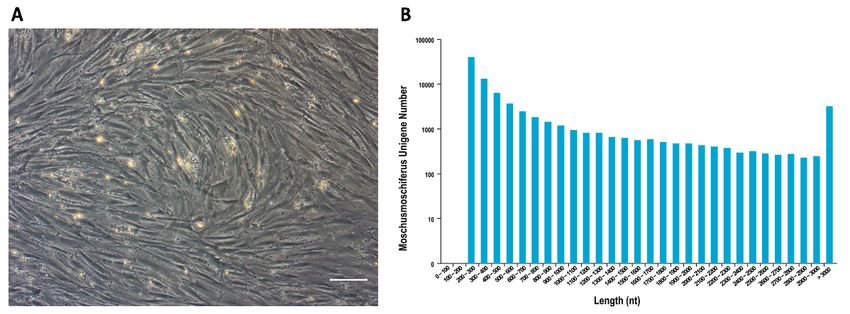
Animals 2023, 13, 17                                                                                        3 of 12

                       acid = 3:1) three times. Cell fixation solution was added dropwise to slides to a height of
                       more than 10 cm. After air drying, cells were stained with staining solution (Giemsa:PBS
                       = 1:9). The chromosomes were observed and photographed under a microscope. The
                       karyotype was subsequently constructed by arranging the sizes of the chromosomes.

                       2.5. Flow Cytometry
                            Flow cytometry was performed according to a protocol described in a previous re-
                       port [16]. Briefly, cells were digested by trypsin and neutralized in medium with 10% FBS
                       and rinsed three times with PBS. A single-cell suspension was made by pipetting cells
                       up and down in PBS. Cells were diluted to a concentration of 1 × 10⁶ cells/mL. Cells
                       were fixed with 2% polyformaldehyde (PFA) for 20 min and washed three times with PBS.
                       Cells were labeled with FITC-conjugated antibodies against CD34, CD166, CD90, CD14,
                       CD 44, CD45, CD71, CD105 and CD29 (Table 1). FITC-conjugated mouse IgG1 was used
                       as the isotype-matched control. Cells were analyzed by FACS flow cytometry using Cell
                       Quest software.

                       Table 1. Antibody sources.

                                  Antibody                       Source                         CAT#
                                   CD105                         abcam                         ab11415
                                   CD166                       eBioscience                     12-1668
                                   CD73                          abcam                        ab157335
                                   CD44                          abcam                        ab269300
                                   CD14                        eBioscience                     11-0149
                                  HLA-DR                         adcam                          ab1182
                                   CD19                        eBioscience                     11-0199
                                   CD34                        eBioscience                     11-0349
                                   CD45                        eBioscience                     11-0459

                       2.6. Differentiation of MSCs
                             For adipogenic differentiation, preformation was performed according to a protocol
                       described in a previous report [17]. Cells at passage 3 were digested, and a suspension of
                       single cells was made. A total of 8 × 10⁴ cells were seeded in a 3.5 cm plate and cultured
                       for 24 h; consequently, the medium was replaced by adipogenic differentiation medium
                       (DMEM containing 10% FBS, 500 µM isobutylmethylxanthine, 1 µM dexamethasone, 10 µM
                       insulin, and 200 µM indomethacin). The medium was changed twice a week. After 21 d,
                       the cells were rinsed twice with PBS and fixed with 4% PFA for 10 min. The cells were
                       stained with staining solution (60% isopropanol containing 0.3% Oil Red) for 20 min and
                       washed twice with 60% ethanol. The cells were observed under an inverted microscope.
                             For osteogenic differentiation, cells were digested with 0.25% trypsin and washed
                       with PBS. Cells (5 × 10⁴ ) were placed in 3.5 cm plates and cultured for 24 h. The culture
                       medium was replaced by aesthetic differentiation medium (DMEM containing 0.1 µM
                       dexamethasone, 300 µM ascorbic acid, and 10 mM β-glycerophosphate) for 3 weeks [18].
                       The medium was changed twice a week. Cells were washed with PBS twice and fixed with
                       4% PFA. Cells were stained with Alizarin Red. After washing with PBS twice, the cells
                       were observed under an inverted microscope.
                             For chondrogenic differentiation, 1 × 10⁶ MSCs at passage 3 were seeded in a 3.5 cm
                       plate. After 24 h, chondrogenic medium (DMEM containing 0.1 µM dexamethasone, 25 µM
                       ascorbic acid 2-phosphate, and 1 ng/mL TGF-β) was added. The medium was changed
                       twice a week. After 21 d, the cells were washed twice with PBS and fixed with 4% PFA. The
                       cells were stained with Alcian Blue and observed under an inverted microscope.

                       2.7. RNA Sequencing of Cells
                           Cells at 3 passages were grown to 80% confluence and were washed twice with PBS.
                       The cells were lysed by adding TRIzol reagent. Total RNA was extracted by a mirVana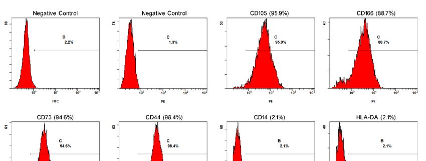
Animals 2023, 13, 17                                                                                                               4 of 12

                       miRNA Isolation Kit (Ambion-1561, Ambion Inc., Austin, TX, USA) following the man-
                       ufacturer’s protocol, and RNA quality was evaluated by a NanoDrop 2000. RNA was
                       reverted to cDNA by PrimeScript 1st Strand cDNA Synthesis Kit (D6110A, TaKaRa, Beijing,
                       China), cDNA was fragmented with an Agencourt AMPure Kit (A63881, Beckman Coul-
                       ter, Inc., Brea, CA, USA), and paired-end 150 bp fragments were sequenced by HiSeqTM
                       2500 instrument. Raw data were screened by Perl script, by which adapters, redundant
                       reads, and low-quality data were removed. Clean data were evaluated, including data
                       quality and GC content. The data were mapped to a eukaryotic reference genome. GO
                       analysis was performed by Blast2GO [19]. GO and KEGG terms were analyzed, and the
                       CDS was predicted.

                       2.8. Extraction of Total RNA and qPCR
                             Cells at 3 passages undergoing adipogenic and osteogenic differentiation in 3.5 cm
                       plates were washed 3 times. Total RNA was extracted by TRIzol reagent, isopropanol,
                       and ethanol. cDNA synthesis was performed by a PrimeScript 1st Strand cDNA Synthesis
                       Kit (D6110A, TaKaRa). qPCR analysis was performed using a PCR mix, and the primers
                       designed by Primer 5.0 based on the predicted CDS (Table S1). qPCR was performed
                       according to the following cycling protocol: 95 ◦ C for 5 min, 45 cycles of 95 ◦ C for 5 s, and
                       58 ◦ C for 10 s. The threshold cycle (Ct) value was normalized to β-actin. ∆∆Ct was used to
                       calculate the differential expression of genes.

                       2.9. Analysis
                            All experiments were performed in triplicate, and data were analyzed by SPSS
                       13.0. The data are shown as means ± standard deviation (SD). Comparative analy-
                       sis was performed by paired t-test, and p-values < 0.05 were considered to indicate
                       significant differences.

                       3. Results
                       3.1. Morphology and Proliferation of MSCs
                              After incubation of MSCs for 48 h, the cells growing adherently under the sparse
 Animals 2023, 13, x
                        hemocytes were visualized by shaking the culture dishes. At first, the morphology   varied,
                                                                                                       5 of 12
                        with some cells appearing spindle-shaped and others round. On day 3 of culture, colonies
                        had formed. On day 7, cells had overspread to the bottom of the culture dishes, and most
                        cells were spindle-shaped. At passage 3, forest musk deer MSCs exhibited the characteristic
                       immunophenotyping by flowcytometry and sequences for gene expression quantification
                        fibroblastic MSC morphology, similar to that observed for rodents and humans (Figure 1A).
                       by qRT-PCR.
                        Cells at passage 9 still showed a strong proliferation ability.

                                    (A)Cell
                        Figure1.1.(A)
                       Figure           Cellmorphology
                                             morphology    of of MSCs
                                                              MSCs      in vitro
                                                                     in vitro     cultured
                                                                              cultured  at 3atpassages.
                                                                                               3 passages.
                                                                                                        CellsCells exhibited
                                                                                                               exhibited      spindle
                                                                                                                         spindle  and and
                       fusiform
                        fusiformshapes.
                                   shapes.Scale
                                            Scalebar:
                                                  bar:100 µm;
                                                        100     (B)(B)
                                                              µm;   Unigenes
                                                                       Unigeneslength  distribution
                                                                                   length           afterafter
                                                                                           distribution   transcriptome   sequencing
                                                                                                               transcriptome   sequencing
                       and
                        anddedenovo
                                novoassemble.
                                        assemble.TheThex-axis  represents
                                                          x-axis           thethe
                                                                  represents    length  of unigenes,
                                                                                    length            and and
                                                                                            of unigenes,   the y-axis  represents
                                                                                                                the y-axis         the the
                                                                                                                            represents
                       number of unigenes within a certain length range.
                        number of unigenes within a certain length range.

                       3.3. Normal Karyotype was Maintained in MSCs
                            As shown in Figure 2, chromosomes in the M phase were collected from MSCs (Fig-
                       ure 2A), and the total number was 58 (Figure 2B). Additionally, after the chromosomes
                       were aligned, the karyotype was observed to be normal. The sex chromosomes were XY,
                       indicating that the sample was derived from forest musk deer. These results demonstrate
                       that the karyotype was normal.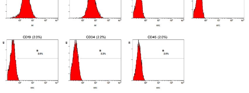
Animals 2023, 13, 17 5 of 12
3.2. RNA Data Were Detected by RNA Sequencing
Passage 3 MSCs’ mRNA was extracted and sequenced. A total of 49,943 unigenes
were spliced in more than 100 bp, and most genes were distributed in the range of 200 bp
to 300 bp (Figure
Figure1B). A Cell
1. (A) totalmorphology
of 23,666 unigenes
of MSCs inwere
vitroannotated
cultured at by nr, Pfam,
3 passages. GO,
Cells and spindl
exhibited
KEGG, whichfusiform
included cell surface markers (e.g., CD105, CD106, CD73, CD44, and CD29),
shapes. Scale bar: 100 µm; (B) Unigenes length distribution after transcriptome seque
unveiling a list
andofdecandidate markers
novo assemble. The that
x-axiscould provide
represents a basisoffor
the length antibody-mediated
unigenes, and the y-axis represen
immunophenotyping
number ofby flowcytometry
unigenes and sequences
within a certain for gene expression quantification
length range.
by qRT-PCR.
3.3. Normal Karyotype was Maintained in MSCs
3.3. Normal Karyotype was Maintained in MSCs
As shown in Figure 2, chromosomes in the M phase were collected from MSCs
As shownurein2A),
Figure
and 2,thechromosomes
total number in wasthe58M phase2B).
(Figure were collected from
Additionally, afterMSCs
the chromoso
(Figure 2A), and the total number was 58 (Figure 2B). Additionally, after
were aligned, the karyotype was observed to be normal. The sex chromosomes the chromo- were
somes were aligned,
indicating thethat
karyotype waswas
the sample observed
derivedtofrom
be normal.
forest muskThe deer.
sex chromosomes
These results demons
were XY, indicating
that thethat the sample
karyotype was derived from forest musk deer. These results
was normal.
demonstrate that the karyotype was normal.
Figure 2. (A) Karyotype analysis of MSCs. Chromosomes were obtained from MSCs at M phase;
Figure 2. (A) Karyotype analysis of MSCs. Chromosomes were obtained from MSCs at M phas
(B) Karyotype analysis showed a normal karyotype. Fifty-eight chromosomes were revealed, includ-
Karyotype analysis showed a normal karyotype. Fifty-eight chromosomes were revealed, inclu
ing the XY sex chromosomes.
the XY sex chromosomes.
3.4. Surface Markers Were Detected by Flowcytometry
3.4. Surface Markers Were Detected by Flowcytometry
To characterize the MSCs, immunophenotyping and immunofluorescence techniques
were applied. TheTo characterize the profiles
immunophenotypic MSCs, immunophenotyping and immunofluorescence
of the positive values obtained for CD105
niques were applied. The immunophenotypic profiles
(95.9%), CD166 (88.7%), CD73 (94.6%), CD44 (98.4%), CD14 (2.1%), HLA-DA of the positive
(2.1%),values
CD19 obtaine
CD105 (95.9%), CD166 (88.7%), CD73 (94.6%), CD44 (98.4%),
(2.0%), CD34 (2.2%), and CD45 (2.0%) are shown in Figure 3. The immunofluorescence CD14 (2.1%), HLA
(2.1%), positive
technique obtained CD19 (2.0%), CD34
results for (2.2%),
CD105,and CD45
CD73, (2.0%)and
CD166, are CD44
shownandin Figure 3. The imm
negative
fluorescence
results for CD14, HLA-DA,technique
CD19, CD34,obtained positive
and CD45. Theresults for CD105,
cells exhibited CD73, CD166,
membrane markersand CD44
negative
similar to those results for CD14, HLA-DA, CD19, CD34, and CD45. The cells exhibited m
of MSCs.
brane markers similar to those of MSCs.
3.5. Differentiation Ability
MSCs have the potential to differentiate into osteoblasts in vitro. After treatment with
osteogenic differentiation medium, the growth of cells gradually slowed and eventually
stopped. Cell morphological changes were observed over the period of treatment. The
cells showed polygonal morphology after culture in the medium for 21 d (Figure 4B). Bone
extracellular matrix deposition was observed by Alizarin Red staining (Figure 4C). For
adipogenic differentiation, the growth of cells gradually decreased and stopped over the
period of exposure to adipogenic differentiation medium. Cell morphology was changed
with treatment in adipogenic differentiation medium for 21 d, the volume of cells became
larger, and lipid vacuoles accumulated in the cytoplasm (Figure 4D). Cytoplasmic lipids
were observed by staining with Oil Red (Figure 4E). For chondrogenic differentiation,Animals 2023, 13, 17 6 of 12
Animals 2023, 13, x
after 21 days of culture, the cells were aggregated and separated by the extracellular
6 of 12
matrix (Figure 4F). Proteoglycans were observed by staining with Alcian Blue (Figure 4G).
These results demonstrated that MSCs could differentiate into osteoblasts, adipocytes,
and chondrocytes.
Figure 3. MSC expressed marker genes. Negative control without fluorescence staining of FITC and
Figure 3. MSC expressed marker genes. Negative control without fluorescence staining of FITC and
PE.
PE. The
The expression
expression of
of CD105,
CD105, CD166,
CD166, CD73,
CD73, CD44,
CD44, CD14,
CD14, HLA-DR,
HLA-DR, CD19,
CD19, CD34,
CD34, and
and CD45
CD45 inin MSCs
MSCs
at
at 3 passages was detected by FACS. MSCs positively expressed CD105, CD166, CD73, CD44, but
3 passages was detected by FACS. MSCs positively expressed CD105, CD166, CD73, CD44, but
negatively
negatively expressed CD14, HLA-DA, CD19, CD34, CD45. B and C in the FACS FACS panels
panels represent
represent
FITC and
FITC and PE-positive
PE-positive cells,
cells, respectively.
respectively.
3.6.
3.5. Expression LevelAbility
Differentiation of mRNA Decreased after Differentiation
To identify
MSCs have the
the gene expression
potential profile after
to differentiate differentiation,
into osteoblasts the After
in vitro. expression levels
treatment of
with
genes in osteoblasts, adipocytes, and chondroblasts were compared with those
osteogenic differentiation medium, the growth of cells gradually slowed and eventually of MSCs
(Figure
stopped. CD29
5).Cell and CD59 were
morphological expressed
changes wereinobserved
MSCs, the expression
over the period of which was higher
of treatment. The
after differentiation, but no significant difference was observed in lipoblasts and osteoblasts
cells showed polygonal morphology after culture in the medium for 21 d (Figure 4B). Bone
(p > 0.05). CD105 expressed at higher levels in three differentiated cells. CD44 with a higher
extracellular matrix deposition was observed by Alizarin Red staining (Figure 4C). For
expression in three differentiated cells, but no significance was observed in lipoblasts
adipogenic differentiation, the growth of cells gradually decreased and stopped over the
(p>0.05). The expression of OPN and RUNX2 was significantly upregulated in osteoblasts.
period of exposure to adipogenic differentiation medium. Cell morphology was changed
ADIPOQ and PPAR were highly expressed in adipocytes. The expression of COL2A1 and
with treatment in adipogenic differentiation medium for 21 d, the volume of cells became
ACAN was significantly increased compared to undifferentiated MSCs. These results sug-
larger, and lipid vacuoles accumulated in the cytoplasm (Figure 4D). Cytoplasmic lipids
gest that the expression profile changed due to the differentiation of MSCs into adipocytes,
were observed by staining with Oil Red (Figure 4E). For chondrogenic differentiation, af-
osteoblasts, and chondroblasts.
ter 21 days of culture, the cells were aggregated and separated by the extracellular matrix
(Figure 4F). Proteoglycans were observed by staining with Alcian Blue (Figure 4G). These
results demonstrated that MSCs could differentiate into osteoblasts, adipocytes, and chon-
drocytes.Animals 2023, 13, x
Animals 2023, 13, 17 7 of 12 7 of 12
Figure 4. Multilineage differentiation potential of MSCs at P4. Cells before differentiation (A). Os-
Figure 4. Multilineage differentiation potential of MSCs at P4. Cells before differentiation (A). Os-
teoblasts derived from MSCs
teoblasts (B)from
derived wereMSCs
stained
(B)with Alizarin
were stainedRed (C).
with Adipocytes
Alizarin derived
Red (C). from MSCs
Adipocytes derived from
(D) were stained with Oil Red (E). Chondrogenic derived from MSCs (F) contained proteoglycans
stained with Alcian Blue (G). Scale bar: 100 µm.blasts (p > 0.05). CD105 expressed at higher levels in three differentiated cells. CD44 with
a higher expression in three differentiated cells, but no significance was observed in lipo-
blasts (p>0.05). The expression of OPN and RUNX2 was significantly upregulated in oste-
oblasts. ADIPOQ and PPAR were highly expressed in adipocytes. The expression of
Animals 2023, 13, 17
COL2A1 and ACAN was significantly increased compared to undifferentiated MSCs. 8 of 12
These results suggest that the expression profile changed due to the differentiation of
MSCs into adipocytes, osteoblasts, and chondroblasts.
Figure5.5.Gene
Figure Geneexpression
expression of
of MSCs
MSCsby
bydifferentiating
differentiatinginto lipoblasts,
into osteoblasts,
lipoblasts, andand
osteoblasts, chondroblasts.
chondroblasts.
Marker gene expression (CD29, CD59, CD105, and CD44) of MSCs after differentiation; marker gene
Marker gene expression (CD29, CD59, CD105, and CD44) of MSCs after differentiation; marker
expression of adipocytes (ADIPOQ and PPAR), osteoblasts (OPN and RUNX2) and chondroblasts
gene expression of adipocytes (ADIPOQ and PPAR), osteoblasts (OPN and RUNX2) and chondrob-
(COL2A1 and ACAN) after the differentiation of MSCs. * p < 0.05, ** p < 0.01, *** p < 0.001, **** p <
lasts (COL2A1 and ACAN) after the differentiation of MSCs. * p < 0.05, ** p < 0.01, *** p < 0.001,
0.0001.
**** p < 0.0001.
4. Discussion
4. Discussion
MSCs are stem cells that can differentiate into other lineages. MSCs can be obtained
MSCs are stem cells that can differentiate into other lineages. MSCs can be obtained
from bone marrow and other organs and tissues, and these cells are non-hematopoietic
from
stembone
cells marrow and other organs
with multipotential and tissues,
differentiation and these MSCs
[5]. Meanwhile, cells are
cannon-hematopoietic
be employed in
stem cells with
cell therapy multipotential
by secreting many celldifferentiation
factors to assist [5]. Meanwhile,
in restoring MSCstissue
damaged can be employed
[20]. MSCs
inare
cell
also applied as donors for cloning animals [21]. Therefore, MSCs are among the seed[20].
therapy by secreting many cell factors to assist in restoring damaged tissue
MSCs are also
cells that may applied asfor
be stored donors
futurefor cloning animals
applications. [21]. Therefore,
For endangered animals, MSCs
MSCsare areamong
an
the seed cells that may be stored for future applications. For endangered
important genetic resource. MSCs have been isolated and characterized from various spe- animals, MSCs
are anincluding
cies, important genetic
humans [5],resource.
experimental MSCs have such
animals beenasisolated and
mice [22], ratscharacterized
[9], and rabbits from
various species, including humans [5], experimental animals
[23], and endangered animals such as giant pandas [12] and red pandas [13]. such as mice [22], rats [9], and
rabbitsIn[23], and endangered animals such as giant pandas [12] and
this study, we successfully isolated MSCs from the bone marrow of forest muskred pandas [13].
InThe
deer. this MSCs
study,appeared
we successfully
to have isolated
a typicalMSCs from theshape
fibroblast-like bone associated
marrow ofwith forest musk
other
deer.
MSCsThe MSCs appeared
[7,12,13,24–26]. MSCstowerehave a typicalisolated
originally fibroblast-like
from bone shape
marrowassociated with other
[5], but isolation
MSCs [7,12,13,24–26].
in other tissues such asMSCs
adipose were originally
tissue [27,28], isolated
umbilicalfrom cordbone
bloodmarrow
[29], and[5], but isolation
placenta [30]
inhave
otherbeen tissues such asAdipose
reported. adiposetissue
tissuecan [27,28], umbilical
be obtained bycord blood
a less [29], method
invasive and placenta
and in[30]
have been reported. Adipose tissue can be obtained by a less invasive method and in larger
quantities than bone marrow and exhibits a similar morphology, immunophenotype, and
differentiation potential [31]. However, we isolated MSCs from bone marrow because the
lower level of fat is stored in the forest musk deer, and it is difficult to separate.
Karyotype analysis confirmed that MSCs contain 58 chromosomes, which is consistent
with the normal karyotype of forest musk deer reported by Chi et al. [32]. We sequenced the
transcriptome to acquire MSC marker gene cDNA sequences of forest musk deer because
of the lack of a reference genome for M. berezovskii, and 49,943 and 23,666 unigenes were
assembled and annotated, respectively.
Since no specific antibodies against forest musk deer are commercially available at
present, we used antibodies against human CD105, CD166, CD73, CD44, CD14, HLA-DA,
CD19, CD34, and CD45 to analyze MSCs by flow cytometry, as these markers are conserved
across species based on RNA-seq data and the same antibody has been used to label markers
in rats, rabbits, and common marmosets [33–36]. According to the International Society
of Cellular Therapy (ISCT), MSCs must have positive expression of CD44, CD90, and
CD105 and lack expression of CD14, CD34, CD45, and HLA-DA surface antigens [37]. The
results of FACS confirmed the presence of MSC surface markers (CD105+/CD44+/CD166+)
and the absence of CD14, CD19, CD34, CD45, and HLA-DA, similar to MSCs from other
species [9,22,38].Animals 2023, 13, 17 9 of 12
MSCs have the ability for multi-differentiation potential, including differentiation into
osteoblasts, adipocytes, chondrocytes [5], muscle cells [39], and neurons [40]. In this study,
MSCs derived from bone marrow were observed to differentiate into osteoblasts, lipoblasts,
and chondroblasts. The RNA-seq data were used for the design of primers used for RT-PCR
in this study. CD44 is the major cell-surface receptor for hyaluron and is generally strongly
expressed in osteoblasts and chondroblasts [41,42], which was observed in our research.
Intense expression of CD105 was observed in osteoblasts and chondroblasts, which res-
onates with a previous study showing that CD105 is important for MSC chondrogenic
differentiation [43]. CD59 is a key complementary regulatory protein that inhibits the
formation of the membrane attack complex (MAC), which promotes inflammatory and
degradation in chondroblasts [44]. CD29, also known as integrin β1, mediates the binding
of chondrocytes to extracellular matrix proteins [45]. We examined the expression level of
the osteogenic, adipogenic, and chondrogenic differentiation marker genes OPN, RUNX2,
ADIPOQ, PPAR, COL2A1, and ACAN by performing RT-PCR [46–48]. These markers were
significantly higher than those of undifferentiated MSCs.
Taken together, the results of this study indicate that cells derived from bone marrow
fulfilled the minimal criteria of MSCs as defined by the ISCT [37]. The results of this
study demonstrate that the cells isolated from the bone marrow of the forest musk deer
were mesenchymal stem cells. MSCs are of considerable significance for biodiversity
conservation efforts and cell therapy for forest musk deer.
5. Conclusions
MSCs are attractive vehicles for developing cell-based therapies and for the study of
diseases. In this study, bone marrow mesenchymal stem cells were isolated from forest
musk deer for the first time. We demonstrated that forest musk deer bone marrow MSCs
have a high degree of similarity to MSCs of other species. The combination of RT-PCR
and flow cytometry resulted in the confirmation of MSC positive expression of surface and
differentiation markers. Forest musk deer bone marrow mesenchymal stem cells exhibit
stemness and differentiation ability. In addition, we constructed a cDNA library of forest
musk deer MSCs, thus permitting functional genomic analysis of the forest musk deer and
building a reference genome. These results lay the foundation for further research to assess
whether bone marrow MSCs can be a promising tool for autologous stem cell therapy and
regenerative medicine in forest musk deer.
Supplementary Materials: The following supporting information can be downloaded at: https:
//www.mdpi.com/article/10.3390/ani13010017/s1, Table S1: Primers for qPCR.
Author Contributions: Conceptualization, W.L., Y.G. and X.Y.; Data curation, W.L. and J.Y.; Formal
analysis, W.L. and Y.G.; Funding acquisition, X.Y.; Investigation, Y.G., M.G. and M.C.; Methodology,
W.L. and Y.G.; Project administration, X.Y.; Resources, J.W. and C.S.; Supervision, X.Y.; Validation,
J.W., J.Y. and C.S.; Visualization, W.L. and Y.G.; Writing—original draft, W.L.; Writing—review and
editing, Y.G., M.G., M.C. and X.Y. All authors have read and agreed to the published version of
the manuscript.
Funding: This research was funded by the National Natural Science Foundation of China, grant num-
ber 30900155; Key R & D plan of Shaanxi Province, grant number 2021NY-014; Opening Foundation of
Shaanxi Key Laboratory for animal conservation of Northwest University, grant number AC2019004.
Institutional Review Board Statement: The experiments performed in this study were approved by
the Animal Ethics Committee of Northwest University in China (NWU-AWC-20200702D).
Informed Consent Statement: Informed consent was obtained from the owner of the animals in-
volved in this study.
Data Availability Statement: The datasets generated in this study can be found in National Center
for Biotechnology Informatics (NCBI) BioSample Accession: SAMN15782435 (https://www.ncbi.
nlm.nih.gov/biosample/?term=SAMN15782435).Animals 2023, 13, 17 10 of 12
Acknowledgments: We specifically thank the staff of the forest musk deer farm for their enthusiastic
support of our study.
Conflicts of Interest: The authors declare no conflict of interest.
References
1. Fan, Z.; Li, W.; Jin, J.; Cui, K.; Yan, C.; Peng, C.; Jian, Z.; Bu, P.; Price, M.; Zhang, X.; et al. The draft genome sequence of forest
musk deer (Moschus berezovskii). Gigascience 2018, 7, giy038. [CrossRef] [PubMed]
2. Qiu, X.; Guo, Q.; Liu, X.; Luo, H.; Fan, D.; Deng, Y.; Cui, H.; Lu, C.; Zhang, G.; He, X.; et al. Pien Tze Huang Alleviates
Relapsing-Remitting Experimental Autoimmune Encephalomyelitis Mice by Regulating Th1 and Th17 Cells. Front. Pharmacol.
2018, 9, 1237. [CrossRef] [PubMed]
3. Tsoi, B.; Chen, X.; Gao, C.; Wang, S.; Yuen, S.C.; Yang, D.; Shen, J. Neuroprotective Effects and Hepatorenal Toxicity of Angong
Niuhuang Wan Against Ischemia-Reperfusion Brain Injury in Rats. Front. Pharmacol. 2019, 10, 593. [CrossRef] [PubMed]
4. Wijnstekers, W. The Convention on International Trade in Endangered Species of Wild Fauna and Flora (CITES)—35 Years of
Global Efforts to Ensure That International Trade in Wild Animals and Plants Is Legal and Sustainable. Forensic Sci. Rev. 2011, 23,
1–8. [PubMed]
5. Pittenger, M.F.; Mackay, A.M.; Beck, S.C.; Jaiswal, R.K.; Douglas, R.; Mosca, J.D.; Moorman, M.A.; Simonetti, D.W.; Craig, S.;
Marshak, D.R. Multilineage Potential of Adult Human Mesenchymal Stem Cells. Science 1999, 284, 143–147. [CrossRef]
6. Mushahary, D.; Spittler, A.; Kasper, C.; Weber, V.; Charwat, V. Isolation, cultivation, and characterization of human mesenchymal
stem cells. Cytom. Part A 2018, 93A, 19–31. [CrossRef]
7. Webb, T.L.; Quimby, J.M.; Dow, S.W. In vitro comparison of feline bone marrow-derived and adipose tissue-derived mesenchymal
stem cells. J. Feline Med. Surg. 2012, 14, 165–168. [CrossRef]
8. Zhuang, Y.; Li, D.; Fu, J.; Shi, Q.; Lu, Y.; Ju, X. Comparison of biological properties of umbilical cord-derived mesenchymal
stem cells from early and late passages: Immunomodulatory ability is enhanced in aged cells. Mol. Med. Rep. 2015, 11, 166–174.
[CrossRef]
9. Woodbury, D.; Schwarz, E.J.; Prockop, D.J.; Black, I.B. Adult rat and human bone marrow stromal cells differentiate into neurons.
J. Neurosci. Res. 2000, 61, 364–370. [CrossRef]
10. Song, Z.; Cong, P.; Ji, Q.; Chen, L.; Nie, Y.; Zhao, H.; He, Z.; Chen, Y. Establishment, Differentiation, Electroporation and Nuclear
Transfer of Porcine Mesenchymal Stem Cells. Reprod. Domest. Anim. 2015, 50, 840–848. [CrossRef]
11. Luo, Y.; Xie, L.; Mohsin, A.; Ahmed, W.; Xu, C.; Peng, Y.; Hang, H.; Zhuang, Y.; Chu, J.; Guo, M. Efficient generation of male
germ-like cells derived during co-culturing of adipose-derived mesenchymal stem cells with Sertoli cells under retinoic acid and
testosterone induction. Stem Cell Res. Ther. 2019, 10, 91. [CrossRef] [PubMed]
12. Liu, Y.; Liu, Y.; Yie, S.; Lan, J.; Pi, J.; Zhang, Z.; Huang, H.; Cai, Z.; Zhang, M.; Cai, K.; et al. Characteristics of mesenchymal stem
cells isolated from bone marrow of giant panda. Stem Cells Dev. 2013, 22, 2394–2401. [CrossRef] [PubMed]
13. An, J.H.; Li, F.P.; He, P.; Chen, J.S.; Cai, Z.G.; Liu, S.R.; Yue, C.J.; Liu, Y.L.; Hou, R. Characteristics of Mesenchymal Stem Cells
Isolated from the Bone Marrow of Red Pandas. Zoology 2020, 140, 125775. [CrossRef] [PubMed]
14. Enes, S.R.; Ahrman, E.; Palani, A.; Hallgren, O.; Bjermer, L.; Malmstrom, A.; Scheding, S.; Malmstrom, J.; Westergren-Thorsson, G.
Quantitative proteomic characterization of lung-MSC and bone marrow-MSC using DIA-mass spectrometry. Sci. Rep. 2017, 7,
9316. [CrossRef]
15. Ayscue, L.H.; Ross, D.W.; Ozer, H.; Rao, K.; Gulley, M.L.; Dent, G.A. Bcr/abl recombinant DNA analysis versus karyotype in the
diagnosis and therapeutic monitoring of chronic myeloid leukemia. Am. J. Clin. Pathol. 1990, 94, 404–409. [CrossRef]
16. Ramos, T.L.; Ignacio Sanchez-Abarca, L.; Muntion, S.; Preciado, S.; Puig, N.; Lopez-Ruano, G.; Hernandez-Hernandez, A.;
Redondo, A.; Ortega, R.; Rodriguez, C.; et al. MSC surface markers (CD44, CD73, and CD90) can identify human MSC-derived
extracellular vesicles by conventional flow cytometry. Cell Commun. Signal. 2016, 14, 2. [CrossRef]
17. Thaweesapphithak, S.; Tantrawatpan, C.; Kheolamai, P.; Tantikanlayaporn, D.; Roytrakul, S.; Manochantr, S. Human serum
enhances the proliferative capacity and immunomodulatory property of MSCs derived from human placenta and umbilical cord.
Stem Cell Res. Ther. 2019, 10, 79. [CrossRef]
18. Fiorentini, E.; Granchi, D.; Leonardi, E.; Baldini, N.; Ciapetti, G. Effects of osteogenic differentiation inducers on in vitro expanded
adult mesenchymal stromal cells. Int. J. Artif. Organs 2011, 34, 998–1011. [CrossRef]
19. Götz, S.; García-Gómez, J.M.; Terol, J.; Williams, T.D.; Nagaraj, S.H.; Nueda, M.J.; Robles, M.; Talón, M.; Dopazo, J.; Conesa, A.
High-throughput functional annotation and data mining with the Blast2GO suite. Nucleic Acids Res 2008, 36, 3420–3435. [CrossRef]
20. Fu, X.; Liu, G.; Halim, A.; Ju, Y.; Luo, Q.; Song, G. Mesenchymal Stem Cell Migration and Tissue Repair. Cells 2019, 8, 784.
[CrossRef]
21. Zhai, Y.; Li, W.; Zhang, Z.; Cao, Y.; Wang, Z.; Zhang, S.; Li, Z. Epigenetic states of donor cells significantly affect the development
of somatic cell nuclear transfer (SCNT) embryos in pigs. Mol. Reprod. Dev. 2018, 85, 26–37. [CrossRef] [PubMed]
22. Tropel, P.; Noël, D.; Platet, N.; Legrand, P.; Benabid, A.-L.; Berger, F. Isolation and characterisation of mesenchymal stem cells
from adult mouse bone marrow. Exp. Cell Res. 2004, 295, 395–406. [CrossRef] [PubMed]
23. Ashton, B.A.; Eaglesom, C.C.; Bab, I.; Owen, M.E. Distribution of fibroblastic colony-forming cells in rabbit bone marrow and
assay of their osteogenic potential by an in vivo diffusion chamber method. Calcif. Tissue Int. 1984, 36, 83–86. [CrossRef] [PubMed]Animals 2023, 13, 17 11 of 12
24. Mohamed-Ahmed, S.; Fristad, I.; Lie, S.A.; Suliman, S.; Mustafa, K.; Vindenes, H.; Idris, S.B. Adipose-derived and bone marrow
mesenchymal stem cells: A donor-matched comparison. Stem Cell Res. Ther. 2018, 9, 168. [CrossRef]
25. Arnhold, S.J.; Goletz, I.; Klein, H.; Stumpf, G.; Beluche, L.A.; Rohde, C.; Addicks, K.; Litzke, L.F. Isolation and characterization of
bone marrow-derived equine mesenchymal stem cells. Am. J. Veter Res. 2007, 68, 1095–1105. [CrossRef]
26. Liu, Q.; Zhu, Y.; Qi, J.; Amadio, P.C.; Moran, S.L.; Gingery, A.; Zhao, C. Isolation and characterization of turkey bone marrow-
derived mesenchymal stem cells. J. Orthop. Res. 2019, 37, 1419–1428. [CrossRef]
27. Zuk, P.A.; Zhu, M.; Mizuno, H.; Huang, J.; Futrell, J.W.; Katz, A.J.; Benhaim, P.; Lorenz, H.P.; Hedrick, M.H. Multilineage cells
from human adipose tissue: Implications for cell-based therapies. Tissue Eng. 2001, 7, 211–228. [CrossRef]
28. Zuk, P.A.; Zhu, M.; Ashjian, P.; De Ugarte, D.A.; Huang, J.I.; Mizuno, H.; Alfonso, Z.C.; Fraser, J.K.; Benhaim, P.; Hedrick, M.H.
Human adipose tissue is a source of multipotent stem cells. Mol. Biol. Cell 2002, 13, 4279–4295. [CrossRef]
29. Bieback, K.; Kern, S.; Klüter, H.; Eichler, H. Critical parameters for the isolation of mesenchymal stem cells from umbilical cord
blood. Stem Cells 2004, 22, 625–634. [CrossRef]
30. In’t Anker, P.S.; Scherjon, S.A.; Kleijburg-van der Keur, C.; de Groot-Swings, G.M.; Claas, F.H.; Fibbe, W.E.; Kanhai, H.H. Isolation
of mesenchymal stem cells of fetal or maternal origin from human placenta. Stem Cells 2004, 22, 1338–1345. [CrossRef]
31. Kern, S.; Eichler, H.; Stoeve, J.; Klüter, H.; Bieback, K. Comparative analysis of mesenchymal stem cells from bone marrow,
umbilical cord blood, or adipose tissue. Stem Cells 2006, 24, 1294–1301. [CrossRef] [PubMed]
32. Chi, J.; Fu, B.; Nie, W.; Wang, J.; Graphodatsky, A.S.; Yang, F. New insights into the karyotypic relationships of Chinese muntjac
(Muntiacus reevesi), forest musk deer (Moschus berezovskii) and gayal (Bos frontalis). Cytogenet. Genome Res. 2005, 108, 310–316.
[CrossRef] [PubMed]
33. Asumda, F.Z.; Chase, P.B. Age-related changes in rat bone-marrow mesenchymal stem cell plasticity. BMC Cell Biol. 2011, 12, 44.
[CrossRef] [PubMed]
34. Nunomura, S.; Shimada, S.; Kametani, Y.; Yamada, Y.; Yoshioka, M.; Suemizu, H.; Ozawa, M.; Itoh, T.; Kono, A.; Suzuki, R.; et al.
Double expression of CD34 and CD117 on bone marrow progenitors is a hallmark of the development of functional mast cell of
Callithrix jacchus (common marmoset). Int. Immunol. 2012, 24, 593–603. [CrossRef] [PubMed]
35. Savic, L.J.; Doemel, L.A.; Schobert, I.T.; Montgomery, R.R.; Joshi, N.; Walsh, J.J.; Santana, J.; Pekurovsky, V.; Zhang, X.; Lin, M.; et al.
Molecular MRI of the Immuno-Metabolic Interplay in a Rabbit Liver Tumor Model: A Biomarker for Resistance Mechanisms in
Tumor-targeted Therapy? Radiology 2020, 296, 575–583. [CrossRef]
36. Yang, H.; Wu, S.; Feng, R.; Huang, J.; Liu, L.; Liu, F.; Chen, Y. Vitamin C plus hydrogel facilitates bone marrow stromal
cell-mediated endometrium regeneration in rats. Stem Cell Res. Ther. 2017, 8, 267. [CrossRef]
37. Dominici, M.; Le Blanc, K.; Mueller, I.; Slaper-Cortenbach, I.; Marini, F.; Krause, D.; Deans, R.; Keating, A.; Prockop, D.; Horwitz, E.
Minimal criteria for defining multipotent mesenchymal stromal cells. The International Society for Cellular Therapy position
statement. Cytotherapy 2006, 8, 315–317. [CrossRef]
38. Russell, K.A.; Chow, N.H.C.; Dukoff, D.; Gibson, T.W.G.; LaMarre, J.; Bette, D.H.; Koch, T.G. Characterization and Immunomodu-
latory Effects of Canine Adipose Tissue- and Bone Marrow-Derived Mesenchymal Stromal Cells. PLoS ONE 2016, 11, e0167442.
[CrossRef]
39. Gu, W.; Hong, X.; Le Bras, A.; Nowak, W.N.; Bhaloo, S.I.; Deng, J.; Xie, Y.; Hu, Y.; Ruan, X.Z.; Xu, Q. Smooth muscle cells
differentiated from mesenchymal stem cells are regulated by microRNAs and suitable for vascular tissue grafts. J. Biol. Chem.
2018, 293, 8089–8102. [CrossRef]
40. Venkatesh, K.; Sen, D. Mesenchymal Stem Cells as a Source of Dopaminergic Neurons: A Potential Cell Based Therapy for
Parkinson’s Disease. Curr. Stem Cell Res. Ther. 2017, 12, 326–347. [CrossRef]
41. Fujii, Y.; Fujii, K.; Nakano, K.; Tanaka, Y. Crosslinking of CD44 on human osteoblastic cells upregulates ICAM-1 and VCAM-1.
FEBS Lett. 2003, 539, 45–50. [CrossRef] [PubMed]
42. Takahashi, N.; Knudson, C.B.; Thankamony, S.; Ariyoshi, W.; Mellor, L.; Im, H.J.; Knudson, W. Induction of CD44 cleavage in
articular chondrocytes. Arthritis Rheum. 2010, 62, 1338–1348. [CrossRef] [PubMed]
43. Narcisi, R.; Cleary, M.A.; Brama, P.A.; Hoogduijn, M.J.; Tüysüz, N.; Berge, D.T.; van Osch, G.J. Long-Term Expansion, Enhanced
Chondrogenic Potential, and Suppression of Endochondral Ossification of Adult Human MSCs via WNT Signaling Modulation.
Stem Cell Rep. 2015, 4, 459–472. [CrossRef] [PubMed]
44. Wang, Q.; Rozelle, A.L.; Lepus, C.M.; Scanzello, C.R.; Song, J.J.; Larsen, D.M.; Crish, J.F.; Bebek, G.; Ritter, S.Y.;
Lindstrom, T.M.; et al. Identification of a central role for complement in osteoarthritis. Nat. Med. 2011, 17, 1674–1679.
[CrossRef] [PubMed]
45. Loeser, R.F. Integrins and chondrocyte-matrix interactions in articular cartilage. Matrix Biol. 2014, 39, 11–16. [CrossRef]
46. Bokui, N.; Otani, T.; Igarashi, K.; Kaku, J.; Oda, M.; Nagaoka, T.; Seno, M.; Taternatsu, K.; Okajima, T.; Matsuzaki, T.; et al.
Involvement of MAPK signaling molecules and Runx2 in the NELL1-induced osteoblastic differentiation. FEBS Lett. 2008, 582,
365–371. [CrossRef]Animals 2023, 13, 17 12 of 12
47. Ambati, S.; Yu, P.; McKinney, E.C.; Kandasamy, M.K.; Hartzell, D.; Baile, C.A.; Meagher, R.B. Adipocyte nuclei captured from VAT
and SAT. BMC Obes. 2016, 3, 35. [CrossRef]
48. de Crombrugghe, B.; Lefebvre, V.; Behringer, R.R.; Bi, W.; Murakami, S.; Huang, W. Transcriptional mechanisms of chondrocyte
differentiation. Matrix Biol. 2000, 19, 389–394. [CrossRef]
Disclaimer/Publisher’s Note: The statements, opinions and data contained in all publications are solely those of the individual
author(s) and contributor(s) and not of MDPI and/or the editor(s). MDPI and/or the editor(s) disclaim responsibility for any injury to
people or property resulting from any ideas, methods, instructions or products referred to in the content.You can also read