KERATINOLYTIC ENZYMES: PRODUCERS, PHYSICAL AND CHEMICAL PROPERTIES. APPLICATION FOR BIOTECHNOLOGY
←
→
Page content transcription
If your browser does not render page correctly, please read the page content below
Reviews
UDC 577.152.32 https://doi.org/10.15407/biotech12.02.027
KERATINOLYTIC ENZYMES:
PRODUCERS, PHYSICAL AND CHEMICAL
PROPERTIES. APPLICATION FOR BIOTECHNOLOGY
K. V. AVDIYUK, L. D. VARBANETS
Zabolotny Institute of Microbiology and Virology
of the National Academy of Sciences of Ukraine, Kyiv
E-mail: varbanets_imv@ukr.net
Received 02.02.2019
Revised 26.02.2019
Accepted 10.05.2019
The aim of the review was to analyze the current ideas on keratinases, a group of proteolytic
enzymes that catalyse the cleavage of keratins, which are highly stable fibrous proteins.
Representatives of various taxonomic groups of microorganisms, including fungi, actinomycetes and
bacteria, are keratinase producers. Modern classification of keratinases according to the MEROPS
database is given. It based on the similarity of the amino acid sequences, which also reflects the
evolutionary interactions between proteolytic enzymes. The MEROPS database combines the
proteases into 62 clans and 264 families now. The studies of physical and chemical properties of
keratinases indicate that the enzymes are active in a wide range of temperature and pH values, with
the optimal action at neutral and alkaline pH and t = 40–70 C. It was shown that microbial
keratinases were predominantly the metallo-, serine- or metallo-serine proteases. They are usually
extracellular, and their synthesis is induced by keratin substrates. The review discusses the practical
use of keratinases. These enzymes have been successfully applied in bioconversion of keratin wastes
to animal feed and nitrogenous fertilizer, as well as in leather, textile, detergent, cosmetic,
pharmaceutical industries. Keratinases are also applicable as pesticides and in the production of
nanoparticles, biofuel, biodegradable films, glues and foils. In addition, keratinases are used in the
degradation of prion proteins which are able to cause a number of human and animal neurodegenerative
diseases of spongiform encephalopathy.
Key words: keratinases, producers, regulation of synthesis, physical and chemical properties.
The keratins are unsoluble fibrillar hard, firm tissues forming animal horns and
proteins that make up the external protective hooves [6].
surfaces in vertebrates and are the structural According to the secondary structure,
components of wool, hoof, horns, hair, nails keratins are grouped in two types:
and feathers [1, 2]. Keratins are known for 1) -keratins contain residues of all
their complex degradation and high stability amino acids and differ from other fibrillar
caused by the firm stabilization of their proteins by their high content of cysteine.
polypeptide chains, tightly packed with Their polypeptide chains are -helices.
hydrogen bonds and hydrophobic interactions The primary structure of the -keratin
[3]. Moreover, the disulfide bonds cross- polypeptide chains is not periodic. The
linking the chains contribute to their stability -keratins are insoluble and elastic due to
and tolerance to the degrading effect of the the presence of numerous disulfide bonds
usual proteases such as pepsin, trypsin and between polypeptide chains. The protein
papain [4, 5]. molecular weight is 10–50 kDa. The main
The term “keratin” (derived from Greek structural element of the mammalian
“kera”, meaning “horn”) was for the first -keratin is a protofibril with a diameter of
time used in 1850 to describe matter from the 2 nm, formed by three interwoven supercoiled
27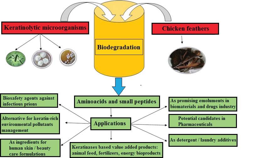
BIOTECHNOLOGIA ACTA, V. 12, No 2, 2019
-spiral segments of polypeptides with not- containing waste. These figures are over 40
coiled chain ends. The -keratins are part million tons per year for countries such as the
of the nails, horns, and hooves of mammals USA, Brazil, and China. Today, one of the main
(hard keratin which contains 18–22% methods of utilization of these wastes is their
cysteine), hair, wool, skin (soft keratin, burning. However, the method has a number
contains up to 14% cysteine) [1, 7]; of shortcomings: it is considerably costly, and
2) -keratins are harder and shaped burning the waste releases a significant amount
as several zigzag-like polypeptide chains of harmful gases in the atmosphere, which is
(-folded sheets), usually anti-parallel to dangerous to the environment [1].
each other, which is stabilized by hydrogen Another method of utilizing feathers
bonds and hydrophobic interactions. These is the chemical treatment under pressure,
proteins contain much less cysteine, but are which reduces their stiffness and increases
rich in amino acids such as glycine, alanine, digestibility. This method, however, is also
and serine (a characteristic repeat of the highly expensive, and involves a breakdown
“GSGAGA” sequence). Unlike -keratin, the of thermosensitive amino acids tryptophan
transverse disulfide bonds are absent between and lysine [11]. Therefore, it is nowadays very
adjacent polypeptide chains of -keratin, and important to develop a highly efficient and
the peptide fibrils are more flexible but not cheap way of processing keratin-containing
elastic. These proteins are found in feathers, raw materials. Alternatively, this issue can be
beaks and claws of birds, in porcupine needles, solved by the enzyme biodegradation, which
in claws and scales of reptiles, in turtle shells not only improves the nutritional value of
and arthropod exoskeletons. The molecular feathers, but also offers mild conditions for
mass of -keratin varies from 10 to 22 kDa [1, the production of valuable products. Microbial
8, 9] (Fig. 1). keratinases (E.C. 3.4.21 / 24 / 99.11) are a
The food industries, especially the meat group of proteolytic enzymes that meet these
market, slaughterhouse and wool industry, requirements because they are capable of
produce millions of tones of biomass that decomposing complex and rich keratin waste
contains keratin [1, 10]. Millions of tons of into more easily digestible components [1, 11,
feathers are emitted into the environment as a 12]. The ability to synthesize these enzymes is
by-product of the poultry processing enterprises, found in some insects (moth larvae), as well as
which makes serious problems as a pollutant, in microorganisms including bacteria, fungi,
as well as sources of the H5N1 virus [11]. The and yeasts, which can be isolated from keratin-
human population constantly increases on containing waste [2, 3, 11, 13].
the planet causing the intensive development It has been found that the keratin-
of these industries, which, in turn, causes containing by-products contain 15–18%
a steady increase in the amount of keratin- nitrogen, 2–5% sulfur, 3.2% minerals, 1.27%
а b
c
Fig. 1. Structure of keratin: a —Hierarchy of -keratin showing the assembly from:
two polypeptide chains (i) to a fibrous structure (iv); b — -keratin with a pleated sheet shape that consists
of antiparallel chains with R-groups that extend between sheets; c — TEM micrograph of -keratin from
a sheep horn displaying the composite structure of a crystalline keratin core within an amorphous keratin
matrix [9]
28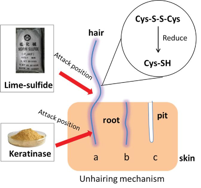
Reviews

fats and 90% protein. Hence, the rich in            and 264 families now (http://merops.sanger.
keratin organic waste can be a natural source       ac.uk/cgi-bin/family_index?type=P#S) [5,
of protein. Natural keratin, obtained from          14]. The classification of keratinases according
biomass, does not contain harmful chemicals         to the MEROPS database is given in Table.
and can be directly used for the production             The authors [18, 19] classified the
of various cosmetics, creams, shampoos,             keratinase of Meiothermus taiwanensis WR-
hair conditioners and biomedical products.          220 and the islandizin enzyme synthesized by
Monomer units of natural keratin, penetrating       Fervidobacterium islandicum, as belonging to
the skin and cuticle of hair, can nourish them      the clan SB, family S8.
without any side effects [1].                           Depending on the nature of the active
    Keratinases from microorganisms are also        center, microbial keratinases can be serine
used to make protein supplements, animal            proteases, metal proteases and serine metal
feeds, skin treatments, and in detergents.          proteases. The exception is yeast keratinases,
And the keratinase produced by the action of        which are asparagine proteases [14].
cleavage products used in the production of             Metal and serine peptidases are
nitrogen fertilizers, glue and foil, biofilms,      endoproteases that cleave peptide bonds
as well as plastics [1, 10, 11, 14]. In addition,   inside the polypeptide chain. Serine proteases
the promising direction is using keratinases        are a functionally rich and diverse group of
in the degradation of prions for the treatment      proteolytic enzymes with nucleophilic serine
of bovine spongiform encephalopathy in              residues (Ser) in the active center. The latter
order to prevent the prion-containing waste         attacks the carbonyl part of the peptide bond to
from contaminating the environment [5,              form an intermediate compound (acyl-enzyme
10, 15]. Keratinases can also be used as an         intermediate). To date, there are more than
active component of pesticides to combat root       333000 serine proteases that are classified
nematodes that cause the formation of galls,        in 53 families and 16 clans. Based on the
thickenings at the roots of plants [16, 17].        structure, serine proteases are divided into two
    Consequently, the growing interest in           categories: trypsin-like and subtilisin-like. The
microbial keratinases in various industries         enzymes of subtilisin subfamily are completely
leads to the search for new keratinolytic           inhibited by phenylmethylsulfonyl fluoride
producers of enzymes with properties that are       (PMSF) and chymostatin [14].
commensurate with commercial needs.                      In metal proteases, a nucleophilic attack on
    Keratinase classification. Proteases are        the peptide bond can be transmitted through
widespread, being involved in many biological       the water molecules coordinated by the two-
reactions occurring both in the cell and in the     valent metal ion (usually Zn (II), sometimes
body as a whole, and they play an important         Co (II), Mn (II)) or the bimetallic center of the
role in the circulation of nitrogen in nature.      enzyme (two ions Zn (II) or one Zn (II) ion and
Their general action mechanism is that of the       one Co (II) or Mn (II) ion). Depending on the
hydrolytic enzymes cleaving peptide bonds           amount of metal ions required for the catalysis,
from the ends of the polypeptide chain (exo-        metalloproteases are divided into two groups:
protease) or within the chain (endo-proteases)      the first group requires two metal ions, and
[5]. According to the amino acid sequence in        the other one needs only one. To date, there
the active centers of enzymes and the catalytic     are about 294000 metalloproteases, united in
action mechanism associated with it, these          73 families and 15 clans [5]. Catalytic activity
enzymes are grouped into asparagine, cysteine,      of metalloproteases is suppressed by chelating
glutamine, aspartate, metal, serine, threonine,     agents and heavy metals [14].
mixed proteases, and those with an unknown              The microbial keratinases of Actinomadura
catalytic action mechanism [14].                    keratinilytica Cpt29 [12, 20], Streptomyces
     In the Enzyme Nomenclature (1979), the         sp. 1382 [21], Actinomadura viridilutea
keratinases of Streptomyces and Trichophyton        DZ50 [22], Purpureocillium lilacinum
were named as Е.С. 3.4.99.11 and 3.4.99.12          [23], Aspergillus parasiticus [24], Bacillus
according to the type of catalyzed reaction         pumilus GRK [25], Bacillus licheniformis
[12, 13]. However, the intense development          RPk [26], Thermoactinomyces sp. YT06 [27],
of molecular biology caused the improvement         Brevibacillus brevis [28], Caldicoprobacter
of the classification system. Based on the          algeriensis [29] are serine proteases.
similarity of the amino acid sequences, which           Keratinases synthesized by Acinetobacter and
also reflects the evolutionary interactions         Bacillus subtilis MTCC (9102) are metalloproteases
between proteolytic enzymes, the MEROPS             [30, 31], while the enzyme of Bacillus parabrevis is
database united the proteases into 62 clans         a serine-metalloprotease [32].

                                                                                                    29
BIOTECHNOLOGIA ACTA, V. 12, No 2, 2019

                                   Keratinases classification (MEROPS)
           Name                   Main name                                MEROPS ID
                                                      Clan SB >> Subclan (none) >> Family S8 >> Subfamily A
         Keratinase                Subtilisin         >> S08.001; Catalytic type — serine;
        (Bacillus sp.)             Carlsberg          Subclass 3.4 (Peptidases) >> Sub-subclass 3.4.21 (Serine
                                                      endopeptidases) >> Peptidase 3.4.21.62
                                  Keratinase
  Keratinase (Doratomyces                             Clan SB >> Subclan (none) >> Family S8 >> Subfamily A
                                 (Doratomyces
        microsporus)                                  >> S08.148; Catalytic type — serine
                                  microsporus)
       Keratinase K1            Keratinase K1
                                                Clan SB >> Subclan (none) >> Family S8 >> Subfamily A
     (Stenotrophomonas        (Stenotrophomonas
                                                >> S08.110; Catalytic type — serine
         maltophilia)             maltophilia)
       Keratinase K2            Subfamily S8A
                                                      Clan SB >> Subclan (none) >> Family S8 >> Subfamily A
     (Stenotrophomonas           unassigned
                                                      >> S08.UPA; Catalytic type — serine
         maltophilia)             peptidases
                                                      Clan MA >> Subclan MA(E) >> Family M4 >> Subfamily
     Keratinase Ker P                                 (none) >> M04.005; Catalytic type — metallo;
                                  Pseudolysin
 (Pseudomonas aeruginosa)                             Subclass 3.4 (Peptidases) >> Sub-subclass 3.4.24
                                                      (Metalloendopeptidases) >> Peptidase 3.4.24.26
     Keratinase KerSMF          Subfamily S8A
                                                      Clan SB >> Subclan (none) >> Family S8 >> Subfamily A
     (Stenotrophomonas           unassigned
                                                      >> S08.UPA; Catalytic type — serine
         maltophilia)             peptidases
                                    SFase-2           Clan PA >> Subclan PA(S) >> Family S1 >> Subfamily E
      Keratinase Sfp2
                                 endopeptidase        >> S01.431; Catalytic type — serine
                                Subfamily S1A
  Keratinase Streptomyces                             Clan PA >> Subclan PA(S) >> Family S1 >> Subfamily A
                                 unassigned
        albidoflavus                                  >> S01.UPA; Catalytic type — serine
                                  peptidases
          KerSMD                Keratinase K1
                                                Clan SB >> Subclan (none) >> Family S8 >> Subfamily A
     (Stenotrophomonas        (Stenotrophomonas
                                                >> S08.110; Catalytic type — serine
         maltophilia)             maltophilia)

    Interestingly, Bacillus halodurans PPKS-2                   Keratinases are produced by both
synthesizes two keratinases which are                       pathogenic and non-pathogenic fungi:
different by the catalytic type: keratinase 1 is            Alternaria, Arthrographis [34], Aspergillus [24,
a disulfide reductase, keratinase 2 is a serine             35], Beauveria, Chrysosporium, Cladobotryum,
protease [33].                                              Cladosporium (C. sphaerospermum) [34],
    Consequently, most of the investigated                  Doratomyces (D. microsporus) [3, 36],
keratinases are serine proteases, the synthesis             Geomyces, Gymnoascus, Malbranchea,
of which is suppressed by phenylmethylsulfonyl              Microsporum, Mucor, Myceliophthora
fluoride (PMSF) and chymostatin [14].                       [34], Paecilomyces (P. marquandii) [36],
    Keratinase producers. Keratinolytic                     Pectinotrichum [34], Penicillium (P. citrinum)
enzymes are synthesized by many bacteria,                   [37], Purpureocillium [23], Renispora
including actinomycetes, as well as fungi.                  [34], Scopulariopsis [7], Sporendonema,
Among the latter, a significant role is played              Trichophyton [34]. The pathogenicity and
by skin fungi, which in saprophytic state are               virulence of some fungi are closely related
able to digest keratin in vitro and use it as a             to the ability of the producers to break down
substrate, and some can penetrate into the                  both hard and soft types of keratin. However,
tissue (in vivo) and cause dermatomycosis in                only non-pathogenic keratinolytics are of an
humans and animals [7].                                     industrial importance, because of the risk of
    There are two types of fungi that inhabit               infection with pathogenic strains.
keratin substrates:                                             Among the Gram-positive bacteria, the
    • keratinolytic fungi, which affect the keratin         ability to synthesize keratinases and cleave
substrate directly and cleave the molecules;                keratin is found in the genera Arthrobacter
    • keratinophilic fungi, which use the                   [38], Bacillus [12], Kocuria [39], Lysobacter
matter that is naturally linked to keratin, or the          [12], Microbacterium [8], Nesternokia [12],
products of keratin destruction under the effect            etc. However the most common producers
of other fungi [7].                                         of keratinases are the representatives of

30
Reviews
the genus Bacillus, namely B. subtilis and such as soy flour [47], starch [49], sucrose
B. licheniformis [26]. Although keratinolysis is [27], and dextrose [50] can also induce the
detected in other species of the genus Bacillus: synthesis of these enzymes. If glucose is used
B. altitudinis [40], B. halodurans [33], as a carbon source, the synthesis of certain
B. tequilensis [41], B. pumilus [25], B. cereus, keratinases is suppressed, which is associated
and B. thuringiensis [42]. Several genera of with the phenomenon of catabolite repression
the Gram-negative bacteria also can synthesize [8, 47]. However, there is no repression of the
keratinases: Acinetobacter [30], Alcaligenes keratinase synthesis in A. parasiticus enzyme
[43], Caldicoprobacter [29], Chryseobacterium, in the presence of a mixture of 1% glucose with
Fervidobacterium [8, 14], Klebsiella [44], 1% keratin [24], similarly to the keratinase of
Stenotrophomonas, Thermoanaerobacter [8, B. thuringiensis Bt407 [49].
14, 45], Vibrio, Xanthomonas [8]. The keratinase synthesis is also influenced
The keratinolytic activity is also found by the presence of organic or inorganic
in several actinomycetes: Actinomadura nitrogen sources. Keratin-containing
(А. keratinilytica, A. viridilutea) [20, 22], substrates may serve as a source of nitrogen
Nocar diopsis [10], Streptomyces pactum, [25]. Still, researchers sometimes use extracts
S. graminofaciens, S. albidflavus, S. flavis [10]. of yeast extract [24, 26, 49] or a combination
Recently, researchers became interested of inorganic nitrogen (potassium nitrate [38],
in thermophilic and hyperthermophilic sodium nitrate [27]) or peptone [50, 51] or
organisms that synthesize keratinases with tryptone to extract a significant amount of
unique properties. The temperature optimum enzyme [51].
of those enzymes is within the range of Another important factor influencing
80–100 C: Aeropyrum, Fervidobacterium the production of enzymes, in addition to the
[46], Microbisporaaerata [10], Pyrococcus content of nutrient medium, is the cultivation
[46], Streptomyces gulbarguensis, S. conditions: the optimum growth temperature
thermoviolaceus, S. thermonitrificans [10], of the producer, the initial pH value of the
Thermoactinomyces [27], Thermoanaerobacter, medium, the intensity of mixing and the
Thermococcus [46], etc. These enzymes are amount of seed material [8, 14].
promising for research, since they can be used Temperature is the determining factor
in high-temperature processing of keratin- in the synthesis of enzymes. However, the
containing raw materials. optimum synthesis temperature depends
Keratinase synthesis regulation. Microbial on whether the culture is mesophilic or
keratinases are predominantly extracellular thermophilic. Most keratinolytic fungi
enzymes that are synthesized in the culture are mesophiles producing keratinases at
medium containing keratin as an inducer [4, temperatures 26 С [37], 28 С [23], or 30 C
22, 41, 47]. Although there are microorganisms [24, 35]. Bacterial keratinases are usually
capable of producing intracellular keratinase, synthesized at a temperature of 25 C [39],
in particular Trichophyton gallinae [48]. The 37 C [25, 26, 33, 38, 50], 50 C [29]. However,
geophilic microscopis fungi Arthroderma there are thermophilic and hyperthermophilic
quadrifidum, A. curreyi and Chrysosporium bacteria Thermoanaerobacter and
pruinosum can synthesize both extracellular Fervidobacterium that secrete keratinases in
and cellular keratinases [14]. the medium at elevated temperatures, 65 C
The microbial production of enzymes is [52] and 70 C [12], respectively.
a complicated and highly regulated process Also an equally important parameter in
that depends on the growth phase of the the synthesis of keratinases is the initial pH
microorganism. Production of keratinase is of the nutrient medium. Most of investigated
most intense at the end of the exponential or in keratinases were produced at neutral [38,
the stationary phase of growth [5, 11, 49]. 49, 50, 51] or alkaline [25, 27, 33, 51] pH
Most keratinases are inducible enzymes, values. Although the fungi P. marquandii,
although some are expressed constitutively D. microsporus [36], A. parasiticus [24] and
[5, 14]. Adding keratin or keratin-containing bacterium B. subtilis KD-N2 [53] synthesized
substances in the culture medium is important keratinases in weakly acidic media with pH
for the induction of keratinase synthesis. The values of 6.0 and 6.5, respectively.
most common source of carbon and nitrogen is The amount of inoculum introduced into
chicken feathers [20, 25, 39], both alone and in the nutrient medium is an important factor
combination with yeast extract [26], a mixture affecting cell growth and the formation of the
of chicken feathers and keratin [24, 38], hair target product. The amount of seed material
keratin [37]. However, non-keratin substrates used in different studies usually varies from
31BIOTECHNOLOGIA ACTA, V. 12, No 2, 2019
1% to 5% [24, 25, 38]. Additionally, if promote using Aspergillus sp. DHE7 in
inoculum portion was up to 10%, that was the environmentally friendly process of
optimal for the synthesis of keratinase by B. bioconversion of keratin waste.
subtilis KD-N2 in a hair-containing medium Quite often, keratinase genes are cloned
[53], while 50% inoculum was needed for the in host cells in order to increase the enzyme
keratinase synthesis by B. subtilis MTSC9102 output, which is necessary for the its
[50] on horns as a substrate at solid phase commercialization. Thus, more than 50%
culturing. of industrially important keratinases are
Most microbial producers of enzymes are produced by heterologous expression in the
aerobic and need oxygen for their growth host cells, which are specially adapted for the
and development. Lack of oxygen can lead to intensive production of target enzymes. E.
adverse changes in the enzyme composition coli is commonly used as an expression system
and loss of the target product, or even kill for recombinant proteins [58]; however, the
the organism. Oxygen requirements during limiting factors in obtaining the enzyme using
enzymatic processes are provided by aeration that host are the accumulation of inactive
and mixing [8]. The degree of aeration for compounds [59], as well as the need for folding
the synthesis of keratinase is: 100 rpm [36], of pro-keratinase in vitro, which essentially
120 rpm [49], 140 rpm [35], 150 rpm [38, affects the yield of the final active product. If
51], 180 rpm [27], 200 rpm [23, 25, 26], expression in the host cells does not provide
220 rpm [37]. the proper folding of the proteins, additional
Microbial production of keratinases is procedures are needed to form disulfide bonds
carried out by submerged (SmF) [8, 29, 54] [5]. Therefore, other microorganisms, including
or solid -state fermentation (SSF) [50, 55, representatives of different species of the genus
56]. Using SPA has several advantages over Bacillus can be used as host cells [60], in which
SmF: the enzymes are cheaper, the minimum the introduction and expression of numerous
energy costs, the stability of output product, copies of keratinase genes leads to the increased
the lower cost of water, and better oxygen enzyme output. However, the use of those
circulation [8]. The SmF method, in contrast to producers may be limited by plasmid instability.
SSF, makes it possible to automate the process In addition, certain Bacillus species synthesize
parameters needed for the optimum growth. a lot of other enzymes inherent in that producer
Thus, the selection of the appropriate (amylase, mannanase, cellulase), which can
nutrient medium and cultivation conditions negatively affect the process of separation
can not only increase the yield of enzymes, and purification of the target product [5].
but also produce preparations with certain Quite often, Pichia pastoris yeasts are used
properties. Thus, the authors [57] investigated as an expression system for the production of
ten microbial isolates on their ability to keratinases, providing an appropriate medium
produce keratinases. The most active isolate, for posttranslational modifications and folding
identified as B. licheniformis ALW1, exhibited of eukaryotic keratinases [59]. These single-
25.2 U/ml keratinase activity. After the celled eukaryotes are easily cultured and
optimization of cultivation conditions, the manipulated, so they are successfully used to
biosynthesis of keratinase increased almost expression many proteases of bacteria, fungi
three times to 72.2 U/ml. and mammals [5].
Fungal producer Aspergillus sp. DHE7 Physical and chemical factors are also used
[11] was selected among the 15 keratinolytic to obtain mutants with elevated keratinase
strains isolated from the poultry farm soil. It activity. Thus, a mutant Streptomyces
showed the highest keratinase yield of 199 ± radiopugnans KR 12 strain was obtained as
4.2 U/ml when incubated for 4 days at 30 C a result of UV irradiation. Its keratinolytic
and pH 6.0 on a 2% chicken feathers substrate. activity was thrice increased compared to wild
The addition of 0.5% sucrose as a supporting strain [61].
source of carbon increased the keratinase Chemical agents such as ethyl-
yield to 226 ± 5.4 U/ml, while introducing methylsulfonate (EMC), N-methyl-N’-nitro-
the additional sources of nitrogen to the N-nitroso-guanidine (MNNG), or ethidium
medium did not affect the enzyme synthesis. bromide, can also be used to induce random
The authors [11] have shown that the best mutations in the DNA molecule [5]. For
substrates for the keratinases synthesis example, the use of MNNG resulted in the
are goat hair (452 ± 12.3 U/ml), turkey production of a mutant B. subtilis strain KD-
feathers (435 ± 9.2 U/ml) and sheep wool N2, whose keratinase activity was 2.5 times
(322 ± 13.4 U/ml). The obtained results greater than that of wild strain [62].
32Reviews
Four amino acid substitutions (N122Y, keratinases synthesized by pathogenic fungi
N217S, A193P, N160C) were introduced into (for example, keratinase 2 of Trichophyton
the keratinase of B. licheniformis BBE11-1 using mentagrophytes) have a molecular weight of
site-directed mutagenesis, which made it possible up to 440 kDa [5].
to increase its catalytic activity by 5.6 times in
comparison with wild-type keratinase [63]. pН- and thermal optimum, рН- and thermal
stability of keratinases
Physical and chemical properties Thermal optimum, pH optimum, as well
of keratinases as thermostability of enzymes are important
Molecular weight of keratinases. Most characteristics that determine the possibility
keratinases isolated from bacteria, fungi, and of their further application.
actinomycetes are monomeric enzymes with Most of microbial keratinases achieve the
molecular weight varies from 14 to 240 kDa highest activity in the alkaline or neutral
[12, 14, 29]. The lowest molecular weights of pH values of 7.0–9.0: keratinase of B. alti-
15–20 kDa and 18 kDa are characteristic of the tudinis RBDV1 [40], B. parabrevis [32],
Streptomyces sp. 1382 [21] and Streptomyces B. brevis [28], Bacillus sp. P45 [67], Bacillus
albidoflavus [5, 64] keratinases, respectively. thuringiensis Bt407 [49] at рН 8.0; keratinase
Keratinases isolated from bacteria, including of B. tequilensis Q7 [41], C. algeriensis [29],
actinomycetes, are characterized by the A. parasiticus [35] at pH 7.0; keratinase of
following molecular weight ratios: 43 kD B. licheniformis RPk [26] at pH 9.0. However,
in B. altitudinis RBDV1 [40], 30 and 66 some enzymes are optimally active outside this
kDa in B. halodurans PPKS-2 [33], 32 kDa values, even at extreme alkaline or slightly
in B. licheniformis RPk [26 ], 28 kDa in B. acidic pH [29]. Thus, keratinase of B. subtilis
tequilensis Q7 [41], 28 kDa in Brevibacillus MTCC (9102) exhibits maximum activity
parabrevis [32], 25 kDa in Acinetobacter in a weakly acidic pH of 6.0 [31]. Several
[30], 33 kDa in Caldicoprobacter algeriensis keratinolytic enzymes are notably stable in a
[29], 19.5 kDa in A. viridilutea DZ50 [22 ], 29 wide range of pH [23, 29, 52, 60]. The enzyme
kDa in A. keratinilytica Cpt29 [20], 35 kDa of A. keratinilytica Cpt29 is active at рН of
in Thermoactinomyces sp. YT06 [27]. Four 3.0 to 10.0, with the optimum рН of 10.0
fractions with keratinase activity have been [20]; keratinases obtained from B. halodurans
isolated from Micrococcus luteus with the PPKS-2 are active at рН of 7.0-13.0 with
following molecular weights: 62 kDa, 139 kDa, the optimum pH of 11.0 [33]. The protease
185 kDa and 229 kDa, respectively [52]. The synthesized by Streptomyces sp. AB1 is stable
highest molecular weight, 240 kDa, is found at pH 4.0–11.0 for 96 hours with the optimum
for the keratinase of Kocuria rosea [14]. High pH of 11.5 [68]. The maximum activity at a
molecular weights are usually characteristic pH of 11.0 was also detected by keratinases of
of keratinases synthesized by thermophiles A. viridilutea DZ50 [22], Acinetobacter [30], B.
or which are metalloproteases [29, 36, 53]. licheniformis ER-15 [65]. The pH of 10.0–10.5
Thus, an anaerobic thermophilic bacterium was optimum for keratinases, synthesized by
Thermoanaerobacter sp. 1004-09, isolated the recombinant strain B. subtilis WB600 [60]
from hot springs of the rift zone of Baikal, and Meiothermus taiwanensis WR-220 [18].
synthesizes keratinase with a molecular weight The highest value of the pH optimum in the
of 150 kDa [53]. alkaline region of 12.5 is characteristic for
Some bacterial producers of keratinases keratinase of Nocardiopsis sp. TOA-1 [69].
synthesize multimeric proteins [14, 29]. For The optimum temperature of keratinase
example, B. licheniformis ER-15 keratinase activity is very diverse and often depends on
is a dimeric enzyme with subunits of 28 and the producer and temperature conditions of
30 kDa [65]. And three keratinolytic enzymes the producer growth [29, 38, 60, 64]. Quite
(K1, K2 and K3) of 48 kDa, 36 kDa and 17 kDa, often, though, the maximum activity of an
respectively, were obtained as a result of the enzyme is observed at a temperature higher
3-stage purification from Stenotrophomonas than the optimum conditions for the growth
maltophilia BBE11-1 [66]. of a microorganism [26, 33, 35]. Thus, the
Fungal keratinases usually have low keratinase of A. viridilutea DZ50 exhibits
molecular weights of 20 to 40 kDa: 21 kDa in maximum activity at a temperature of 80 C,
P. citrinum [54], 37 kDa in Purpureocillium while the producer growth temperature is
lilacinum [23], 36 kDa in A. parasiticus [35], 45 C [22]; enzyme synthesized by Nocardiopsis
33 kDa in P. marquandii, and 30 kDa in sp. TOA-1, which grows at 30 C, has a
D. microsporus [36]. Although it is noted that temperature optimum at 60 C [69].
33BIOTECHNOLOGIA ACTA, V. 12, No 2, 2019
Enzymes of thermophilic microorganisms There also are thermolabine keratinases.
A. keratinilytica Cpt29, Streptomyces sp. For example, keratinase of Bacillus sp. P45
strain AB1, A. viridilutea DZ50, B. altitudinis showed maximum activity at 55 C, but it
RBDV1 have a temperature optimum at 70 C had low thermal stability and was completely
[20], 75 C [68], 80 C [22], and 85 C [40], inactivated after 10 min at 50 C [67].
respectively, whereas mesophilic cultures Investigating the influence of temperature
exhibit maximum activity at 40 C [28, 31, 38, on the activity of various keratinases, it was
60]. The lowest optimum temperature of 30 C shown that they exhibit activity both in the
is characteristic for enzymes synthesized by wide [18, 20, 31, 40, 52, 60, 68], and narrow
B. tequilensis Q7 [41] and P. citrinum values of temperatures [54].
[54] and the highest for keratinases of Substrate specificity. Microbial
hyperthermophilic microorganisms isolated keratinases isolated from different sources
from marine hydrothermal sources that are characterized by different properties
exhibited maximum activity at a temperature depending on the producer. Keratinases
of 100 C and above [18, 46]. synthesized by fungi, actinomycetes, and
Many keratinases exhibit maximum bacteria are capable of cleaving a wide range
activity at a temperature of 50–55 C, of substrates from soft keratin (e.g., corneous
particularly those of M. luteus [70], A. para- layer [65]) to hard keratin of feathers [20, 27,
siticus [35], Acinetobacter [30], C. algeriensis 39, 43, 44, 65, 66, 70], wool [2, 30, 32], human
[29], Bacillus sp. P45 [67], B. thuringiensis hair [36] and animal hair, hoof [66], horn [13],
Bt407 [49]. Certain enzymes exhibited azokeratin [68], keratin azure [28, 67, 68, 70].
thermal optimum at a temperature of 60 C, Also, certain keratinases degrade collagen [13,
such as produced by B. licheniformis RPk 71], elastin [28, 59], gelatin [13, 28, 60, 70],
[26], B. parabrevis [32], Thermoanaerobacter albumin [28, 60, 71], hemoglobin [28, 65],
1004-09 [52], Nocardiopsis sp. TOA-1 [69]. fibrin [65], and azocasein [28, 35].
Keratinases, active at high temperatures, are Keratinase substrate specificity depends
used in various industries and in medicine. on the chemical properties of the substrates.
The enzyme thermostability is an important Since keratin consists of 50–60% hydrophobic
characteristic for most biotechnological and aromatic amino acids, keratinases
processes. Hence, thermophilic and predominantly cleave peptide bonds containing
hyperthermophilic microorganisms are of P1 hydrophobic and aromatic amino acid
particular interest as a potential source of residues in P1 position [4, 59]. The study of
new thermostable enzymes. There are several hydrolysis using an oxidized insulin B-chain
advantages in using thermostable enzymes as a substrate showed that the keratinases
in industrial processes: reducing the risk of Thermoanaerobacter sp. 1004-09 and
of microbial contamination, reducing the P. aeruginosa KP1 and KP2 selectively
viscosity and mixing speed, increasing the hydrolyze the bonds formed by phenylalanine,
solubility of the substrate and the diffusion valine, tyrosine or leucine [72]. Substrate
rate [19, 46]. specificity of these keratinases was determined
The thermal activity and thermal stability using synthetic substrates, such amino
of most keratinases is found to increase in acid derivatives as p-nitroanilides (pNA),
the presence of Ca2+ ions [13, 26, 28, 49], and p-nitrophenyl esters (ONp), and derivatives
those of several other enzymes in the presence of 7-amino-4-methylcumarin (AMC). The
of Mg2+ [13, 40, 71] or Mn2+ ions [20, 40, 70]. residues in the P2 and P3 positions also play
Interestingly, Ca2+ ions did not affect the a role. The keratinase KerS14 of B. subtilis
activity of keratinase Thermoanaerobacter predominantly cleaves peptide bonds with Arg
sp. 1004-09, but significantly increased its in position P1, Gly and Ala in position P2, Gln
thermal stability from 10 min to 13 h at a or Glu at position P3 [14].
temperature of 94 С [52]. Study of the ability of R. marquandii
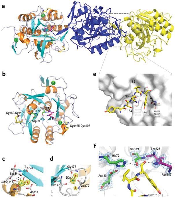
The keratinase of Meiothermus taiwanensis and D. microsporus keratinases to hydrolyze
WR-220 is known to contain two calcium various substrates showed that the enzymes
binding sites [18] (Fig. 2). The elimination of cleaved -keratin of skin and nails, to a lesser
Ca2+ ions from such site causes a significant extent that of hair and wool, but could not
reduction in the thermostability of enzymes. cleave chicken feathers -keratin. It was
The role of Ca2+ ions may be related to the established that the presence of reducing
stabilization of the activated keratinase agents stimulated the enzymatic hydrolysis of
form and the protection of its structure from keratin. When 1mM dithiothreitol was added,
autolysis [29, 49]. the keratinase activity of R. marquandii and
34Reviews
a
b e
f
c d
Fig. 2. Crystal structure of rMtaKer:
a — Stereoview of three mature rMtaKer monomers is shown in the asymmetric unit. The trimers are
named chain A (orange/cyan), chain B (blue), and chain C (yellow); b — The core structure of mature rMtaKer
is made of a seven-stranded parallel sheet flanked by six -helices (orange) and five -sheets (cyan). The
calcium ions are shown as green spheres and two intramolecular disulfide bridges are highlighted in yellow.
The catalytic triad (Asp, His, and Ser) is labeled by green sticks and the oxyanion hole residues are marked
by magenta sticks; c — Structure of the 1Ca-binding site forms a pentagonal bipyramidal geometry with
five residues and one water molecule (red ball); d — Structure of the second Ca-binding site is coordinated to
three residues and two water molecules; e — The end part of the C-terminus (Tyr-Glu-Asn-Leu-Tyr) from C
chain binds at the substrate-binding cleft (S1–S4) on the B chain of rMtaKer as shown in gray. The substrate
is displayed as a yellow stick model; f — Tyr282 seals the hydrophobic pocket of S1 site and several hydrogen
bonds are also observed along the pocket surfaces and around the active site [18]
35BIOTECHNOLOGIA ACTA, V. 12, No 2, 2019
D. microsporus increased two and three times, at work and the ability to genetically change.
respectively [36]. This is due to the ability of In addition, microbial enzymes also have many
reducing agents to decrease the amount of advantages over chemical compounds and animal
disulfide bonds in keratin threads, thereby enzymes, such as high activity, broad substrate
promoting the access of enzymes to the specificity, and ability to biodegrade [1, 4, 10].
substrate for proteolytic attack [36]. Keratinases, due to their broad substrate
Keratinase produced by P. citrinum PC-54-91 specificity and the ability to split the substrate
VILAR cleaves -keratin and almost does not resistant to hydrolysis, are used in many
hydrolyze collagen [54]. industrial processes (Fig. 3).
Keratinase, synthesized by B. pumilus FH9, Annually, a very large amount of keratin
showed broad substrate specificity, splitting waste is formed worldwide. The main
both soluble and insoluble substrates. The environmental pollutants are poultry, leather
enzyme showed the highest proteolytic activity and textile industries, and a lot of waste in
on casein, serum bovine albumin, gelatin, the form of feathers, hair, bristles, horns
collagen, and to a lesser degree was able to and hooves is obtained from livestock and
hydrolyze feathers, wool and horns [13]. slaughterhouses [14, 17, 45].
Hence, there are keratinases with a narrow, However, there are some restrictions
and with a broad substrate specificity [13, 28, on the use of keratin waste in the European
66, 70]. Union (EU). Following the outbreak of bovine
Practical use of keratinases. Microbial spongiform encephalopathy (BSE) in the
enzymes make up a significant proportion of United Kingdom, the European Union and
industrial catalysts, of which about 65% of the United States of America introduced
the market is occupied by hydrolases. Among strict rules regarding the use of animal by-
the latter, there is a very important group of products, namely grouping them into three
proteases with a wide range of applications [5]. categories in accordance with the level of
The increased commercial interest in microbial risk of transmission of pathogens and toxic
enzymes is due to the fact that microorganisms substances. Only keratin waste of the third
are their inexhaustible source due to their category can be processed and used for the
natural diversity, ease of cultivation, safety production of feed.
Fig. 3. Schematic representation of current and potential applications of microbial keratinases [10]
36Reviews
According to the Regulation (EC) No BioResource International (BRI) produces
1774/2002 of the European parliament, the third enzyme preparations Versazyme® and
waste category includes the products of animal Valkerase® containing keratinases of B.
origin that 1) are obtained from animal carcasses, licheniformis and used to degrade the keratin
2) are not used for human consumption, 3) will waste [72].
not transmit pathogens to humans and animals. The hydrolysed feathers can also be used
A large amount of waste produced by animal to produce biohydrogen or as fertilizers in
processing plants can be used as a substrate for organic farming promoting the slow release
bioenergy and high-value products if these wastes of nitrogen, improving plant growth, and
are pre-treated properly [14]. enhancing the activity of microorganisms in
In 2012, around 8.5 billion tons of bird the soil, structuring it, and also increasing the
feathers were produced worldwide [5]. Bird moisture-retaining capacity of the soil [5].
feathers consist of keratin (predominantly Thus, hydrolyzates of bovine horns and
-keratin) and contain a significant amount hooves produced using P. marquandii can be
of serine, glutamic acid, proline and a small utilized as fertilizers because they contain a
amount of methionine, histidine and lysine. significant amount of amino acids (with the
One of the ways to process feather waste is exception of proline and tryptophan), and
transforming them into flour which can then significantly differ from other fertilizers
be used as raw material in the production of by a positive effect on plant growth. The
biofuel, as an ingredient of bioplastics and as hydrolyzates obtained as a result of P. wooson-
a feed for animals. The traditional method of gensis TKB2 splitting the bird feathers
treating feathers involves high temperature contribute to seed germination and seedlings
and pressure, is energy-intensive and causes growth [14, 47].
the loss of several essential amino acids The transformation of keratin waste
(methionine, lysine, histidine and tryptophan). into biofuel is a far-reaching direction for
An alternative method is to treat feathers with producing clean energy that can partially
keratinases and obtain hydrolysates, which, meet the global demand for energy. Feather
in their nutritional value and digestibility, hydrolyzate produced by B. lichenifomis
exceed the products obtained by chemical and keratinase can then be used by Thermococcus
mechanical processing [3, 5, 14, 46]. High- litoralis culture to obtain biogas [3, 14, 46].
quality amino acids thus obtained can be added The keratinases are most widely used in
to feed birds, ruminants, pigs and fish [5, 47]. the leather industry, which is one of the oldest
Another way to dispose of keratin waste and fastest growing industries in the world,
is composting, during which organic keratin important in the modern economy. At the same
waste is gradually converted into inorganic time, it represents one of the world’s largest
nitrogen (ammonium and nitrate) and sulfur sources of pollution, since skin treatment
(sulfate), which can then be easily absorbed by requires the use of toxic substances hazardous
plants [14]. However, this process is very long- to the environment, including workers of such
term because of the stability of the substrate enterprises [5, 14, 42, 73]. The production of
to the action of most proteolytic enzymes. At leather involves several stages, one of which
first, within one to four weeks, bacteria and is the removal of epidermis, hair, and wool.
actinomycetes develop within the compost and That is done with chemical reagents (alkali,
are gradually replaced by fungi. Keratinolytic sulfides) which destroy disulfide bonds in the
strains are detected around the sixth week, molecules of structural proteins and sadly
their growth correlates with mineralization pollute the surrounding nature. The use of
of nitrogen and sulfur. To accelerate and keratinases is an alternative solution (Fig.
intensify the process of composting, it is 4), which leads to a decrease in the level of
possible to inoculate compost with keratinases environmental pollution, and improves the
of microorganisms [14]. Various studies have quality of the final product. The enzymes
shown that the addition of B. licheniformis significantly reduce the time and cost of
and Streptomyces sp. greatly improved the “dehairing”, simplify the general scheme
degradation of chicken feathers compost and of leather treatment and the final product
obtaining valuable products that can later be is of higher quality. Proteolytic enzymes
used in agriculture [14]. are increasingly used to soften the leather
In laboratory conditions, many and in preparing it to the tanning process.
microorganisms were shown to break down Keratinases can remove animal hair, but they
chicken feathers and other keratin-containing should not exhibit collagenase activity in order
products [20, 28, 44, 55, 65, 67]. not to damage the leather. The enzymes most
37BIOTECHNOLOGIA ACTA, V. 12, No 2, 2019
often used in the leather industry are produced As part of complex proteolytic
by Bacillus sp., Pseudomonas stutzeri, preparations, keratinases are used in soaking,
Cladicoprobacter algeriensisi, Acinetobacter liming and softening of skins. Patented
sp., Paenibacillus woosongensis, Vibrio special preparations for “dehairing” consist
metschnikovii, Microbacterium sp. kr10 and of a mixture of enzymes isolated from
various fungal species: Aspergillus tamarii, micromycetes, streptomycetes and bacteria of
Penicillium chrysogenum and Trichoderma the genus Bacillus [37].
harzianum [5, 14]. For example, the keratinase Keratinases, particularly those
of Bacillus safensis LAU 13 fully dehairs the isolated from B. licheniforms, B. cereus,
goat skin in 12 hours without obvious damage Chryseobacterium L99 and Pseudomonas
compared to the chemical method which does sp., are important in the textile industry,
not allow for complete dehairing and involves since they can improve the fiber resistance to
leather damage [47]. The keratinase of shrinkage and staining [47, 60]. Applying an
B. brevis US575 effectively dehairs the skins of enzyme complex of keratinase, cutinase, lipase
rabbits, sheep and cows [28]. and transglutaminase can greatly improve the
At one of the stages of leather treatment, process of wool processing [72].
chromium sulphate (CrSO4) is used to stabilize The most promising direction is using
it. The compound is only partially bound and keratinases in the detergent industry, because
most of it gets into sewage. The permissible most of the enzymes are alkaline proteases
level of Cr in sewage in most countries of the stable at relatively high temperatures
world is less than 2 mg/l, so it is necessary and sufficiently tolerant of surface active
to increase the absorption rate of Cr in the compounds [20, 25, 32, 35, 67]. Being
processing. Adding keratin hydrolyzate (2– substrate specific, keratinases are able to
3% w/w) of horn flour makes it possible to remove contamination over a short period
reduce Cr in sewage from 35% to 10%. The low of time without damaging the structure
molecular weight keratin peptides contained in and strength of the fibers. They are used to
the hydrolyzates react with Cr to form the Cr- hydrolyze the keratin derivatives on collars
keratin complex, which, when interacting with and cuffs [10, 14]. The alkaline keratinase of P.
collagen, enhances absorption of Cr [14]. woosongensis TKB2 effectively removes blood
Fig. 4. Two unhairing strategies (lime sulfide and keratinase) used in beamhouse process. Though those two
strategies share similar unhairing mechanism of reducing S-S bond to dissolve hair protein, lime sulfide
attacks the hair shaft outside the skin, while keratinolytic protease attacks the hair root to produce shaft
free skins:
a — whole hair in skin without treatment; b — unhairing with lime sulfide and hair shaft was still
remaining in the hair pit; c — unhairing with keratinase and skins are free from hair shaft [73]
38Reviews
stains from surgical clothes, and egg yolk The keratinase ability to hydrolyse
and chocolate from ordinary clothing. And B. keratin can also be used to heal wounds.
thuringiensis TS2 keratinase degrades not only The avascular nature of the wounds in the
blood stains and egg yolk, but also effectively third degree of burns can interfere with the
removes hair from goat skin [3]. effective diffusion of systemic antimicrobial
Keratinases can also be applied in the agents in the wound where the number
detergent industry in the treatment of of microorganisms is usually very high.
wastewater, generated by the laundry washing Enzymatic treatment of the wound increases
and containing a large amount of keratin waste the penetration of local antibiotics and
and dirt [3, 5, 14]. The commercial product stimulates wound healing [3, 14].
BioGuard Plu, manufactured by RuShay Inc., is In addition, the alkaline keratinases of
used to clean drainage pipes and septic tanks [72]. B. pumilus and Staphylococcus auricularis
Also, keratinases are widely used in the are known to inhibit the formation of
biomedical, pharmaceutical and cosmetic biofilms by 86% and 50%, respectively,
industries, in the preparation of vaccines, the and also remove 0.4013 g and 0.3823
production of bioactive peptides, therapeutic g of silver from 1 g of X-ray and photo
serums, the creation of cosmetic products film, respectively. Alkaline proteases of
(nutritional creams, lotions, anti-dandruff Aspergillus versicolor and B. subtilis ATCC
shampoos); callus removal, degradation of 6633 also provide a good recovery of silver
keratinized skin, its epilation, removal of from X-ray films [14, 47].
keratin in the treatment of psoriasis and acne New applications of keratinases are
[3, 5, 10, 14, 72]. associated with the removal of ear sulfur,
Proteos Biotech produces commercial whitening of pearls, purification of contact
preparations: Keratoclean® Hydra PB, lenses and the participation of microbial
Pure100 Keratinase, Keratoclean Sensitive PB keratinases in the formation of silver
and Keatopeel PB for callus removal and acne nanoparticles [14, 47].
treatment [72]. Several groups of researchers studied
Keratinases are used to cleave dead skin the potential of keratinases as agents of
cells, improve blood circulation and thoroughly biocontrol. Keratinase produced by S.
and deeply cleanse it, prepare skin for maltophilia R13 was effective against
absorption of nutrients: masks, creams, as well several fungal pathogens, including
as ampouled preparations and serums, which Fusarium solani, F. oxysporum, Mucor
are extremely useful for dry and sensitive skin sp. and A. niger, which cause diseases of
[1, 42]. valuable plants and cultures. Keratinase
The hair consists mainly of keratin (90%) synthesized by Thermoactinomyces also
and a small amount of lipids (1–9%). Keratin showed antimicrobial properties in relation
hydrolyzates are effective restorers in the hair to plant pathogens. Keratinase of Bacillus
care process. Most of the keratin hydrolysates sp. 50-3, as already mentioned, is effective
used for this purpose are chemically against agricultural pests such as nematodes
hydrolyzed or thermally treated hooves, horns [16, 17]. Also, this enzyme can be used
and wool, although recently using of microbial against mosquitoes that are the carriers of
keratinases has become popular. Treatment of many tropical diseases [14].
chicken feathers with keratinolytic enzymes of In recent years, the attention of scientists
S. maltophilia resulted in hydrolyzates which and physicians in different countries of the
had a positive effect on the hair, as evidenced world is tied to such unusual animal proteins
by the strength, shine, softness of both normal as prions. Prion is a nerve cell protein,
and damaged hair [14, 46]. necessary for its vital functions and normal
The two most common diseases for which functioning, which as a result of mutations
keratinase are used are onychomycosis and becomes neurotoxic and capable of killing
psoriasis. The nail plate consists essentially these cells, that is, it becomes an infectious
of 80% of “hard” keratin and 20% of soft unit. In this case, the PrPC cell prion protein
keratin. For effective local nail treatment, structurally converts into an incorrect
it is necessary to relax the hard nail plate fold form, known as PrP Sc. The usual PrP C
keratin. For example, P. marquandii keratinase protein is approximately 45% -helix and
increases the delivery of preparations by partial only 3% -sheets, and the abnormal PrP Ss
hydrolysis of nail plates [14]. FixaFungus and conformer is about 30% -helix and 45%
Preteos Biotech produce FixaFungus ™ and -sheets. Such prion can cause a number of
Kernail-Soft PB, used to treat nails [72]. neurodegenerative diseases in humans and
39BIOTECHNOLOGIA ACTA, V. 12, No 2, 2019
animals with the formation of spongiform a remarkable synergistic enzymatic preparation
encephalopathy, which also belong to a group composed of keratinase and biosurfactant
of slow infections and are characterized derived from Pseudomonas aeruginosa
by damage to the central nervous system NCIMB 8626, ME7 scrapie prion was degraded
(CNS), muscle, lymphoid and other systems, to undetectable levels at 65 C in 10 min.
and always lethal. The prevalence of prion Interestingly biosurfactant alone showed no
diseases increases with each passing year. detectable activity on ME7 scrapie prion. Time-
The prion can enter the environment in course degradation analysis showed progressive
several ways: improper disposal after attenuation of PrPSc signal at 50 C over time.
death, wrongly done disposal of biological Test of residual infectivity by standard cell
materials, or sewage at slaughterhouses culture assay showed that this enzymatic
and in hospitals, as well as in case of waste method completely destroyed standard sheep
processing of bone flour of infected animals. scrapie prion (SSBP/1) at 65 C in 1 h. The mean
The basic methods used to eliminate prions, survival time of mice challenged with enzyme
such as combustion, thermal and alkaline digested inoculum significantly increased from
hydrolysis, are very rigid and energy- 278 ± 9 days to 334 ± 42 days compared to those
consuming and negatively affect medical inoculated intraperitoneally with neat ME7
instruments. Since the prion structure is scrapie (p = 0.008 at 95 % confidence interval).
highly similar to -keratinous feathers, Furthermore, 47 % of all the mice in enzyme-
keratinases are capable of hydrolyzing it digested group lacked detectable levels of PrPSc.
[14, 72], which provides an environmentally These results suggest a substantial reduction
friendly and sustainable alternative for in the infectious titre or complete destruction
prion degradation. Various studies have of ME7 prion infectivity by the enzymatic
been carried out on the use of microbial preparation. Therefore, this mild enzymatic
keratinases isolated from Bacillus sp., treatment method has potential applications for
Streptomyces sp., Nocardiopsis sp. TOA- prion decontamination.
1 and thermophilic microorganisms such Currently, three commercial keratinase
as Thermoanaerobacter, Thermosipho and enzyme preparations are used to degrade
Thermococcus sp. for the degradation of infectious prion proteins: Versazyme® (BRI),
prions [3, 5, 14]. Pure100 Keratinase ™ (Proteos Biotech),
Keratinase produced by B. licheniformis and Prionzyme ™ (Genencor International).
PWD-1 can degrade the brain tissue of Prionzyme ™ removes prions from medical
cattle infected with bovine spongiform and dental instruments with its effective
encephalopathy and sheep scrapie in the enzymatic decontamination technology [72].
presence of a detergent and at elevated Thus, in recent years, interest in
temperatures (> 100 C) [10]. Thirty two the study of keratinases has increased
microbial strains were isolated on feather meal significantly. This, above all, is due
agar from primary effluent and farmyard to the annual growth of poultry farms
wastes [45]. One of the isolates, a Gram positive and livestock production, as well as the
bacterium, demonstrated significant keratinase continuous expansion of the use of these
activity (11.00 ± 0.71 U/ml). The isolate enzymes. The keratinase preparations today
was identified by 16S rDNA and designated are quite expensive (such as the keratinase
as Bacillus licheniformis N22. The growth produced by Merck) and can not meet the
conditions for optimum keratinase synthesis in growing needs of these enzymes. In Ukraine,
a minimal growth medium (MGM) were found only one keratinase preparation, ENZIM
to be pH 8.5, 50 C, 1.1 % (w/v) feather meal (ENZIM Group, Corporation, Ladyzhyn,
substrate and at incubation time of 32 h. The Vinnytsia Region) is available, but there
molecular weight of purified keratinase was 28 is no information on the basic physico-
KDa as measured by SDS-PAGE and confirmed chemical characteristics of this enzyme.
by MALDITOF-MS. Optimum keratinase Therefore, the search for new producers of
activity was obtained at pH 8.5 and 50 C. keratinases and study of their properties
This keratinase fully degraded recalcitrant are an important area of scientific research,
melanised feather in 48 h, and also digested which has not only a fundamentally scientific
ME7 scrapie prion at 65 C in 2 h to levels of aspect but also a significant ecological and
PrPSc undetectable by Western blot analysis. In biotechnological potential.
40You can also read